Systematic Review

Spontaneous Stone Passage Rates of Ureteric Stones After Stenting for Acute Renal Colic: A Systematic Review

1
Department of Surgery, The University of Melbourne, Austin Health, Heidelberg, VIC 3010, Australia
2
Department of Urology, Austin Health, Heidelberg, VIC 3084, Australia
3
Akeeko Medical, Heidelberg, VIC 3084, Australia
*
Author to whom correspondence should be addressed.
Soc. Int. Urol. J. 2025, 6(5), 65; https://doi.org/10.3390/siuj6050065
Submission received: 18 August 2025 / Revised: 28 September 2025 / Accepted: 16 October 2025 / Published: 21 October 2025

Abstract

Background/Objectives: Renal colic poses a significant burden on patients and healthcare systems. Negative ureteroscopy in the setting of stented patients is reported at up to 14%, resulting in unnecessary surgeries and inefficiencies. While ureteral stents have demonstrated efficacy in relieving obstruction, their exact effect on spontaneous stone passage (SSP) is unclear. Hence, a systematic evaluation of the literature was performed to identify the impact of ureteral stents on spontaneous stone passage rates. Methods: A systematic search was conducted in MEDLINE, Embase, and PubMed (January 1989–February 2025) to identify studies investigating indwelling ureteric stents and SSP. Two independent reviewers screened the abstracts and full texts, with a third resolving conflicts. Quality assessment was conducted using The Risk Of Bias In Non-randomized Studies—of Interventions (ROBINS-I) and Cochrane Risk of Bias 2 (RoB-2) tools. Results: A total of 2437 patients in 14 studies investigating SSP in stented patients were included. One included study was a randomised controlled trial, but the rest were observational (n = 13). Three studies compared stented and control groups, whereas 11 studies only investigated patients with stents. Mean/median overall stone sizes ranged from 4.7 to 7.8 mm in diameter. Overall, SSP rates with stents varied significantly, ranging from 1.7 to 42.3%, in the setting of variable stone size, location, duration of follow-up, and method of stone passage detection. When comparing stented and non-stented patients, two studies demonstrated impaired SSP rates in stented patients (13.9% vs. 26.8% and 14% vs. 20%), but only one of these differences was statistically significant. Three studies comparing patients with retrograde ureteral stents and nephrostomies found increased SSP rates in nephrostomy cohorts (p < 0.001). Conclusions: Stone passage rates with stents vary widely due to heterogeneity in study design, patient characteristics, and follow-up. Some studies suggest that stents may impair passage; however, evidence remains inconclusive due to the limited availability of high-quality comparative data. This study underscores the need for larger prospective trials to clarify the actual impact of stenting on stone passage.

Graphical Abstract

1. Introduction

Ureteric stone-related renal colic is a prevalent urological condition that poses a significant health burden on patients and healthcare systems worldwide. The reported prevalence of renal colic varies from 5 to 15 per cent depending on the geographic location, and recurrences of further episodes are common [1]. Patients may present with severe flank pain caused by obstructing ureteric stones. Pain results from ureteric spasms, localised inflammation, and renal capsular swelling [2]. Various treatment strategies are available, ranging from conservative management to surgical interventions.
Ureteral stents (also referred to as JJ or double J stents) are indwelling devices designed to maintain ureteral patency and facilitate urine drainage in the event of an obstruction. They remain an integral aspect of managing patients with ureteric stones. While ureteral stents have demonstrated efficacy in enhancing urinary flow and relieving symptoms related to obstruction, their effect on the spontaneous passage of ureteric calculi remains unclear. Decisions regarding ureteral stenting are often influenced by factors such as stone size, location, duration, renal function, and symptoms.
Negative ureteroscopy remains a persistent issue in ureteric colic management, with published real-world occurrence rates between 4 and 14% [3]. Although seemingly indolent, negative ureteroscopies add a significant cost burden to healthcare systems in the setting of unnecessary procedures and place patients at risk of procedurally associated harm and complications. The standard practice of ureteral stone management involves routine staged ureteroscopic lithotripsy or stone extraction post-ureteral stent placement. Although there is evidence of the ‘Negative ureteroscopy’ rates for elective surgery in non-stented patients, a systematic analysis of trials investigating spontaneous stone passage (SSP) rates in the setting of indwelling ureteral stents or other forms of urinary diversion does not exist. A comprehensive understanding of the effect of ureteric stenting and urinary diversion on spontaneous stone passage rates would be instructive in determining the relevance and feasibility of pre-ureteroscopy re-imaging. As such, we systematically and rigorously evaluated the literature to investigate the SSP rates in patients who have undergone ureteral stenting or other urinary diversion for renal colic.

2. Materials and Methods

This systematic review is registered with the International Prospective Register of Systematic Reviews (PROSPERO ID—CRD42023461064) and conducted in accordance with the Preferred Reporting Items for Systematic Reviews and Meta-Analyses (PRISMA) Guidelines.

2.1. Search Strategy

We performed a systematic search in the MEDLINE, Embase, and PubMed databases from 1989 until February 2025. Medical subject headings and keywords were combined with Boolean operators to identify articles investigating the relationship between ureteric stenting and spontaneous passage of ureteric stones, with or without comparison groups. Two independent reviewers screened titles and abstracts to identify potentially relevant articles. Discrepancies were resolved by a third reviewer.

2.2. Study Selection

Studies were included if they assessed the overall stone passage rates of patients with ureteric stents inserted for renal colic, with clearance confirmed by any measurement of stone passage, regardless of the method or timing of follow-up. Measurements of interval stone passage included computed tomography (CT) or ureteroscopy (URS). Both observational (prospective/retrospective cohort studies) and interventional (randomised controlled trials) study designs were included if they satisfied the inclusion and exclusion criteria.
Studies investigating patients with single or multiple ureteric stones were included. Any method of ureteric stenting was included, including retrograde ureteric stenting via cystoscopy and anterograde ureteric stenting via percutaneous nephrostomy (PCN). Studies investigating nephrostomy alone (without anterograde JJ stents) were also included for comparison.
Studies were excluded if (1) patients did not undergo or have a ureteric stent (2) the population included did not have ureteric stones as the indication for stent (3) the population did not include patients without stone clearance (4) interval passage of ureteric stones was not assessed (5) it was a duplicate study, (6) Study language other than English, (7) it was an animal study and (8) Abstracts without data.
Two reviewers (SL, PG) independently screened titles and abstracts to identify relevant articles. The full texts of relevant articles were then evaluated against the strict inclusion and exclusion criteria. Disagreements between the reviewers were resolved by a third reviewer (DT).

2.3. Data Extraction

Data extracted by two independent reviewers included the type and location of the study, year of publication, study patient population, patient characteristics, stone characteristics (size, location), stone passage or retention rates, stone size and location, duration of obstruction and follow-up duration.

2.4. Data Synthesis

A meta-analysis was planned if the studies were sufficiently homogeneous in terms of populations, interventions, comparators, and outcomes. If meta-analysis was deemed inappropriate due to substantial clinical or methodological heterogeneity, a narrative synthesis was undertaken in accordance with established methods as outlined by the SWiM (Synthesis Without Meta-analysis) reporting guidelines [4]. For narrative synthesis, studies would be grouped by the interventions performed (i.e., stents vs. nephrostomy), and findings summarised descriptively as well as in tabular form. All relevant outcome measurements (i.e., percentages, means, medians, odds ratios) addressing our review question were described and outlined. Differences in outcomes were examined in relation to study intervention, patient stone characteristics (i.e., size and location) and timing of outcome measurement.

2.5. Quality Assessment
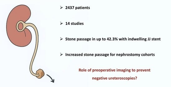
Quality assessment was performed by two independent blinded authors (SL, PG) using The Risk Of Bias In Non-randomised Studies—of Interventions (ROBINS-I) assessment tool for observational studies and the Cochrane Risk of Bias-2 (RoB-2) tool for randomised controlled trials. The ROBINS-I tool is used to assess the risk of bias across several domains, including confounding, patient selection, intervention, missing data, and measurement of outcomes. It is used in non-randomised studies, including single-arm trials and observational cohort studies, and is applicable to most of the articles included in this review. The Cochrane Risk of Bias-2 tool is a validated assessment tool for interventional trials that investigates several domains, including risk of bias from randomisation, deviations from the selected intervention, missing outcome data, measurement of outcomes, and reported results. All conflicts were resolved by discussion.

3. Results

The electronic search yielded 3631 results (Figure 1). After removing duplicates, conducting title/abstract screening and full-text screening, 14 studies were included for investigation [5,6,7,8,9,10,11,12,13,14,15,16,17,18]. The reasons for article exclusion during full-text review included being a duplicate study (n = 5) where both the conference abstract and full-text article was included and not identified as duplicates during the importation of files, incorrect outcomes (n = 7), incorrect intervention (n = 9), incorrect study design (n = 2) incorrect patient population (n = 2), and abstract with insufficient data (n = 6).

3.1. Quality Assessment

Results from the quality assessment are summarised in Figure 2. Across all observational studies, there was generally a high risk of bias. The risk of bias was assessed as moderate, serious, and critical for five, eight, and one studies, respectively. No included studies had an overall low risk of bias. In general, there was a moderate or high risk of bias across studies for confounding, selection, measurement of outcomes and reported results. Confounding and selection bias were frequently precipitated by the inclusion of patients with specific subsets of stone characteristics. The risk of bias assessment for one randomised controlled trial, using the Cochrane Risk of Bias tool, resulted in an overall high risk, primarily due to deviations from the intended interventions caused by participant attrition. In addition, there was significant inconsistency and heterogeneity in study design, stone characteristics, population, follow-up, and primary outcomes. Quantitative analysis of publication bias was not performed due to the absence of a meta-analysis. However, publication bias was assessed subjectively as high.

3.2. Study Characteristics

The fourteen studies conducted between 1990 and 2024 consisted of 2437 patients (Table 1). One included study was a randomised controlled trial [17], while the rest were observational, either prospective cohort studies [5,10,11,13,14,18] (n = 6) or retrospective cohort studies [6,7,8,9,12,15,16] (n = 7). All studies investigated patients with stents in situ and reported SSP. The sample sizes of the included studies varied from 42 to 401 patients. The included studies had a number of primary objectives, including assessing spontaneous stone passage rates in stented patients as either a primary outcome or a secondary outcome, with a separate primary aim. Inclusion criteria across studies were patients presenting with renal or ureteric colic with a single ureteric stone (n = 5) or otherwise unspecified (n = 9). One study excluded pelvic-ureteric junction stones due to the risk of retropulsion, a further study included only patients with infected urine, and one study included patients with acute calcular anuria. A final study included those with prior failed primary ureteroscopic access attempts.
Included are studies that investigated SSP in a single group of stented patients (n = 8); with two arms comparing stented and non-stented patients (n = 3); and comparing passage rates in stented patients to patients undergoing nephrostomy (n = 3). These subgroups are highlighted in Table 1.
Due to considerable clinical and methodological heterogeneity among the included studies, as well as a low number of studies with multiple comparator arms (i.e., stent vs. nephrostomy), a meta-analysis was not performed. Variability was noted in patient populations, follow-up duration, outcome measurement, and intervention protocols. As such, heterogeneity was deemed to be high.

3.3. Patient and Stone Characteristics

Various methods were used to outline stone sizes and locations (Table 2). Most articles provided the overall mean or median stone sizes with maximum diameters in millimetres. Some provided sizes with maximum diameters of passed and retained stones. Others provided subgroups of stone sizes and the number of patients fitting within these groups rather than providing average sizes. Some studies with more than one arm also provided average sizes for different intervention groups. Similarly, the stone location was identified using a range of methods. The stone location was commonly classified as the proximal, middle and distal ureter. Mean/median overall stone sizes ranged from 4.7 to 7.8 mm in diameter. Passed stone sizes ranged from 3.2 to 8.4mm, and retained stone sizes ranged from 6 to 9.4mm (Table 2). Fourteen studies investigated patients with single obstructing ureteric stones, and two included those with multiple (ipsilateral or contralateral) stones.

3.4. Spontaneous Stone Passage Rates

Results on follow-up and stone passage are highlighted in Table 3. Overall, spontaneous stone passage rates varied significantly between 1.7% and 42.3%. Spontaneous stone passage occurred in 2 patients (1.7%) in a study that followed patients for only 96 h after JJ stent insertion [17]. Additionally, spontaneous stone passage was not a primary outcome of this study. In contrast, another study [5] found a high stone passage rate of 42.3% with a follow-up duration of three months and a mean stone size of 8.4mm and 9.4mm for passed and retained stones, respectively [5]. Of the remaining studies, spontaneous passage rates ranged from 8% to 38.6%. Follow-up in these trials varied from 21 days to 3 months.
In published trials, significant associations were found between various patient and stone characteristics and stone passage. Individual analyses in included studies found associations between spontaneous stone passage and stone size (n = 10), location (n = 5), density (n = 3), and duration of stent (n = 2). A reasonably powered retrospective cohort study (n = 209) found statistically significant associations with all the above on univariate analysis, but only stone size and location remained significant on multivariate analysis [9]. Two similarly powered retrospective cohort studies found stone passage rates of 14.4 and 26.2%, respectively, finding significant associations between stone size and passage on logistic regression analysis [12,16]. A published abstract of 401 patients demonstrated significant differences in spontaneous stone passage rates according to both size (p = 0.002) and location (p < 0.001), with passage rates of 75% and 10.3% for stones < 2.9 mm and 7–8.9 mm, respectively, as well as passage rates of 70.8% and 16.7% for distal and proximal stones, respectively [6]. Multivariate logistic regression analysis on another retrospective cohort of 216 patients revealed that stone size, location, and stent dwell time were independent predictive variables of stone passage [8].

3.5. Comparison of Stented, Non-Stented and Nephrostomy Groups

Only three studies included both stented and non-stented subgroups [7,15,18]. One retrospective cohort study (n = 219) found a statistically significant lower stone passage rate in stented compared with non-stented patients (13.9 vs. 26.8%, odds ratio [OR] = 0.43, p = 0.019) on multivariate analysis [7]. Another retrospective cohort study (n = 194) found lower stone passage rates in patients with stents (14 vs. 20%). This result was not statistically significant (p = 0.30). However, in this study, patients in the JJ stent subgroup were less likely to have distal stones (p = 0.03) [15]. The third study, conducted in 1990, had a small sample size (n = 42) and found that three patients in the JJ stent group passed their stone, while none in the unstented cohort of 12 patients did [18].
Three studies (one abstract, two published papers) compared stone passage rates in patients with retrograde ureteral stents and nephrostomies. All three found statistically significant relationships between PCN and stone passage when compared to stent groups. An observational prospective cohort study (n = 42) followed patients for 30–40 days and found 25% stone passage rates for the stented group compared to 38.9% in the PCN group [10]. In the multivariable logistic regression analysis, the PCN group had an odds ratio of 6.667 for spontaneous stone passage (p = 0.047). Another study, which was larger (n = 239), had only 96 h of follow-up [17]. It found a statistically significantly higher stone passage rate in patients with nephrostomy (7.6% vs. 1.7%). A published abstract (n = 74) found higher odds of stone passage in patients undergoing nephrostomy (OR = 2.31) [14]. Two studies investigating quality-of-life measures found improved parameters in PCN groups over stent groups [10,17].

4. Discussion

Renal colic, resulting from ureteric obstruction by urinary calculi, represents a significant clinical and economic burden, owing to its association with acute pain, risk of infection, and potential renal impairment [19]. Ureteral stents are routinely utilised to decompress the obstructed system, alleviate symptoms, preserve renal function, and facilitate passive ureteral dilation prior to definitive ureteroscopic stone clearance.
While it is assumed that stone migration does not occur following stent placement, findings from this review suggest otherwise. However, results must be interpreted with caution due to a high level of heterogeneity across study designs, population characteristics and results. Studies on stented patients report a wide range of spontaneous passage rates, from as low as 1.7% (with short follow-up) to as high as 42.3% in studies with longer observation periods. These findings highlight that stone passage can occur despite the presence of a stent, particularly for smaller, distal stones and with longer stent dwell times [5,8]. Despite this, comparative studies [7,15] show that patients with stents tend to have lower rates of spontaneous stone passage compared to those managed without stents. The precise mechanism by which indwelling JJ stents may impair stone transit remains unclear. Early experimental data demonstrated reduced ureteral peristalsis and delayed stone transit in the presence of stents [20,21]. Mechanistically, stents may obstruct stone migration by occupying the ureteral lumen and diverting urinary flow away from the stone, thereby preventing its migration. Additionally, retrograde stent placement may cause proximal stone displacement due to guidewire manipulation, irrigation, or contrast injection, further reducing passage likelihood [10,21,22].
Reviewed studies indicate that a proportion of stented patients may be stone-free at the time of planned ureteroscopy and underscore the need to re-evaluate routine assumptions regarding stone persistence following stent placement. It is important to distinguish between studies reporting stone passage in stented patients and those comparing stenting with other management strategies, such as conservative treatment (unstented cohort) or nephrostomy. These study designs address different clinical questions—one examines expectant management after stenting, and the other guides the initial intervention choice (i.e., conservative management without JJ stent vs. stented vs. nephrostomy). Recognising this distinction is essential for applying evidence appropriately in clinical decision-making. While several predictive tools, including AI-based calculators like the Multi-centre cohort study evaluating the role of Inflammatory Markers In patients presenting with acute ureteric Colic (MIMIC) [23] and STONE Score [24], exist to estimate spontaneous stone passage in non-stented patients, no such models currently exist for patients with indwelling ureteral stents—highlighting a key gap in clinical decision support.
The conventional management of renal colic typically follows a staged approach: initial ureteral stenting to relieve obstruction, followed by ureteroscopic lithotripsy and subsequent stent removal. The rationale for definitive stone removal surgery, e.g., URS post-stenting, is multifactorial. First, it is commonly believed that JJ stents impair ureteral peristalsis, reducing the likelihood of spontaneous stone passage. Second, radiological assessment of stone presence is limited in the setting of an indwelling stent, as the radiopaque stent can obscure adjacent calculi—particularly on non-contrast CT, which already has limited ability to differentiate between similarly dense structures. One retrospective study reported only 67% accuracy in detecting stones in stented patients [25]. Third, patient-reported passage is notoriously unreliable, as it depends on the patient’s awareness and ability to identify stone expulsion. These factors collectively reinforce the prevailing paradigm of routinely performing URS after stenting.
However, our findings and identified literature suggest that a meaningful subset of stented patients may pass their stones spontaneously and may, therefore, undergo unnecessary ureteroscopy. Real-world evidence supports this: a 2019 systematic review reported negative URS rates as high as 14% [3]. Subjecting patients who have undergone spontaneous stone passage [5] to URS exposes them to unnecessary anaesthesia and procedural risks (e.g., infection, ureteral trauma, and stricture), which leads to unnecessary healthcare costs—especially concerning given the narrow operative windows required to avoid stent encrustation and morbidity [26,27]. Although indirectly related, the spontaneous passage of ureteral fragments post-definitive lithotripsy in the presence of ureteral stents also further supports the notion that stone passage is possible and likely with ureteral stents in situ. Aghamir et al. found a spontaneous fragment passage rate of 45.7% post-lithotripsy in stented patients [28]. In addition, Schatloff et al. also found stone fragment passage rates of up to 87% post lithotripsy, inclusive of patients with stents [29].
Further comparative studies suggest that PCN is associated with significantly higher spontaneous stone passage rates than JJ stenting, as well as improved quality-of-life outcomes and fewer lower urinary tract symptoms [10,14,17,30]. Although the exact mechanism remains speculative, animal studies indicate that nephrostomies may preserve ureteral peristalsis and facilitate passage by avoiding intraluminal obstruction [21]. Unlike stents, PCNs do not occupy the ureter, potentially allowing more natural urinary flow and stone transit—though this has not been definitively confirmed in human studies. Furthermore, nephrostomies allow for more accurate preoperative CT imaging to assess the residual stone burden [28]. Despite the associated mobility and hygiene challenges, nephrostomies offer a suitable alternative to ureteral stenting in carefully selected and appropriately counselled patients. Additionally, the effect of stent size on SSP rates is unclear, which may pose a potential avenue for research.
The key predictors of spontaneous passage are stone size, stone location, and the interval between stenting and reassessment. These findings suggest that selective re-imaging prior to URS, particularly for patients with small (<5 mm), distal stones or longer stent dwell times (>7 days) could help reduce unnecessary interventions. Supporting this, Brodie et al. (2022) found that pre-URS imaging reduced the rate of negative URS from 14% to 6% [12]. Pre-URS imaging is also cost-effective—performing imaging in eight patients to avoid one unnecessary ureteroscopy results in a net saving of £1683.56 per URS avoided, according to National Health Service (NHS) cost data [12]. A non-contrast CT performed 24–48 h before planned URS may help identify patients who are already stone-free. However, the known limitations of imaging in the presence of a stent, especially false negatives, require cautious interpretation [25].
Despite the findings discussed, several important limitations must be acknowledged. The included studies are generally of variable methodological quality, with a high risk of bias—particularly selection bias and confounding. Most were single-centre, retrospective cohort studies with inconsistent follow-up durations, which may have introduced selection bias and affected true cumulative stone passage rates.
Another key limitation is the significant heterogeneity across studies—in terms of design, patient populations, stone characteristics, outcome definitions, and reporting standards. This variability contributes to inconsistencies in the reported outcomes and precludes meaningful pooling of data for a robust meta-analysis. As such, while the evidence supports the potential for spontaneous stone passage with stenting, definitive conclusions remain limited.
Overall, this review highlights the presence of spontaneous stone passage in the setting of indwelling ureteral stents, despite the absence of high-quality data. Hence, carefully selected patients may benefit from appropriate pre-operative imaging to reduce unnecessary ureteroscopies. Importantly, this review highlights the need for well-designed prospective studies, ideally propensity-matched cohorts or randomised controlled trials, to validate current observations and provide stronger evidence to guide clinical practice. Future research should explore strategies such as routine URS versus selective re-imaging or stent removal in patients with small (<5 mm), distal ureteric stones. Primary outcomes could include rates of negative ureteroscopy, with secondary endpoints evaluating quality of life and cost-effectiveness, helping to optimise patient selection for intervention following JJ stent insertion.

5. Conclusions

Spontaneous stone passage after JJ stent insertion occurs in a meaningful minority of patients. However, routine subsequent ureteroscopy in patients with indwelling stents for obstructing ureteral stones remains standard practice. Reassessment—through selective re-imaging prior to URS may substantially reduce unnecessary procedures, lower costs, and minimise patient risk. Furthermore, percutaneous nephrostomy offers a potential alternative in appropriate clinical scenarios by enhancing stone passage whilst improving patient comfort. Despite this, the risk of bias, retrospective study designs, and significant heterogeneity in the data limit the strength of our findings. This systematic review underscores the need for larger, prospective, comparative, propensity-matched trials to determine the effect of indwelling ureteral stents on the spontaneous passage of ureteral stones.

Author Contributions

S.L.: conceptualisation, methodology, formal analysis, investigation, data curation, writing—original draft, visualisation. P.G.: formal analysis, investigation, data curation. D.T.: investigation. D.B.: writing—review and editing, supervision. O.P.: writing—review and editing, supervision. J.I.: conceptualisation, methodology, formal analysis, writing—review and editing, supervision. All authors have read and agreed to the published version of the manuscript.

Funding

The authors received no direct or indirect financial support for this study.

Data Availability Statement

Not applicable.

Conflicts of Interest

Sean Lim: No conflict of interest to declare. Oneel Patel: Oneel Patel is employed by Akeeko Medical, which develops medical devices. The author has disclosed this relationship in accordance with ethical research guidelines, and there was no direct or indirect financial support from Akeeko Medical related to this study. Joseph Ischia: Joseph Ischia is CEO and founder of Akeeko Medical, which develops medical devices. The author has disclosed this relationship in accordance with ethical research guidelines, and there was no direct or indirect financial support from Akeeko Medical related to this study. Patrick Gordon: No conflict of interest to declare. Daryl Thompson: No conflict of interest to declare. Damien Bolton: Damien Bolton is an investor in Akeeko Medical, which develops medical devices. The author has disclosed this relationship in accordance with ethical research guidelines, and there was no direct or indirect financial support from Akeeko Medical related to this study.

References

  1. Pathan, S.A.; Mitra, B.; Bhutta, Z.A.; Qureshi, I.; Spencer, E.; Hameed, A.A.; Nadeem, S.; Tahir, R.; Anjum, S.; Cameron, P.A. A comparative, epidemiological study of acute renal colic presentations to emergency departments in Doha, Qatar, and Melbourne, Australia. Int. J. Emerg. Med. 2018, 11, 1. [Google Scholar] [CrossRef] [PubMed]
  2. Patti, L.; Leslie, S.W. Acute Renal Colic; StatPearls: Treasure Island, FL, USA, 2023. [Google Scholar]
  3. Rice, P.; Prattley, S.; Somani, B.K. ‘Negative Ureteroscopy’ for Stone Disease: Evidence from a Systematic Review. Curr. Urol. Rep. 2019, 20, 13. [Google Scholar] [CrossRef]
  4. Campbell, M.; McKenzie, J.E.; Sowden, A.; Katikireddi, S.V.; Brennan, S.E.; Ellis, S.; Hartmann-Boyce, J.; Ryan, R.; Shepperd, S.; Thomas, J.; et al. Synthesis without meta-analysis (SWiM) in systematic reviews: Reporting guideline. BMJ 2020, 368, l6890. [Google Scholar] [CrossRef] [PubMed]
  5. Taha, D.E.; Elshal, A.M.; Zahran, M.H.; Harraz, A.M.; El-Nahas, A.R.; Shokeir, A.A. After urgent drainage of an obstructed kidney by internal ureteric stenting; is ureteroscopic stone extraction always needed? Arab J. Urol. 2015, 13, 258–263. [Google Scholar] [CrossRef]
  6. Trachsel, Y.; Förster, B.; John, H.; Schregel, C. MP14-12 spontaneous passage rate of ureteral stones and predictive factors after pre-stenting. J. Urol. 2022, 207 (Suppl. 5), e236. [Google Scholar] [CrossRef]
  7. González-Padilla, D.A.; González-Díaz, A.; Peña-Vallejo, H.; Santos Pérez de la Blanca, R.; Teigell-Tobar, J.; Hernández-Arroyo, M.; Abad-López, P.; Rodriguez-Antolin, A.; Cabrera-Meiras, F. Long surgical waiting list times are associated with an increased rate of negative ureteroscopies. Can. Urol. Assoc. J. 2021, 15, 407–411. [Google Scholar] [CrossRef] [PubMed]
  8. Stojkova Gafner, E.; Grüter, T.; Furrer, M.A.; Bosshard, P.; Kiss, B.; Vartolomei, M.D.; Roth, B. A treatment strategy to help select patients who may not need secondary intervention to remove symptomatic ureteral stones after previous stenting. World J. Urol. 2020, 38, 2955–2961. [Google Scholar] [CrossRef]
  9. Kuebker, J.M.; Robles, J.; Kramer, J.J.; Miller, N.L.; Herrell, S.D.; Hsi, R.S. Predictors of spontaneous ureteral stone passage in the presence of an indwelling ureteral stent. Urolithiasis 2019, 47, 395–400. [Google Scholar] [CrossRef]
  10. de Sousa Morais, N.; Pereira, J.P.; Mota, P.; Carvalho-Dias, E.; Torres, J.N.; Lima, E. Percutaneous nephrostomy vs ureteral stent for hydronephrosis secondary to ureteric calculi: Impact on spontaneous stone passage and health-related quality of life—A prospective study. Urolithiasis 2019, 47, 567–573. [Google Scholar] [CrossRef]
  11. Kailasa, A.K.; Bhuvanagiri, A.K.; Kannan, S.; Thangavelu, M.; Ahiaku, E.K.; Alexandrou, K. Negative ureteroscopy following emergency ureteric stenting. Eur. Urol. Suppl. 2019, 18, e2475–e2476. [Google Scholar] [CrossRef]
  12. Brodie, A.; Johnston, T.; Lloyd, P.; Hemsworth, L.; Barabas, M.; Keoghane, S. Reducing the rate of negative ureteroscopy: Predictive factors and the role of preoperative imaging. Ann. R. Coll. Surg. Engl. 2022, 104, 588–593. [Google Scholar] [CrossRef]
  13. Roy, J.; Zargar-Shoshtari, K. Emergency ureteroscopy versus stenting—Does infected urine matter? BJU Int. 2019, 123 (Suppl. 2), 5–6. [Google Scholar] [CrossRef]
  14. Rodrigues, R.M.; Silva, B.; Morais, N.; Pereira, J.P.; Anacleto, S.; Passos, P.; Torres, J.; Dias, E.; Lima, E.; Mota, P. MP15-05 percutaneous nephrostomy, ureteral stent or primary ureteroscopy with stone removal for the treatment of hydronephrosis secondary to ureteric calculi: A prospective evaluation of the impact on complications, stone management and health-related quality of life. J. Urol. 2020, 203 (Suppl. 4), e205–e206. [Google Scholar] [CrossRef]
  15. Baumgarten, L.; Desai, A.; Shipman, S.; Eun, D.D.; Pontari, M.A.; Mydlo, J.H.; Reese, A.C. Spontaneous passage of ureteral stones in patients with indwelling ureteral stents. Can. J. Urol. 2017, 24, 9024–9029. [Google Scholar]
  16. Nogara, A.; Lucignani, G.; Turetti, M.; Silvani, C.; Marmiroli, A.; Nizzardo, M.; Gadda, F.; Zanetti, S.P.; Longo, F.; De Lorenzis, E.; et al. Prevalence and predictors of stone passage after double J stenting for symptomatic ureteral stones: A cross-sectional, real-life study. World J. Urol. 2024, 42, 8. [Google Scholar] [CrossRef]
  17. Hasan, A.M.; Abdelhamid, A.M.; Ahmed, M.A.; Reyad, A.M. Percutaneous nephrostomy tube versus double J ureteric stent for the management of non-septic calcular anuria in adults: Prospective randomized study. Arab J. Urol. 2024, 23, 1–8. [Google Scholar] [CrossRef] [PubMed]
  18. Jones, B.J.; Ryan, P.C.; Lyons, O.; Grainger, R.; McDermott, T.E.; Butler, M.R. Use of the double pigtail stent in stone retrieval following unsuccessful ureteroscopy. Br. J. Urol. 1990, 66, 254–256. [Google Scholar] [CrossRef] [PubMed]
  19. Zhang, L.; Zhang, X.; Pu, Y.; Zhang, Y.; Fan, J. Global, Regional, and National Burden of Urolithiasis from 1990 to 2019: A Systematic Analysis for the Global Burden of Disease Study 2019. Clin. Epidemiol. 2022, 14, 971–983. [Google Scholar] [CrossRef] [PubMed]
  20. Ryan, P.; Lennon, G.; McLEAN, P.; Fitzpatrick, J. The effects of acute and chronic JJ stent placement on upper urinary tract motility and calculus transit. Br. J. Urol. 1994, 74, 434–439. [Google Scholar] [CrossRef]
  21. Lennon, G.M.; Thornhill, J.A.; Grainger, R.; McDermott, T.E.D.; Butler, M.R. Double Pigtail Ureteric Stent versus Percutaneous Nephrostomy: Effects on Stone Transit and Ureteric Motility. Eur. Urol. 1997, 31, 24–29. [Google Scholar] [CrossRef]
  22. Mosayyebi, A.; Manes, C.; Carugo, D.; Somani, B.K. Advances in Ureteral Stent Design and Materials. Curr. Urol. Rep. 2018, 19, 35. [Google Scholar] [CrossRef]
  23. Shah, T.T.; Gao, C.; Peters, M.; Manning, T.; Cashman, S.; Nambiar, A.; Cumberbatch, M.; Lamb, B.; Peacock, A.; Van Son, M.J.; et al. Factors associated with spontaneous stone passage in a contemporary cohort of patients presenting with acute ureteric colic: Results from the Multi-centre cohort study evaluating the role of Inflammatory Markers In patients presenting with acute ureteric Colic (MIMIC) study. BJU Int. 2019, 124, 504–513. [Google Scholar]
  24. Gupta, K.; Ricapito, A.; Lundon, D.; Khargi, R.; Connors, C.; Yaghoubian, A.J.; Gallante, B.; Atallah, W.M.; Gupta, M. Harnessing Artificial Intelligence to Predict Spontaneous Stone Passage: Development and Testing of a Machine Learning-Based Calculator. J. Endourol. 2025, 39, 738–747. [Google Scholar] [CrossRef]
  25. Tang, V.; Attwell-Heap, A. Computed tomography versus ureteroscopy in identification of renal tract stone with ureteral stent in situ. Ann. R. Coll. Surg. Engl. 2011, 93, 639–641. [Google Scholar] [CrossRef] [PubMed]
  26. Scarneciu, I.; Lupu, S.; Pricop, C.; Scarneciu, C. Morbidity and impact on quality of life in patients with indwelling ureteral stents: A 10-year clinical experience. Pak. J. Med. Sci. 2015, 31, 522–526. [Google Scholar] [CrossRef]
  27. Tomer, N.; Garden, E.; Small, A.; Palese, M. Ureteral Stent Encrustation: Epidemiology, Pathophysiology, Management and Current Technology. J. Urol. 2021, 205, 68–77. [Google Scholar] [CrossRef]
  28. Aghamir, S.M.K.; Mohammadi, A.; Farahmand, H.; Meysamie, A.P. Effects of Prophylactic Insertion of Double-J Stents to Decrease Episodes of Renal Colic in Patients with Recurrent Ureteral Stones. J. Endourol. 2008, 22, 435–438. [Google Scholar] [CrossRef] [PubMed]
  29. Schatloff, O.; Lindner, U.; Ramon, J.; Winkler, H.Z. Randomized Trial of Stone Fragment Active Retrieval Versus Spontaneous Passage During Holmium Laser Lithotripsy for Ureteral Stones. J. Urol. 2010, 183, 1031–1036. [Google Scholar] [CrossRef] [PubMed]
  30. Shoshany, O.; Erlich, T.; Golan, S.; Kleinmann, N.; Baniel, J.; Rosenzweig, B.; Eisner, A.; Mor, Y.; Ramon, J.; Winkler, H. Ureteric stent versus percutaneous nephrostomy for acute ureteral obstruction-clinical outcome and quality of life: A bi-center prospective study. BMC Urol. 2019, 19, 79. [Google Scholar] [CrossRef]
Figure 1. PRISMA flow diagram illustrating the study selection process. A total of 3631 records were identified through database searches (Embase, PubMed, and MEDLINE). After removal of 1168 duplicates (8 manually and 1160 by Covidence), 2463 studies were screened. Of these, 2418 were excluded based on title and abstract review. Forty-five full-text articles were assessed for eligibility, with 30 excluded for reasons including duplicate publication (n = 5), wrong outcomes (n = 7), wrong intervention (n = 9), wrong study design (n = 2), wrong patient population (n = 2), or insufficient data in the abstract (n = 6). Fourteen studies were included in the final review.
Siuj 06 00065 g001
Figure 2. Risk of bias assessment for included studies using the ROBINS-I tool (A) for observational studies and the Cochrane Risk of Bias (RoB) tool (B) for the randomised controlled trial. The ROBINS-I tool evaluates seven domains: confounding, selection of participants, classification of interventions, deviations from intended interventions, missing data, measurement of outcomes, and selection of the reported result. The Cochrane RoB tool assesses five domains specific to randomised studies. Abbreviations: ROBINS-I = Risk Of Bias In Non-randomised Studies of Interventions; RoB = Risk of Bias.
Siuj 06 00065 g002
Table 1. Baseline characteristics of included studies evaluating spontaneous stone passage. The table summarises study design, patient population, intervention groups (e.g., ureteral stent, percutaneous nephrostomy [PCN], or non-stented control), sample sizes, mean or median patient age, and methods used to assess stone passage (e.g., non-contrast computed tomography [CT], ureteroscopy [URS], or clinical confirmation of stone passage). Abbreviations: RCT = Randomised Controlled Trial; URS = Ureteroscopy; CT = Computed Tomography; PCN = Percutaneous Nephrostomy; PUJ = Pelviureteric Junction; N/A = Not Available.
StudyStudy TypePatient PopulationIntervention GroupsSample SizeAge (Mean)Measurement of Outcome
Taha 2015 [5]Prospective cohortUreteric colicSingle arm (stone passage with stent)19653.7CT
Trachsel 2022 [6]Retrospective Cohort (Abstract)Ureteric colicSingle arm401N/ACT or URS
Gonzalez-Padilla 2021 [7]Retrospective CohortSingle ureteral stoneStent vs. no stentStent: 137
Control: 82
Median 57.6CT or URS
Stojkova Gafner 2020 [8]Retrospective CohortSingle ureteral stoneSingle arm 216Median 54Presenting filtered stone or CT
Kuebker 2019 [9]Retrospective CohortStented for a single ureteral stone. Excluded PUJ stones.Single arm20951.2CT (n = 3) or URS (n = 206)
de Sousa Morais 2019 [10]Prospective cohortEmergency stent or PCN for ureteric colicStent vs. PCNStent: 32
PCN: 18
57.6CT
Kailasa 2019 [11]Prospective cohort (Abstract)Ureteric colicSingle arm4454URS
Brodie 2022 [12]Retrospective CohortUreteric colicSingle arm25756CT or URS
Roy 2019 [13]Prospective cohort (Abstract)Single ureteral stone with infected urineSingle arm47N/AUnspecified
Rodrigues 2020 [14]Prospective cohort (Abstract)Ureteric colicStent vs. PCNStent: 53
PCN: 21
N/AUnspecified
Baumgarten 2017 [15]Retrospective CohortUreteric or renal colicStent vs. no stentStent: 119
Control: 75
<30: 25
30–40: 26
40–50: 44
>50: 89
URS
Nogara 2024 [16]Retrospective CohortSingle ureteral stoneSingle arm249Median 56CT
Hasan 2024 [17]RCTAcute calcular anuria. Solitary and dual kidneysStent vs. PCNStent: 121
PCN: 118
Median 48Unspecified
Jones 1990 [18]Prospective Cohort StudyPrior failed URS for ureteric colicStent vs. no stentStent: 30
Control: 12
Stent: 42
Control: 38
CT or URS
Table 2. Summary of stone characteristics across included studies evaluating spontaneous stone passage. The table presents sample size, stone location (proximal, mid, distal ureter or renal pelvis), number of stones (single vs. multiple), stone size (reported as mean, median, surface area or volume), and stone density (measured in Hounsfield Units (HUs) or volume, where applicable). Where available, data are stratified by intervention groups such as ureteral stenting, percutaneous nephrostomy (PCN), or non-stented controls. Abbreviations: HU = Hounsfield Units; IQR = Interquartile Range; SD = Standard Deviation; mm2 = square millimetres.
StudySample Size (n)Stone LocationSingle vs. Multiple StonesStone Size (mm)Stone Density (HU)
Taha 2015 [5]196Upper: 98
Middle: 69
Lower: 28
UnspecifiedPassed: 8.4 ± 2.8
Retained: 9.4 ± 2.8
Passed: 485.6 (SD 308)
Retained: 685.9 (SD 314)
Trachsel 2022 [6]401-Unspecified4.7 (IQR 3.3–5.6)-
Gonzalez-Padilla 2021 [7]Stent: 137
Control: 82
Proximal: 46 (21%)
Mid: 46 (21%)
Distal:127 (58%)
SingleMedian 7 (IQR 5–8)Median 744 (IQR 528–910)
Stojkova Gafner 2020 [8]216Proximal: 56 (26)
Mid: 55 (25)
Distal: 105 (49)
SingleMedian 5 (Range 2–11)Median 710
Kuebker 2019 [9]209Proximal: 119 (57)
Mid: 31 (15)
Distal: 59 (28)
Single6.6 ± 2.5-
de Sousa Morais 2019 [10]Stent: 32
PCN: 18
Proximal: 27 (54.0)
Distal: 23
Stent:
Single: 13 (72.2)
Multiple: 5 (27.8)
PCN:
Single: 20 (62.5)
Multiple: 12 (37.5)
Stent:
Median 47.0 mm2 (IQR 21.5–84.2 mm2)
PCN:
Median 92.0 mm2 (IQR 55.5–128.2 mm2)
Median 500
(IQR 390–656)
Kailasa 2019 [11]44Proximal: 12
Mid: 14
Distal: 18
Unspecified--
Brodie 2022 [12]257Proximal: 160 (62)
Mid: 32 (13)
Distal: 64 (25)
Unspecified7.8699
Roy 2019 [13]47-Single7 (Median)-
Rodrigues 2020 [14]Stent: 53
PCN: 21
-UnspecifiedStent:
7
PCN:
8
-
Baumgarten 2017 [15]Stent: 119
Control: 75
Stent:
Renal Pelvis: 22 (18)
Proximal Ureter: 42 (35)
Mid: 19 (16)
Distal: 32 (27)
Unknown: 4 (3)
Control:
Renal Pelvis: 27 (36)
Proximal Ureter: 17 (23)
Mid: 10 (13)
Distal: 21 (28)
Stent:
Single: 108 (91)
Multiple: 11 (9)
Control:
Single: 70 (93)
Multiple: 5 (7)
Stent:
<4 mm: 13 (11)
4–8 mm: 63 (53)
>8 mm: 37 (31)
Unknown: 6 (5)
Control:
<4 mm: 3 (4)
4–8 mm: 36 (48)
>8 mm: 35 (47)
Unknown: 1 (1)
-
Nogara 2024 [16]249Proximal: 102 (41.0)
Mid: 48 (19.3)
Distal: 99 (39.8)
Single7.1 (Median)Median: 671
Hasan 2024 [17]239Stent:
48 (Median)
PCN:
52 (Median)
UnspecifiedStent:
1334 mm3 (Volume)
PCN:
1326 mm3 (Volume)
Stent:
880
PCN:
893
Jones 1990 [18]Stent: 30
Control: 12
Stent:
Upper: 18 (60.0)
Mid: 7 (23.3)
Lower: 5 (16.7)
Control:
Upper: 7 (58.3)
Mid: 3 (25.0)
Lower: 2 (16.7)
UnspecifiedStent:
9
Control:
7.4
-
Table 3. Summary of spontaneous stone passage (SSP) outcomes across studies evaluating stented, non-stented, and nephrostomy (PCN) patient cohorts. The table includes follow-up duration, SSP rates by treatment group, and key findings or statistically significant predictors. Asterisks (*) denote statistically significant differences (p < 0.05). Abbreviations: SSP = Spontaneous Stone Passage; PCN = Percutaneous Nephrostomy; URS = Ureteroscopy; UVA = Univariate Analysis; MVA = Multivariate Analysis; NCCT = Non-Contrast Computed Tomography; QoL = Quality of Life.
StudyFollow-Up DurationSpontaneous Stone Passage (Stented)Stone Passage (PCN)Stone Passage (Non-stented)Other Pertinent Findings
Taha 2015 [5]3 Months (0.5–6.3)83 (42.3)--Opacity (p < 0.001), stone width (p = 0.038), and positive urine culture (p = 0.046) associated with greater probability of SSP.
Trachsel 2022 [6]Median 26 days (IQR 22–31)Overall: 95 (23.7)
0–2.9 mm: 75%
3–4.9 mm: 37.1%
5–6.9 mm: 19.1
7–8.9 mm: 10.3%
>9 mm: 8.2%
Distal: 70.8%
Middle: 12.5%
Proximal: 16.7%
--Size (p = 0.002) and location (p < 0.001) associated with greater likelihood of SSP.
Gonzalez-Padilla 2021 [7]Median 74 (IQR 45–127)19 * (13.9)-22 * (26.83)Lower SSP for stented patients on MVA (OR = 0.43, p = 0.019). Size, location, radioopacity associated with greater likelihood of SSP(p < 0.05).
Stojkova Gafner 2020 [8]Median 4 weeks (Range 1–14)74 (34.3)--Smaller size (p < 0.001), distal stone location (p = 0.046) and stent dwell time (p = 0.02) independent predictors of SSP.
Kuebker 2019 [9]Mean 21 (SD 15.2)17 (8)--Stone diameter, location, and stent duration associated with SSP on UVA (p < 0.01). Stone diameter and stent duration significantly associated on MVA
de Sousa Morais 2019 [10]30–40 days (Protocol)8 * (25)7 * (38.9)-Stone size (p = 0.012) associated with SSP. SSP higher for PCN (OR 6.667) adjusting for size and location. PCN was better tolerated and associated with better QoL.
Kailasa 2019 [11]Not provided17 (38.6)--Stone size significantly associated with negative URS (p = 0.02).
Brodie 2022 [12]Not provided37 (14.4)--Logistic regression showed stone size as a predictor of negative URS (p = 0.01).
Roy 2019 [13]Not provided5 (10.6)---
Rodrigues 2020 [14]Not provided---Spontaneous stone passage higher for PCN than RUS (OR = 2.31). on multivariable analysis
Baumgarten 2017 [15]Stented:
<30 days: 18 (15)
31–60 days: 41 (34)
61–100 days: 34 (29)
>100 days: 22 (18)
Unknown: 4 (3)
Control:
<30 days: 20 (27)
31–60 days: 26 (35)
61–100 days: 10 (13)
>100 days: 16 (21)
Unknown: 3 (4)
17 (14) 15 (20)SSP lower in stented groups but not statistically significant (p = 0.30). Stone size significant predictor of SSP on MVA (p = 0.01).
Nogara 2024 [16]1 month (Fixed)65 (26.2) Stone diameter, density and location significantly associated with SSP (p < 0.001).
Hasan 2024 [17]96 h2 * (1.7)9 * (7.6) PCN associated with higher passage rates (p = 0.028) and QoL scores (p < 0.001) compared to stent.
Jones 1990 [18]Unspecified3 (10.0) 0 (0)-
Disclaimer/Publisher’s Note: The statements, opinions and data contained in all publications are solely those of the individual author(s) and contributor(s) and not of MDPI and/or the editor(s). MDPI and/or the editor(s) disclaim responsibility for any injury to people or property resulting from any ideas, methods, instructions or products referred to in the content.

