Interesting Images

Iatrogenic Small Intestine Perforation During Suprapubic Catheter Change

1
Department of Urology, All India Institute of Medical Sciences, Patna 801507, Bihar, India
2
Department of Radiodiagnosis, All India Institute of Medical Sciences, Patna 801507, Bihar, India
3
Department of General Surgery, All India Institute of Medical Sciences, Patna 801507, Bihar, India
*
Author to whom correspondence should be addressed.
Soc. Int. Urol. J. 2025, 6(1), 1; https://doi.org/10.3390/siuj6010001
Submission received: 21 August 2024 / Accepted: 27 August 2024 / Published: 11 February 2025
A gentleman in his 70s presented to the emergency department with diffuse abdominal pain, abdominal distention, and anuria of 3-days duration. The patient had undergone a suprapubic catheter insertion 18 months earlier for a pan anterior urethral stricture, as the patient was not willing to have major reconstructive surgery at his age, and he was on a monthly suprapubic catheter exchange programme at a primary care facility. This is a usual practice in our area, as the incidence of sediment formation and encrustation is high, especially in patients with a foreign body in situ. This is generally attributed to our geographical location having a high prevalence of urinary stone formation and use of latex urinary catheters [1]. The most recent change of the suprapubic catheter in his village was 3 days before presentation to our unit. The patient had no urine output from the suprapubic catheter subsequently. On further evaluation with ultrasonography, the urinary bladder was found to be distended with 600 mL of urine, and there was increased echogenicity of both kidneys. Kidney function tests were deranged with a serum creatinine of 3.92 mg/dL. An urgent non-contrast computed tomography of the abdomen was performed which revealed a distended urinary bladder with suprapubic catheter traversing the plane between the anterior abdominal wall and the urinary bladder, perforating the small intestine. The inflated balloon of the catheter was within the lumen of the small intestine (Figure 1). The patient underwent an emergency laparotomy through a lower midline vertical incision. On entering the peritoneal cavity, the suprapubic catheter was seen perforating the small intestine, 45 cm proximal to the ileocaecal junction. The inflated catheter balloon in the lumen had resulted in the dilatation of the proximal intestinal segment (Figure 2). The intestine distal to the perforation was collapsed. The perforated segment was resected, and a double-layered end-to-end anastomosis was performed. A fresh suprapubic cystostomy was created, and a new suprapubic catheter was placed. After surgery, the patient had an uneventful recovery. His kidney function tests normalised within 10 days of surgery. He remains under regular follow-up. When last seen 3 months after surgery, he was healthy and under a regular monthly suprapubic catheter change from our centre.
The indications for long-term suprapubic catheterisation include neuropathic bladders, impaired mobility or cognition of the patient, or complex urethral strictures for which the patient is unfit or unwilling to undergo the major reconstructive procedure [2]. The practice of changing the catheter every month while common in our area is not practiced worldwide because while latex catheters are cheaper and commonly available in our area, they are more prone to encrustation and blockage, so they need to be replaced earlier than silicone catheters, which are easily available worldwide and can be kept in situ for a longer duration [3]. Most complications associated with suprapubic catheterisation occur at the time of insertion when there is injury to the visceral organs due to either perforation by the trocar, loss of suprapubic tract, or insertion at a higher position in an underfilled bladder [4]. Studies have shown the incidence of bowel perforation to be 2.4–2.7% after suprapubic catheterisation [5,6]. However, bowel perforation following suprapubic catheter exchange is an extremely rare event [7,8,9]. During a suprapubic catheter change, complications arise when the tract (fistula) is not well-formed and the new catheter does not pass easily through the tract without resistance. However, in our case, since the tract was mature and 18 months old, a guidewire was not used during the exchange. The old catheter was not clamped to keep the bladder full before removal so the bladder would have been decompressed at the time of exchange. In this case, the gap between the bladder and anterior abdominal wall indicated that the original placement of the catheter was possibly too high. After decompression of the bladder following the suprapubic catheter removal, the tract size increased due to traction, and the tract may have been lost, and the catheter might have found a way into the gap between the bladder and the anterior abdominal wall.
A strict protocol for the suprapubic catheter change should be followed, and the person performing it should have adequate skill and training. The standard procedure for a suprapubic catheter exchange performed at our centre is as follows:
  • The patient should be placed in the supine position, and the area from the umbilicus to the midthigh should be adequately exposed and cleaned.
  • The bladder should be filled with approximately 250 mL through the existing catheter in order to distend the bladder and push the anterior bladder against the anterior abdominal wall.
  • During withdrawal of the previous catheter, its length and direction of entry into the bladder should be noted, and the insertion of the new catheter within the tract should be in the same direction and up to approximately same length.
  • The free flow of urine should be confirmed after the new suprapubic catheter insertion. The patient should be kept under observation for about 30 min after the suprapubic catheter exchange to confirm continuous drainage. There should be no pain during or after the catheter exchange.

Author Contributions

Conceptualization, N.K. and K.R.; methodology, N.K. and S.S.; software, S.S.; validation, N.K., S.K. and K.R.; formal analysis, K.R.; investigation, N.K.; resources, S.S.; data curation, S.K.; writing—original draft preparation, N.K.; writing—review and editing, K.R.; visualization, S.S. and S.K.; supervision, N.K.; project administration, N.K. All authors have read and agreed to the published version of the manuscript.

Funding

This research received no external funding.

Institutional Review Board Statement

The study did not require ethical approval.

Informed Consent Statement

Written informed consent has been obtained from the patient to publish this case.

Data Availability Statement

Not applicable.

Conflicts of Interest

The authors declare no conflict of interest.

References

  1. Kalpande, S.; Saravanan, P.R.; Saravanan, K. Study of factors influencing the encrustation of indwelling catheters: Prospective case series. Afr. J. Urol. 2021, 27, 50. [Google Scholar] [CrossRef]
  2. Jane Hall, S.; Harrison, S.; Harding, C.; Reid, S.; Parkinson, R. British Association of Urological Surgeons suprapubic catheter practice guidelines—Revised. BJU Int. 2020, 126, 416–422. [Google Scholar] [CrossRef] [PubMed]
  3. Oyortey, M.A.; Essoun, S.A.; Ali, M.A.; Abdul-Rahman, M.; Welbeck, J.; Dakubo, J.C.B.; Mensah, J.E. Safe duration of silicon catheter replacement in urological patients. Ghana. Med. J. 2023, 57, 66–74. [Google Scholar] [CrossRef] [PubMed] [PubMed Central]
  4. Ahluwalia, R.S.; Johal, N.; Kouriefs, C.; Kooiman, G.; Montgomery, B.S.I.; Plail, R.O. The surgical risk of Suprapubic catheter insertion and long-term sequelae. Ann. R. Coll. Surg. Engl. 2006, 88, 210–213. [Google Scholar] [CrossRef] [PubMed]
  5. Sheriff, M.K.M.; Foley, S.; McFarlane, J.; Nauth-Misir, R.; Craggs, M.; Shah, P.J.R. Long-term suprapubic catheterisation: Clinical outcome and satisfaction survey. Spinal Cord. 1998, 36, 171–176. Available online: https://pubmed.ncbi.nlm.nih.gov/9554016/ (accessed on 7 December 2023). [CrossRef] [PubMed]
  6. Cundiff, G.; Bent, A.E. Suprapubic catheterization complicated by bowel perforation. Int. Urogynecol J. Pelvic Floor. Dysfunct. 1995, 6, 110–113. [Google Scholar] [CrossRef]
  7. Wu, C.-C.; Su, C.-T.; Lin, A.C.-M. Terminal ileum perforation from a misplaced percutaneous suprapubic cystostomy. Eur. J. Emerg. Med. 2007, 14, 92–93. [Google Scholar] [CrossRef] [PubMed]
  8. Mongiu, A.K.; Helfand, B.T.; Kielb, S.J. Small bowel perforation during suprapubic tube exchange. Can. J. Urol. 2009, 16, 4519–4521. [Google Scholar] [PubMed]
  9. Witham, M.D.; Martindale, A.D. Occult transfixation of the sigmoid colon by suprapubic catheter. Age Ageing 2002, 31, 407–408. [Google Scholar] [CrossRef] [PubMed]
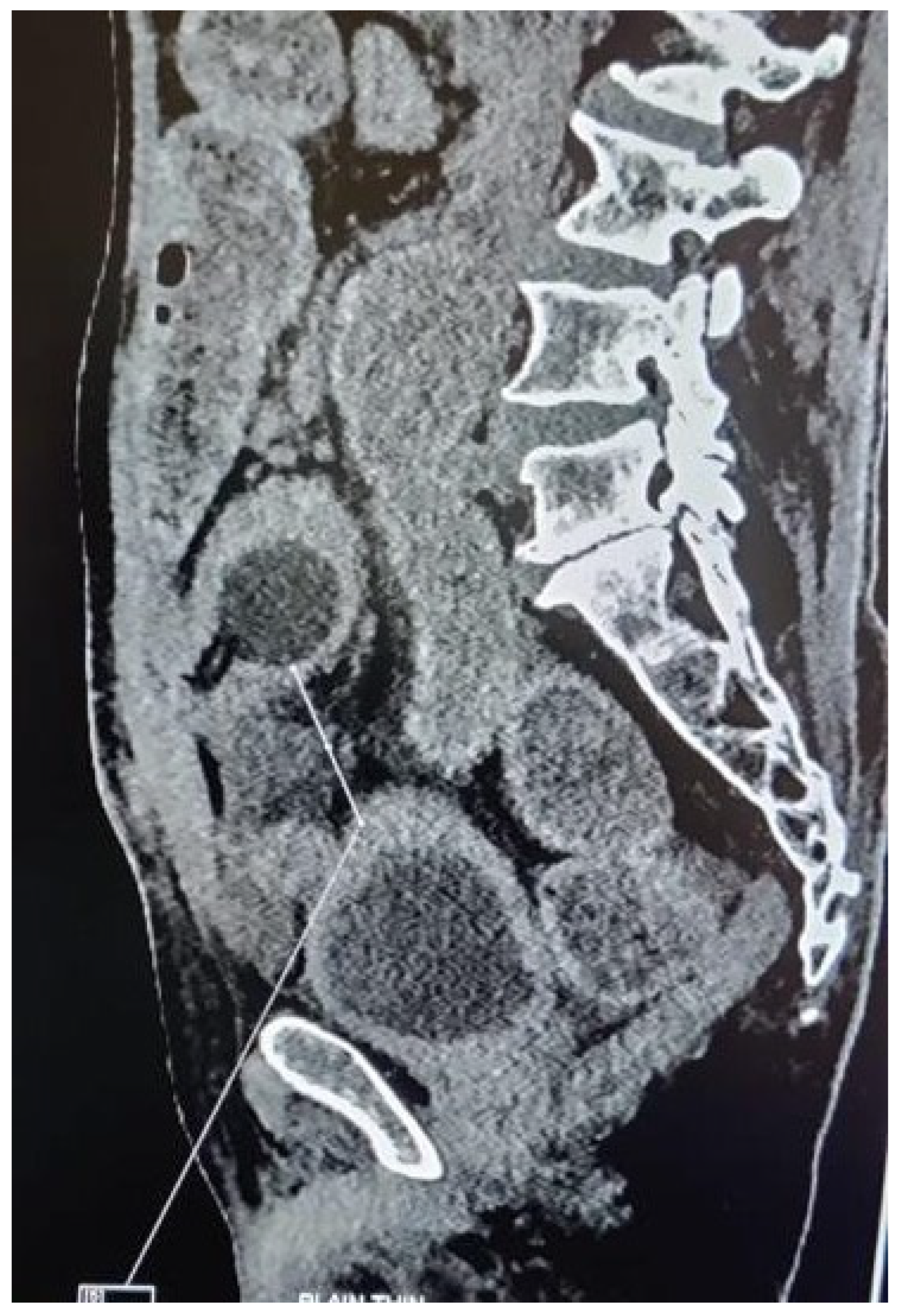
Figure 1. NCCT abdomen and pelvis in sagittal section showing the distended urinary bladder and bulb of the suprapubic Foley’s catheter inflated inside the small intestine lumen.
Siuj 06 00001 g001
Figure 2. Line diagram showing (a) the normal suprapubic catheter position and (b) the catheter going between the urinary bladder and anterior abdominal wall into the peritoneal cavity and perforating the bowel ((a,b) created by Naveen Kumar). Intraoperative finding of (c) the catheter perforating the small intestine and (d) the bulb of the suprapubic Foley’s catheter inflated inside the small intestine lumen.
Siuj 06 00001 g002
Disclaimer/Publisher’s Note: The statements, opinions and data contained in all publications are solely those of the individual author(s) and contributor(s) and not of MDPI and/or the editor(s). MDPI and/or the editor(s) disclaim responsibility for any injury to people or property resulting from any ideas, methods, instructions or products referred to in the content.