Systematic Review

Urinary Tract Obstruction Secondary to Fungal Balls: A Systematic Review

by
Kenneth Keen Yip Chew
1,2,*,
Maryaan Kas
1,2 and
Pascal Mancuso
1,2
1
Liverpool Hospital, Sydney, NSW 2170, Australia
2
Faculty of Medicine and Health, University of New South Wales, Sydney, NSW 2052, Australia
*
Author to whom correspondence should be addressed.
Soc. Int. Urol. J. 2024, 5(3), 227-236; https://doi.org/10.3390/siuj5030034
Submission received: 24 January 2024 / Revised: 19 April 2024 / Accepted: 25 April 2024 / Published: 18 June 2024

Abstract

:
Objectives: To understand the clinical characteristics, risk factors, diagnosis, treatment, and outcomes of urinary tract obstruction caused by fungal balls. Materials and Methods: A comprehensive search was conducted across PubMed, Embase, and Medline following the PRISMA guidelines, including case reports and case series. Study quality and risk of bias were assessed using the Oxford Centre for Evidence-Based Medicine (CEBM) document. The systematic review process was aimed at gathering and synthesising all available research evidence on the topic. Results: We included 54 articles reporting on 57 patients, primarily males (61.4%) with a median age of 56 years. The most common presenting symptoms were flank pain (71.9%) and fevers (59.6%). All diagnoses were made on the presence of yeast in urine or intra-operative tissue cultures and the presence of obstruction on imaging. The most common pathogen isolated were those of the Candida species (61.5%) followed by Aspergillus (33.3%). Multimodal management was the mainstay approach with the use of systemic and local antifungal therapy in combination with surgical drainage of the obstruction. The mortality rate was around 12.3%. Conclusions: Management of renal or ureteral obstruction caused by fungal balls requires a multimodal and multidisciplinary approach, comprising systemic antifungal therapy, drainage procedures, and in some instances, surgical intervention. The growing prevalence of antifungal resistance and the high mortality potential of fungal uropathy necessitates ongoing research into the optimal diagnostic and treatment modalities for this condition.

1. Introduction

Fungal uropathies, characterized by a spectrum of conditions affecting the urinary tract due to fungal infections, represent an area of growing clinical concern in urology. While urinary tract infections (UTIs) are conventionally associated with bacterial pathogens, an increasing prevalence of fungal UTIs, particularly in immunocompromised patients and those with urinary catheters or indwelling devices, has been noted over recent years [1]. Among these, the formation of fungal balls causing ureteral and renal obstruction has emerged as a significant challenge to urological care.
Fungal bezoars, or mycetomas, are conglomerates of fungal hyphae, host cell debris, and urinary sediment that can obstruct the urinary tract, often complicating the clinical course of fungal UTIs [2]. The most common pathogen implicated is the Candida species, although Aspergillus and other rare fungal species have also been reported [3]. While fungal balls in the urinary tract are well documented in literature, most cases pertain to bladder or urethral obstructions. Comparatively, there is a dearth of studies on ureteral and renal obstructions caused by fungal balls, leading to diagnostic and therapeutic uncertainty.
Obstructive uropathy secondary to fungal balls can have significant implications, with the potential for severe hydronephrosis, renal damage, and progression to life-threatening sepsis. As urologists, the need for a clearer understanding of this condition is essential. The potential impact on patient outcomes, clinical resources, and therapeutic intervention necessitates a thorough investigation into fungal uropathies, particularly their predisposing factors, diagnostic hallmarks, and treatment strategies.
The difficulty in diagnosis is further compounded by the fact that patients may often present with non-specific symptoms, overlapping with bacterial UTIs or other urological conditions [4]. The majority of fungal UTIs are acquired via the ascending route with a small minority of patients with candiduria acquired via the hematogenous route. The presence of candiduria can signal diverse pathological states, ranging from benign colonisation of indwelling catheters to fungal balls [5]. This leads to a wide spectrum of clinical disease with the majority of patients presenting with asymptomatic candiduria [6]. Furthermore, standard urine cultures may not always yield a fungal pathogen, thus delaying or misdirecting treatment [7]. Hence, the necessity of maintaining a high index of suspicion and the need for definitive diagnostic criteria becomes evident.
Treatment for fungal balls causing ureteral and renal obstruction is complex and often necessitates a multimodal approach involving systemic antifungal therapy, drainage procedures, and in some cases, surgical intervention [7]. With the rise of antifungal resistance and potential complications of invasive procedures, optimal management strategies have yet to be defined.
This systematic review aims to fill a crucial knowledge gap in this domain by synthesising available evidence on renal tract obstruction secondary to fungal bezoars. The goal is to enhance our understanding, thereby guiding evidence-based decision-making in clinical practice. By illuminating this often overlooked cause of obstructive uropathy, we aim to both improve the recognition of fungal obstruction and analyse the optimal management of this condition, to ultimately improve patient care and outcomes.

2. Materials and Methods

A comprehensive search was conducted in accordance with the Preferred Reporting Items for Systematic Reviews and Meta-analyses (PRISMA) guidelines using the following databases: PubMed, Embase, and Medline. We searched these databases from inception until December 2023. The reference lists of the included studies and relevant reviews were manually checked to identify additional potential studies.
The search strategy combined key terms related to fungal balls and ureteral/renal obstruction with terms chosen with the assistance of our local health district librarian. The terms used were “fungal balls” OR “fungal bezoars” OR “Candida” OR “Aspergillus” OR “trichosporon” OR “saccharomyces” AND “ureteral obstruction” OR “renal obstruction” NOT “neonates” OR “infants”. An English language restriction was imposed.

2.1. Eligibility Criteria

Studies were eligible for inclusion if they met the following criteria:
  • Original articles reporting on patients diagnosed with upper urinary tract obstruction from fungal balls/bezoars.
  • Studies providing data on at least one of the following outcomes: patient demographics, clinical presentation, mode of diagnosis, treatment modalities, and patient outcomes.
  • Case reports, case series, observational studies (cohort- and case-control), and clinical trials were included.
Articles were excluded if they met the following criteria:
  • Reviews, commentaries, editorials, letters, conference abstracts, and expert opinions.
  • Studies not providing specific data on outcomes for patients.
  • Studies on paediatric or neonatal patients (aged < 18 years).

2.2. Data Extraction and Quality Assessment

Data were extracted using a pre-specified data extraction form. The following information was obtained from each study: first author’s name, year of publication, study design, patients’ baseline characteristics, clinical findings, diagnostic methods, treatment methods, and outcomes.
The methodological quality of the included studies was assessed by two independent authors (K.C. and M.K.). Any disagreements between reviewers were resolved through discussion or consultation with a third reviewer if necessary.

2.3. Data Synthesis and Analysis

Given the nature of the included studies being primarily case reports and case series, a meta-analysis was not feasible. Instead, data were synthesised qualitatively. Patient characteristics, such as age, gender, and presenting symptoms, were described. In addition, details about diagnostic procedures, pathologic findings, treatments, and outcomes were collated. Descriptive statistics were used to summarise the data.

2.4. Risk of Bias across Studies

The risk of bias in case reports and case series was evaluated using the Oxford Centre for Evidence-Based Medicine: Levels of Evidence (CEBM) document. All articles were of level 4 evidence.
Given that case reports and case series are inherently high in bias and low in evidence level, the overall evidence generated by this review should be interpreted with caution.

2.5. Ethics Approval and Consent to Participate

As this study was a systematic review, it did not involve direct contact with patients or alteration to patient care and thus did not require ethics committee approval.

2.6. Availability of Data and Material

All data generated or analysed during this study are included in this published article or are available from the corresponding author upon request.

3. Results

3.1. Study Selection and Characteristics

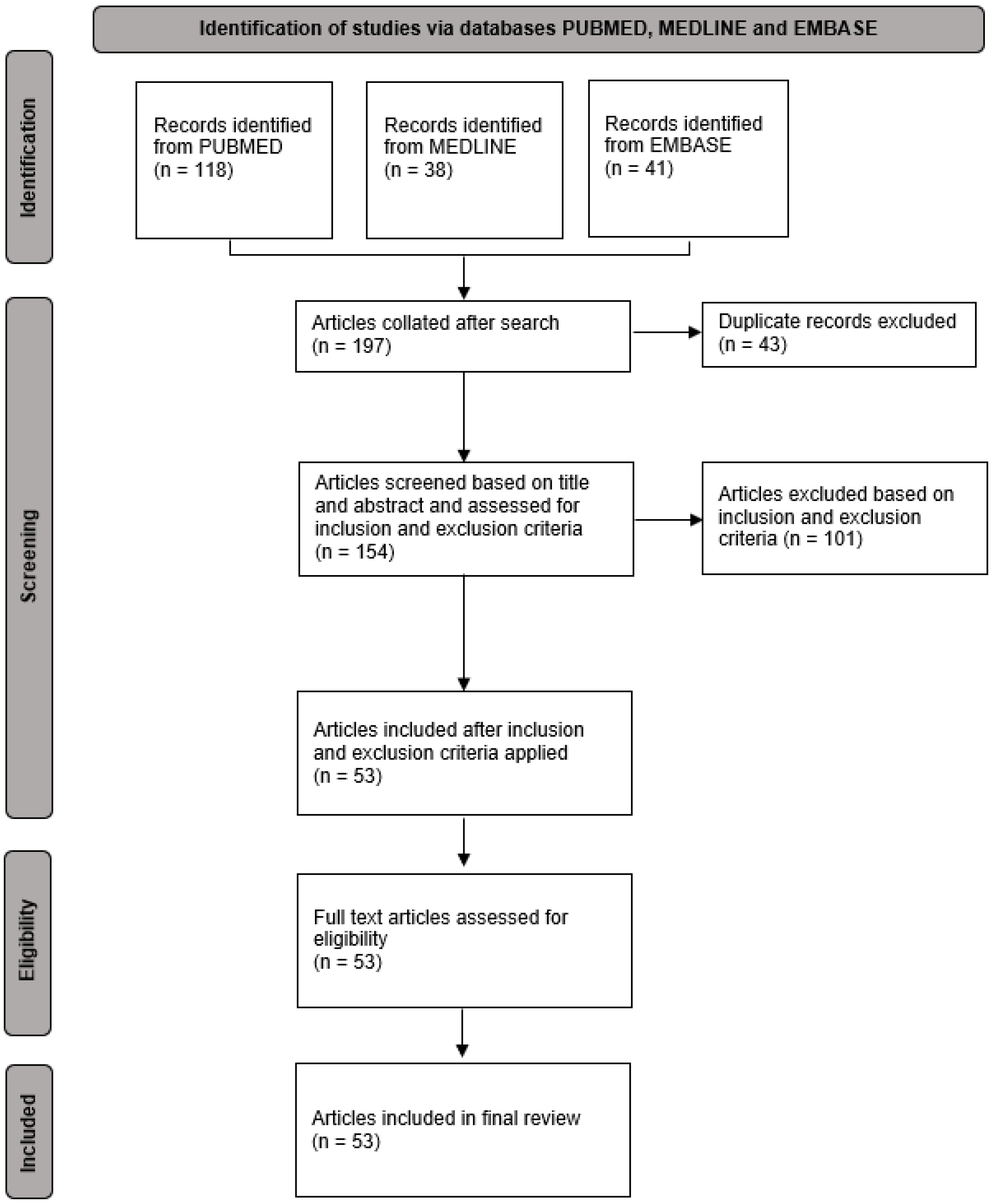
The initial search yielded a total of 197 studies which were screened for eligibility. Of these, 144 were excluded due to various reasons such as irrelevant topics, not reporting specific data on patients, or not being original articles. A total of 53 articles, all case reports and case series, met our inclusion criteria and were included in the final qualitative synthesis [2,7,8,9,10,11,12,13,14,15,16,17,18,19,20,21,22,23,24,25,26,27,28,29,30,31,32,33,34,35,36,37,38,39,40,41,42,43,44,45,46,47,48,49,50,51,52,53,54,55,56,57,58]. The PRISMA flow diagram of the study selection process is shown in Figure 1.

3.2. Patient Characteristics and Clinical Presentation

The 53 articles included a total of 56 patients with fungal balls causing ureteral or renal obstruction, with ages ranging from 26 to 80 (median age: 55 years). Of these, 34 (60.7%) were males and 22 (39.3%) were females. The most common presenting complaint was flank pain (n = 41, 73.2%), followed by fevers (n = 34, 60.7%) and haematuria (n = 10, 17.9%). Importantly, 44 (78.6%) of the patients were immunosuppressed due to pre-existing conditions or medications.

3.3. Diagnostic Procedures and Pathologic Findings

All patients were diagnosed based on the presence of yeast in urine culture or intra-operative tissue culture, and the presence of obstruction on imaging. The most common imaging modality was renal tract ultrasound (n = 30, 53.6%) and computed tomography (n = 29, 51.8%), followed by magnetic resonance imaging or intravenous pyelography.
Of the 56 patients included, 47 patients (83.9%) were diagnosed with an acute kidney injury with a median creatinine of 324 umol/L, and 35 patients (62.5%) had a leucocytosis.
The most common pathogen isolated were those of Candida species (n = 34, 60.7%), followed by Aspergillus (n = 19, 33.9%).

3.4. Treatment and Outcomes

While there was variation in the approach to management across case reports, antifungal therapy was a mainstay of therapy in the majority of patients (n = 52, 92.9%). The most common agents utilised were Amphotericin B (n = 24, 46.2%), followed by Fluconazole (n = 19, 26.5%), either in isolation or in combination with other antifungal or antibacterial agents.
Figure 1. PRISMA flow diagram for systematic review of fungal balls causing renal or ureteral obstruction.
Siuj 05 00034 g001
Additionally, there were differences in the route of antifungal therapy, with oral, intravenous, and local (i.e., nephrostomy tube or intravesical irrigation) administration methods used. The majority of cases utilised a combination of these routes (n = 33, 58.9%).
From a procedural standpoint, percutaneous nephrostomy tubes (n = 27, 48.2%) and ureteric stents/catheters (n = 26, 46.4%) were the most common interventions utilised (n = 27, 48.2%). In nine cases (16.1%), both percutaneous nephrostomy tubes and ureteric stents were employed. In a smaller subset of patients, more invasive urological interventions were required, ranging from percutaneous nephrolithotomy (PCNL) (n = 3, 5.3%) and pyelotomy/pyelostomy (n = 7, 12.3%) to nephrectomy (n = 5, 8.8%).
Most patients responded well to a combination of antifungal therapy and surgical management (n = 50, 89.3%), with resolution of obstruction and improvement in clinical parameters.
Treatment was unsuccessful in seven patients (12.5%), who died despite intervention. However, in these patients, mortality was secondary to other complications during the hospital admission (e.g., pulmonary embolism, circulatory failure, small bowel infarction, pneumonia) rather than a pure sequela of their fungal infection.

3.5. Quality of Included Studies

As per the Centre for Evidence-Based Medicine (CEBM) Critical Appraisal Tools, the quality of the included studies was varied. Many case reports and series lacked details about patient demographics, specific interventions, or precise clinical outcomes, contributing to the risk of bias. It is important to interpret the results keeping this limitation in mind.

4. Discussion

The results of this systematic review illustrate the need for a heightened clinical awareness of fungal balls as a cause of ureteral and renal obstructions, particularly in immunocompromised patients. This need is further underscored by the rising prevalence of UTIs, especially resistant strains. Equally, the potential for fungal balls to complicate the clinical course of these infections through avenues such as severe renal damage or life-threatening sepsis necessitates that they are targeted carefully and effectively. This compilation of available evidence points to an increased awareness of this pathology, particularly in immunocompromised patients, a demographic that appears more susceptible to this complication.
The study population consisted of a slightly higher proportion of males and spanned a wide age range (26–82 years), suggesting a broad patient profile for this condition. Most patients presented with symptoms such as flank pain and fevers, and in a minority of cases, haematuria. This non-specific symptomology, has significant overlap with those patients presenting with ascending bacterial urinary tract infections which adds to the diagnostic complexity of fungal balls causing ureteric obstruction. With 78.6% of individuals in this review identified as immunocompromised (most commonly due to diabetes), this highlights the need for heightened suspicion of fungal balls in patients that present with these symptoms and with evidence of obstruction with no clear cause.
The diagnosis was made with the finding of an obstruction on imaging in conjunction with urinary culture or intra-operative findings of fungus. Although the presence of obstruction was easily identified on either computed tomography or ultrasound, importantly, urine cultures demonstrating funguria were crucial in making the diagnosis and commencing timely management with the use of antifungals in conjunction with relieving obstruction. It is important to note that pre-operative imaging can often be misleading, as all patients in the review were diagnosed with obstruction on imaging, but none were diagnosed with obstruction due to fungal balls purely based on imaging. In addition, pre-operative imaging can often be misleading and mimic other reasons for obstructions such as ureteric or renal clots, radiolucent calculi, or obstruction of unknown cause.
From a therapeutic standpoint, there is a lack of uniform guidelines regarding optimal management. The majority of patients underwent multimodal therapy, combining systemic antifungal therapy—the local administration of antifungals with surgical drainage. The most common systemic antifungal agents used were Amphotericin B and Fluconazole. In the majority of cases that underwent local irrigation with antifungals, Amphotericin B was the most commonly used agent. The administration of Amphotericin B requires a specific dosing and exerts a concentration-dependent fungicidal activity and requires daily monitoring [59]. The administration, dosing, and monitoring should be guided by the clinician’s local guidelines and early consultation with their local infectious diseases team should be undertaken.
In terms of surgical interventions, percutaneous nephrostomy tubes and ureteric stents were mainstay approaches. In contrast, more invasive surgical procedures like percutaneous nephrolithotomy, pyelotomy/pyelostomy, and partial/total nephrectomies were relatively less frequently reported. This disparity could reflect an informal therapeutic hierarchy, where less invasive interventions are the first-line solutions, with more aggressive surgical measures reserved for refractory or severe cases.
The combination of antifungal therapy and surgical management was generally successful, as evidenced by a high success rate (87.5%). However, the substantial mortality rate (12.5%) despite treatment highlights the severe clinical implications of this condition. Even though the causes of death were often secondary to other complications, it is plausible that the fungal infection and associated interventions might have contributed to the overall burden of illness.
While not explicitly addressed in these case reports and series, it is prudent to review and potentially adjust diabetic medications, particularly those in the sodium–glucose cotransporter-2 (SGLT2) inhibitor class. These medications have been linked to an increased risk of urinary tract infections, and consideration should be given to their discontinuation [60]. Such decisions should be made in consultation with your local endocrinology team.
A significant limitation of this review is the inherent low level of evidence and high risk of bias in the included studies, primarily case reports and case series. Moreover, the variable quality of the studies, as per the Centre for Evidence-Based Medicine (CEBM) Critical Appraisal Tools, further restricts the conclusiveness of the findings. Due to the nature of the included studies, the evidence generated should be interpreted with caution.
This systematic review highlights the critical need for vigilance in suspecting fungal balls in patients with obstructive uropathy, especially in those with compromised immune systems. The treatment process requires a multifaceted approach, but effective management can markedly enhance patient outcomes. Currently, a multidisciplinary and phased therapeutic strategy is considered the most effective, emphasizing the early use of antifungals, decompression of the urinary collecting system, and engagement with local infectious diseases teams. We propose this treatment algorithm for addressing fungal balls that cause obstruction, with the understanding that any intervention for this complex condition should involve collaboration among the local infectious diseases team, urology team, and pharmacists (Figure 2).

Supplementary Materials

The following supporting information can be downloaded at: https://www.mdpi.com/article/10.3390/siuj5030034/s1.

Author Contributions

K.K.Y.C.—Conceptualisation, Methodology, Writing—Original draft, Writing—Review and Editing; M.K.—Conceptualisation, Methodology, Data curation; P.M.—Supervision. Writing—Review and Editing. All authors have read and agreed to the published version of the manuscript.

Funding

This research received no external funding.

Data Availability Statement

The data supporting the findings of this study are available within the article and/or its Supplementary Material.

Conflicts of Interest

The authors declare no conflicts of interest.

References

  1. Ng, K.P.; Kuan, C.S.; Kaur, H.; Na, S.L.; Atiya, N.; Velayuthan, R.D. Candida species epidemiology 2000–2013: A laboratory-based report. Trop. Med. Int. Health 2015, 20, 1447–1453. [Google Scholar] [CrossRef] [PubMed]
  2. Tan, W.P.; Turba, U.C.; Deane, L.A. Renal Fungus Ball: A Challenging Clinical Problem. Urol. J. 2016, 84, 113–115. [Google Scholar] [CrossRef] [PubMed]
  3. Kauffman, C.A. Diagnosis and management of fungal urinary tract infection. Infect. Dis. Clin. N. Am. 2014, 28, 61–74. [Google Scholar] [CrossRef] [PubMed]
  4. Pappas, P.G.; Kauffman, C.A.; Andes, D.R.; Clancy, C.J.; Marr, K.A.; Ostrosky-Zeichner, L.; Reboli, A.C.; Schuster, M.G.; Vazquez, J.A.; Walsh, T.J.; et al. Clinical Practice Guideline for the Management of Candidiasis: 2016 Update by the Infectious Diseases Society of America. Clin. Infect. Dis. 2016, 62, e1–e50. [Google Scholar] [CrossRef] [PubMed]
  5. Wise, G.J.; Silver, D.A. Fungal infections of the genitourinary system. J. Urol. 1993, 149, 1377–1388. [Google Scholar] [CrossRef] [PubMed]
  6. Sobel, J.D. Candiduria: A randomized, double-blind study of treatment with fluconazole and placebo. The National Institute of Allergy and Infectious Diseases (NIAID) Mycoses Study Group. Clin. Infect. Dis. 2000, 30, 19–24. [Google Scholar] [CrossRef]
  7. Kueter, J.C.; A MacDiarmid, S.; Redman, J.F. Anuria due to bilateral ureteral obstruction by Aspergillus flavus in an adult male. Urology 2002, 59, 601. [Google Scholar] [CrossRef] [PubMed]
  8. Zhu, L.; Chen, X.; Wu, J.; Yang, F.; Weng, X. Aspergillus vertebral osteomyelitis and ureteral obstruction after liver transplantation. Transpl. Infect. Dis. 2011, 13, 192–199. [Google Scholar] [CrossRef] [PubMed]
  9. Scerpella, E.G.; Alhalel, R. An unusual cause of acute renal failure: Bilateral ureteral obstruction due to Candida tropicalis fungus balls. Clin. Infect. Dis. 1994, 18, 440–442. [Google Scholar] [CrossRef]
  10. Paul, S.; Singh, V.; Sankhwar, S.; Garg, M. Case Report: Renal aspergillosis secondary to renal intrumentation in immunocompetent patient. BMJ Case Rep. 2013, 11, 2013. [Google Scholar]
  11. Napodano, R.J.; Bansal, S. Gross hematuria: A rare manifestation of primary renal candidiasis. Postgrad. Med. 1980, 67, 253–257. [Google Scholar] [CrossRef] [PubMed]
  12. Najafi, N.; Shokohi, T.; Basiri, A.; Parvin, M.; Yadegarinia, D.; Taghavi, F.; Hedayati, M.T.; Abdi, R. Aspergillus terreus-related ureteral obstruction in a diabetic patient. Iran. J. Kidney Dis. 2013, 7, 151–155. [Google Scholar] [PubMed]
  13. Mitchell, K.M. Coexisting bacterial pyelonephritis and bilateral ureteral fungus balls in a diabetic patient. Case Rep. 1990, 77, 596–599. [Google Scholar]
  14. Levin, D.L.; Zimmerman, A.L.; Ferder, L.F.; Shapiro, W.B.; Wax, S.H.; Porush, J.G. Acute renal failure secondary to ureteral fungus ball obstruction in a patient with reversible deficient cell-mediated immunity. Clin. Nephrol. 1975, 4, 202–210. [Google Scholar] [PubMed]
  15. Johnson, J.R.; Ireton, R.C.; Lipsky, B.A. Emphysematous pyelonephritis caused by Candida albicans. J. Urol. 1986, 136, 80–82. [Google Scholar] [CrossRef] [PubMed]
  16. Garcia, H.; Guitard, J.; Peltier, J.; Tligui, M.; Benbouzid, S.; Elhaj, S.A.; Rondeau, E.; Hennequin, C. Caspofungin irrigation through percutaneous calicostomy catheter combined with oral flucytosine to treat fluconazole-resistant symptomatic candiduria. J. Med. Mycol. 2015, 25, 87–90. [Google Scholar] [CrossRef]
  17. Eisenberg, R.L.; Hedgcock, M.W.; Shanser, J.D. Aspergillus mycetoma of the renal pelvis associated with ureteropelvic junction obstruction. J. Urol. 1977, 118, 466–467. [Google Scholar] [CrossRef] [PubMed]
  18. Dubert, M.; Loi, V.; Tligui, M.; Hertig, A. Unusual presentation of more common disease/injury: Rapidly progressive kidney failure induced by fungal mycelia obstructing indwelling ureteral stents. BMJ Case Rep. 2012, 2012, bcr2012007504. [Google Scholar] [CrossRef] [PubMed]
  19. Comings, D.E.; Turbow, B.A.; Callahan, D.H.; Waldstein, S.S. Obstructing aspergillus case to the renal pelvis. Report of a case in a patient having diabetes mellitus and Addison’s disease. Arch. Intern. Med. 1962, 110, 255–261. [Google Scholar] [CrossRef]
  20. Cohen, G.H. Obstructive Uropathy Caused By Ureteral Candidiasis. J. Urol. 1973, 110, 285–287. [Google Scholar] [CrossRef]
  21. Choi, H.; Kang, I.S.; Kim, H.S.; Lee, Y.H.; Seo, I.Y. Invasive Aspergillosis Arising from Ureteral Aspergilloma. Yonsei Med. J. 2011, 52, 866–868. [Google Scholar] [CrossRef]
  22. Béland, G.; Piette, Y. Urinary tract candidiasis: Report of a case with bilateral ureteral obstruction. Can. Med. Assoc. J. 1973, 108, 472–476. [Google Scholar] [PubMed]
  23. Beilke, M.; Kirmani, N. Candida Pyelonephritis Complicated by Fungaemia in Obstructive Uropathy. BJU Int. 1988, 62, 7–10. [Google Scholar] [CrossRef] [PubMed]
  24. Ahuja, A.; Aulakh, B.S.; Cheena, D.K.; Garg, R.; Singla, S.; Budhiraja, S. Aspergillus Fungal Balls Causing Ureteral Obstruction. Urol. J. 2009, 6, 127–129. [Google Scholar] [PubMed]
  25. Abdeljaleel, O.A.; Alnadhari, I.; Mahmoud, S.; Khachatryan, G.; Salah, M.; Ali, O.; Shamsodini, A. Treatment of Renal Fungal Ball with Fluconazole Instillation Through a Nephrostomy Tube: Case Report and Literature Review. Am. J. Case Rep. 2018, 19, 1179–1183. [Google Scholar] [CrossRef] [PubMed]
  26. Smaldone, M.C.; Cannon, G.M.; Benoit, R.M. Bilateral ureteral obstruction secondary to aspergillus bezoar. J. Endourol. 2006, 20, 318–320. [Google Scholar] [CrossRef]
  27. Shimada, S.; Nakagawa, H.; Shintaku, I.; Saito, S.; Arai, Y. Acute renal failure as a result of bilateral ureteral obstruction by Candida albicans fungus balls. Int. J. Urol. 2006, 13, 1121–1122. [Google Scholar] [CrossRef]
  28. Yang, C.-C.; Lee, M.-C.; Tzeng, Y.-H.; Chang, C.-C.; Tsai, S.-H. An unusual cause of renal failure: Bilateral ureteral obstruction by Candida tropicalis fungus balls. Kidney Int. 2007, 71, 373. [Google Scholar] [CrossRef] [PubMed]
  29. Ullah, S.R.; Jamshaid, A.; Zaidi, S.Z. Renal aspergilloma presenting with pelvi-ureteric junction Obstruction (PUJO). J. Pak. Med. Assoc. 2016, 66, 903–904. [Google Scholar]
  30. Walzer, Y.; Bear, R.A. Ureteral obstruction of renal transplant due to ureteral candidiasis. Urology 1983, 21, 295–297. [Google Scholar] [CrossRef]
  31. Vuruskan, H.; Ersoy, A.; Girgin, N.; Ozturk, M.; Filiz, G.; Yavascaoglu, I.; Oktay, B. An unusual cause of ureteral obstruction in a renal transplant recipient: Ureteric aspergilloma. Transplant. Proc. 2005, 37, 2115–2117. [Google Scholar] [CrossRef] [PubMed]
  32. Stein, J.; Latz, S.; Ellinger, J.; Fechner, G.; Safi, M.; Krausewitz, P.; Müller, S.; Weyer, K.; Müller, S.C. Fungaemia caused by obstructive renal candida bezoars leads to bilateral chorioretinitis: A case report. BMC Urol. 2018, 18, 21. [Google Scholar] [CrossRef] [PubMed]
  33. Veroux, M.; Corona, D.; Giuffrida, G.; Gagliano, M.; Tallarita, T.; Giaquinta, A.; Zerbo, D.; Cappellani, A.; Veroux, P.F. Acute renal failure due to ureteral obstruction in a kidney transplant recipient with candida albicans contamination of preservation fluid. Transpl. Infect. Dis. 2009, 11, 266–268. [Google Scholar] [CrossRef] [PubMed]
  34. O’Kane, D.; Kiosoglous, A.; Jones, K. Candida dubliniensis encrustation of an obstructing upper renal tract calculus. BMJ Case Rep. 2013, 2013. [Google Scholar] [CrossRef] [PubMed]
  35. Modi, P.; Goel, R. Synchronous endoscopic management of bilateral kidney and ureter fungal bezoar. Urol. Int. 2007, 78, 374–376. [Google Scholar] [CrossRef]
  36. Senneville, E.; Jana, F.; Gerard, Y.; Bourez, J.M.; Alfandari, S.; Chidiac, C.; Mouton, Y. Bilateral ureteral obstruction due to Saccharomyces cerevisiae Fungal Balls. Clin. Infect. Dis. 1996, 23, 636–637. [Google Scholar] [CrossRef]
  37. Onozawa, K.; Miyake, N.; Iwasaki, N.; Nishida, R.; Chong, Y.; Shimoda, S.; Shimono, N.; Akashi, K. A case of Candida albicans fungus balls in the urinary tract appeared during the course of antifungal treatment for Candida endophthalmitis. J. Infect. Chemother. 2015, 21, 687–690. [Google Scholar] [CrossRef]
  38. Sadagah, L.; Alharbi, M.; Alshomrani, M.; Almalki, A. Renal Allograft Aspergillus Infection Presenting With Obstructive Uropathy: A Case Report. Transplant. Proc. 2017, 49, 193–197. [Google Scholar] [CrossRef]
  39. Pérez-Arellano, J.; Angel-Moreno, A.; Belón, E.; Francès, A.; Santana, O.; Martín-Snchez, A. Isolated renoureteric aspergilloma due to Aspergillus flavus: Case report and review of the literature. J. Infect. 2001, 42, 163–165. [Google Scholar] [CrossRef]
  40. Keane, P.F.; McKENNA, M.; Johnston, S.R. Fungal Bezoar causing ureteric obstruction. BJU Int. 1993, 72, 247–248. [Google Scholar] [CrossRef]
  41. Schelenz, S.; Ross, C.N. Limitations of caspofungin in the treatment of obstructive pyonephrosis due to Candida glabrata infection. BMC Infect. Dis. 2006, 6, 126. [Google Scholar] [CrossRef] [PubMed]
  42. Kamel, G.; Stephan, A.; Barbari, A.; Kilani, H.; Karam, A.; Zeineh, S.; Salmeh, P.; Husni, R.; Mokhbat, J.; Khoury, J. Obstructive anuria due to fungal bezoars in a renal graft recipient. Transplant. Proc. 2003, 35, 2692–2693. [Google Scholar] [CrossRef] [PubMed]
  43. Krishnamurthy, R.; Aparajitha, C.; Abraham, G.; Shroff, S.; Sekar, U.; Kuruvilla, S. Renal aspergillosis giving rise to obstructive uropathy and recurrent anuric renal failure. Geriatr. Nephrol. Urol. 1998, 8, 137–139. [Google Scholar] [CrossRef] [PubMed]
  44. Liu, J.; Jiang, Y.; Liu, Z.; Song, Q.; Li, Z. A Case of Misdiagnosed as Upper Urinary Tract Obstruction Caused by the Fungal Ball. Infect. Drug Resist. 2022, 15, 6109–6114. [Google Scholar] [CrossRef] [PubMed]
  45. Lee, S.W. An aspergilloma mistaken for a pelviureteral stone on nonenhanced CT: A fungal bezoar causing ureteral obstruction. Korean J. Urol. 2010, 51, 216–218. [Google Scholar] [CrossRef] [PubMed]
  46. Bibler, M.R.; Gianis, J.T. Acute ureteral colic from an obstructing renal aspergilloma. Clin. Infect. Dis. 1987, 9, 790–794. [Google Scholar] [CrossRef] [PubMed]
  47. Gerridzen, R.G.; Wesley-James, T. Acute ureteral obstruction from candidal cystitis requiring bilateral percutaneous nephrostomies. Urology 1988, 32, 444–446. [Google Scholar] [CrossRef]
  48. Berlanga, G.A.; Machen, G.L.; Lowry, P.S.; Brust, K.B. Management of a Renal Fungal Bezoar Caused by Multidrug-Resistant Candida Glabrata. Bayl. Univ. Med. Cent. Proc. 2016, 29, 416–417. [Google Scholar] [CrossRef]
  49. Jiang, S.H.; Myers, R.L.; Walters, G.D. Candida tropicalis bezoar as a cause of obstructive nephropathy. Kidney Int. 2011, 79, 690. [Google Scholar] [CrossRef]
  50. Das, M.K.; Pakshi, R.R.; Kalra, S.; Elumalai, A.; Theckumparampil, N. Fungal Balls Mimicking Renal Calculi: A Zebra among Horses. J. Endourol. Case Rep. 2019, 5, 167–170. [Google Scholar] [CrossRef]
  51. Abbass, K.; Jaffery, A.T.; Markert, R.J.; Saklayen, M.G.; Khan, N.A. Fungus balls due to Candida tropicalis. Int. Urol. Nephrol. 2011, 44, 1293–1294. [Google Scholar] [CrossRef] [PubMed]
  52. Hiyama, Y.; Takahashi, S.; Masumori, N. Management of fungus balls as a result of Candida albicans. Int. J. Urol. 2018, 25, 635–636. [Google Scholar] [CrossRef] [PubMed]
  53. Di Paola, G.; Mogorovich, A.; Fiorini, G.; Cuttano, M.G.; Manassero, F.; Selli, C. Candida bezoars with urinary tract obstruction in two women without immunocompromising conditions. Sci. World J. 2011, 11, 1168–1172. [Google Scholar] [CrossRef] [PubMed]
  54. Arend, S.M.; Kuijper, E.J.; Vaal, B.J.; Fijter, J.W.; Wout, J.W.V. Successful treatment of fungus balls due to fluconazole-resistant Candida sake obstructing ureter stents in a renal transplant patient. Eur. J. Clin. Microbiol. Infect. Dis. 2005, 25, 43–45. [Google Scholar] [CrossRef] [PubMed]
  55. Abuelnaga, M.; Khoshzaban, S.; Badr, M.R.; Chaudry, A. Successful Endoscopic Management of a Renal Fungal Ball using Flexible Ureterorenoscopy. Case Rep. Urol. 2019, 2019, 9241928. [Google Scholar] [CrossRef] [PubMed]
  56. Arichi, N.; Yasumoto, H.; Ogawa, K.; Nagami, T.; Anjiki, H.; Nakamura, S.; Mitsui, Y.; Hiraoka, T.; Sumura, M.; Shiina, H. Case report of a ureteral obstruction by Candida Albicans Fungus balls detected by magnetic resonance imaging in kidney transplant recipient. Exp. Clin. Transplant. 2014, 12, 559–561. [Google Scholar] [PubMed]
  57. Zeineddine, N.; Mansour, W.; El Bitar, S.; Campitelli, M.; Mobarakai, N. Fungal Bezoar: A Rare Cause of Ureteral Obstruction. Case Rep. Infect. Dis. 2017, 2017, 6454619. [Google Scholar] [CrossRef] [PubMed]
  58. Yoon, Y.K.; Kang, E.H.; In, K.H.; Kim, M.J. Unilateral ureteral obstruction caused by Aspergillus, subgenus Nidulantes in a patient on steroid therapy: A case report and review of the literature. Med. Mycol. 2010, 48, 647–652. [Google Scholar] [CrossRef]
  59. Noor, A.; Preuss, C.V. Amphotericin B. [Updated 2024 Feb 28]. StatPearls [Internet]. Available online: https://www.ncbi.nlm.nih.gov/books/NBK482327/ (accessed on 29 July 2023).
  60. Tanrıverdi, M.; Baştemir, M.; Demirbakan, H.; Ünalan, A.; Türkmen, M.; Tanrıverdi, G. Association of SGLT-2 inhibitors with bacterial urinary tract infection in type 2 diabetes. BMC Endocr. Disord. 2023, 23, 211. [Google Scholar] [CrossRef]
Figure 2. Proposed treatment algorithm for fungal ball obstruction of the upper urinary tract. * In the absence of standardized protocols, we propose a local approach that involves administering amphotericin B deoxycholate, at a concentration of 50 mg per 1000 mL of sterile water, delivered at a rate of 40 mL per hour over a 24 h period. Follow-up imaging is recommended one week post administration to evaluate the therapeutic response. It is important to emphasise that the selection, administration, and duration of antifungal irrigation should be determined based on radiological findings and in consultation with the local infectious diseases team. # This algorithm is intended to assist clinicians in managing fungal ball obstruction of the urinary tract. It should not replace clinical judgment, local guidelines, or established local protocols.
Siuj 05 00034 g002
Disclaimer/Publisher’s Note: The statements, opinions and data contained in all publications are solely those of the individual author(s) and contributor(s) and not of MDPI and/or the editor(s). MDPI and/or the editor(s) disclaim responsibility for any injury to people or property resulting from any ideas, methods, instructions or products referred to in the content.