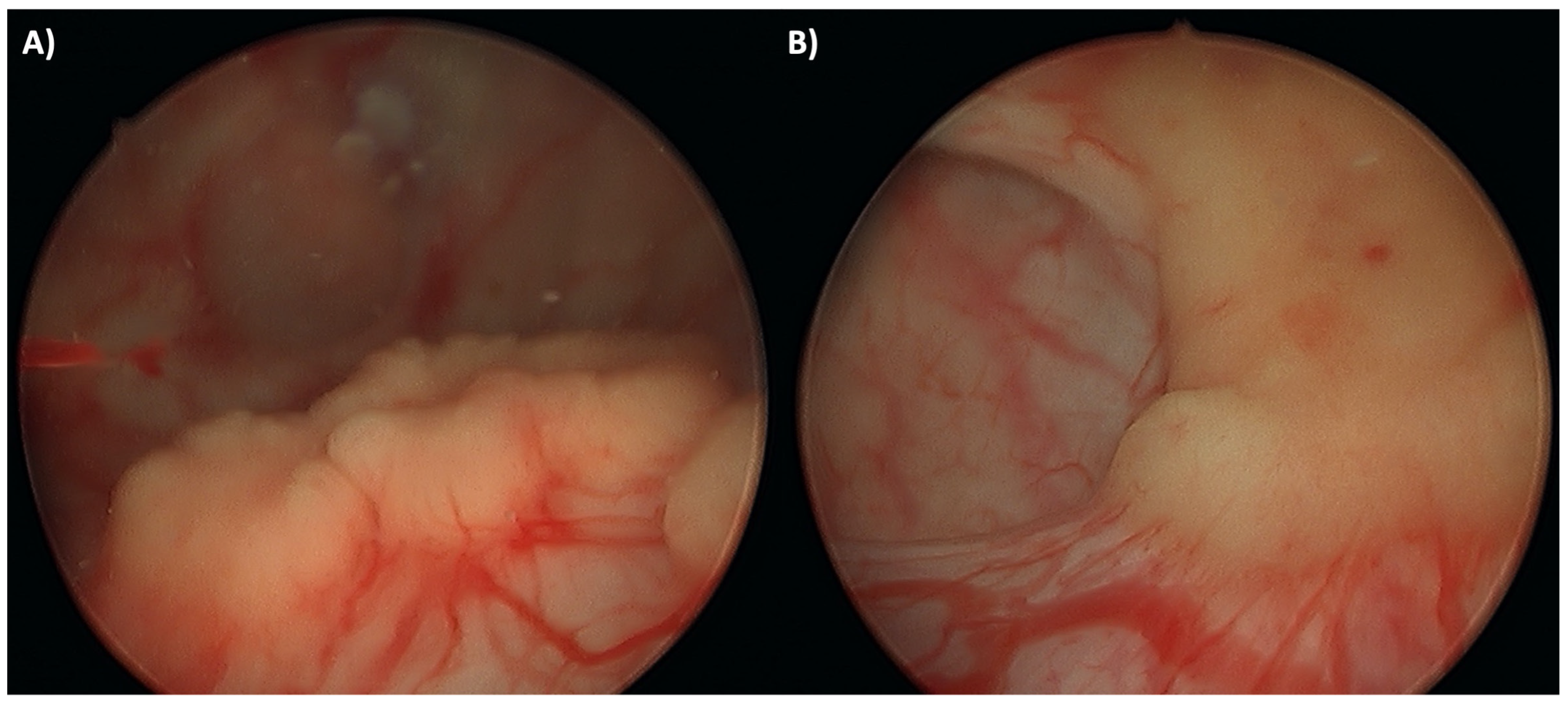
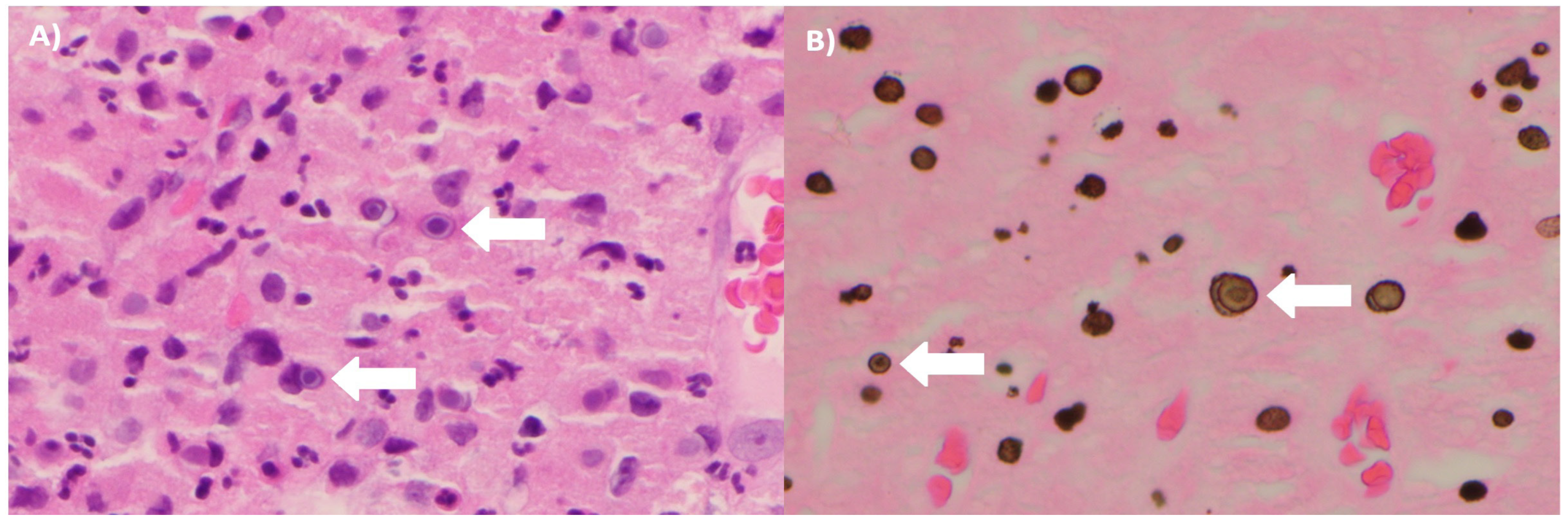
Malakoplakia Causing Poor Bladder Compliance and Bilateral Hydroureteronephrosis


Informed Consent Statement
Conflicts of Interest
References
- Kogulan, P.K.; Smith, M.; Seidman, J.; Chang, G.; Tsokos, M.; Lucey, D. Malakoplakia involving the abdominal wall, urinary bladder, vagina, and vulva: Case report and discussion of malakoplakia-associated bacteria. Int. J. Gynecol. Pathol. 2001, 20, 403–406. [Google Scholar] [CrossRef]
- Bylund, J.; Pais, V.M., Jr. A case of acute renal failure caused by bilateral, multifocal malacoplakia lesions of the bladder and ureters. Nat. Clin. Pract. Urol. 2008, 5, 516–519. [Google Scholar] [CrossRef]
- Sanchez, L.M.; Sanchez, S.I.; Bailey, J.L. Malacoplakia presenting with obstructive nephropathy with bilateral ureter involvement. Nat. Rev. Nephrol. 2009, 5, 418–422. [Google Scholar] [CrossRef] [PubMed]
- Cavallone, B.; Serao, A.; Audino, P.; Di Stasio, A.; Tiranti, D.; Vota, P. Bilateral hydroureteronephrosis with renal failure caused by malacoplakia. Urologia 2017, 85, 36–37. [Google Scholar] [CrossRef] [PubMed]
- Stamatiou, K.; Chelioti, E.; Tsavari, A.; Koulia, K.; Papalexandrou, A.; Efthymiou, E.; et al. Renal failure caused by malakoplakia lesions of the urinary bladder. Nephrourol. Mon. 2014, 6. [Google Scholar] [CrossRef] [PubMed]
This is an open access article under the terms of a license that permits non-commercial use, provided the original work is properly cited. © 2022 The Authors. Société Internationale d'Urologie Journal, published by the Société Internationale d'Urologie, Canada.
Share and Cite
Pham, C.T.; Edwards, M.; Chung, A.S.J.; Chalasani, V. Malakoplakia Causing Poor Bladder Compliance and Bilateral Hydroureteronephrosis. Soc. Int. Urol. J. 2022, 3, 281-282. https://doi.org/10.48083/QFCW5582
Pham CT, Edwards M, Chung ASJ, Chalasani V. Malakoplakia Causing Poor Bladder Compliance and Bilateral Hydroureteronephrosis. Société Internationale d’Urologie Journal. 2022; 3(4):281-282. https://doi.org/10.48083/QFCW5582
Chicago/Turabian StylePham, Cecile T., Melanie Edwards, Amanda S. J. Chung, and Venu Chalasani. 2022. "Malakoplakia Causing Poor Bladder Compliance and Bilateral Hydroureteronephrosis" Société Internationale d’Urologie Journal 3, no. 4: 281-282. https://doi.org/10.48083/QFCW5582
APA StylePham, C. T., Edwards, M., Chung, A. S. J., & Chalasani, V. (2022). Malakoplakia Causing Poor Bladder Compliance and Bilateral Hydroureteronephrosis. Société Internationale d’Urologie Journal, 3(4), 281-282. https://doi.org/10.48083/QFCW5582