Case Report

Association of Long-Term Speech Therapy and Neuromodulation in Primary Progressive Aphasia: Lessons from a Case Report

1
Department of Neuroscience and Movement Science, Faculty of Science and Medicine, University and Hospital of Fribourg, 1700 Fribourg, Switzerland
2
Department of Neurology, The First Affiliated Hospital, Sun Yat-sen University, Guangzhou 510080, China
3
Leenards Memory Center, Centre Hospitalier Universitaire Vaudois, 1011 Lausanne, Switzerland
*
Author to whom correspondence should be addressed.
Clin. Transl. Neurosci. 2022, 6(3), 17; https://doi.org/10.3390/ctn6030017
Submission received: 23 February 2022 / Revised: 19 June 2022 / Accepted: 14 July 2022 / Published: 19 July 2022
(This article belongs to the Section Neurorehabilitation)

Abstract

:
Primary progressive aphasia (PPA) is a neurodegenerative disorder with a progressive loss of language. Long-term support requires speech therapy but also individually set training programs. Here we propose an 8-month individualized speech-training program which alternates 3-week periods of transcranial direct current stimulation (tDCS) treatment with intensive daily language exercises and a 3-week period without tDCS treatment and a less intensive language exercise from home in a patient with non-fluent variant PPA (nfvPPA). The endpoints were the following: adherence to this program, language data after 8 months, questionnaires related to emotional valence, and brain volume changes. The results showed a persistent adherence after 8 months and a positive compliance reported by both the patient and the partner. The language evaluation showed a clinical stabilization. Moreover, a significant and positive influence of tDCS on mood was observed. This is, to our knowledge, the first ever published report of a combined neuromodulation and language training during the course of 8 months. Our finding suggests the feasibility of programs integrating hospital speech therapy, home training, and tDCS modulation in PPA. Further studies should be conducted in order to disentangle the contextual influences on language performance from the tDCS intervention effects and to address the observation of an initial improvement and a subsequent stabilization effect of language performances.

1. Introduction

Primary progressive aphasia (PPA) is a neurodegenerative disease whose prominent clinical feature is language impairment. We can further distinguish between non-fluent or agrammatic, logopenic, and semantic variants, which cover different neuropathological diagnostic categories (Alzheimer’s disease; frontotemporal dementia; and, more rarely, cortico-basal degeneration, progressive supranuclear palsy, and Pick’s disease) [1]. Non-fluent variant PPA (nfvPPA), in particular, is characterized [1] by (i) agrammatism and/or apraxia of speech; (ii) impaired comprehension of complex sentences and/or preserved comprehension of single words and/or preserved object knowledge; and (iii) left posterior frontal and insular atrophy or hypoperfusion/hypometabolism in the same region.
It has a progressive course of up to 14 years, with agrammatism as a prominent feature. However, in some cases, it can evolve with a dominant presence of apraxia of speech (DAoS) compared to agrammatism, or even solely with the presence of Progressive Primary Apraxia of Speech (PPAoS) in the absence of agrammatism [2]. PPAoS is characterized by a slow speech rate, sound distortions, segmentation of syllables, and increased difficulty with long words [3]. Particularly, naming capacities often show a marked decline (for example 20% decline over 2 years in naming tasks) [4].
Many therapies for PPA are based on word retrieval, using both phonological and semantic treatment, or sometimes both [5,6]. A classical therapy for nfvPPA is script training. Working on personalized scripts allows the patient to target the two main symptoms of nfvPPA, namely agrammatism and apraxia of speech [7]. Lastly, implementing an assistive augmentative communication (AAC) device is essential to ensure functional communication as the disease is progressing [8,9].
Several tDCS therapy strategies have been proposed to support communication and slow the diseases’ progression, which classically involves the left prefrontal region, including the inferior frontal gyrus. Besides the established focused speech therapy [7], several non-invasive brain stimulation techniques (NIBS) have been proposed, giving some interesting, although inconsistent, results in post-stroke aphasia [10]. Two of the most common NIBS that are being investigated in post-stroke aphasia research are TMS and tDCS. The rationale behind their application in aphasia rehabilitation is that both methods employ an electric field to influence neurons’ activity, with the first inducing neuron action potentials and the latter producing significant cortical network excitability [11]. Repetitive TMS applied to the left inferior and superior frontal gyrus has been shown to improve language production and spontaneous speech, together with increasing left parietotemporal and left frontal areas metabolism [12]. Moreover, tDCS has been essentially used in vascular aphasia, but degenerative nfvPPA has also been proposed for neuromodulation. Written naming [13] and verb comprehension were the only tasks that improved significantly after right parietal tDCS for 5 days, and improvement lasted 2 weeks. Furthermore, tDCS over the left IFG, along with written naming and spelling therapy for 2 weeks, gave rise to a greater improvement compared to the sham coupled with the same therapy. The generalization of tDCS gains persisted for 2 months. In another study, tDCS over the left IFG during 10–14 days improved written verb naming compared to sham [14,15]. Interestingly, a lower resting-state fMRI connectivity in the left IFG and temporal areas following tDCS stimulation compared to sham condition [16] has been observed, and the changes correlate with language outcomes’ improvement. However, most of the studies are of rather short therapy [13]. In their systematic review, Coemans et al. (2021) [17] reported that 13 of the 17 studies focused stimulation sites on the main locus of atrophy in nfvPPA patients either with an anode placed in the left inferior frontal, corresponding to the F7 electrode in the EEG 10–20 electrode position system or in the left temporoparietal areas, and the cathode placed on the right fronto-orbital area, forehead, or right shoulder. Interestingly, a longitudinal study showed improved sentence processing and neurocognitive process changes after 18 months of speech therapy in a patient with nfvPPA [18]. Moreover, these studies mainly focused on written and oral lexical access rather than measures of speech or language fluency. A systematic review [19] included 16 studies and analyzed the factors that predicted the maintenance of language performance over time. Continuous practice following therapy, length of the treatment, and frequency of the sessions were the different factors found.
A meta-analysis focusing on tDCS and TMS effects added to speech therapy showed interesting results concerning the use of tDCS for PPA [20]. A significant effect was found on language performance in patients who received tDCS or TMS compared with those who received the sham in addition to speech therapy. However, future research needs to be conducted, particularly to assess which variables can generate positive gains for persons with PPA.
Thus, there are a certain number of encouraging results in the tDCS stimulation of patients with nfvPPA. However, these data reflected rather short interventions [21], considering that nfvPPA develops over more than 6 years. For this reason, we were interested in assessing the effects of a longer therapeutic approach. In particular, our aim was to assess if our therapy is sustainable, that is, to be correctly followed if it lasts more than 6 months and if the initial improvement persists with longer speech training. Given this configuration, we adapted an initial hospital training followed by home training for the patient with alternation of 3 weeks with and without tDCS therapy. Some studies showed that patients with PPA may exhibit mood and/or behavioral symptoms, even in the early stages of the disease [22,23,24]. The study by Tarun et al. [22] found that nfvPPA patients are more likely to present with appetite changes. On the other hand, there were some studies indicating that tDCS can improve mood in healthy individuals [25,26,27]. To the best of our knowledge, no study on the effects of tDCS on mood in patients with PPA was reported. Our main interest was to confirm a good adherence to therapy, a language improvement, or at least non-deterioration, over 8 months, and to analyze mood and behavioral questionnaires with the prediction that such changes in subjective markers would be long lasting. Additionally, the imaging characteristic of nfvPPA is asymmetrical atrophy in cerebral regions involved in language processing, including the inferior frontal gyrus and the insula cortex, with extension to the superior temporal gyrus [28]. A study on longitudinal imaging changes in PPA indicated that the severest atrophic progression within a 1-year follow-up occurred in the basal ganglia in nfvPPA patients [29]. Hence, we were also interested in whether this patient presented nfvPPA-specific atrophy patterns in structural MRI and comparing brain morphometry before and after the program.

Case Report: Medical History and Clinical Examination

Mr. LA is a 75-years-old native English speaker and former right-handed security professional born in the USA and currently living in Switzerland. He complained of frustrating speech impediments, which started in 2016, for which he had speech therapy in Switzerland and in Hong Kong. In 2019, language had become more effortful and less accessible. During his first evaluation, language had become less spontaneous, and he spoke in shorter sentences, without agrammatism. The volume of his voice was weaker. He had the impression of having a frozen face and jaw without dysphagia. His wife reported difficulty in finding the words, sometimes with a stammer, stumbling on the first syllable. Writing was less spontaneous. Speaking French had become almost impossible. There were neither personality changes nor delusional symptoms. He did not mention memory or planning disorders, and the activities of daily living were preserved. He complained of some motor slowing, but neither muscle stiffness nor a history of cramps or myoclonus cramps was present. He had no difficulty with the fine motility of his hand to open boxes, cut meat, and shave. The axial motility to get up from a chair, turn over in bed, and put on shirts with sleeves or trousers was normal.
Neurological examination in September 2020 disclosed a well-oriented patient in time and space. His score on the Montreal Cognitive Assessment was 29/30, and 16/18 on the Frontal Assessment Battery. His working memory, recent verbal, and episodic memory were also well preserved. No difficulty in descriptions or semantic associations were observed. He recognized famous faces well. He had neither perceptual nor associative difficulties with visual gnosis. Meaningless gestures were at 4/6; pantomimes, and meaningful gestures were well executed by both hands. Mathematical calculation was rapid. There was no emotional lability. The rest of the neurological examination was normal. His brain MRI showed moderate bi-frontal atrophy.
The subject’s language capacity was tested in September 2020 and was deemed to be non-fluent, with short sentences and a tendency toward breathlessness, and he had a moderate apraxia of speech (AoS). Spontaneous and elicited speech were non-fluent yet informative, marked by latencies before speaking, with the need to close his eyes before speaking. Word naming did not show any paraphasia or semantic disorder. Comprehension, repetition, and written language were normal. Writing was a bit slow but not micrographic. Speech was characterized by a slowed speech rate (83 words/minute) and syntactically simple utterances (MLU of 9.4, with a mean of 9.2 [30]). There were also rare phonetic distortions. In a picture description test, iterations at the beginning of sentences were present. Noun and action naming were quantitatively preserved. Sentence and text reading was below standard, characterized by substitutions of visually related words (e.g., “then” for “the”), self-corrections, and disfluencies. The rest of language evaluation is summarized in Table 1.

2. Materials and Methods

2.1. Therapy Program—Design

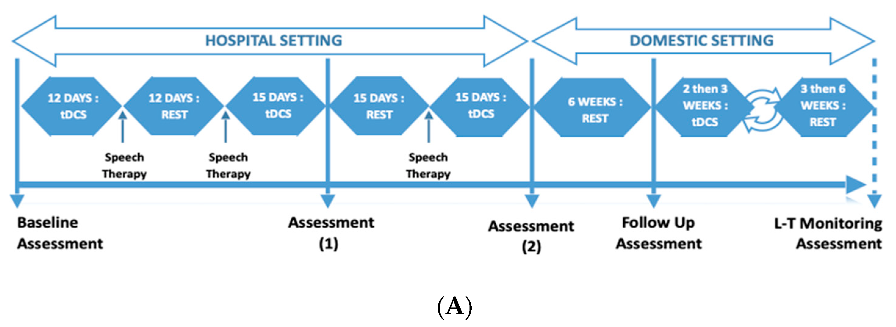
This was a repeated measures design where LA underwent three rounds of alternating tDCS and rest phases (Figure 1A). The tDCS phase comprised tDCS treatment accompanied with intense language exercises (15 days, 5 days per week), followed by the rest phase comprising minimal language exercises (15 days, alternative days) (see Figure 1B for measured key endpoints). This was over a total period of 3 months in a hospital setting. After the third round, there was a 6-week rest phase, which was followed by the second part of the program. This followed a similar design (alternating tDCS phase and rest phase) in a domestic setting and using a commercially available tDCS stimulator.

2.2. tDCS Treatment

During the tDCS phases, fixed at 5:00 p.m. in order to be consistent and control the influence of fatigue, LA received a twenty-minute tDCS stimulation while performing multiple types of language exercises. The rest phases consisted of 15 consecutive days of rest (or up to 6 weeks in the domestic setting part), with 30–45 min of language therapy every two days.
In the second part of the program, the procedures were similar, with the exception that the exercises and the stimulation were performed at LA’s home with occasional clinical contact (once per round) and the overall support of his partner. LA received anodal tDCS stimulation over the left IFG paired with language therapy, and tDCS was administered with the DC-Stimulator Plus®, NeuroConn. Two relatively small anodes and cathodes (5 × 5 cm2) were used to increase focality [31]. The electrodes were inserted in saline-soaked sponges (0.17 mL/cm2) before being positioned. The anode was placed over the left Broca Area (F7), individually located with the Beam A8 system [32]. The cathode was positioned over the right inferior frontolateral gyrus [23] (AF8). We decided to place the returning electrode (cathode) around AF8 in order to have a better control of the electric current flow over the surface of the left inferior frontal gyrus (Figure 2). In addition, the tDCS from NeuroConn is designed to place electrodes on brain and not on the shoulder because of the length of cable [33]. A standard 1.5 mA stimulation was first planned, but during the pilot trial, the patient asked to decrease the intensity. Thus, 1 mA intensity was chosen in order to compromise between the literature and the patient’s feelings [17]. The current was ramped up for 30 s at the beginning and ramped down for 15 s at the end of the stimulation.

2.3. Exercises during tDCS Phase

The tDCS phase consisted of 3 exercises during 20 min at each tDCS session: a tongue twister, text reading (300–500 words), and the creation of 30 sentences based on an image presented on a screen for 5 s. Directly after the tDCS phase, when the stimulation was over, LA continued the language exercises for another twenty-five minutes [15] and performed one of the 3 following exercises: (a) construction of 15 sentences based on 3 words one of which was a conjunction, (b) construction of 3 sentences based on an image for 15 different images, and (c) description of action words based on an image and construction of a sentence by using the same word for 20 images. The procedure for subsequent tDCS phases was identical to the first, except for the rest phase, which lasted 15 days. During the third tDCS phase, a diadochokinetic exercise was added as an alternative to the tongue-twister exercise. In addition, the length of text reading exercise was increased to 400–600 words. All other instructions were identical.
The difference between the tasks used during and after stimulation resides in LA’s main language impairment. Indeed, LA’s productions contained sound distortions because of his apraxia of speech. Plus, his speech rate was slow. The three tasks used during stimulation target those symptoms and allows him to work on coarticulation, phonetic accuracy, and fluency in response to a stimulus. The remaining tasks target syntactic processing, which is, at this point, slightly impaired (mild difficulties in sentence repetition and a grammatical error in a written picture description).
After each round, language and speech were assessed by using different tasks from those used during training. Baseline language assessment comprised elicit spontaneous speech samples and a text reading. To collect elicit spontaneous speech, we used a narration task (Frog, Where Are You?) and a picture description (Birthday Cake by Brookshire). For text reading, we used the text from YAA-R battery.
Using different tasks between training phases and baseline assessment allowed us to see to which point those demanding language exercises could generalize to a more natural context, meaning elicited speech. This is the reason why we chose a narrative task and a picture description one. This way, we were able to collect measures such as MLU and number of words per minute, measures being interesting for syntactic complexity and fluency. In addition, we chose a reading text to assess the number of mistakes made and fluency.

2.4. Exercises during Rest Phase

Every two days, LA performed one of the following exercises: construction of a sentence based on 3 words, one of which is a conjunction; creation of 3 sentences based on an image; and description of action words based on an image and construction of a sentence. He then performed the 3 following exercises: creation of a sentence based on image which was shown for 5–6 s, reading tongue twisters, and text reading of 300–500 words.

2.5. Image Acquisition, Processing, and Data Analysis

Whole-brain images were acquired on a 3T MR scanner (MAGNETOM Skyra, Siemens Healthcare, Erlangen, Germany) at 15-month intervals, 3 months before the beginning and two months after the end of the study, respectively. The acquisition protocol included high-resolution 3D T1-weighted sequence. MorphoBox prototype was used to automatically segment brain regions and measure regional brain volumes [34]. The total intracranial volume normalized volume (TIV-normalized volume) was adopted as the measurement of the volumes of different brain regions and compared with the normative ranges adjusted for head size, age, and sex. Brain regions with volume deviations from the normative ranges were considered to be abnormal [35].

2.6. Outcomes

2.6.1. Adherence to Treatment and Subjective Feeling

Considering the duration and the discipline required for the therapy, one of our objectives was to assess the patient’s motivation, his punctuality, his regularity of attendance in the sessions. It is also interesting to identify times when motivation may have fluctuated. We also added descriptions from both of the experimenters’ points of view and the neurologists, and the patient’s subjective feelings and expectations, as well as his partner’s.

2.6.2. Language Assessment

An exhaustive aphasiological evaluation was conducted in September 2020 before the therapy started, and one month after the end of the 8 months of intermittent training. The Psycholinguistic Assessment of Language Processing in Aphasia (PALPA) was used to evaluate word and sentence repetition, spoken words, and spoken sentence–picture matching, as well as logatom, word, and sentence reading [36]. Word and action-word retrieving abilities were assessed by using the Grémots battery. A task of picture description was employed to evaluate the mean length utterance. Picture matching (Pyramid and Palm Trees Test, 1992) and text reading performances (YAA-R/5) were evaluated [37]. Non-verbal semantic language (PPTT) was not assessed at the end of the intervention [38]. During the tDCS intervention, some additional follow-ups were conducted in order to assess the evolution of performances during the therapy. Categorical and phonological fluencies were assessed four times with Isaac’s test [39] and “P” letter, respectively [40], during clinical performed between December 2020 and September 2021. The number of words per minute and MLU were assessed by means of a picture narration task (Frog where are you?), as well as a picture description task (Birthday Cake by Nicholas & Brookshire, 1993). Text-reading performances (YAA-R/5) also were assessed [37] during the language assessments. Assessment Timepoints and Table of measured endpoints are shown in Figure 1.

2.6.3. Multidimensional Mood State Questionnaire (MDBF)

We used the English version of the MDBF, originally devised by Steyer et al. (1997) [41]. The questionnaire was adapted to the patients’ needs, and the scoring scale was augmented. Three scales are provided: good–bad, awake–tired, and calm–nervous. This questionnaire was measured daily.

2.6.4. Brain Atrophy

The atrophy rates of different brain regions between 2020 and 2021 were measured by calculating the % volume change with the following formula: (1-Normalized Volume_2021/Normalized Volume_2020) × 100. The brain regions found to be possibly atrophic in nfvPPA in the previous literature [29,42] and detected to be abnormally atrophic in this patient by MorphoBox prototype were taken as the regions of interest (ROIs). An atrophy rate of >4% (following data from Lombardi et al., 2021) was identified as abnormal.

2.6.5. Statistical Analysis

Statistical analyses were conducted with RStudio (2009–2019) version 1.2.5033. Language effects were evaluated by comparing September 2020 and September 2021 logopedic assessment. Language analyses on syntactic complexity were conducted by using TAASC [43,44]. Moreover, to have an idea of the effect size of the intervention, we conducted a Non-Overlap of All Pairs (NAP) analysis [45]. The NAP method allows us to compare the degree of measures overlap between two phases, with the assumption that the more the performances for the two phases overlap, the less likely the intervention had an effect [46]. The influence of tDCS stimulation on mood was investigated by the mean of t-tests in a repeated measures paradigm.

2.7. Written Informed Consent

The participant’s written consent, expressing his willingness to participate in the study, was obtained, and the study was approved by the Swiss Vaud cantonal human research ethics committee, Switzerland. See the Institutional Review Board Statement section.
3. Results

3.1. Adherence and Subjective Feelings

No session was missed, except when an accident prevented sessions during two weeks; thus, punctuality was always respected, and the questionnaires were duly completed. Furthermore, the patient was motivated and cooperative during all the months of therapy. The experimenters noticed his investment in the sessions, as well as his willingness to complete the tasks. It was observed that adherence and motivation (based on attendance and punctuality) were excellent throughout the 8 months, where it was considered relatively highest during the tDCS phases. Subjectively, it was expressed by the patient and observed by the partner that the motivation was highest during the tDCS phases compared to the rest phases, whether it was in hospital or domestic settings (Appendix A Table A1).

3.1.1. Subjective Feeling (LA)

Once I had heard of tDCS therapy, I was keenly interested to participate in this treatment. After the first round of treatment, I noticed a marked improvement in my speech: More ease in speaking and less difficulty in finding my words. The second and third rounds were less effective but nonetheless beneficial. It was not tiring and did not lose my enthusiasm in the treatments. As I mentioned above, I feel it has improved. The daily regimental structure of treatments did not bother me either.

3.1.2. Adherence (YB)

LA remained motivated throughout the 3 rounds in adhering to the protocol. It needs to be specified that this type of therapy, tDCS in combination with speech training would only suit persons who are disciplined and positively motivated. His motivation did not waver at all during this case study, though it appeared to be highest during the active tDCS phases.

3.2. Language Pre- and Post-tDCS Intervention Program

Language assessment was conducted at the end of the tDCS intervention (see Table 2). Clinical oral expression appreciation overall showed a maintenance of performance. Particularly, mean language utterance (MLU) at the end of the intervention improved from 9.4 to 10.1. Word repetition, word retrieval, and action-word retrieval were also stable. Categorical fluency showed a performance superior to the norm (z-score = 1.67), and the phonological performance (z-score = −1) remained stable and in the norm. Sentence repetition remained stable and below the norm. The other performances remained stable.
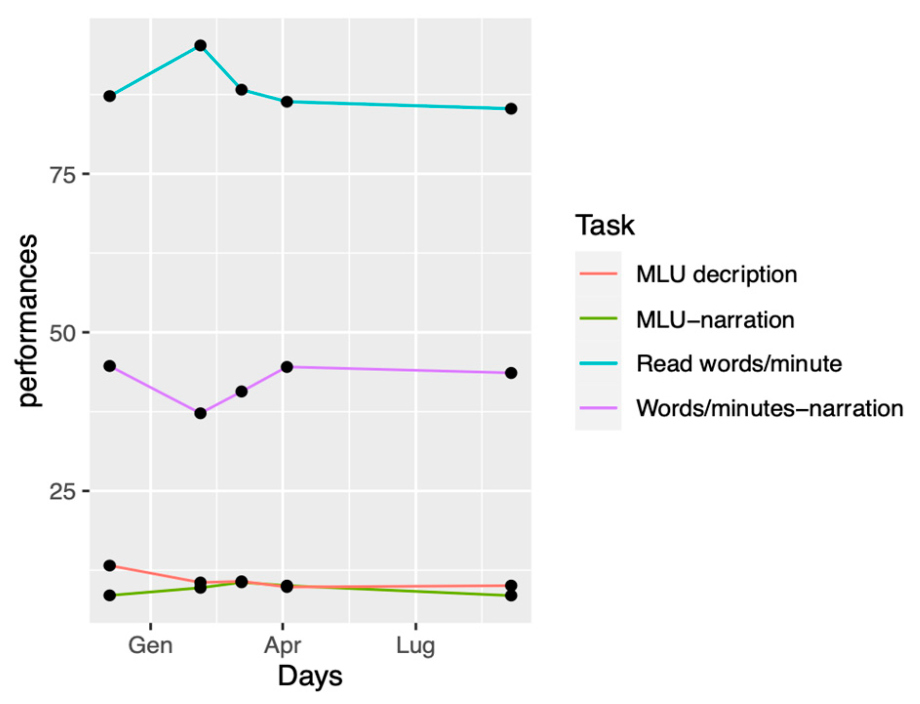
The NAP analysis was conducted on the follow-up data (Appendix B, Table A2). The NAP showed that 75% of MLU measures during tDCS were greater than the MLU measured before tDCS intervention (Figure 3). MLU in the picture description task showed no effect size. Reading performances in terms of words/per minute showed a medium effect size, where 50% of measures performed during the tDCS intervention were greater than values collected before the beginning of the therapy. No effect size was found for picture description task and words/minute in the picture narration task.
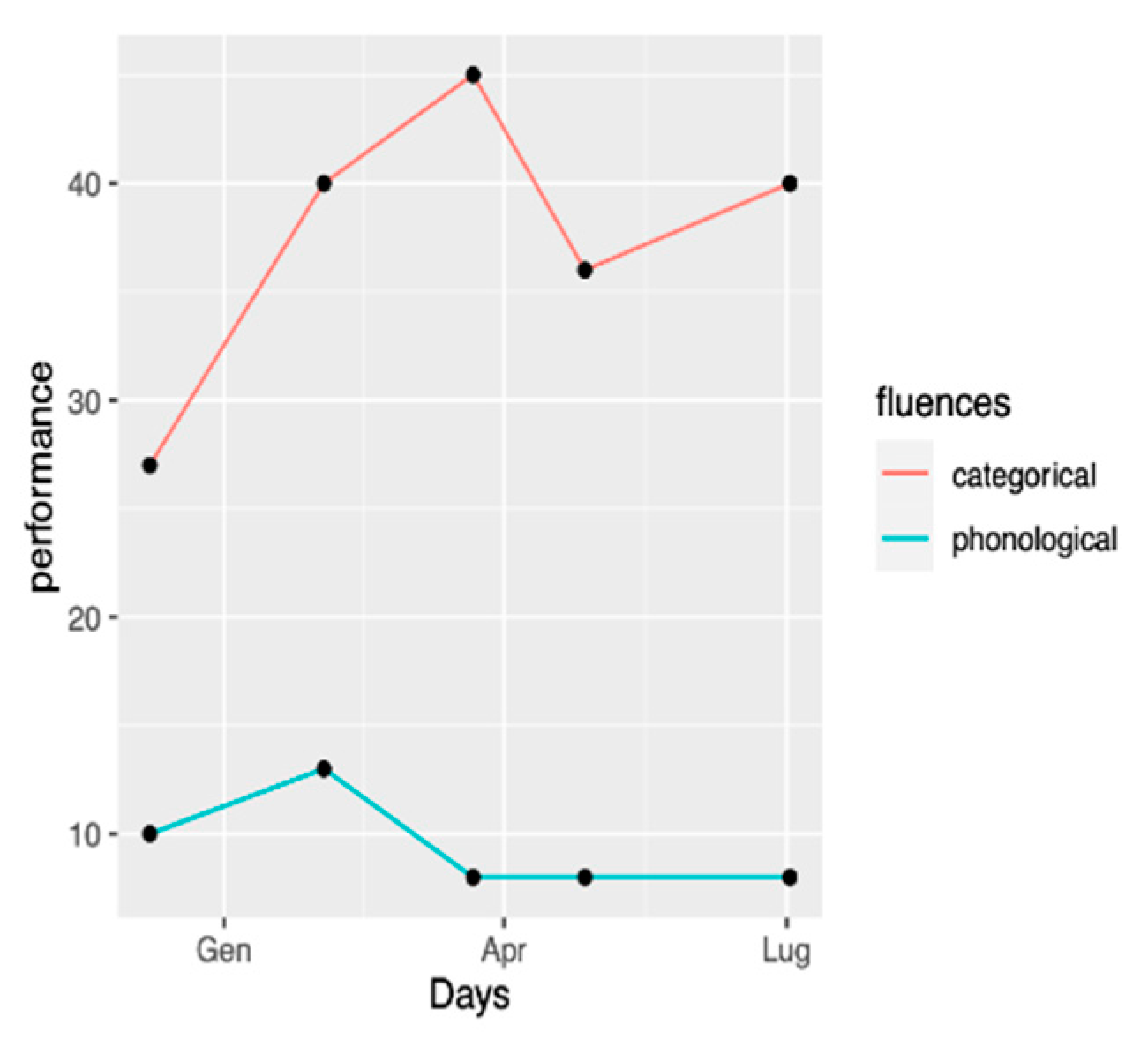
Regarding verbal fluency, a large effect size was found for categorical fluency, with all measures during tDCS intervention larger than performances found at the beginning of the intervention. A weak size-effect was found for phonological fluency, as 1 out of 4 of the tDCS phase values were improved compared to before the therapy performances (Appendix B, Figure A1).

3.3. Language Analyses after Each tDCS Round

After each round of tDCS, LA underwent a few language assessments, including a picture description task (Birthday Cake by Brookshire), a narration task (Frog, Where Are You?), and a text reading.
At each timepoint, we assessed syntactic complexity with the program TAASSC [43] and, more specifically, the Syntactic Complexity Analysis [44].
We chose three analyses that we found relevant: Mean Length of Utterance (MLU), Mean Length of T-Unit (MLT), and Mean Length of Clause (MLC). Results on the narration task (Frog, Where Are You?) can be found on Table 3, and the results for the picture description task can be found on Table 4. Mean Length of Utterance (MLU) is the number of words per sentence. MLC is the average words per clause. A clause includes a subject and a finite verb. It is an indicator of intra-clausal complexity [43]. Finally, a T-Unit is an independent clause and any dependent clause attached to it. It adds an extra specificity on syntactic complexity compared to MLC.
MLS, MLT, and MLC on a picture description and narration task (Frog, Where Are You?) are stable over time, which is a positive finding in a neurodegenerative disease. Statistically, none of the measures on each time point for the picture description and the narration task (Table 5 and Table 6) is significant.

3.4. Mood

Repeated t-tests were employed to investigate a difference in mood during the rest phases and the tDCS phases (see Table 7). The analyses revealed a significant difference in MDBF scales, t (20) = 2.82, p < 0.05, indicating that mood was significantly boosted (M = 41.72, SD = 11.15) when LA was given the tDCS therapy compared to when he was in the rest phase (M = 28.05, SD = 16.85).

3.5. Brain Morphometry from Structural MRI

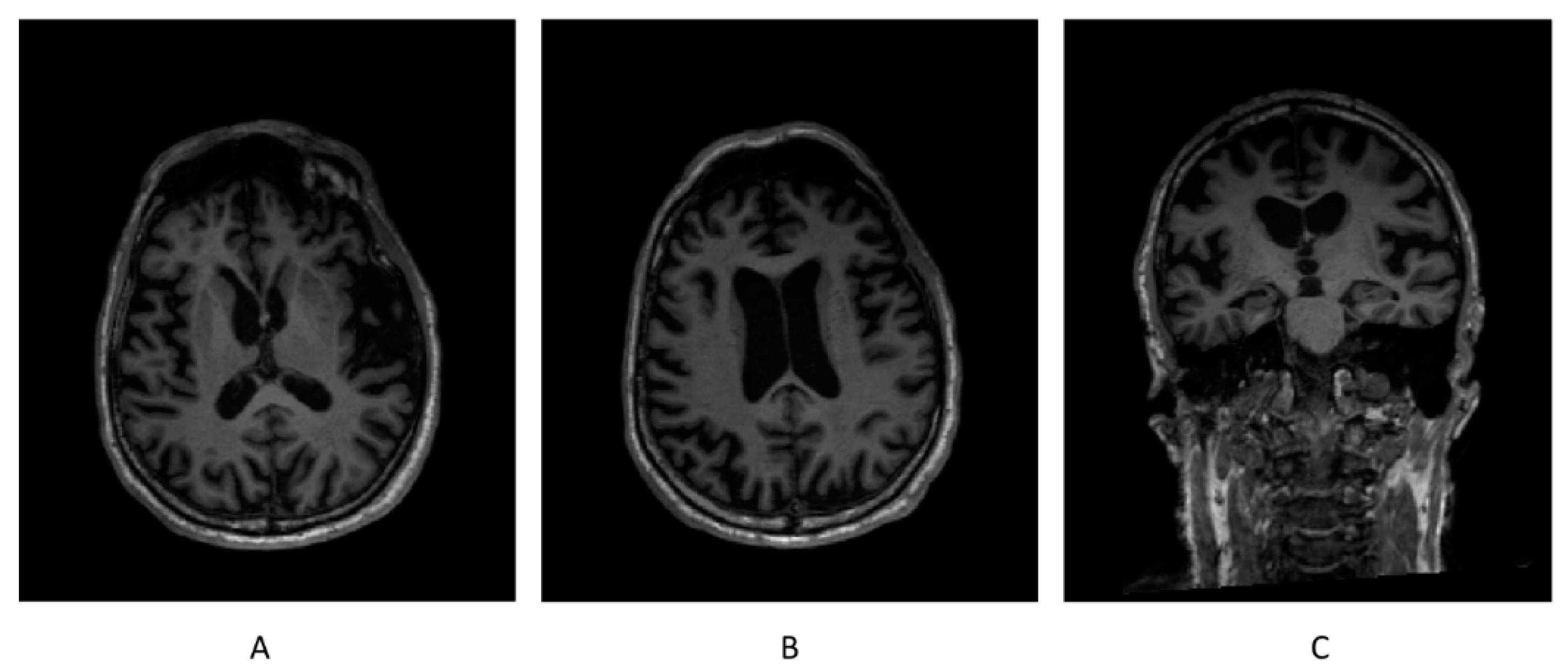
Brain morphometry from the structural MRI conducted before the protocol did not find any brain region having a volume out of the normative range. Brain morphometry from the structural MRI attained on 1 October 2021 revealed that the volumes of both gray matter (GM) and white matter (WM) in bilateral frontal lobes were below the normative ranges (TIV-normalized volume of GM in left frontal lobe = 5.28%, and normative range = 5.40–6.59%; TIV-normalized volume of GM in right frontal lobe = 5.28%, and normative range = 5.34–6.46%; TIV-normalized volume of WM in left frontal lobe = 4.07%, and normative range = 4.41–5.67%; and TIV-normalized volume of WM in right frontal lobe = 4.29%, and normative range = 4.37–5.55%), while the volumes of other brain regions were within the normative ranges (Figure 4).
The ROIs whose atrophy rates were calculated included both GM and WM in bilateral frontal lobes, bilateral parietal lobes, and bilateral temporal lobes, as well as bilateral insula and bilateral striatum. The atrophy rate of the whole brain was also calculated in contrast with the ROIs. As a result, both the GM and WM in the left frontal lobe, the GM in left parietal lobe and right insula, WM in right frontal lobe, and WM in right parietal lobe showed abnormally high atrophy rates (>4%). Neither the temporal lobes nor striatum had any significant atrophy rate (Table 8).

4. Discussion

This case report of 8 months of therapy alternating intensive training with tDCS and lighter training without neuromodulation in a patient with nfvPPA brought the following results: (i) LA showed high involvement over 8 months, with only 2 weeks of missed training (due to interferential medical condition) in 8 months (240 days); (ii) two evaluations one year apart showed stabilization of language performances; (iii) LA showed high involvement over 8 months, with only 3 days of missed training (due to interferential medical condition) in 8 months (240 days); and (iv) a significant atrophy occurred in bilateral frontal lobes, including GM and WM.

4.1. Adherence to Treatment and Mood

One of the aims of the present case study was to examine the clinical applicability of long-term tDCS therapy by observing the patient’s adherence to the intervention. Moreover, the patient was directly involved by integrating his expectations, as well as his subjective feelings concerning the different phases of the treatment and its effects. These data inform us about the feasibility of this type of treatment. The patient did not report any decrease in motivation or fatigue during the treatment, suggesting that its duration and intensity were appropriate. These findings are consistent with our observations. In other words, both the patient and his partner were completely committed to the treatment. It is important to note that the patient is self-motivated and rigorous by nature, characteristics that are essential to the success of a project requiring a high level of involvement such as this.
The patient described a positive subjective feeling regarding the language aspects, such as more ease in speaking and a decrease in the difficulty of finding words. The experimenters also noted improved fluency of speech over the course of the sessions and the patient’s spontaneity in discussing a variety of topics. The analysis results revealed a difference in mood depending on whether LA was in a tDCS or a rest phase, with the first condition showing a significantly higher mood compared to the latter. Of interest, it has been shown that tDCS on the frontal region can induce an improvement in mood [27]. In addition, regular social interactions with experimenters and physical activity in conjunction with travel to sessions could also have positively influenced mood. Therefore, we must take into consideration that other factors besides tDCS therapy may have had an impact on the improvement in mood experienced by the patient.
In response to our objective of a long-term feasibility analysis, we affirm that the therapy was sustainable and correctly followed despite its intensity and duration of 8 months. The results showed a persistent adherence after 8 months and a positive compliance reported by both the patient and partner. The language evaluation showed a clinical stabilization of language performance. Moreover, a significant and positive influence of tDCS on mood was observed. This is, to our knowledge, the first-ever published report of a combined neuromodulation and language training during the course of 8 months. A quite long longitudinal treatment study for PPAoS, involving repeated exercises over the span of 8 months, was performed. In the first month, it occurred twice per week, for 45 min. During the remaining 7-month treatment period, sessions occurred at home practice for 10-to-15 min sessions 3 times per week. The results indicated that language was stable over 8 months [47]. Neuromodulation was not performed in Meyer’s study, which focused on phonological and orthographic treatment. Our finding tends to confirm the feasibility of programs integrating hospital speech therapy, home training, and tDCS modulation in primary progressive aphasia. Nonetheless, it is necessary to highlight that this type of therapy requires considerable discipline, motivation, and endurance and would thereby not be suitable for all patients.

4.2. Language Effect

Language performances were assessed over a period of one year. Particularly, we were interested in evaluating the long-lasting duration effects of tDCS stimulation on language performances. Comparisons between September 2020 and September 2021 assessments showed an overall stabilization of language performances. On the one hand, overall comprehension performances remain unattained, and such a stabilization cannot be associated with the therapy regimen. However, several outcomes were stable after the intervention. Studies suggest that improvement, generalization, and maintenance of those gains occur in different PPA forms [7,48]. The particularity of our case report is that LA showed maintenance of non-treated items, suggesting a possible role of tDCS in effect generalization, as suggested by Cotelli et al. [49].
Previous studies using tDCS treatments over nfvPPA, using left inferior frontal or tempo parietal anodal stimulations, and right fronto-orbital or facial area, show an effect on word-retrieving performances [49]. Particularly, oral naming productions [13,49], as well as written naming productions [13,50], are reported to improve greatly after tDCS sessions, and this is the case for a number of studies. This is not the case in our study, as the patient showed a comparable score of performances in oral picture naming tasks during all the therapy and because word retrieving was not impaired (see Table 1 and Table 2). However, the finding of a stabilization of overall performances is encouraging, as PPA usually shows a relatively rapid degeneration of clinical performances over a short period of time [4]. Phonological and semantic fluency also remained in the range of normality [39,40], showing a certain stabilization. Previous studies have already shown the beneficial effect on verbal fluency, with the categorical one improving more than the phonological one [51] in healthy subjects and in nfvPPA patients [52] of tDCS stimulation over the left IFG. Moreover, LA’S mean MLS for the picture narration task, which was assessed during each follow-up session, in comparison to groups found in Tetzloff et al.’s (2018) study (addressing nfvPPA, DAoS, and PPAoS), seems to be nearer to the latter group compared to DAoS or nfvPPA [2]. This pattern is coherent with LA’s language assessment: there is no evidence for agrammatism but rather mild AoS. These results are also coherent with LA’s complaints about effortful speech. A benefit of tDCS on oral expression rate and mean language utterance have already been suggested in other studies [52]. Interestingly, and in contrast to picture narration task, LA’s MLU for the picture description task did not show any improvement, but rather a stabilization. To our knowledge, there is no study addressing MLU dissociations in these two tasks. Nevertheless, it could be a result of a gain of flexibility for narration, as it is possible to construct inferences and to implement more articulated utterances compared to a description task. Further addressing this question would be appropriate from a methodological point of view.
Concerning the duration of the protocol, LA trained for 72 tDCS sessions over 8 months. Such a protocol can be considered a training and maintenance protocol [11] and is much longer than other maintenance protocols used with success in post-stroke aphasia (5 to 10 stimulation sessions executed over a period from 2 weeks to 2 months, [53,54], in the treatment of Primary Progressive Aphasia (1 to 15 combined neuromodulation and speech therapy session [20], in stroke-induced motor impairment (maximum of 40 sessions [55], or in chronic migraines (up to 12) [56]. We have proposed such protocols for the eventual long-term effects of stimulations and to avoid the paradoxical exhausting effect though eventual too-intense long-term training. Further studies are needed to identify which the long-term attrition to neuromodulation protocols’ parameters of stimulation (for example monohemispheric or bihemispheric stimulation location, stimulation intensity, session duration, interval between sessions, and time of the protocols) can be adapted maximize or at least confirm a consistent effect. We need to individualize tDCS protocols in order to couple language therapy with neuromodulation.
Finally, it is also worth noting that, over the course of the intervention, the biggest clinical improvement was seen during the first part of the training, when LA underwent tDCS stimulation in a hospital setting. The second part of the therapy seems to be less effective, as language performances stabilized. The question arises whether training at home is less effective rather than being immersed in a hospital setting. Nevertheless, it is also true that this issue has been observed in other tDCS short-term interventions on nfvPPA patients [21] that did not show an improvement during the second session of tDCS. Therefore, our data are in line with such a mild clinical improvement of language performances in the early phase of therapy, followed by a certain stabilization. Further investigating is needed in order to assess the causes that underlie the depletion of this effect and to eventually disentangle contextual influences on language outcomes.

4.3. Volume Change of the Brain

The bilateral frontal atrophy involving GM and WM found in the second session of MRI was partly consistent with the structural neuroimaging characteristics of nfvPPA [29,57]. Such works addressing this in the literature also reported atrophy of temporal lobes and striatum; however, the volumes of these regions were still within the normative ranges in LA’s case. Further longitudinal structural MRI measurements are needed to trace the possible atrophy of the other brain regions in the future. Atrophy rates in different brain regions between the two sessions of MRI might predict the pathological atrophy emerging in the future. The brain regions with abnormally high atrophy rates in this case overlapped with the results of previous studies. For instance, notable atrophy rates in both GM and WM in the left frontal lobe were consistent with the findings by Lombardi et al. [29], but not the normal atrophy rates of bilateral temporal lobes and striatum. Such variability between this single case and other cohort studies is reasonable, since the most convincing method of morphometry, Voxel-based morphometry (VBM), cannot be applied in a single case. Nevertheless, the atrophy rate of the whole brain in this patient, 2.38%, was consistent with the result of a cohort study, 2.6 (1.2)%/year in the patients with nfvPPA [58], suggesting that the tDCS stimulations had no structural effect. In addition, we also found from the morphometry results of 2021 that the TIV-normalized volumes of the majority of the brain regions in the left hemisphere were smaller than the opposite regions in the right hemisphere, except the frontal GM and the parietal WM. This imaging characteristic of nfvPPA has been stated in the previous meta-analysis [57]. Interestingly, this asymmetry was not so pronounced in the morphometry results of 2020.

4.4. Limits and Future Directions

As this case report took shape during the development and the implementation of the intervention, different limitations are present. First of all, the design of the intervention provided a period of stimulation in a hospital setting and a second period at the patient’s home. Therefore, it did not allow us to evaluate the influence of the context on the language performances. In fact, the stabilization of the improvement of language deficits overlaps with the beginning of the home-setting stimulation. It is also very important to note that, in this study, the design did not allow us to control for the influence of a possible placebo effect [59]. Mood susceptibility to placebo effects has been observed in different contexts [60], including language outcomes [15]. A double-blind designed study which integrates a sham tDCS condition would be appropriate to disentangle improvements due to tDCS from placebo-induced effects.

5. Conclusions

Here we presented a tDCS intervention on a patient diagnosed with nfvPPA for 8 months in a mixed (hospital and domestic) setting. Besides confirming a non-degradation of certain aspects of language, we wanted to investigate the feasibility of a longer tDCS intervention compared to already published ones. We also shed some light on the patient’s subjective and positive feelings reported during the stimulation period. This case report, together with previously published studies, suggests an interesting role of tDCS for PPA as a complementary approach to focused speech therapy. Future double-blind designed studies should be considered to investigate the different influences of both context and tDCS on language stabilization.

Author Contributions

Conceptualization, J.-M.A., A.H., Y.B. and L.A.; methodology, J.-M.A.; formal analysis, A.H., Y.Z., L.G. and L.C.; investigation, A.H., Y.Z., L.C., L.G. and E.D.; data curation, A.H., Y.Z. and L.G.; writing—original draft preparation, L.G.; writing—review and editing, L.C. and E.D.; supervision, J.-M.A.; funding acquisition, J.-M.A. and Y.Z. All authors have read and agreed to the published version of the manuscript.

Funding

This research was funded by the Swiss National Science Foundation (SNF grant 325130_182594), the Institutional support of University of Fribourg and Lausanne Hospital and China Scholarship Council (CSC) (No. 202006385028) to Yifan Zheng.

Institutional Review Board Statement

The participant’s written consent was obtained, and the study was approved by the Swiss Vaud cantonal human research ethics committee, Switzerland, reference no. 2019-02178.

Informed Consent Statement

Informed consent was obtained from all subjects involved in the study. Written informed consent was obtained from the patient(s) to publish this paper.

Data Availability Statement

The data presented in this study are available upon request from the corresponding author. The data are not publicly available due to their containing information that could compromise the privacy of the research participant.

Acknowledgments

We are grateful to Michael Mouthon for the reviewing of the MRI section.

Conflicts of Interest

The authors declare no conflict of interest. The funders had no role in the design of the study; in the collection, analyses, or interpretation of data; in the writing of the manuscript; or in the decision to publish the results.

Appendix A

Table A1. Adherence during the different therapy phases. During hospital setting, tDCS phases (labeled tDCS) were performed at the hospital, while rest phases (labeled “rest”) took place at home. During the home setting, all phases took place at home. Note that the patient described improved mood and motivation during the stimulation phases.
Date of SessionAttendancePunctualityPatient/Partner’s Comments
HOSPITAL SETTING
ROUND 1 (tDCS Phase)8 to 23 Dec 2020100%1700 +/− 15 minMinor itching at the side of electrodes. At end of tDCS session, LA and partner felt his ease of speaking was improved and more motivated.
ROUND 2 (Rest Phase)25 Dec 2020 to 10 Jan 2021100%1400 +/− 2 hLA was not as motivated to do training as he was in hospital setting and without tDCS treatment.
ROUND 2 (tDCS Phase)11 to 29 Jan 2021100%1700 +/− 15 minMood and motivation to speak is noted during tDCS sessions compared to rest phases.
ROUND 3 (Rest Phase)4 to 21 Feb 2021100%1400 +/− 2 hSimilar as with previous rest phase, not as motivated.
ROUND 3 (tDCS Phase)22 Feb to 12 Mar 2021100%1700 +/− 15 minMood and motivation to speak is continually noted during tDCS sessions compared to rest phases.
DOMESTIC SETTING
Long-Term I (6 weeks Rest Phase)13 Mar to 21 Apr 202165%1400 +/− 24 hTraining stopped for 2 weeks: Recovery from an accident resulting in small fracture of patient’s right hand. Mood and motivation did go down during these 2 weeks.
Long-term I (2 weeks tDCS Phase)28 Apr to 7 May 2021100%1130 +/− 30 minDuring these sessions, feeling that mood and motivation does improve, though not as clearly marked as in hospital setting.
Long-term II (6 weeks Rest Phase)11 May to 18 Jun 2021100%1400 +/− 2 hFeeling that 6 weeks rest from tDCS was too long. Motivation wanes does wane, though adherence is still maintained.
Long-Term II (2 weeks tDCS Phase)21 Jun to 1 Jul 2021100%1130 +/− 30 minAfter this session, decided to resume to 3 weeks rest phase and 3 weeks of tDCS, as per in hospital setting.
Long-Term III (3 weeks Rest Phase)2 to 20 Jul 2021No Training Agreed to stop language training during 3 weeks rest phase.
Long-Term III (3 weeks tDCS Phase)21 Jul to 5 Aug 2021100%1130+/− 30 minOverall good adherence to tDCS sessions, whether hospital or domestic setting. Preference is 3 weeks on and 3 weeks off.

Appendix B

Table A2. Mean language utterance and fluency assessed during each tDCS follow-up.
12.2002.2103.2104.2109.21
Picture narration (MLU)8.569.7410.6210.18.54
Picture narration (words/minute)44.737.2540.6944.5543.6
Picture description (MLU)13.2510.5710.759.8810.1
Text reading aloud (words/minute)87.2595.2388.2586.3685.24
Figure A1. Phonological and categorical fluencies.
Ctn 06 00017 g0a1

References

  1. Gorno-Tempini, M.L.; Hillis, A.E.; Weintraub, S.; Kertesz, A.; Mendez, M.; Cappa, S.F.; Ogar, J.M.; Rohrer, J.D.; Black, S.; Boeve, B.F.; et al. Classification of primary progressive aphasia and its variants. Neurology 2011, 76, 1006–1014. [Google Scholar] [CrossRef] [PubMed] [Green Version]
  2. Tetzloff, K.A.; Utianski, R.L.; Duffy, J.R.; Clark, H.M.; Strand, E.A.; Josephs, K.A.; Whitwell, J.L. Quantitative Analysis of Agrammatism in Agrammatic Primary Progressive Aphasia and Dominant Apraxia of Speech. J. Speech Lang. Hearing Res. 2018, 61, 2337–2346. [Google Scholar] [CrossRef] [PubMed]
  3. Jung, Y.; Duffy, J.R.; Josephs, K.A. Primary Progressive Aphasia and Apraxia of Speech. Skull Base 2013, 33, 342–347. [Google Scholar] [CrossRef] [PubMed] [Green Version]
  4. Rogalski, E.; Cobia, D.; Harrison, T.M.; Wieneke, C.; Weintraub, S.; Mesulam, M.M. Progression of language decline and cortical atrophy in subtypes of primary progressive aphasia. Neurology 2011, 76, 1804–1810. [Google Scholar] [CrossRef] [Green Version]
  5. Jokel, R.; Graham, N.L.; Rochon, E.; Leonard, C. Word retrieval therapies in primary progressive aphasia. Aphasiology 2014, 28, 1038–1068. [Google Scholar] [CrossRef]
  6. Savage, S.A.; Ballard, K.J.; Piguet, O.; Hodges, J.R. Bringing words back to mind—Improving word production in semantic dementia. Cortex 2013, 49, 1823–1832. [Google Scholar] [CrossRef] [PubMed]
  7. Henry, M.L.; Hubbard, H.I.; Grasso, S.M.; Mandelli, M.L.; Wilson, S.M.; Sathishkumar, M.T.; Fridriksson, J.; Daigle, W.; Boxer, A.L.; Miller, B.L.; et al. Retraining speech production and fluency in non-fluent/agrammatic primary progressive aphasia. Brain 2018, 141, 1799–1814. [Google Scholar] [CrossRef] [Green Version]
  8. Pattee, C.; Von Berg, S.; Ghezzi, P. Effects of alternative communication on the communicative effectiveness of an individual with a progressive language disorder. Int. J. Rehabil. Res. 2006, 29, 151–153. [Google Scholar] [CrossRef]
  9. Góral-Półrola, J.; Półrola, P.; Mirska, N.; Mirski, A.; Herman-Sucharska, I.; Pąchalska, M. Augmentative and Alternative Communication (AAC) for a patient with a nonfluent/agrammatic variant of PPA in the mutism stage. Ann. Agric. Environ. Med. 2016, 23, 182–192. [Google Scholar] [CrossRef] [Green Version]
  10. Biou, E.; Cassoudesalle, H.; Cogné, M.; Sibon, I.; De Gabory, I.; Dehail, P.; Aupy, J.; Glize, B. Transcranial direct current stimulation in post-stroke aphasia rehabilitation: A systematic review. Ann. Phys. Rehabil. Med. 2019, 62, 104–121. [Google Scholar] [CrossRef]
  11. Lefaucheur, J.-P.; Wendling, F. Mechanisms of action of tDCS: A brief and practical overview. Neurophysiol. Clin. Neurophysiol. 2019, 49, 269–275. [Google Scholar] [CrossRef] [PubMed]
  12. Pytel, V.; Cabrera-Martín, M.N.; Delgado-Álvarez, A.; Ayala, J.L.; Balugo, P.; Delgado-Alonso, C.; Yus, M.; Carreras, M.T.; Carreras, J.L.; Matías-Guiu, J.; et al. Personalized repetitive transcranial magnetic stimulation for primary progressive aphasia. J. Alzheimer’s Dis. 2021, 84, 151–167. [Google Scholar] [CrossRef] [PubMed]
  13. Fenner, A.S.; Webster, K.T.; Ficek, B.; Frangakis, C.E.; Tsapkini, K. Written Verb Naming Improves After tDCS Over the Left IFG in Primary Progressive Aphasia. Front. Psychol. 2019, 10, 1396. [Google Scholar] [CrossRef] [Green Version]
  14. Cotelli, M.; Manenti, R.; Petesi, M.; Brambilla, M.; Cosseddu, M.; Zanetti, O.; Miniussi, C.; Padovani, A.; Borroni, B. Treatment of Primary Progressive Aphasias by Transcranial Direct Current Stimulation Combined with Language Training. J. Alzheimer’s Dis. 2014, 39, 799–808. [Google Scholar] [CrossRef]
  15. Tsapkini, K.; Webster, K.T.; Ficek, B.N.; Desmond, J.E.; Onyike, C.U.; Rapp, B.; Frangakis, C.E.; Hillis, A.E. Electrical brain stimulation in different variants of primary progressive aphasia: A randomized clinical trial. Alzheimer’s Dement. Transl. Res. Clin. Interv. 2018, 4, 461–472. [Google Scholar] [CrossRef] [PubMed]
  16. Ficek, B.N.; Wang, Z.; Zhao, Y.; Webster, K.T.; Desmond, J.E.; Hillis, A.E.; Frangakis, C.; Faria, A.V.; Caffo, B.; Tsapkini, K. The effect of tDCS on functional connectivity in primary progressive aphasia. NeuroImage Clin. 2018, 19, 703–715. [Google Scholar] [CrossRef]
  17. Coemans, S.; Struys, E.; Vandenborre, D.; Wilssens, I.; Engelborghs, S.; Paquier, P.; Tsapkini, K.; Keulen, S. A Systematic Review of Transcranial Direct Current Stimulation in Primary Progressive Aphasia: Methodological Considerations. Front. Aging Neurosci. 2021, 13, 568. [Google Scholar] [CrossRef] [PubMed]
  18. Thompson, C.K.; Barbieri, E.; Mack, J.E.; Wilkins, A.; Xie, K.Y. Plasticity of sentence processing networks: Evidence from a patient with agrammatic variant of primary progressive aphasia (PPA). Neurocase 2021, 27, 39–56. [Google Scholar] [CrossRef]
  19. Cadório, I.; Lousada, M.; Martins, P.; Figueiredo, D. Generalization and maintenance of treatment gains in primary progressive aphasia (PPA): A systematic review. Int. J. Lang. Commun. Disord. 2017, 52, 543–560. [Google Scholar] [CrossRef]
  20. Nissim, N.R.; Moberg, P.J.; Hamilton, R.H. Efficacy of Noninvasive Brain Stimulation (tDCS or TMS) Paired with Language Therapy in the Treatment of Primary Progressive Aphasia: An Exploratory Meta-Analysis. Brain Sci. 2020, 10, 597. [Google Scholar] [CrossRef]
  21. Wang, J.; Wu, D.; Chen, Y.; Yuan, Y.; Zhang, M. Effects of transcranial direct current stimulation on language improvement and cortical activation in nonfluent variant primary progressive aphasia. Neurosci. Lett. 2013, 549, 29–33. [Google Scholar] [CrossRef] [PubMed]
  22. Singh, T.D.; Duffy, J.R.; Strand, E.A.; Machulda, M.M.; Whitwell, J.L.; Josephs, K.A. Neuropsychiatric symptoms in primary progressive aphasia and apraxia of speech. Dement. Geriatr. Cogn. Disord. 2015, 39, 228–238. [Google Scholar] [CrossRef] [Green Version]
  23. Modirrousta, M.; Price, B.H.; Dickerson, B.C. Neuropsychiatric symptoms in primary progressive aphasia: Phenomenology, pathophysiology, and approach to assessment and treatment. Neurodegener. Dis. Manag. 2013, 3, 133–146. [Google Scholar] [CrossRef] [Green Version]
  24. Medina, J.; Weintraub, S. Depression in Primary Progressive Aphasia. J. Geriatr. Psychiatry Neurol. 2007, 20, 153–160. [Google Scholar] [CrossRef]
  25. Austin, A.; Jiga-Boy, G.M.; Rea, S.; Newstead, S.; Roderick, S.; Davis, N.J.; Clement, R.M.; Boy, F. Prefrontal electrical stimulation in non-depressed reduces levels of reported negative affects from daily stressors. Front. Psychol. 2016, 7, 315. [Google Scholar] [CrossRef] [PubMed] [Green Version]
  26. Newstead, S.; Young, H.; Benton, D.; Jiga-Boy, G.; Sienz, M.L.A.; Clement, R.M.; Boy, F. Acute and repetitive fronto-cerebellar tDCS stimulation improves mood in non-depressed participants. Exp. Brain Res. 2018, 236, 83–97. [Google Scholar] [CrossRef] [PubMed] [Green Version]
  27. Marshall, L.; Mölle, M.; Hallschmid, M.; Born, J. Transcranial Direct Current Stimulation during Sleep Improves Declarative Memory. J. Neurosci. 2004, 24, 9985–9992. [Google Scholar] [CrossRef] [Green Version]
  28. Bisenius, S.; Neumann, J.; Schroeter, M.L. Validating new diagnostic imaging criteria for primary progressive aphasia via anatomical likelihood estimation meta-analyses. Eur. J. Neurol. 2016, 23, 704–712. [Google Scholar] [CrossRef]
  29. Lombardi, J.; Mayer, B.; Semler, E.; Anderl-Straub, S.; Uttner, I.; Kassubek, J.; Diehl-Schmid, J.; Danek, A.; Levin, J.; Fassbender, K.; et al. Quantifying progression in primary progressive aphasia with structural neuroimaging. Alzheimer’s Dement. 2021, 17, 1595–1609. [Google Scholar] [CrossRef]
  30. Kemper, S.; Sumner, A. The structure of verbal abilities in young and older adults. Psychol. Aging 2001, 16, 312–322. [Google Scholar] [CrossRef]
  31. Nitsche, M.A.; Cohen, L.G.; Wassermann, E.M.; Priori, A.; Lang, N.; Antal, A.; Paulus, W.; Hummel, F.; Boggio, P.S.; Fregni, F.; et al. Transcranial direct current stimulation: State of the art 2008. Brain Stimul. 2008, 1, 206–223. [Google Scholar] [CrossRef] [PubMed]
  32. Beam, W.; Borckardt, J.J.; Reeves, S.T.; George, M.S. An efficient and accurate new method for locating the F3 position for prefrontal TMS applications. Brain Stimul. 2009, 2, 50–54. [Google Scholar] [CrossRef] [Green Version]
  33. Radman, N.; Britz, J.; Buetler, K.; Weekes, B.S.; Spierer, L.; Annoni, J.-M. Dorsolateral prefrontal transcranial direct current stimulation modulates language processing but does not facilitate overt second language word production. Front. Neurosci. 2018, 12, 490. [Google Scholar] [CrossRef] [PubMed]
  34. Yan, S.; Qian, T.; Maréchal, B.; Kober, T.; Zhang, X.; Zhu, J.; Lei, J.; Li, M.; Jin, Z. Test-retest variability of brain morphometry analysis: An investigation of sequence and coil effects. Ann. Transl. Med. 2020, 8, 12. [Google Scholar] [CrossRef] [PubMed]
  35. Roche, A.; Maréchal, B.; Kober, T.; Krueger, G.; Hagmann, P.; Maeder, P.; Meuli, R. Assessing brain volumes using MorphoBox prototype. Magn. Flash 2017, 68, 33–38. [Google Scholar]
  36. Kay, J.; Lesser, R.; Coltheart, M. Psycholinguistic assessments of language processing in aphasia (PALPA): An introduction. Aphasiology 1996, 10, 159–180. [Google Scholar] [CrossRef]
  37. Warmington, M.; Stothard, S.E.; Snowling, M.J. Assessing dyslexia in higher education: The York adult assessment battery-revised. J. Res. Spéc. Educ. Needs 2013, 13, 48–56. [Google Scholar] [CrossRef]
  38. Howard, D.; Patterson, K.E. The Pyramids and Palm Trees Test; Thames Valley Test Company: Suffolk, UK, 1992; Available online: https://eprints.ncl.ac.uk (accessed on 1 September 2020).
  39. Isaacs, B.; Akhtar, A.J. The set test: A rapid test of mental function in old people. Age Ageing 1972, 1, 222–226. [Google Scholar] [CrossRef]
  40. Raoux, N.; Le Goff, M.; Auriacombe, S.; Dartigues, J.-F.; Amieva, H. Fluences verbales sémantiques et littérales: Normes en population générale chez des sujets âgés de 70 ans et plus issus de la cohorte PAQUID. Rev. Neurol. 2010, 166, 594–605. [Google Scholar] [CrossRef]
  41. Steyer, R.; Schwenkmezger, P.; Notz, P.; Eid, M. Der Mehrdimensionale Befindlichkeitsfragebogen (MDBF) [The Multidimensional Mood State Questionnaire]; Hogrefe: Göttingen, Germany, 1997. [Google Scholar]
  42. Gorno-Tempini, M.L.; Dronkers, N.F.; Rankin, K.P.; Ogar, M.J.; Phengrasamy, L.; Rosen, H.J.; Johnson, J.K.; Weiner, M.W.; Miller, B.L. Cognition and anatomy in three variants of primary progressive aphasia. Ann. Neurol. 2004, 55, 335–346. [Google Scholar] [CrossRef] [Green Version]
  43. Kyle, K. Measuring Syntactic Development in L2 Writing: Fine Grained Indices of Syntactic Complexity and Usage-Based Indices of Syntactic Sophistication; Georgia State University: Atlanta, GA, USA, 2016. [Google Scholar] [CrossRef]
  44. Lu, X. Automatic analysis of syntactic complexity in second language writing. Int. J. Corpus Linguist. 2010, 15, 474–496. [Google Scholar] [CrossRef]
  45. Parker, R.I.; Vannest, K. An Improved Effect Size for Single-Case Research: Nonoverlap of All Pairs. Behav. Ther. 2009, 40, 357–367. [Google Scholar] [CrossRef] [PubMed]
  46. Krasny-Pacini, A.; Evans, J. Single-case experimental designs to assess intervention effectiveness in rehabilitation: A practical guide. Ann. Phys. Rehabil. Med. 2018, 61, 164–179. [Google Scholar] [CrossRef] [PubMed]
  47. Meyer, A.M.; Tippett, D.C.; Turner, R.S.; Friedman, R.B. Long-Term maintenance of anomia treatment effects in primary progressive aphasia. Neuropsychol. Rehabil. 2018, 29, 1439–1463. [Google Scholar] [CrossRef]
  48. Flurie, M.; Ungrady, M.; Reilly, J. Evaluating a Maintenance-Based Treatment Approach to Preventing Lexical Dropout in Progressive Anomia. J. Speech Lang. Hearing Res. 2020, 63, 4082–4095. [Google Scholar] [CrossRef]
  49. Cotelli, M.; Manenti, R.; Ferrari, C.; Gobbi, E.; Macis, A.; Cappa, S.F. Effectiveness of language training and non-invasive brain stimulation on oral and written naming performance in Primary Progressive Aphasia: A meta-analysis and systematic review. Neurosci. Biobehav. Rev. 2020, 108, 498–525. [Google Scholar] [CrossRef] [PubMed]
  50. Zhao, K.; Zhao, J.; Zhang, M.; Cui, Q.; Fu, X. Neural Responses to Rapid Facial Expressions of Fear and Surprise. Front. Psychol. 2017, 8, 761. [Google Scholar] [CrossRef] [PubMed]
  51. Cattaneo, Z.; Pisoni, A.; Papagno, C. Transcranial direct current stimulation over Broca’s region improves phonemic and semantic fluency in healthy individuals. Neuroscience 2011, 183, 64–70. [Google Scholar] [CrossRef]
  52. Gervits, F.; Ash, S.; Coslett, H.B.; Rascovsky, K.; Grossman, M.; Hamilton, R. Transcranial direct current stimulation for the treatment of primary progressive aphasia: An open-label pilot study. Brain Lang. 2016, 162, 35–41. [Google Scholar] [CrossRef] [Green Version]
  53. Lefaucheur, J.-P.; Antal, A.; Ayache, S.S.; Benninger, D.H.; Brunelin, J.; Cogiamanian, F.; Cotelli, M.; De Ridder, D.; Ferrucci, R.; Langguth, B.; et al. Evidence-based guidelines on the therapeutic use of transcranial direct current stimulation (tDCS). Clin. Neurophysiol. 2017, 128, 56–92. [Google Scholar] [CrossRef]
  54. Meinzer, M.; Darkow, R.; Lindenberg, R.; Flöel, A. Electrical stimulation of the motor cortex enhances treatment outcome in post-stroke aphasia. Brain 2016, 139, 1152–1163. [Google Scholar] [CrossRef]
  55. Van Hoornweder, S.; Vanderzande, L.; Bloemers, E.; Verstraelen, S.; Depestele, S.; Cuypers, K.; van Dun, K.; Strouwen, C.; Meesen, R. The effects of transcranial direct current stimulation on upper-limb function post-stroke: A meta-analysis of multiple-session studies. Clin. Neurophysiol. 2021, 132, 1897–1918. [Google Scholar] [CrossRef]
  56. Hodaj, H.; Payen, J.-F.; Mick, G.; Vercueil, L.; Hodaj, E.; Dumolard, A.; Noëlle, B.; Delon-Martin, C.; Lefaucheur, J.-P. Long-term prophylactic efficacy of transcranial direct current stimulation in chronic migraine. A randomised, patient-assessor blinded, sham-controlled trial. Brain Stimul. 2022, 15, 441–453. [Google Scholar] [CrossRef] [PubMed]
  57. Schroeter, M.L.; Raczka, K.; Neumann, J.; von Cramon, D.Y. Towards a nosology for frontotemporal lobar degenerations—A meta-analysis involving 267 subjects. NeuroImage 2007, 36, 497–510. [Google Scholar] [CrossRef]
  58. Rohrer, J.D.; Clarkson, M.J.; Kittus, R.; Rossor, M.N.; Ourselin, S.; Warren, J.D.; Fox, N.C. Rates of Hemispheric and Lobar Atrophy in the Language Variants of Frontotemporal Lobar Degeneration. J. Alzheimer’s Dis. 2012, 30, 407–411. [Google Scholar] [CrossRef] [PubMed] [Green Version]
  59. Fonteneau, C.; Mondino, M.; Arns, M.; Baeken, C.; Bikson, M.; Brunoni, A.R.; Burke, M.J.; Neuvonen, T.; Padberg, F.; Pascual-Leone, A.; et al. Sham tDCS: A hidden source of variability? Reflections for further blinded, controlled trials. Brain Stimul. 2019, 12, 668–673. [Google Scholar] [CrossRef] [PubMed]
  60. Oken, B.S. Placebo effects: Clinical aspects and neurobiology. Brain 2008, 131, 2812–2823. [Google Scholar] [CrossRef] [PubMed] [Green Version]
Figure 1. (A) Design of the study. (B) Measured key endpoints.
Ctn 06 00017 g001aCtn 06 00017 g001b
Figure 2. The current distribution in the brain with the anode (1) placed over the left Broca Area (F7) and the cathode (2) placed over the right inferior frontolateral gyrus (AF8).
Ctn 06 00017 g002
Figure 3. MLU and words/minute reading performances.
Ctn 06 00017 g003
Figure 4. High-resolution 3D T1-weighted sequence of the structural brain MRI on 1 October 2021. (A) Axial slice displaying normal bilateral basal ganglia, (B) axial slice displaying atrophic bilateral frontal lobes, and (C) coronal slice displaying normal bilateral hippocampus.
Ctn 06 00017 g004
Table 1. Language evaluation.
DomainTask
Oral expressionPicture description
Word repetition (PALPA)
Sentence repetition (PALPA)
Word retrieving (Grémots)
Action word retrieving (Grémots)
Oral comprehensionSpoken word-picture matching (PALPA)
Spoken sentence-picture matching (PALPA)
Written expressionWritten picture description
Reading aloudWord reading (PALPA)
Logatom reading (PALPA)
Sentence reading (PALPA)
Text reading (YAA-R/5)
Nonverbal semanticPicture-matching (PPTT)
Oral-lingual-facial praxis
Table 2. Table resuming language performances before and at the end of the tDCS intervention program.
DomainTaskResults September 2020Results September 2021
Oral expressionPicture descriptionMLU = 9.4MLU = 10.1
Word repetition (PALPA)80/8080/80
Sentence repetition (PALPA)32/36 *33/36 *
Word retrieving (Grémots)32/36, c1032/36, c25
Action word retrieving (Grémots)34/36, c5032/36, c25
Oral comprehensionSpoken word-picture matching (PALPA)40/4040/40
Spoken sentence-picture matching (PALPA)58/6059/60 + 1 SC
Written expressionWritten picture description1 orthographic and 1 grammatical mistake1 orthographic mistake
Reading aloudWord reading (PALPA)24/2424/24
Logatom reading (PALPA)15/1814/18 + 2 SC
Sentence reading (PALPA)32/3632/36
Text reading (YAA-R/5)87.85 * words/min85.24 * words/min
Nonverbal semantic Picture-matching (PPTT)48/52 M-
Oral-lingual-facial praxis 430/435410.5/435
Note: Table summarizing language assessment before and at the end of tDCS training. Values that are below the norms (<2 SD from normative mean values) are reported with an asterisk *.
Table 3. Narration task.
MLSMLTMLC
September 20208.66108.03
December 202012.6110.478.88
February 202111.2311.238.11
April 202111.1610.937.88
Table 4. Picture description.
MLSMLTMLC
September 20209.1410.669.14
December 20201114.0614.06
February 202110108
April 202110107.14
Table 5. Picture description.
Time Point 1 − Time Point 2tdfP
T0 − T13.56720.07
T0 − T22.47820.132
T0 − T32.32920.145
T1 − T20.52620.651
T1 − T30.73120.541
T2 − T3120.423
Note: T0 = September 2020; T1 = December 2020; T2 = February 2021; T3 = April 2021.
Table 6. Narration task (Frog, Where Are You?).
Time Point 1 − Time Point 2tdfP
T0 − T10.4120.0722
T0 − T20.03420.976
T0 − T30.31920.78
T1 − T2−0.69820.557
T1 − T3−0.36820.748
T2 − T32.8720.103
Note: T0 = September 2020; T1 = December 2020; T2 = February 2021; T3 = April 2021.
Table 7. Mood scores during the tDCS phases compared to the rest phases.
MET
tDCS41.72 *11.15
REST28.05 *16.85
Note: Mood scores measured with the MDBF total scale. Significant (p < 0.05) differences in mood are reported with an *.
Table 8. Atrophy rates of the ROIs in an interval of 1 year and 4 months.
ROITIV-Normalized Volume in 2020 (%)TIV-Normalized Volume in 2021 (%)Atrophy Rate (%)
Whole brain71.569.82.38
Frontal GM L5.535.284.52 *
Frontal GM R5.495.283.83
Parietal GM L3.613.454.43 *
Parietal GM R3.473.470
Temporal GM L4.174.121.2
Temporal GM R4.564.540.44
Insula L0.330.330
Insula R0.410.394.88 *
Frontal WM L4.454.078.54 *
Frontal WM R4.624.297.14 *
Parietal WM L3.353.243.28
Parietal WM R3.33.115.76 *
Temporal WM L2.242.230.45
Temporal WM R2.32.280.87
Striatum L0.870.870
Striatum R0.890.881.12
Note: Atrophy rates of the ROIs. Significant atrophy rates are reported with an *.
Publisher’s Note: MDPI stays neutral with regard to jurisdictional claims in published maps and institutional affiliations.