Review

Cluster Headache Pathophysiology—A Disorder of Network Excitability?

1
Department of Neurology, University Hospital Zurich, 8091 Zurich, Switzerland
2
Department of Neurology and Neurorehabilitation, Zurzach Care, 5330 Bad Zurzach, Switzerland
3
Department of Neuroradiology and Clinical Neuroscience Center, University Hospital Zurich, University of Zurich, 8091 Zurich, Switzerland
*
Author to whom correspondence should be addressed.
Clin. Transl. Neurosci. 2021, 5(2), 16; https://doi.org/10.3390/ctn5020016
Submission received: 22 June 2021 / Revised: 18 August 2021 / Accepted: 2 September 2021 / Published: 9 September 2021
(This article belongs to the Section Headache)

Abstract

:
Patients’ accounts of cluster headache attacks, ictal restlessness, and electrophysiological studies suggest that the pathophysiology involves Aδ-fibre nociceptors and the network processing their input. Continuous activity of the trigeminal autonomic reflex throughout the in-bout period results in central sensitization of these networks in many patients. It is likely that several factors force circadian rhythmicity upon the disease. In addition to sensitization, circadian changes in pain perception and autonomic innervation might influence the excitability of the trigeminal cervical complex. Summation of several factors influencing pain perception might render neurons vulnerable to spontaneous depolarization, particularly at the beginning of rapid drops of the pain threshold (“summation headache”). In light of studies suggesting an impairment of short-term synaptic plasticity in CH patients, we suggest that the physiologic basis of CH attacks might be network overactivity—similarly to epileptic seizures. Case reports documenting cluster-like attacks support the idea of distinct factors being transiently able to induce attacks and being relevant in the pathophysiology of the disorder. A sustained and recurring proneness to attacks likely requires changes in the activity of other structures among which the hypothalamus is the most probable candidate.

1. Introduction

Cluster headache (CH) is a primary headache disorder characterised by excruciating and strictly unilateral pain that, generally, centres near one eye [1,2]. During the attacks, which—if left untreated—may last up to three hours, most patients pace around restlessly with ipsilateral lacrimation, rhinorrhoea, and possibly other symptoms indicative of an increased parasympathetic innervation. In episodic cluster headache, which is the most common variant, attacks occur in bouts, which, on average, last about eight weeks and eventually pass into a remission period of variable duration. In chronic CH, attacks continue without remission [3,4].
Attacks occur spontaneously, but not randomly—the time of day, as well as age and sex, modulate the probability of their occurrence [5,6,7]. To understand the origin of the pain, researchers used imaging techniques—positron emission tomography (PET) and functional magnetic resonance imaging (fMRI)—to track brain activity during the attacks [8,9]. They did not identify an attack generator but documented increased activity in the posterior hypothalamus, thalamus, insular cortex, as well as anterior cingulate cortex.
Interestingly, the processing of physiological nociceptive input implicates similar areas of the central nervous system, ref. [10] and the time of day as well as age and sex, are influencing factors, too [11,12,13,14,15,16,17,18].
In this review, we hypothesize that sensitization, as well as circadian changes in pain perception, autonomic innervation, hypothalamic activity—and possibly some other parameters—modulate neuronal excitability and force rhythmicity upon the disease (see Figure 1). The resulting hyperexcitability might then trigger spontaneous depolarization, which—favoured or prolonged by altered synaptic plasticity [19,20,21]—will result in excruciating pain. Hence, ill-regulated excitability rather than malfunctioning cortical structures might be the culprit of attack generation.
We review the evidence supporting our hypothesis and discuss the idea in detail. It is our hope to contribute towards a better understanding of the pathophysiology of CH, and to stimulate new hypotheses for future studies.

2. Pain and Restlessness

Pain experienced in CH attacks resembles epicritic pain [22,23] because of its sharp, drilling or stabbing character, refs. [24,25] and therefore suggests an implication of the network handling nociceptive afferents from Aδ-fibre nociceptors. Interestingly, perceiving pain as epicritic might result in restlessness—a behaviour that usually accompanies CH attacks [22,26].
Epicritic pain feels like being inflicted by an exterior (and therefore escapable) cause; protopathic pain, on the other hand, appears to be related to an interior (and therefore inescapable) source [22]. Given that the former may lead to aggressive fight behaviour, and the latter to quiescence, the restlessness that often accompanies CH attacks may be a “fight reaction”—an unconditioned reflex reaction to being threatened [22]. Conversely, the dull pain of migraine attacks might correspond to a protopathic type of pain, which leads to quiescence, and therefore the withdrawal of many patients.
In a rat model, distinct areas of the periaqueductal grey (PAG)—activated by sensory input from Aδ-fibre or C-fibre nociceptors—mediate behavioural responses to pain, which are restlessness and quiescence, respectively [27,28]. However, in a study using functional magnetic resonance imaging (MRI), no activation of the PAG was described during CH attacks [8]. Instead, there was increased activity in the posterior hypothalamus [8,9].
In humans, electric stimulation of the posterior hypothalamus can generate aggressive behaviour that may correspond to a fight reaction [22,29]. Consequently, ictal restlessness may be a form of human “fight reaction”, ref. [22] caused by the resemblance of the attacks to epicritic pain caused by external stimuli.

3. Sensitization

Pain does not arise from the conscious realization of unfiltered sensory afferents—the nervous system modulates its afferents before the conscious mind becomes aware of them.
Sensitization amplifies pain caused by nociceptive input, ref. [30] and two types—peripheral and central—are distinguished, depending on whether it occurs in the peripheral or the central nervous system [30].
Peripheral sensitization results from factors mediating inflammation, the so-called “inflammatory soup”, containing—amongst others—substance P, calcitonin gene-related peptide (CGRP), and tumour necrosis factor α [31]. Sensitization in the central nervous system results in increased responsiveness to nociceptive stimulation (hyperexcitability) and lowered thresholds for pain perception [30,32].
One possibility to quantify central sensitization is to measure the threshold and latency of the nociceptive flexion reflex (NFR)—a polysynaptic reflex that is mediated chiefly by Aδ-fibres [33,34]. Two studies using this method reported sensitization on the side of the headache during the in-bout period and—albeit to a smaller extent—during the out-bout period [35,36]. Notably, patients with chronic CH had no such finding compatible with central sensitization (see below).
Two studies measured the corneal pain threshold testing the nociceptive blink reflex in patients with episodic CH and a mixed sample of patients with episodic and chronic CH. They also reported bilateral sensitization that was more pronounced ipsilaterally to the attacks [20,37].
Quantitative sensory testing (QST) revealed lower thresholds for pinprick pain (mediated by Aδ-fibre nociceptors [18]) bilaterally on the back of the hand in the active period of CH; in the face, there was a trend towards a lower pinprick pain threshold [38]. Other pain thresholds (mediated exclusively or partly by C-fibre nociceptors [18]) were not affected. The presence of Aδ-fibre sensitization in skin areas not affected by the pain indicates so-called secondary hyperalgesia that is a correlate of central sensitization and the result of Aδ-nociceptor-activation [39].
Consequently, these studies suggest that the pathophysiology of CH attacks implicates networks processing Aδ-fibre input. Persistent pain between attacks and allodynia, which are prevalent among CH patients, refs. [40,41,42] indicate increased sensitivity. The chronology of central sensitization in CH is unstudied; in particular, we do not know whether sensitization precedes the bout or is a consequence thereof. However, given that so-called subsyndromal “shadow attacks” often herald the beginning of a cluster bout, ref. [43] we speculate that “full-blown” attacks occur when central sensitization increases.

4. The Influence of Prophylactic Treatment

Several drugs influence the frequency of CH attacks, and, interestingly, many of them raise the pain threshold and further support the idea of pain networks being central to the appearance of attacks.
Verapamil blocks N-type voltage-gated calcium channels (VGCC) that occur in presynaptic nerve terminals and play a role in transmitting painful stimuli [44]. In animal models, verapamil had analgesic effects that were probably chiefly due to a reduced release of neurotransmitters contributing to central sensitization [45,46].
Also, valproic acid sometimes reduces the frequency of CH attacks [47]; studies showed that treated animals had a reduced reaction to inflammatory and—to a lesser extent to—neurogenic pain [48,49,50]. Furthermore, melatonin raises the pain threshold possibly through modulation of descending pathways, refs. [51,52] and substitution prevented attacks in some patients with episodic CH [53,54]. No benefit was found in patients suffering from chronic CH [54,55].
Lithium often reduces the attack frequency in CH, refs. [56,57,58] and it attenuated allodynia for heat, cold, and mechanical stimuli in animal experiments [59,60,61]. Finally, animal studies on CGRP receptor antagonists suggest an impact on hyperalgesia, as well [62].
Topiramate seems to be an exception as it does not influence pain thresholds in the area of the trigeminal nerve, ref. [63] although it reduced the attack frequency in several—but not all [64]—studies [65,66,67,68]. Reduction in BOLD (blood oxygen level-dependent) activity in pain-processing areas like the thalamus, insular cortex, amygdala, mid-cingulate cortex, cerebellum and mesencephalon led to the hypothesis of the efficacy of topiramate being a consequence of its capacity to increase cerebral gamma-aminobutyric acid (GABA) levels [63,66,69].
Consequently, several drugs, which influence pain perception, seem to prevent attacks in both episodic and chronic CH, although one study reported no sensitization in chronic CH [36]. Thus, either influencing the pain threshold has therapeutic effects irrespective of sensitization, or these drugs have additional effects that help to prevent attacks.

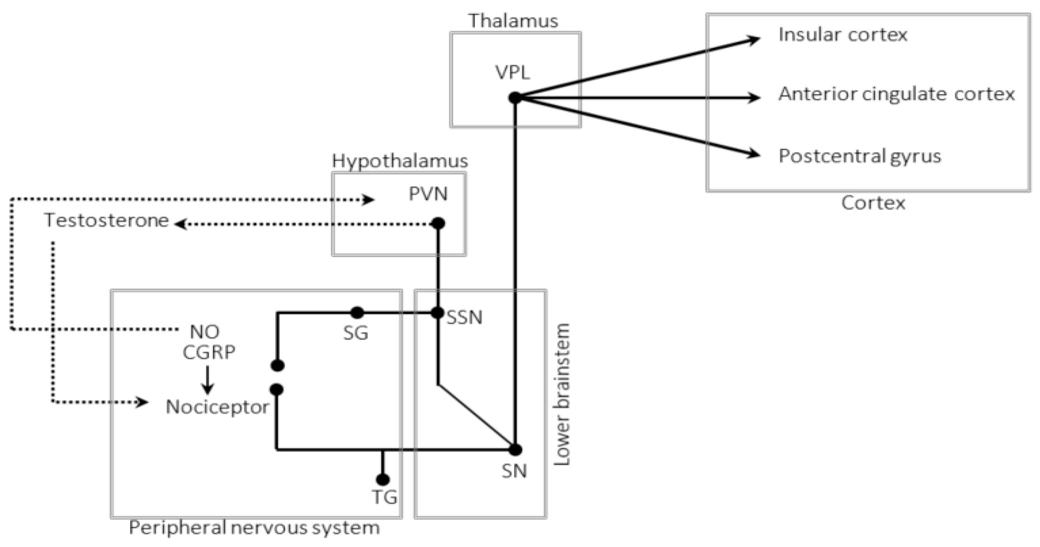
5. The Trigeminal–Autonomic Reflex

Common symptoms of CH attacks, such as lacrimation and rhinorrhoea, as well as an increased serum concentration of vasoactive intestinal polypeptide (VIP) suggest elevated parasympathetic activity [70]. This increase results from an activation of the trigeminal–autonomic reflex, whose afferent fibres run in the trigeminal and efferent fibres in the facial nerve [70].
It is unknown which part of the reflex arc is activated first in spontaneous attacks; stimulation of neither part invariably leads to attacks [71,72]. The matter is further complicated by reports suggesting that the trigeminal nerve may not be necessary at all to suffer attacks [73,74]. Stimulation of the sphenopalatine ganglion, on the other hand, does prevent and abort attacks in many patients, suggesting that these nerve fibres do exert an influence on ictal pathophysiology and influence the probability of attacks [75].
Activation of the efferent reflex arc is associated with the release of neurotransmitters such as CGRP and nitric oxide (NO) [70,76].
Studies on CH attack frequency investigating the effects of antagonists directed against CGRP or its receptors yielded mixed results. While one substance met the primary endpoint in episodic CH, ref. [77] another did not [78]. In patients with chronic CH, these drugs produced no significant reduction in attack frequency, which is in contrast to the effects in chronic migraine, ref. [79] suggesting that the CGRP-system exerts a smaller influence on attack frequency in CH than in migraine. The lower concentration of CGRP in chronic than in episodic CH, ref. [80] suggests a reduced activity of the efferent reflex arc in these patients.
NO is a potent vasodilator capable of triggering CH attacks [81] and possibly inducing—if administered for a sufficiently long time—even bouts [82]. It is often released during inflammatory processes and leads to central sensitization [83,84,85,86]. Sometimes, inflammation suffices to induce pain of similar phenotype temporarily. Case reports documented an association of local inflammations, such as sinusitis, ref. [87] facial herpes simplex, ref. [88] and herpes zoster in the ophthalmic division of the trigeminal nerve [89] with cluster-like attacks.
Treatment with warfarin led to remission in some CH patients, possibly by inhibiting NO production [90]. Corticosteroids, which are highly effective in preventing CH attacks, inhibit NO production as well [91,92]. However, since the effect of corticosteroids usually does not outlast the intake of the drug for extended periods, ref. [93] NO is likely a mediator in advanced stages of the pathophysiologic cascade—but not the attack generator itself.
Between the attacks, there is an elevated concentration of CGRP and products of NO in the blood and cerebrospinal fluid (CSF) of CH patients, respectively [70,94]. Given the short half-life of these molecules, we suspect that the trigeminal–autonomic reflex is permanently active—not just during the pain phase. Such an increased activity might cause aseptic inflammation and sustain sensitization [76,83].
Consequently, “shadow attacks”, ref. [43] and autonomic symptoms preceding the attack, ref. [95] might increase sensitization until reaching the threshold necessary to activate nociceptors. Thus, if and for how long autonomic symptoms precede the pain phase might depend on the “baseline sensitization”. It would be interesting to investigate the correlation between the time autonomic symptoms precede the pain, the extent of central sensitization, and the concentrations of CGRP and NO products in future studies.
Assuming that the efferent arc of the trigeminal autonomic reflex is less active in chronic than in episodic CH, we may also suppose that less NO and CGRP are released. However, data supporting this hypothesis are still lacking. If true, such a reduced secretion could explain why sensitization is present in episodic CH, but not in chronic CH [36].
An inter-individually differing activity of the reflex may have therapeutic implications as well. Guidelines recommend both sumatriptan and oxygen as an abortive treatment for CH attacks [96]. However, while sumatriptan aborts attacks in episodic and chronic CH equally well, oxygen is less effective in chronic CH [97].
In an animal model, oxygen and naratriptan reduced the flow of the lacrimal gland that stimulation of the superior salivatory nucleus had elicited, ref. [98] suggesting that both affect the efferent arc of the trigeminal autonomic reflex. However, triptans inhibit not only the efferent arc but also trigeminal input, suggesting an influence on the afferent arc, ref. [99] which has not been described for oxygen. Consequently, altering the activity of the efferent reflex arm might be less efficacious in chronic CH because of its relatively smaller importance in the pathophysiology of an attack.
These findings and considerations suggest that, in patients transitioning from episodic to chronic CH, the trigeminal autonomic reflex loses importance concerning attack frequency and attack generation. Consequently, changes in the pain threshold induced by reflex activity cannot be the only mechanisms contributing to the onset of an attack.

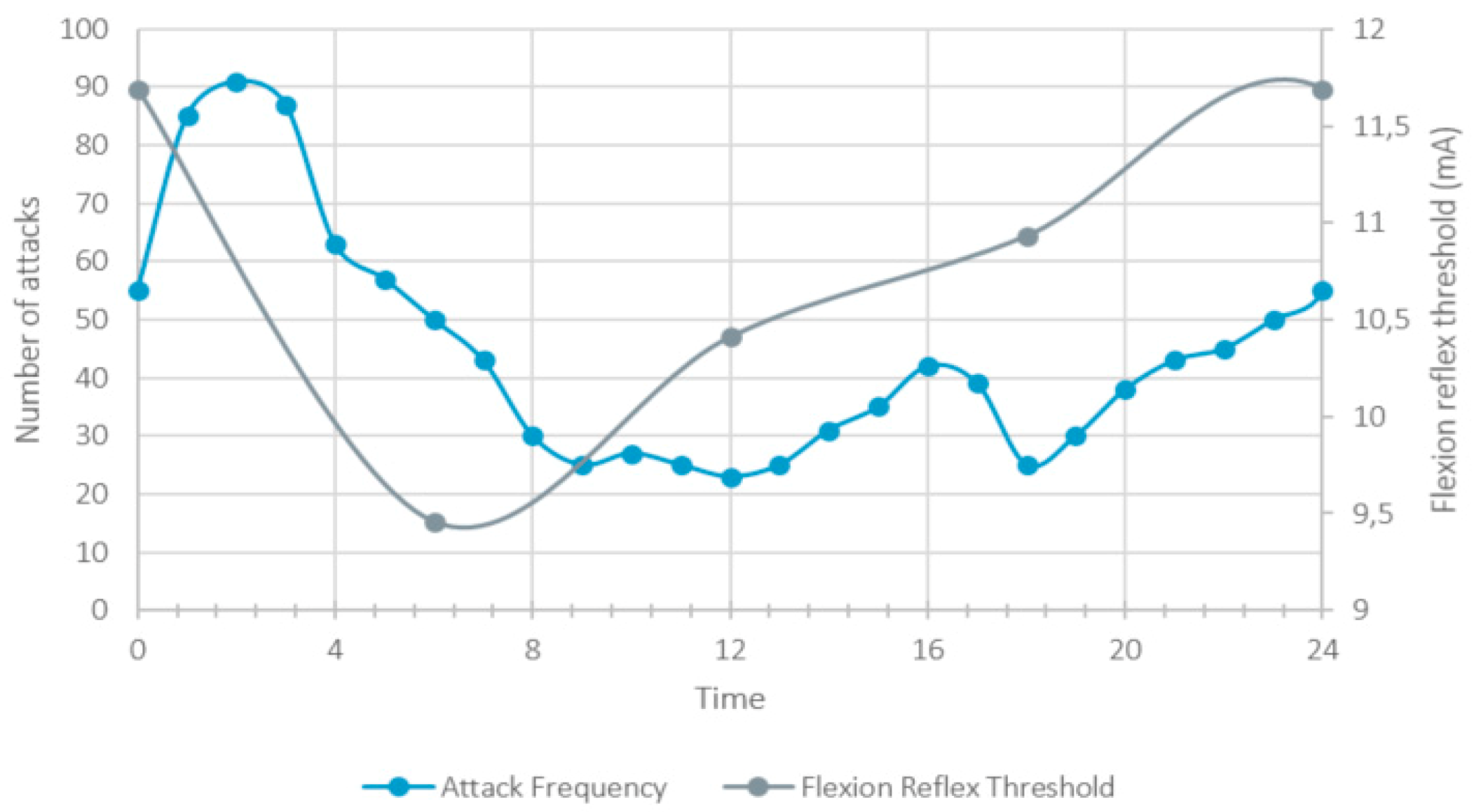
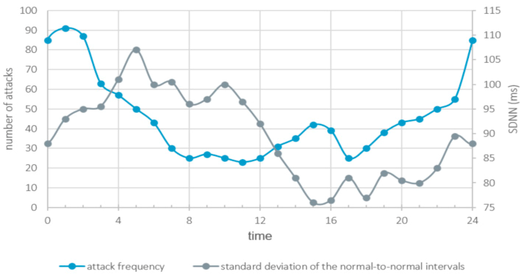
6. Circadian Rhythmicity

Several studies analysing pain perception reported circadian fluctuations with differing maxima for different types of pain [11,12,13,14,15,16,17]. The circadian rhythms of epicritic and protopathic pain are phase-shifted [17,100]; the detection threshold for epicritic pain is lowest at noon, while the threshold for the detection of protopathic pain is lowest at night with a minimum around midnight [100]. The mechanism regulating circadian changes in pain perception is not known.
Similarly, CH attacks do not occur randomly. In many patients, CH has circadian and circannual rhythmicity [101]. While the risk of an attack is lowest around noon, it reaches an interim high at 4 p.m. and rises to its maximum shortly after midnight (see Figure 2) [5].
Investigating circadian changes in the NRF, ref. [36] Nappi et al. reported the highest flexion reflex thresholds at midnight, and the lowest at 6 a.m. in patients suffering from episodic CH, suggesting a steep fall after midnight—similar to the circadian rhythmicity of CH attacks (see Figure 2). In chronic CH, however, they detected no circadian rhythmicity.
Consequently, changes in the probability of occurrence of CH attacks parallel changes in the threshold for epicritic pain [11,14] These findings are compatible with the idea of shared neuronal networks processing epicritic pain and CH attacks, and suggest that circadian changes in the threshold of epicritic pain might be linked to the rhythmicity of CH attacks.
Furthermore, Figure 2 indicates that attacks occur most frequently when the pain threshold drops—not so much when it is low (by absolute values)—suggesting that relative changes in pain threshold might be more important than absolute values.

7. The Autonomic Nervous System

Autonomic symptoms precede attacks in almost half of the CH patients, ref. [95] and Horner’s syndrome sometimes accompanies them [102]. Hence, an altered autonomic innervation is a hallmark of the disorder, but its significance is incompletely understood.
Postganglionic sympathetic lesions cause Horner’s syndrome similar to what CH patients often experience during and sometimes in-between attacks [95,102,103]. Occasionally, interictal sympathetic dysfunction in the face also leads to Harlequin syndrome, characterized by reduced ipsilateral facial sweating and flushing [104].
Based on arteriographic findings, some authors hypothesized that ictal swelling of the carotid artery, possibly mediated by increased activity of the locus coeruleus, ref. [105] could damage or temporarily incapacitate sympathetic nerve fibres [106,107]. Consequently, Horner’s and Harlequin syndrome would be a consequence of an attack, rather than its cause.
The authors of one article reporting an attack mimicking cluster headache (caused by a dissection of the carotid artery) implied that a sharp decrease in sympathetic innervation, can cause pain resembling that of a CH attack [108]. Consequently and given that sympathetic innervation may reduce the activity of Aδ-fibre nociceptors, refs. [109,110] a sudden reduction may render patients prone to experiencing pain similar to CH attacks.
Based on the cited case report, ref. [108] we speculate that a persisting sympathetic deficit, indicated by an enduring Horner’s syndrome, may increase the probability of suffering CH attacks. However, there is no published evidence indicating whether the extent of the sympathetic deficit influences attack frequency or bout duration. As attacks did not persist in this patient, ref. [108] an autonomic deficit itself is insufficient to lead to sustained disease activity.
In addition to a local reduction in the sympathetic innervation during attacks, there is a physiological and systemic decrease in the sympathetic activity at night, when attacks most commonly occur [111]. Counterintuitively, the authors of a case series suggest that an even further reduction in the sympathetic innervation through sympathetic blocks may prevent and abort attacks [112]. Thus, it remains open for discussion whether these circadian changes have an impact on attack generation at all.
Changes in the heart rate variability indicate that circadian rhythmicity underlies the systemic parasympathetic activity. The parasympathetic tone decreases during the day and increases at night until shortly after awakening (see Figure 3) [113,114].
Reduced parasympathetic activity is associated with increased pain perception in men, but not in women [115]. Could these sex-related differences account for the differences in the circadian rhythm of CH attacks between women and men [6]? Could increasing parasympathetic innervation lead to a rapid decrease in attack frequency between 2 and 4 a.m. and between 5 and 6 p.m. (see Figure 3)?
One study reported a systemic reduction in the parasympathetic innervation both in-bout and out-bout [116]. This alteration might contribute to increased pain sensitivity.
In summary, a relative decrease in local sympathetic and systemic parasympathetic innervation is present in some CH patients and might influence pain perception. Little do we know about what causes these alterations of the autonomic nervous system; it can be hypothesized that the central nervous system—the hypothalamus being the most likely candidate—generates these long-lasting changes in pain perception and autonomic innervation.

8. The Hypothalamus

Changes in autonomous nervous system activity are not the only indications of the involvement of the hypothalamus in CH. Further arguments are (i) perturbations of the circadian rhythm of serum cortisol during cluster periods, refs. [117] (ii) a reduction in serum melatonin during cluster periods, ref. [117,118] as well as a (iii) reduction in testosterone—which leads to a reduced pain threshold [119] possibly through testosterone-sensitive TRPM8 receptors [120,121]—(iv) of prolactin, ref. [122] and (v) of TSH [123]. Moreover, fMRI studies showed decreased resting-state functional connectivity of the hypothalamus and an increased activity ipsilateral to the attacks [124,125,126]. Finally, the positive effect on attack frequency of continuous hypothalamic stimulation may support the idea of hypothalamic involvement [126]. However, there are persisting doubts about the precise structures that stimulation needs to target to achieve therapeutic success [127,128].
The diversity of these findings suggests an implication of hypothalamic nuclei. A likely candidate is the paraventricular nucleus (PVN) that controls neuroendocrine and autonomic function, ref. [129] projects to the superior salivatory nucleus in rats, ref. [130] and whose stimulation led to hypoalgesia in an animal model [131].
The PVN consists of magnocellular and parvocellular neurons; the former project to the posterior pituitary gland and influence the secretion of vasopressin and oxytocin; the latter provide superior control of the autonomous nervous system and secrete hormones controlling the anterior pituitary gland [129]. The above-listed studies reported reduced concentrations of several hormones that hint at a decreased activity of parvocellular neurons.
Interestingly, blocking NO led to an increased release of adrenocorticotrophic hormone and corticosterone in the PVN of rats, ref. [132] suggesting that an increase in NO—as seen in CH94—might do the opposite. Furthermore, NO donors applied to the PVN reduced the sympathetic innervation [133]. Consequently, the PVN can induce neuroendocrine changes similar to those seen in CH; NO donors, reported to induce attacks and bouts, refs. [81,82] could exert their effect in this area.
The paraventricular nucleus receives direct innervation from the suprachiasmatic nucleus that harbours the “inner clock” [134,135]. There are seasonal changes in the activity of this nucleus—but not in the PVN—which might contribute to the circannual activity of CH.134
Furthermore, this area might play a role in elevated body weight, and hypertension, ref. [136] which are slightly more common in CH patients than in controls [137]. The available evidence does not allow drawing definite conclusions, though; further research is necessary.

9. Homeostatic Plasticity and the Occurrence of Attacks

Several factors regulating pain perception are altered in CH patients, suggesting that their pain networks may be—intermittently or continuously—closer to depolarisation. Among these are sensitisation, a reduced autonomic innervation, as well as lowered testosterone, melatonin, and cortisol levels (see above). Moreover, depression [138] and sleep deprivation, refs. [139,140] which reduce the pain threshold as well, are also common [5,141]. Finally, circadian changes in pain perception also influence depolarisation probability.
Thus, in CH, there is a sustained decrease in the pain threshold—with additional super-imposed transitory decreases. Although the site of action is not precisely known for some of the implicated factors, it seems reasonable to believe that most influence nociception on the level of the brainstem and the peripheral nervous system (see Figure 1). Consequently, these factors modulate the firing rate of neurons signalling activation of nociceptors to the thalamus and cortex.
Studies reporting that stimulation of the trigeminal nerve or the sphenopalatine ganglion failed to elicit attacks in some patients during the in-bout period support the idea of several factors being necessary for an attack to occur [72,142].
We hypothesize that when the summation of several influencing factors maximally reduced the pain threshold, even weak activation of nociceptors might lead to the perception of intense pain with excitatory influences strongly outweighing inhibiting factors.
Neuronal excitability is generally tightly regulated because failure may lead to the synchronous firing of large numbers of neurons manifesting as epileptic seizures. For this reason, these controlling mechanisms—referred to as homeostatic plasticity—incited great interest among researchers in the pathophysiology of epilepsy [143].
The pathophysiological cascade of CH attacks outlined above implies network overactivity and malfunctioning of homeostatic plasticity in analogy to epileptic seizures. Interestingly, analysing habituation and the nociceptive blink reflex in patients with episodic CH in- and out-bout, Perrotta et al. found evidence for faulty synaptic plasticity in CH patients, indicative of malfunctioning short-term depression (STD) [19].
Short-term synaptic plasticity influences the transmission of presynaptic action potentials to post-synaptic structures. Depletion of presynaptic vesicles, post-synaptic desensitization, and feedback activation of presynaptic receptors contribute to STD that limits transmission foremost at high depolarisation frequencies [144].
CH patients fail to habituate to high-frequency nociceptive stimuli, refs. [19,20,21] and commonly report rapidly increasing pain intensity during, on average, the first nine minutes of an attack [24]. Thus, there is neither electrophysiological nor clinical evidence of a short-term depression of synaptic transmission. On the contrary, the time course of pain intensity suggests potentiation.
Untreated attacks usually last between 45 min and 3 h [25]. It is possible that after this period, some threshold-lowering influences reduce their intensity, but the emergence of long-term depression is likely as well. A refractory period following attacks during which no attacks can occur spontaneously and whose duration correlates with the length of an attack, ref. [145] might indicate long-term changes in excitability. Moreover, the association of attacks with steep falls in the pain threshold—not with low thresholds in general—might imply that long-term depression prevents attacks but takes some time to set in. We are not aware of any study analysing long-term depression in CH.
While we do hypothesize that the depolarization of a network that processes input from Aδ-fibre nociceptors is common to all CH attacks, we also suspect that causal factors and their degree of influence differ between patients.
Lee et al. reported that circadian rhythmicity of the attacks occurs less frequently with an increasing number of bouts [101]. Thus, circadian changes in pain threshold become somewhat dispensable in the pathophysiology CH after some time suggesting attack-induced changes in other brain areas, possibly the hypothalamus. Moreover, the persistence of attacks in patients with chronic CH is intriguing given the absence of sensitization [36]. This implies that upon transitioning from episodic to chronic CH, proneness to depolarisation does not depend on sensitization anymore. Independence from sensitization may also explain why sectioning the trigeminal nerve does not prevent all patients from having attacks [73,74].
Furthermore, the occasional absence of autonomic symptoms [25] suggests that even increased parasympathetic activity may sometimes be dispensable.

10. Summary and Conclusions

Patients’ accounts of CH attacks, ictal restlessness, and some electrophysiological studies suggest that the pathophysiology of CH involves Aδ-fibre nociceptors and networks processing their input. Continuous activity of the trigeminal autonomic reflex throughout the in-bout period might result in central sensitization within these networks in many patients.
It seems possible that an interplay of several factors allows for spontaneous depolarization and forces circadian rhythmicity upon the disease. In addition to sensitization, circadian changes in pain perception and autonomic innervation might be responsible for hyperexcitability of the trigeminal cervical complex (see Figure 1). It is likely that the relative importance of individual factors varies throughout the disorder—in particular when transitioning from episodic to chronic CH, or vice versa.
Summation of several factors influencing pain perception might render neurons vulnerable to spontaneous depolarization, particularly at the beginning of rapid drops of the pain threshold (“summation headache”). In light of studies suggesting an impairment of short-term synaptic plasticity in CH patients, we suggest that a key element in CH attack generation might be network overactivity—similar to epileptic seizures.
Case reports documenting cluster-like attacks are compatible with our idea of several factors being involved in inducing attacks. A sustained and recurring proneness to attacks could be explained by changes in the activity of supra-tentorial neuronal structures among which the hypothalamus is the most likely candidate. The patterns of hormonal changes that commonly occur in CH patients support this idea.
Imaging studies provided evidence of network disruption and altered hypothalamic functional connectivity that back the idea of multifactorial disease [146]. Likewise, the effect of acute and preventive medication provides insights into the pathophysiology of the disorder [147]. However, these studies did not indicate where these alterations add together to trigger an attack. The strength of our hypothesis is that it indicates the attack generator more precisely than previous articles did.
Moreover, the predicted network overactivity may explain the excruciating pain that accompanies the attacks. Further, it might also explain the difference between very little to no pain during “shadow attacks” and excruciating pain, as only network overactivity would lead to intense pain. Finally, the strict unilaterality suggests that attacks originate from areas with little to no exchange of fibres representing different sides of the body such as the brain stem.
However, no study observed the predicted brain stem mechanisms (probably located in or mediated by the SSN or SN) yet. One approach to test the hypothesis could be the registration of the effects of repeated stimulation of Aδ-fibre nociceptors on functional imaging at different times. Based on the pathophysiologic mechanisms postulated above, we would expect that time determines the evoked brain stem activity.
Although we believe that much of the available data support our hypotheses, we need to mention some further limitations. The sample sizes in some of the cited studies were small, comprised mainly of men and not all researchers distinguished episodic from chronic CH. Furthermore, we cannot know whether the cited studies accidentally investigated subpopulations of CH patients that do not represent all patients. Some of the hypotheses presented in this article are speculative and await confirmation.

Author Contributions

H.P. planned the review, collected and interpreted the data, and drafted the manuscript. P.S.S., L.M. and A.R.G. revised the manuscript. All authors have read and agreed to the published version of the manuscript.

Funding

The authors disclosed receipt of the following financial support for the research, authorship, and/or publication of this article: H.P. was funded by the Werner Dessauer Stiftung. P.S.S. received financial support from the Research Fund of the ZURZACH Care group.

Institutional Review Board Statement

Not applicable.

Informed Consent Statement

Not applicable.

Data Availability Statement

Not applicable.

Conflicts of Interest

The authors declare no potential conflict of interest.

Abbreviations

CGRPcalcitonin gene-related peptide
CHCluster headache
CSFcerebrospinal fluid
fMRIfunctional magnetic resonance imaging
GABAgamma-aminobutyric acid
NFRnociceptive flexion reflex
NOnitric oxide
PAGperiaqueductal grey
PETpositron emission tomography
PVNparaventricular nucleus
QSTquantitative sensory testing
SDNNstandard deviation of the normal-to-normal intervals
STDshort-term depression
VGCCvoltage-gated calcium channels
VIPvasoactive intestinal polypeptide

References

  1. Schröder, P.; Gaul, C.; Drabik, A.; Molsberger, A. Pain localization in cluster headache patients: Onset, peak, and radiation. Acta Neurol. Scand. 2021, 143, 441–449. [Google Scholar] [CrossRef] [PubMed]
  2. Burish, M.J.; Pearson, S.M.; Shapiro, R.E.; Zhang, W.; Schor, L.I. Cluster headache is one of the most intensely painful human conditions: Results from the International Cluster Headache Questionnaire. Headache J. Head Face Pain 2021, 61, 117–124. [Google Scholar] [CrossRef] [PubMed]
  3. Olesen, J. Headache Classification Committee of the International Headache Society (IHS) The International Classification of Headache Disorders, 3rd edition. Cephalalgia 2018, 38, 1–211. [Google Scholar]
  4. Pedersen, A.S.; Snoer, A.; Barloese, M.; Petersen, A.; Jensen, R.H. Prevalence of pre-cluster symptoms in episodic cluster headache: Is it possible to predict an upcoming bout? Cephalalgia 2021, 41, 799–809. [Google Scholar] [CrossRef] [PubMed]
  5. Barloese, M.; Lund, N.; Petersen, A.; Rasmussen, M.; Jennum, P.; Jensen, R. Sleep and chronobiology in cluster headache. Cephalalgia 2015, 35, 969–978. [Google Scholar] [CrossRef]
  6. Lund, N.; Barloese, M.; Petersen, A.; Haddock, B.; Jensen, R. Chronobiology differs between men and women with cluster headache, clinical phenotype does not. Neurology 2017, 88, 1069–1076. [Google Scholar] [CrossRef]
  7. Lee, M.J.; Choi, H.A.; Shin, J.H.; Park, H.R.; Chung, C.-S. Natural course of untreated cluster headache: A retrospective cohort study. Cephalalgia 2017, 38, 655–661. [Google Scholar] [CrossRef]
  8. Morelli, N.; Rota, E.; Gori, S.; Guidetti, D.; Michieletti, E.; DE Simone, R.; Di Salle, F. Brainstem activation in cluster headache: An adaptive behavioural response? Cephalalgia 2013, 33, 416–420. [Google Scholar] [CrossRef] [Green Version]
  9. May, A.; Bahra, A.; Büchel, C.; Frackowiak, R.; Goadsby, P.J. Hypothalamic activation in cluster headache attacks. Lancet 1998, 352, 275–278. [Google Scholar] [CrossRef]
  10. Legrain, V.; Iannetti, G.D.; Plaghki, L.; Mouraux, A. The pain matrix reloaded: A salience detection system for the body. Prog. Neurobiol. 2011, 93, 111–124. [Google Scholar] [CrossRef] [Green Version]
  11. Frederickson, R.C.; Burgis, V.; Edwards, J.D. Hyperalgesia induced by naloxone follows diurnal rhythm in responsivity to painful stimuli. Science 1977, 198, 756–758. [Google Scholar] [CrossRef]
  12. Göbel, H.; Cordes, P.; Christiani, K. Algesimetric experiment on pain sensitivity in the pericranial musculature. Circadian rhythms and corresponding psychic variables. Schmerz 1989, 3, 209–218. [Google Scholar] [CrossRef]
  13. Göbel, H.; Cordes, P. Circadian Variation of Pain Sensitivity in Pericranial Musculature. Headache J. Head Face Pain 1990, 30, 418–422. [Google Scholar] [CrossRef]
  14. Sandrini, G.; Alfonsi, E.; Bono, G.; Facchinetti, F.; Montalbetti, L.; Nappi, G. Circadian variations of human flexion reflex. Pain 1986, 25, 403–410. [Google Scholar] [CrossRef]
  15. Aviram, J.; Shochat, T.; Pud, D. Pain Perception in Healthy Young Men Is Modified by Time-Of-Day and Is Modality Dependent. Pain Med. 2015, 16, 1137–1144. [Google Scholar] [CrossRef] [PubMed]
  16. Labrecque, G.; Vanier, M.-C. Biological rhythms in pain and in the effects of opioid analgesics. Pharmacol. Ther. 1995, 68, 129–147. [Google Scholar] [CrossRef]
  17. Bruguerolle, B.; Labrecque, G. Rhythmic pattern in pain and their chronotherapy. Adv. Drug Deliv. Rev. 2007, 59, 883–895. [Google Scholar] [CrossRef] [PubMed]
  18. Rolke, R.; Baron, R.; Maier, C.; Tölle, T.R.; Treede, R.-D.; Beyer, A.; Binder, A.; Birbaumer, N.; Birklein, F.; Bötefür, I.C.; et al. Quantitative sensory testing in the German Research Network on Neuropathic Pain (DFNS): Standardized protocol and reference values. Pain 2006, 123, 231–243. [Google Scholar] [CrossRef] [PubMed]
  19. Perrotta, A.; Coppola, G.; Anastasio, M.G.; De Icco, R.; Ambrosini, A.; Serrao, M.; Parisi, V.; Evangelista, M.; Sandrini, G.; Pierelli, F.; et al. Trait- and Frequency-Dependent Dysfunctional Habituation to Trigeminal Nociceptive Stimulation in Trigeminal Autonomic Cephalalgias. J. Pain 2018, 19, 1040–1048. [Google Scholar] [CrossRef] [PubMed]
  20. Coppola, G.; Di Lorenzo, C.; Bracaglia, M.; Di Lenola, D.; Parisi, V.; Perrotta, A.; Serrao, M.; Pierelli, F. Lateralized nociceptive blink reflex habituation deficit in episodic cluster headache: Correlations with clinical features. Cephalalgia 2014, 35, 600–607. [Google Scholar] [CrossRef]
  21. Holle, D.; Zillessen, S.; Gaul, C.; Naegel, S.; Kaube, H.; Diener, H.-C.; Katsarava, Z.; Obermann, M. Habituation of the nociceptive blink reflex in episodic and chronic cluster headache. Cephalalgia 2012, 32, 998–1004. [Google Scholar] [CrossRef] [PubMed]
  22. Montagna, P.; Pierangeli, G.; Cortelli, P. The Primary Headaches as a Reflection of Genetic Darwinian Adaptive Behavioral Responses. Headache J. Head Face Pain 2010, 50, 273–289. [Google Scholar] [CrossRef] [PubMed]
  23. Beissner, F.; Brandau, A.; Henke, C.; Felden, L.; Baumgartner, U.; Treede, R.D.; Oertel, B.G.; Lotsch, J. Quick discrimination of A(delta) and C fiber mediated pain based on three verbal descriptors. PLoS ONE 2010, 5, e12944. [Google Scholar] [CrossRef] [PubMed] [Green Version]
  24. Torelli, P.; Manzoni, G.C. Pain and behaviour in cluster headache. A prospective study and review of the literature. Funct. Neurol. 2004, 18, 205–210. [Google Scholar]
  25. Schurks, M.; Kurth, T.; De Jesus, J.; Jonjic, M.; Rosskopf, D.; Diener, H.-C. Cluster Headache: Clinical Presentation, Lifestyle Features, and Medical Treatment. Headache J. Head Face Pain 2006, 46, 1246–1254. [Google Scholar] [CrossRef]
  26. Blau, J. Behaviour during a cluster headache. Lancet 1993, 342, 723–725. [Google Scholar] [CrossRef]
  27. Keay, K.A.; Bandler, R. Distinct central representations of inescapable and escapable pain: Observations and speculation. Exp. Physiol. 2002, 87, 275–279. [Google Scholar] [CrossRef] [Green Version]
  28. Parry, D.; Macmillan, F.; Koutsikou, S.; McMullan, S.; Lumb, B. Separation of A- versus C-nociceptive inputs into spinal–brainstem circuits. Neuroscience 2008, 152, 1076–1085. [Google Scholar] [CrossRef]
  29. Bejjani, B.; Houeto, J.; Hariz, M.; Yelnik, J.; Mesnage, V.; Bonnet, A.; Pidoux, B.; Dormont, D.; Cornu, P.; Agid, Y. Aggressive behavior induced by intraoperative stimulation in the triangle of Sano. Neurology 2002, 59, 1425–1427. [Google Scholar] [CrossRef]
  30. Woolf, C.J. Central sensitization: Implications for the diagnosis and treatment of pain. Pain 2011, 152, S2–S15. [Google Scholar] [CrossRef] [PubMed]
  31. Basbaum, A.I.; Bautista, D.M.; Scherrer, G.; Julius, D. Cellular and Molecular Mechanisms of Pain. Cell 2009, 139, 267–284. [Google Scholar] [CrossRef] [PubMed] [Green Version]
  32. Woolf, C.J. Evidence for a central component of post-injury pain hypersensitivity. Nature 1983, 306, 686–688. [Google Scholar] [CrossRef] [PubMed]
  33. Skljarevski, V.; Ramadan, N.M. The nociceptive flexion reflex in humans—Review article. Pain 2002, 96, 3–8. [Google Scholar] [CrossRef]
  34. Arendt-Nielsen, L.; Brennum, J.; Sindrup, S.; Bak, P. Electrophysiological and psychophysical quantification of temporal summation in the human nociceptive system. Graefes Arch. Clin. Exp. Ophthalmol. 1994, 68, 266–273. [Google Scholar] [CrossRef]
  35. Sandrini, G.; Antonaci, F.; Lanfranchi, S.; Milanov, I.; Danilov, A.; Nappi, G. Asymmetrical reduction of the nociceptive flexion reflex threshold in cluster headache. Cephalalgia 2000, 20, 647–652. [Google Scholar] [CrossRef]
  36. Nappi, G.; Sandrini, G.; Alfonsi, E.; Cecchini, A.P.; Micieli, G.; Moglia, A. Impaired Circadian Rhythmicity of Nociceptive Reflex Threshold in Cluster Headache. Headache J. Head Face Pain 2002, 42, 125–131. [Google Scholar] [CrossRef] [PubMed]
  37. Sandrini, G.; Alfonsi, E.; Ruiz, L.; Pavesi, G.; Micieli, G.; Manzoni, G.C.; Mancia, D.; Nappi, G. Impairment of corneal pain perception in cluster headache. Pain 1991, 47, 299–304. [Google Scholar] [CrossRef]
  38. Ladda, J.; Straube, A.; Förderreuther, S.; Krause, P.; Eggert, T. Quantitative Sensory Testing in Cluster Headache: Increased Sensory Thresholds. Cephalalgia 2006, 26, 1043–1050. [Google Scholar] [CrossRef] [PubMed]
  39. Hsieh, M.-T.; Donaldson, L.F.; Lumb, B.M. Differential contributions of A- and C-nociceptors to primary and secondary inflammatory hypersensitivity in the rat. Pain 2015, 156, 1074–1083. [Google Scholar] [CrossRef]
  40. Marmura, M.J.; Pello, S.J.; Young, W.B. Interictal pain in cluster headache. Cephalalgia 2010, 30, 1531–1534. [Google Scholar] [CrossRef]
  41. Ashkenazi, A.; Young, W.B. Dynamic Mechanical (Brush) Allodynia in Cluster Headache. Headache J. Head Face Pain 2004, 44, 1010–1012. [Google Scholar] [CrossRef] [PubMed] [Green Version]
  42. Göbel, C.H.; Karstedt, S.; Heinze, A.; Koch, B.; Göbel, H. Phenotype of Cluster Headache: Clinical Variability, Persisting Pain Between Attacks, and Comorbidities—An Observational Cohort Study in 825 Patients. Pain Ther. 2021, 1–17. [Google Scholar] [CrossRef]
  43. Ekbom, K. Abortive attacks in episodes of cluster headache. Arch. Neurobiol. 1974, 37, 191–195. [Google Scholar]
  44. Zamponi, G.W.; Lewis, R.J.; Todorovic, S.M.; Arneric, S.P.; Snutch, T.P. Role of voltage-gated calcium channels in ascending pain pathways. Brain Res. Rev. 2009, 60, 84–89. [Google Scholar] [CrossRef] [PubMed] [Green Version]
  45. Tamaddonfard, E.; Erfanparast, A.; Taati, M.; Dabbaghi, M. Role of opioid system in verapamil-induced antinociception in a rat model of orofacial pain. Veter. Res. Forum Int. Q. J. 2014, 5, 49–54. [Google Scholar]
  46. Vanegas, H.; Schaible, H.-G. Effects of antagonists to high-threshold calcium channels upon spinal mechanisms of pain, hyperalgesia and allodynia. Pain 2000, 85, 9–18. [Google Scholar] [CrossRef]
  47. Hering, R.; Kuritzky, A. Sodium valproate in the treatment of cluster headache: An open clinical trial. Cephalalgia 1989, 9, 195–198. [Google Scholar] [CrossRef]
  48. Ximenes, J.C.M.; Gonçalves, D.D.O.; Siqueira, R.M.P.; Neves, K.R.T.; Cerqueira, G.S.; Correia, A.O.; Félix, F.H.C.; Leal, L.K.A.M.; Brito, G.A.D.C.; Naffah-Mazzacorati, M.D.G.; et al. Valproic acid: An anticonvulsant drug with potent antinociceptive and anti-inflammatory properties. Naunyn Schmiedebergs Arch. Pharmacol. 2013, 386, 575–587. [Google Scholar] [CrossRef]
  49. Kunder, S.K.; Bairy, L.K.; Arivazhahan, A. Effect of Sodium Valproate and Docosahexaenoic Acid on Pain in Rats. J. Clin. Diagn. Res. 2017, 11, FF05–FF08. [Google Scholar] [CrossRef]
  50. Hobo, S.; Eisenach, J.C.; Hayashida, K.-I. Up-regulation of spinal glutamate transporters contributes to anti-hypersensitive effects of valproate in rats after peripheral nerve injury. Neurosci. Lett. 2011, 502, 52–55. [Google Scholar] [CrossRef] [PubMed] [Green Version]
  51. Stefani, L.C.; Muller, S.; Torres, I.L.; Razzolini, B.; Rozisky, J.R.; Fregni, F.; Markus, R.; Caumo, W. A Phase II, Randomized, Double-Blind, Placebo Controlled, Dose-Response Trial of the Melatonin Effect on the Pain Threshold of Healthy Subjects. PLoS ONE 2013, 8, e74107. [Google Scholar] [CrossRef]
  52. Miranda-Páez, A.; Zamudio, S.R.; Vázquez-León, P.; Sandoval-Herrera, V.; Villanueva-Becerril, I.; Carli, G. Effect of melatonin injection into the periaqueductal gray on antinociception and tonic immobility in male rats. Horm. Behav. 2017, 89, 23–29. [Google Scholar] [CrossRef]
  53. Leone, M.; D’Amico, D.; Moschiano, F.; Fraschini, F.; Bussone, G. Melatonin versus placebo in the prophylaxis of cluster headache: A double-blind pilot study with parallel groups. Cephalalgia 1996, 16, 494–496. [Google Scholar] [CrossRef] [PubMed]
  54. Peres, M.; Rozen, T.D. Melatonin in the Preventive Treatment of Chronic Cluster Headache. Cephalalgia 2001, 21, 993–995. [Google Scholar] [CrossRef] [PubMed]
  55. Pringsheim, T.; Magnoux, E.; Dobson, C.F.; Hamel, E.; Aubé, M. Melatonin as adjunctive therapy in the prophylaxis of cluster headache: A pilot study. Headache J. Head Face Pain 2002, 42, 787–792. [Google Scholar] [CrossRef]
  56. Steiner, T.J.; Hering, R.; Couturier, E.; Davies, P.; Whitmarsh, T.E. Double-Blind Placebo-Controlled Trial of Lithium in Episodic Cluster Headache. Cephalalgia 1997, 17, 673–675. [Google Scholar] [CrossRef] [PubMed]
  57. Stochino, M.E.; Deidda, A.; Asuni, C.; Cherchi, A.; Manchia, M.; Del Zompo, M. Evaluation of Lithium Response in Episodic Cluster Headache: A Retrospective Case Series. Headache J. Head Face Pain 2012, 52, 1171–1175. [Google Scholar] [CrossRef]
  58. Reilly, D. Lithium Vs Placebo in Cluster Headache. Cephalalgia 1998, 18, 1. [Google Scholar] [CrossRef]
  59. Weinsanto, I.; Mouheiche, J.; Laux-Biehlmann, A.; Aouad, M.; Maduna, T.; Petit-Demoulière, N.; Chavant, V.; Poisbeau, P.; Darbon, P.; Charlet, A.; et al. Lithium reverses mechanical allodynia through a mu opioid-dependent mechanism. Mol. Pain 2018, 14, 1744806917754142. [Google Scholar] [CrossRef]
  60. Banafshe, H.R.; Mesdaghinia, A.; Arani, M.N.; Ramezani, M.H.; Heydari, A.; Hamidi, G.A. Lithium attenuates pain-related behavior in a rat model of neuropathic pain: Possible involvement of opioid system. Pharmacol. Biochem. Behav. 2012, 100, 425–430. [Google Scholar] [CrossRef]
  61. Shimizu, T.; Shibata, M.; Wakisaka, S.; Inoue, T.; Mashimo, T.; Yoshiya, I. Intrathecal lithium reduces neuropathic pain responses in a rat model of peripheral neuropathy. Pain 2000, 85, 59–64. [Google Scholar] [CrossRef]
  62. Greco, R.; Mangione, A.; Siani, F.; Blandini, F.; Vairetti, M.; Nappi, G.; Sandrini, G.; Buzzi, M.; Tassorelli, C. Effects of CGRP receptor antagonism in nitroglycerin-induced hyperalgesia. Cephalalgia 2013, 34, 594–604. [Google Scholar] [CrossRef]
  63. Hebestreit, J.M.; May, A. Topiramate modulates trigeminal pain processing in thalamo-cortical networks in humans after single dose administration. PLoS ONE 2017, 12, e0184406. [Google Scholar] [CrossRef] [PubMed] [Green Version]
  64. Leone, M.; Dodick, D.; Rigamonti, A.; D’Amico, D.; Grazzi, L.; Mea, E.; Bussone, G. Topiramate in Cluster Headache Prophylaxis: An Open Trial. Cephalalgia 2003, 23, 1001–1002. [Google Scholar] [CrossRef] [PubMed]
  65. Wheeler, S.D.; Carrazana, E.J. Topiramate-treated cluster headache. Neurology 1999, 53, 234. [Google Scholar] [CrossRef] [PubMed]
  66. Förderreuther, S.; Mayer, M.; Straube, A. Treatment of Cluster Headache with Topiramate: Effects and Side-Effects in Five Patients. Cephalalgia 2002, 22, 186–187. [Google Scholar] [CrossRef] [PubMed]
  67. Láinez, M.J.A.; Pascual, J.; Pascual, A.M.; Santonja, J.M.; Ponz, A.; Salvador, A. Topiramate in the Prophylactic Treatment of Cluster Headache. Headache J. Head Face Pain 2003, 43, 784–789. [Google Scholar] [CrossRef] [PubMed]
  68. Huang, W.Y.; Lo, M.C.; Wang, S.J.; Tsai, J.J.; Wu, H.M. Topiramate in prevention of cluster headache in the Taiwanese. Neurol. India 2010, 58, 284–287. [Google Scholar] [CrossRef]
  69. Kuzniecky, R.; Hetherington, H.; Ho, S.; Pan, J.; Martin, R.; Gilliam, F.; Hugg, J.; Faught, E. Topiramate increases cerebral GABA in healthy humans. Neurology 1998, 51, 627–629. [Google Scholar] [CrossRef]
  70. Goadsby, P.J.; Edvinsson, L. Human in vivo evidence for trigeminovascular activation in cluster headache Neuropeptide changes and effects of acute attacks therapies. Brain 1994, 117, 427–434. [Google Scholar] [CrossRef]
  71. Goadsby, P.J. Cluster headache and the trigeminal-autonomic reflex: Driving or being driven? Cephalalgia 2017, 38, 1415–1417. [Google Scholar] [CrossRef] [PubMed] [Green Version]
  72. Möller, M.; Haji, A.A.; Hoffmann, J.; May, A. Peripheral provocation of cranial autonomic symptoms is not sufficient to trigger cluster headache attacks. Cephalalgia 2017, 38, 1498–1502. [Google Scholar] [CrossRef] [PubMed]
  73. Matharu, M.; Goadsby, P.J. Persistence of attacks of cluster headache after trigeminal nerve root section. Brain 2002, 125, 976–984. [Google Scholar] [CrossRef] [PubMed]
  74. Jarrar, R.; Black, D.; Dodick, D.; Davis, D. Outcome of trigeminal nerve section in the treatment of chronic cluster headache. Neurology 2003, 60, 1360–1362. [Google Scholar] [CrossRef]
  75. Jürgens, T.P.; Barloese, M.; May, A.; Láinez, J.M.; Schoenen, J.; Gaul, C.; Goodman, A.M.; Caparso, A.; Jensen, R.H. Long-term effectiveness of sphenopalatine ganglion stimulation for cluster headache. Cephalalgia 2016, 37, 423–434. [Google Scholar] [CrossRef]
  76. Goadsby, P.J.; Uddman, R.; Edvinsson, L. Cerebral vasodilatation in the cat involves nitric oxide from parasympathetic nerves. Brain Res. 1996, 707, 110–118. [Google Scholar] [CrossRef]
  77. Goadsby, P.J.; Dodick, D.W.; Leone, M.; Bardos, J.N.; Oakes, T.M.; Millen, B.A.; Zhou, C.; Dowsett, S.A.; Aurora, S.K.; Ahn, A.H.; et al. Trial of Galcanezumab in Prevention of Episodic Cluster Headache. N. Engl. J. Med. 2019, 381, 132–141. [Google Scholar] [CrossRef]
  78. Teva Branded Pharmaceutical Products R&D, Inc. A Study to Explore the Long-Term Safety and Efficacy of Fremanezumab (TEV-48125) for the Prevention of Cluster Headache (ENFORCE). Available online: https://clinicaltrials.gov/ct2/show/NCT03107052 (accessed on 4 December 2020).
  79. Dodick, D.W.; Goadsby, P.J.; Lucas, C.; Jensen, R.; Bardos, J.N.; Martinez, J.M.; Zhou, C.; Aurora, S.K.; Yang, J.Y.; Conley, R.R.; et al. Phase 3 randomized, placebo-controlled study of galcanezumab in patients with chronic cluster headache: Results from 3-month double-blind treatment. Cephalalgia 2020, 40, 935–948. [Google Scholar] [CrossRef]
  80. Snoer, A.; Vollesen, A.L.H.; Beske, R.P.; Guo, S.; Hoffmann, J.; Fahrenkrug, J.; Jørgensen, N.R.; Martinussen, T.; Jensen, R.H.; Ashina, M. Calcitonin gene-related peptide and disease activity in cluster headache. Cephalalgia 2019, 39, 575–584. [Google Scholar] [CrossRef] [Green Version]
  81. Costa, A.; Ravaglia, S.; Sances, G.; Antonaci, F.; Pucci, E.; Nappi, G. Nitric Oxide Pathway and Response to Nitroglycerin in Cluster Headache Patients: Plasma Nitrite and Citrulline Levels. Cephalalgia 2003, 23, 407–413. [Google Scholar] [CrossRef] [PubMed]
  82. Ekbom, K.; Sjöstrand, C.; Svensson, D.A.; Waldenlind, E. Periods of Cluster Headache Induced by Nitrate Therapy and Spontaneous Remission of Angina Pectoris During Active Clusters. Cephalalgia 2004, 24, 92–98. [Google Scholar] [CrossRef]
  83. Steinberg, A.; Remahl, A.I.M.N. Role of Nitric Oxide in Cluster Headache. Curr. Pain Headache Rep. 2012, 16, 185–190. [Google Scholar] [CrossRef]
  84. Lin, Q.; Paleček, J.; Palečková, V.; Peng, Y.B.; Wu, J.; Cui, M.; Willis, W.D. Nitric oxide mediates the central sensitization of primate spinothalamic tract neurons. J. Neurophysiol. 1999, 81, 1075–1085. [Google Scholar] [CrossRef]
  85. Kitto, K.F.; Haley, J.E.; Wilcox, G.L. Involvement of nitric oxide in spinally mediated hyperalgesia in the mouse. Neurosci. Lett. 1992, 148, 1–5. [Google Scholar] [CrossRef]
  86. Meller, S.T.; Gebhart, G.F. Nitric oxide (NO) and nociceptive processing in the spinal cord. Pain 1993, 52, 127–136. [Google Scholar] [CrossRef]
  87. Balgetir, F.; Avcı, D.; Gönen, M.; Taşcı, I. Acute Rhinosinusitis as an Infrequent Cause of Symptomatic Cluster Headache: Report of Seven Cases. J. Oral Facial Pain Headache 2019, 39, 408–412. [Google Scholar] [CrossRef] [PubMed]
  88. Joseph, R.; Rose, F.C. Cluster headache and herpes simplex: An association? Br. Med. J. (Clin. Res. Ed.) 1985, 290, 1625–1626. [Google Scholar] [CrossRef] [PubMed] [Green Version]
  89. Sacquegna, T.; D’Alessandro, R.; Cortelli, P.; de Carolis, P.; Baldrati, A. Cluster Headache After Herpes Zoster Ophthalmicus. Arch. Neurol. 1982, 39, 384. [Google Scholar] [CrossRef] [PubMed]
  90. Hakim, S.M. Warfarin for Refractory Chronic Cluster Headache: A Randomized Pilot Study. Headache J. Head Face Pain 2011, 51, 713–725. [Google Scholar] [CrossRef]
  91. Linehan, J.D.; Kolios, G.; Valatas, V.; Robertson, D.A.F.; Westwick, J. Effect of corticosteroids on nitric oxide production in inflammatory bowel disease: Are leukocytes the site of action? Am. J. Physiol. Liver Physiol. 2005, 288, G261–G267. [Google Scholar] [CrossRef] [Green Version]
  92. Obermann, M.; Nägel, S.; Ose, C.; Sonuc, N.; Scherag, A.; Storch, P.; Gaul, C.; Böger, A.; Kraya, T.; Jansen, J.-P.; et al. Safety and efficacy of prednisone versus placebo in short-term prevention of episodic cluster headache: A multicentre, double-blind, randomised controlled trial. Lancet Neurol. 2021, 20, 29–37. [Google Scholar] [CrossRef]
  93. Antonaci, F.; Costa, A.; Candeloro, E.; Sjaastad, O.; Nappi, G. Single High-Dose Steroid Treatment in Episodic Cluster Headache. Cephalalgia 2005, 25, 290–295. [Google Scholar] [CrossRef] [PubMed]
  94. Steinberg, A.; Wiklund, N.P.; Brundin, L.; Remahl, A.I.M.N. Levels of nitric oxide metabolites in cerebrospinal fluid in cluster headache. Cephalalgia 2010, 30, 696–702. [Google Scholar] [CrossRef] [PubMed] [Green Version]
  95. Snoer, A.; Lund, N.; Beske, R.; Jensen, R.; Barloese, M. Pre-attack signs and symptoms in cluster headache: Characteristics and time profile. Cephalalgia 2018, 38, 1128–1137. [Google Scholar] [CrossRef]
  96. Andrée, C.; Barone-Kaganas, I.; Biethahn, S.; Böttger, K.; Dozier, C.; Emmenegger, M.J.; Flügel, D.; Galli, U.; Gantenbein, A.R.; Gobbi, C.; et al. Therapieempfehlungen für Primäre Kopfschmerzen; Schweizerische Kopfwehgesellschaft SKG: Basel, Switzerland, 2019; Available online: https://headache.ch/download/Content_attachments/FileBaseDoc/SKG_Therapieempfehlungen_DE_19_WEB.pdf (accessed on 4 December 2020).
  97. Pearson, S.M.; Burish, M.J.; Shapiro, R.E.; Yan, Y.; Schor, L.I. Effectiveness of Oxygen and Other Acute Treatments for Cluster Headache: Results From the Cluster Headache Questionnaire, an International Survey. Headache J. Head Face Pain 2019, 59, 235–249. [Google Scholar] [CrossRef] [Green Version]
  98. Akerman, S.; Holland, P.; Summ, O.; Lasalandra, M.P.; Goadsby, P. A translational in vivo model of trigeminal autonomic cephalalgias: Therapeutic characterization. Brain 2012, 135, 3664–3675. [Google Scholar] [CrossRef] [Green Version]
  99. Hamel, E. The Biology of Serotonin Receptors: Focus on Migraine Pathophysiology and Treatment. Can. J. Neurol. Sci. J. Can. Sci. Neurol. 1999, 26, 2–6. [Google Scholar] [CrossRef] [Green Version]
  100. Strempel, H. Orcadian cycles of epicritic and protopathic pain threshold. J. Interdiscip. Cycle Res. 1977, 8, 276–280. [Google Scholar] [CrossRef]
  101. Lee, M.J.; Cho, S.-J.; Park, J.W.; Chu, M.K.; Moon, H.-S.; Chung, P.-W.; Chung, J.-M.; Sohn, J.-H.; Kim, B.-K.; Kim, S.-K.; et al. Temporal changes of circadian rhythmicity in cluster headache. Cephalalgia 2019, 40, 278–287. [Google Scholar] [CrossRef]
  102. Drummond, P.D.; Lance, J.W. Pathological sweating and flushing accompanying the trigeminal lacrimal reflex in patients with cluster headache and in patients with a confirmed site of cervical sympathetic deficit. Evidence for parasympathetic cross-innervation. Brain 1992, 115, 115. [Google Scholar] [CrossRef]
  103. Havelius, U. A Horner-like syndrome and cluster headache. What comes first? Acta Ophthalmol. Scand. 2001, 79, 374–375. [Google Scholar] [CrossRef]
  104. Van Oosterhout, W. Patient with Cluster Headache and Harlequin Sign—Related or Not? Headache J. Head Face Pain 2020, 60, 1761–1766. [Google Scholar] [CrossRef]
  105. Goadsby, P.; Lambert, G.; Lance, J. Effects of locus coeruleus stimulation on carotid vascular resistance in the cat. Brain Res. 1983, 278, 175–183. [Google Scholar] [CrossRef]
  106. Ekbom, K.; Greitz, T. Carotid Angiography in Cluster Headache. Acta Radiol. Diagn. 1970, 10, 177–186. [Google Scholar] [CrossRef]
  107. Drummond, P. Mechanisms of Autonomic Disturbance in the Face During and Between Attacks of Cluster Headache. Cephalalgia 2006, 26, 633–641. [Google Scholar] [CrossRef] [PubMed]
  108. Straube, A.; Freilinger, T.; Rüther, T.; Padovan, C. Two Cases of Symptomatic Cluster-Like Headache Suggest the Importance of Sympathetic/Parasympathetic Balance. Cephalalgia 2007, 27, 1069–1073. [Google Scholar] [CrossRef]
  109. Bülbring, E.; Whitteridge, D. The effect of adrenaline on nerve action potentials. J. Physiol. 1941, 99, 201–207. [Google Scholar] [CrossRef] [Green Version]
  110. Shyu, B.C.; Olausson, B.; Huang, K.H.; Widerström, E.; Andersson, S.A. Effects of sympathetic stimulation on C-fibre responses in rabbit. Acta Physiol. Scand. 1989, 137, 73–84. [Google Scholar] [CrossRef] [PubMed]
  111. Jung, B.-C.; Dave, A.S.; Tan, A.Y.; Gholmieh, G.; Zhou, S.; Wang, D.C.; Akingba, A.G.; Fishbein, G.A.; Montemagno, C.; Lin, S.-F.; et al. Circadian variations of stellate ganglion nerve activity in ambulatory dogs. Hear. Rhythm. 2006, 3, 78–85. [Google Scholar] [CrossRef] [PubMed]
  112. Albertyn, J.; Barry, R.; Odendaal, C.L. Cluster headache and the sympathetic nerve. Headache J. Head Face Pain 2004, 44, 183–185. [Google Scholar] [CrossRef] [PubMed]
  113. Massin, M.M.; Maeyns, K.; Withofs, N.; Ravet, F.; Gérard, P.; Healy, M.J.R. Circadian rhythm of heart rate and heart rate variability. Arch. Dis. Child. 2000, 83, 179–182. [Google Scholar] [CrossRef] [PubMed] [Green Version]
  114. Vandewalle, G.; Middleton, B.; Rajaratnam, S.; Stone, B.M.; Thorleifsdottir, B.; Arendt, J.; Dijk, D.-J. Robust circadian rhythm in heart rate and its variability: Influence of exogenous melatonin and photoperiod. J. Sleep Res. 2007, 16, 148–155. [Google Scholar] [CrossRef] [PubMed]
  115. Nahman-Averbuch, H.; Dayan, L.; Sprecher, E.; Hochberg, U.; Brill, S.; Yarnitsky, D.; Jacob, G. Sex differences in the relationships between parasympathetic activity and pain modulation. Physiol. Behav. 2016, 154, 40–48. [Google Scholar] [CrossRef]
  116. Meineri, P.; Pellegrino, G.; Rosso, M.G.; Grasso, E. Systemic autonomic involvement in episodic cluster headache: A comparison between active and remission periods. J. Headache Pain 2005, 6, 240–243. [Google Scholar] [CrossRef] [Green Version]
  117. Waldenlind, E.; Gustafsson, S.A.; Ekbom, K.; Wetterberg, L. Circadian secretion of cortisol and melatonin in cluster headache during active cluster periods and remission. J. Neurol. Neurosurg. Psychiatry 1987, 50, 207–213. [Google Scholar] [CrossRef] [PubMed] [Green Version]
  118. Chazot, G.; Claustrat, B.; Brun, J.; Jordan, D.; Sassolas, G.; Schott, B. A Chronobiological Study of Melatonin, Cortisol Growth Hormone and Prolactin Secretion in Cluster Headache. Cephalalgia 1984, 4, 213–220. [Google Scholar] [CrossRef] [PubMed]
  119. Gharaghozlu, N.; Ahmadi, R.; Khakpour, B.; Alvany, A.; Keshavarz, X. Effects of Testosterone on Pain Threshold. In Proceedings of the International Conference on Chemical, Agricultural and Medical Sciences, Antalaya, Turkey, 2–3 May 2014. [Google Scholar]
  120. Gkika, D.; Lolignier, S.; Grolez, G.P.; Bavencoffe, A.; Shapovalov, G.; Gordienko, D.; Kondratskyi, A.; Meleine, M.; Prival, L.; Chapuy, E.; et al. Testosterone-androgen receptor: The steroid link inhibiting TRPM8-mediated cold sensitivity. FASEB J. 2020, 34, 7483–7499. [Google Scholar] [CrossRef] [Green Version]
  121. Kobayashi, K.; Fukuoka, T.; Obata, K.; Yamanaka, H.; Dai, Y.; Tokunaga, A.; Noguchi, K. Distinct expression of TRPM8, TRPA1, and TRPV1 mRNAs in rat primary afferent neurons with adelta/c-fibers and colocalization with trk receptors. J. Comp. Neurol. 2005, 493, 596–606. [Google Scholar] [CrossRef]
  122. Waldenlind, E.; Gustafsson, S.A. Prolactin in Cluster Headache: Diurnal Secretion, Response to Thyrotropin-Releasing Hormone, and Relation to Sex Steroids and Gonadotropins. Cephalalgia 1987, 7, 43–54. [Google Scholar] [CrossRef]
  123. Léoné, M.; Patruno, G.; Vescovi, A.; Bussone, G. Neuroendocrine Dysfunction in Cluster Headache. Cephalalgia 1990, 10, 235–239. [Google Scholar] [CrossRef]
  124. Yang, F.-C.; Chou, K.-H.; Fuh, J.-L.; Lee, P.-L.; Lirng, J.-F.; Lin, Y.-Y.; Lin, C.-P.; Wang, S.-J. Altered hypothalamic functional connectivity in cluster headache: A longitudinal resting-state functional MRI study. J. Neurol. Neurosurg. Psychiatry 2014, 86, 437–445. [Google Scholar] [CrossRef] [PubMed]
  125. Morelli, N.; Pesaresi, I.; Cafforio, G.; Maluccio, M.R.; Gori, S.; Di Salle, F.; Murri, L. Functional magnetic resonance imaging in episodic cluster headache. J. Headache Pain 2008, 10, 11–14. [Google Scholar] [CrossRef] [PubMed] [Green Version]
  126. Leone, M.; Franzini, A.; Broggi, G.; Bussone, G. Hypothalamic stimulation for intractable cluster headache: Long-term experience. Neurology 2006, 67, 150–152. [Google Scholar] [CrossRef] [PubMed]
  127. Seijo-Fernandez, F.; Saiz, A.; Santamarta, E.; Nader, L.; Alvarez-Vega, M.A.; Lozano, B.; Seijo, E.; Barcia, J.A. Long-Term Results of Deep Brain Stimulation of the Mamillotegmental Fasciculus in Chronic Cluster Headache. Ster. Funct. Neurosurg. 2018, 96, 215–222. [Google Scholar] [CrossRef]
  128. Nowacki, A.; Schober, M.; Nader, L.; Saryyeva, A.; Nguyen, T.A.K.; Green, A.L.; Pollo, C.; Krauss, J.K.; Fontaine, D.; Aziz, T.Z. DBS for chronic cluster headache: Meta-analysis of individual patient data. Ann. Neurol. 2020, 88, 956–969. [Google Scholar] [CrossRef]
  129. Ferguson, A.V.; Latchford, K.J.; Samson, W.K. The paraventricular nucleus of the hypothalamus—A potential target for integrative treatment of autonomic dysfunction. Expert Opin. Ther. Targets 2008, 12, 717–727. [Google Scholar] [CrossRef]
  130. Li, C.; Fitzgerald, M.E.; LeDoux, M.S.; Gong, S.; Ryan, P.; Del Mar, N.; Reiner, A. Projections from the hypothalamic paraventricular nucleus and the nucleus of the solitary tract to prechoroidal neurons in the superior salivatory nucleus: Pathways controlling rodent choroidal blood flow. Brain Res. 2010, 1358, 123–139. [Google Scholar] [CrossRef] [Green Version]
  131. Yang, J.; Chen, J.-M.; Yang, Y.; Liu, W.-Y.; Song, C.-Y.; Lin, B.-C. Investigating the Role of Hypothalamic Paraventricular Nucleus in Nociception of the Rat. Int. J. Neurosci. 2008, 118, 473–485. [Google Scholar] [CrossRef] [PubMed]
  132. Rivier, C.; Shen, G. In the rat, endogenous nitric oxide modulates the response of the hypothalamic-pituitary-adrenal axis to interleukin-1 beta, vasopressin, and oxytocin. J. Neurosci. 1994, 14, 1985–1993. [Google Scholar] [CrossRef] [Green Version]
  133. Patel, K.P. Role of Paraventricular Nucleus in Mediating Sympathetic Outflow in Heart Failure. Hear. Fail. Rev. 2000, 5, 73–86. [Google Scholar] [CrossRef]
  134. Hofman, M.A.; Swaab, D.F. Seasonal changes in the suprachiasmatic nucleus of man. Neurosci. Lett. 1992, 139, 257–260. [Google Scholar] [CrossRef] [Green Version]
  135. Lee, H.S.; Billings, H.J.; Lehman, M.N. The Suprachiasmatic Nucleus: A Clock of Multiple Components. J. Biol. Rhythm. 2003, 18, 435–449. [Google Scholar] [CrossRef] [PubMed]
  136. Qin, C.; Li, J.; Tang, K. The Paraventricular Nucleus of the Hypothalamus: Development, Function, and Human Diseases. Endocrinology 2018, 159, 3458–3472. [Google Scholar] [CrossRef]
  137. Lund, N.; Petersen, A.; Snoer, A.; Jensen, R.H.; Barloese, M. Cluster headache is associated with unhealthy lifestyle and lifestyle-related comorbid diseases: Results from the Danish Cluster Headache Survey. Cephalalgia 2019, 39, 254–263. [Google Scholar] [CrossRef]
  138. Woo, A.K. Depression and Anxiety in Pain. Rev. Pain 2010, 4, 8–12. [Google Scholar] [CrossRef] [Green Version]
  139. Schuh-Hofer, S.; Wodarski, R.; Pfau, D.B.; Caspani, O.; Magerl, W.; Kennedy, J.D.; Treede, R.-D. One night of total sleep deprivation promotes a state of generalized hyperalgesia: A surrogate pain model to study the relationship of insomnia and pain. Pain 2013, 154, 1613–1621. [Google Scholar] [CrossRef] [PubMed]
  140. Sardi, N.F.; Lazzarim, M.K.; Guilhen, V.A.; Marcílio, R.S.; Natume, P.S.; Watanabe, T.C.; Lima, M.M.; Tobaldini, G.; Fischer, L. Chronic sleep restriction increases pain sensitivity over time in a periaqueductal gray and nucleus accumbens dependent manner. Neuropharmacology 2018, 139, 52–60. [Google Scholar] [CrossRef]
  141. Liang, J.-F.; Chen, Y.-T.; Fuh, J.-L.; Li, S.-Y.; Liu, C.-J.; Chen, T.-J.; Tang, C.-H.; Wang, S.-J. Cluster headache is associated with an increased risk of depression: A nationwide population-based cohort study. Cephalalgia 2013, 33, 182–189. [Google Scholar] [CrossRef]
  142. Schytz, H.W.; Barløse, M.; Guo, S.; Selb, J.; Caparso, A.; Jensen, R.; Ashina, M. Experimental activation of the sphenopalatine ganglion provokes cluster-like attacks in humans. Cephalalgia 2013, 33, 831–841. [Google Scholar] [CrossRef] [PubMed]
  143. Lignani, G.; Baldelli, P.; Marra, V. Homeostatic Plasticity in Epilepsy. Front. Cell. Neurosci. 2020, 14, 197. [Google Scholar] [CrossRef]
  144. Zucker, R.S.; Regehr, W.G. Short-term synaptic plasticity. Annu. Rev. Physiol. 2002, 64, 355–405. [Google Scholar] [CrossRef] [PubMed] [Green Version]
  145. Ekbom, K. Nitrolglycerin as a provocative agent in cluster headache. Arch. Neurol. 1968, 19, 487–493. [Google Scholar] [CrossRef] [PubMed]
  146. Ferraro, S.; Nigri, A.; Demichelis, G.; Pinardi, C.; Chiapparini, L.; Giani, L.; Cecchini, A.P.; Leone, M. Understanding Cluster Headache Using Magnetic Resonance Imaging. Front. Neurol. 2020, 11, 535. [Google Scholar] [CrossRef] [PubMed]
  147. Wei, D.Y.; Goadsby, P.J. Cluster headache pathophysiology—Insights from current and emerging treatments. Nat. Rev. Neurol. 2021, 17, 308–324. [Google Scholar] [CrossRef] [PubMed]
Figure 1. Summary of the different pain threshold-influencing mechanisms potentially implicated in the pathophysiology of cluster headache. The afferent part of the trigeminal–autonomic reflex runs through the trigeminal nerve and the trigeminal ganglion to the brain stem; the efferent part originates in the superior salivatory nucleus, runs through the facial nerve and the sphenopalatine ganglion, and modulates the release of CGRP and NO that lower the pain threshold (their concentration is increased in episodic cluster headache). The paraventricular nucleus of the hypothalamus also modulates pain perception possibly through its connection with the superior salivatory nucleus; this nucleus might relay the circadian rhythmicity of pain perception. Furthermore, testosterone (reduced during the active period of cluster headache) also influences pain perception possibly through modulation of nociceptors. Not depicted are pain-modulating descending pathways; their activity is influenced by melatonin (reduced concentration during the active period of cluster headache). Moreover, we did not depict the sympathetic nervous system that also has circadian rhythmicity. Please see text for further details. Solid lines depict anatomical connections; dashed lines refer to the action of hormones CGRP—calcitonine gene-related peptide; NO—nitrogen oxide; PVN—paraventricular nucleus; SG—sphenopalatine ganglion; SN—spinal nucleus of the trigeminal nerve; SSN—superior salivatory nucleus; TG—trigeminal ganglion; VPL—ventral posterolateral nucleus of the Thalamus.
Ctn 05 00016 g001
Figure 2. Correlation between time and attack frequency as well as time and the flexion reflex threshold in patients with episodic cluster headache; the The left vertical axis refers to the number of attacks in a sample of cluster headache patients,; the right refers to the flexion reflex threshold (mA). Data derived from Nappi et al. [36] and Barloese et al. [5].
Ctn 05 00016 g002
Figure 3. Correlation between time and attack frequency as well as time and the standard deviation of the normal-to-normal intervals (SDNN) in healthy adults; the left vertical axis refers to the number of attacks in a sample of cluster headache patients; the right refers to the standard deviation of the heart rate, which indicates parasympathetic innervation; data derived from Vandewalle et al. [114] and Barloese et al. [5].
Ctn 05 00016 g003
Publisher’s Note: MDPI stays neutral with regard to jurisdictional claims in published maps and institutional affiliations.

