Article

A SERS Substrate for Ultrafast Photosynthetic Au Nanoparticle Growth on WO3 Nanowires

1
Beijing Key Laboratory of Microstructure and Property of Advanced Materials, College of Materials Science and Engineering, Beijing University of Technology, Beijing 100124, China
2
College of Physics and Optoelectronic Engineering, Beijing University of Technology, Beijing 100124, China
3
State Key Laboratory of Chemistry for NBC Hazards Protection, Beijing 102205, China
4
Institute of High Energy Physics, Chinese Academy of Sciences, Beijing 100049, China
*
Authors to whom correspondence should be addressed.
Colloids Interfaces 2025, 9(5), 70; https://doi.org/10.3390/colloids9050070 (registering DOI)
Submission received: 11 September 2025 / Revised: 1 October 2025 / Accepted: 11 October 2025 / Published: 14 October 2025
(This article belongs to the Special Issue State of the Art of Colloid and Interface Science in Asia)

Abstract

The practical adoption of surface-enhanced Raman scattering (SERS) technology is often hampered by the high cost, complex fabrication, and poor reproducibility of conventional substrates, which typically rely on noble metals or inefficient semiconductors. Herein, we address key challenges in the practical commercialization of surface-enhanced Raman scattering (SERS) technology by reporting a facile, scalable, and environmentally benign strategy for fabricating a hybrid SERS substrate. This approach integrates Au nanoparticles (NPs) with hydrothermally synthesized WO3 nanowires through a green photoreduction process, which is rapid, organic-solvent-free, and amenable to large-scale production. The design of the Au/WO3 nanocomposite capitalizes on the synergistic effect between electromagnetic (EM) enhancement from Au NPs and chemical mechanism (CM) enhancement via charge transfer involving the WO3 semiconductor. This synergy empowers the substrate with exceptional SERS activity, enabling the sensitive detection of Rhodamine 6G (R6G) down to 10−11 M and yielding an enhancement factor (EF) of 4.09 × 10⁶. More importantly, this EM-CM synergy proves critical for detecting molecules with weak affinity, such as the nerve agent simulant dimethyl methylphosphonate (DMMP), achieving a significant signal enhancement of 102–103 times, which is notably challenging for conventional plasmonic substrates. Beyond sensitivity, the substrate exhibits excellent reproducibility and operational stability, which are paramount for real-world applications. This work presents a nanohybrid strategy that successfully balances scalability, stability, and sensitivity, offering a reliable and cost-effective pathway for advancing SERS technologies toward practical implementation.

1. Introduction

Surface-Enhanced Raman Scattering (SERS) represents a groundbreaking spectroscopic methodology, offering exceptional sensitivity and specificity for the detection of trace substances, achieving even single-molecule-level resolution. This technique has found broad utility across biomedical applications (ultra-early tumor marker diagnosis, rapid pathogen identification), environmental monitoring (in situ pollutant detection), materials science (real-time tracking of catalytic interfaces), and public safety (identification of explosives and narcotics). By transcending the sensitivity constraints of conventional Raman spectroscopy, SERS opens new avenues for elucidating molecular behaviors at interfaces [1,2,3,4,5,6,7,8,9,10,11,12]. The enhancement mechanisms underpinning SERS are predominantly divided into electromagnetic enhancement (EM) and chemical enhancement (CM) [13]. EM stems from localized surface plasmon resonance (LSPR) excited in noble metal nanostructures (Au and Ag nanoparticles) upon laser illumination, generating highly confined electromagnetic “hotspots” at nanogaps or sharp topographic features. These hotspots can amplify Raman scattering by factors of 106–1010, with efficiency strongly correlated to the nanostructures’ morphology, size, and excitation wavelength [14,15,16]. Despite their promising performance, conventional noble-metal-based SERS substrates are hampered by intrinsic limitations including high material cost, scarcity, and susceptibility to chemical degradation (oxidation and aggregation), leading to performance fade under prolonged or harsh operational conditions. In light of this, semiconductor-based SERS substrates such as CuO, TiO2, and ZnO have emerged as promising alternatives [17,18,19,20]. These materials operate mainly through the CM mechanism, which involves charge transfer (CT) between analyte molecules and the substrate. Chemical adsorption or strong interfacial interaction facilitates coupling between molecular orbitals and semiconductor band states, modulating molecular polarizability and enhancing the Raman cross-section by approximately 10 to 103 times. This enhancement is highly sensitive to the electronic structure alignment at the molecule–substrate interface [21,22,23], nevertheless, the enhancement factor provided by pure semiconductor substrates remains comparatively modest. Recent research focus has accordingly shifted toward hybrid systems that exploit synergistic effects between EM and CM mechanisms: the plasmonic component provides intense electromagnetic confinement, while the semiconductor facilitates charge transfer and improves chemical specificity [13,24]. This combined approach effectively surmounts the sensitivity limitations of traditional Raman techniques, establishing a robust foundation for single-molecule detection and real-time analysis of interfacial reaction dynamics [25,26,27].
Against the backdrop of ongoing innovations in semiconductor-based SERS substrates, tungsten trioxide (WO3) has garnered significant interest owing to its pronounced chemical enhancement properties. Abundant oxygen vacancy defects, a tunable wide bandgap (2.4–2.8 eV), and strong molecular adsorption capacity collectively facilitate efficient charge transfer (CT), establishing WO3 as a promising platform for SERS applications [28]. Nevertheless, the intrinsic lack of electromagnetic enhancement (EM) in pure WO3 limits its detection sensitivity. To overcome this limitation, the strategic decoration of gold nanoparticles (Au NPs) onto WO3 substrates has been proposed, forming Au/WO3 heterostructures [29]. On one hand, the localized surface plasmon resonance (LSPR) excited in Au NPs generates high-intensity electromagnetic hotspots, offering EM enhancement factors on the order of 105–107. On the other hand, the Schottky junction formed at the Au–WO3 interface induces directional electron transfer from WO3 to Au until their Fermi levels equilibrate. This process creates a built-in electric field at the interface, which efficiently promotes charge separation and optimizes the adsorption energy and polarizability of probe molecules, thereby augmenting the chemical enhancement (CM) by 10–103 times [30,31]. Through this synergistic coupling of EM-dominated field amplification and CM-enhanced interfacial interactions, the composite substrate achieves an overall enhancement factor exceeding 106, surpassing the performance of either individual component, while combining the stability and cost benefits of WO3 with the superior EM gain of Au [24,32]; however, traditional Au/WO3 composite synthesis routes rely on complex polymer stabilizers/capping agents/structure-directing additives [33,34], leading to toxicity [35], SERS deactivation [36], and background noise or unwanted signals in SERS analysis [37]. In this study, we propose an innovative ultraviolet-light-driven in situ reduction strategy that leverages the photocatalytic activity of WO3 nanowires. Under UV irradiation, photogenerated holes (h+) in the valence band oxidize sacrificial agents (e.g., methanol), while conduction-band electrons (e) directly reduce Au3+ ions from HAuCl4 solution, leading to the deposition of Au NPs. This approach enables rapid (within 20 min), organic-free, and in situ deposition of Au NPs onto WO3 surfaces [38]. By eliminating exogenous reductants and stabilizers, this method ensures an atomically clean Au/WO3 interface free from molecular contamination. Furthermore, the photo-induced electron flow during synthesis is consistent with the electron transfer direction established in the resultant Schottky junction, which preferentially anchors Au nanoparticles at oxygen vacancy sites and promotes the formation of high-density, well-defined Schottky junctions. This not only enhances charge transfer efficiency but also allows precise regulation of Au NP size and distribution, thereby optimizing LSPR coupling and EM/CM synergy. The resulting SERS substrate exhibits ultrahigh sensitivity supported by dual enhancement mechanisms, offering a novel and scalable route toward industrial fabrication of high-performance detection chips.
Rhodamine 6G (R6G) is widely employed as a standard probe molecule for evaluating SERS substrate performance, owing to its large Raman scattering cross-section, well-defined fingerprint vibrational peaks (612 cm−1, 1360 cm−1, 1510 cm−1), and strong adsorption affinity. Its cationic nature and high photostability ensure robust signal reproducibility, while its suitability for single-molecule detection allows precise assessment of electromagnetic and chemical enhancement (EM/CM) synergy, establishing R6G as an internationally recognized benchmark for quantitative SERS characterization [39]. Dimethyl methylphosphonate (DMMP) serves as an ideal simulant for highly toxic nerve agents such as Sarin and Soman, due to its structural and physicochemical similarity—particularly the presence of phosphoester bonds—coupled with low toxicity. SERS technology meets the critical demands of high sensitivity, specificity, and rapid response required for DMMP detection. As a safe and effective surrogate, DMMP plays an essential role in the development and validation of chemical defense technologies, contributing significantly to national and public security [40].
In this work, WO3 nanorods were synthesized via a facile hydrothermal route and further functionalized with Au nanoparticles through a UV-induced in situ photoreduction process. This photosynthesis approach is rapid, organic-solvent-free, and readily scalable, providing a green and industrially compatible route for SERS substrate fabrication. The resulting Au/WO3 substrate exhibits significantly enhanced SERS performance, attributed to the synergistic effect between the electromagnetic enhancement from Au nanoparticles and the chemical enhancement facilitated by the WO3 semiconductor. The substrate also demonstrates high signal stability, excellent long-term durability, and exceptional reproducibility, as reflected by a relative standard deviation as low as 4.05%. Furthermore, it achieves a detection limit of 10−4 M for dimethyl methylphosphonate (DMMP) at room temperature, showcasing its practical potential for the detection of low-adsorption-affinity molecules. This work highlights a feasible and reliable nanohybrid strategy that integrates scalable synthesis, dual enhancement mechanisms, and operational stability, offering a promising pathway toward the practical application of SERS technology.

2. Materials and Methods

All reagents employed in this study are listed in Supplementary Materials Table S1. Unless otherwise specified, all chemicals were used without further purification. The entire experiment was conducted at room temperature, with solutions prepared using deionized water (DI).

2.1. Preparation of Tungsten Oxide

As shown in Scheme S1 (Supporting Information): WCl6 powder (0.75 g, Aladdin Biochemical Technology Co., Ltd., Shanghai, China) was dissolved in 60 mL of ethanol until the solution turns golden yellow and then transferred to a 100 mL Teflon-lined autoclave. After reacting at 220 °C for 20 h, the deep blue flocculated product was washed repeatedly with ethanol and deionized water at least five times. It was then dried in a 50 °C vacuum oven for 5 h to yield the final powdered product.

2.2. Preparation of Au/WO3

Disperse 10 mg of the obtained product in a solution of 45 mL water and 5 mL ethanol. Add 0.3 mL chloroauric acid (0.98 wt%, Aladdin Biochemical Technology Co., Ltd., Shanghai, China) and sonicate for 30 min. Expose the mixture to intense UV light irradiation for 900 s. The resulting sample was an orange-red product. After multiple centrifugation washes with ethanol and deionized water, it is dried in a 50 °C vacuum oven for 3 h to obtain the final product.

2.3. SERS Measurement

Silicon Wafer Cleaning: Place several 0.5 × 0.5 cm silicon wafers in ethanol (≥99.7%) and deionized water for repeated cleaning at least five times. Apply ultrasonic treatment during each cleaning cycle to remove residual organic matter from the surface.
Substrate Sol–Gel Coating: Secure the silicon wafers onto transparent microscope slides. Dispense 10 μL of WO3 and Au/WO3 sol–gel onto each wafer and allow to dry naturally at room temperature until evaporation is complete.
Adsorption of target molecules: Add 10 μL of R6G (Aladdin Biochemical Technology Co., Ltd., Shanghai, China) ethanol solution at different concentrations and dimethyl methylphosphonate (Aladdin Biochemical Technology Co., Ltd., Shanghai, China) to separate dried samples. Allow natural evaporation at room temperature to ensure tight binding of target molecules to the WO3 sol (For SERS measurements, prepare a 0.1 M stock solution of the analyte in ethanol. Dilute the stock solution to obtain analyte solutions in the concentration range of 10−4 to 10−11 M. Store these solutions protected from light at 4 °C until use.).
The R6G/substrate and DMMP/substrate samples were placed under laser micro-area confocal Raman spectroscopy (Renishaw inVia plc, Wotton-under-Edge, Gloucestershire, UK) at 532 nm/785 nm excitation, with light intensities of 1% and 10% respectively, and exposure times of 10 s each for Raman spectroscopy detection. The 520 cm−1 peak corresponds to the silicon substrate. To prevent interference from the silicon peak, analysis was confined to the region beyond 600 cm−1.

3. Results and Discussion

3.1. Morphology and Characterization of WO3 and Au/WO3

In Figure 1A the phase structures of the as-prepared WO3 and Au/WO3 samples were characterized by X-ray diffraction (XRD). The diffraction pattern of WO3 exhibits major peaks at 2θ = 23.2°, 26.5°, 35.5°, 47.2°, and 55.4°, which correspond to the (002), (120), (121), (004), and (420) crystal planes of monoclinic WO3 (JCPDS 00-020-1324), respectively. For the Au/WO3 composite, additional distinct peaks are observed at 2θ = 38.2°, 44.3°, 64.7°, and 77.7°, which can be assigned to the (111), (200), (220), and (311) planes of face-centered cubic Au (JCPDS 03-065-8601) [41]. These XRD results confirm the successful formation of the Au/WO3 heterostructure without altering the crystalline framework of WO3. The morphology and elemental composition of the as-synthesized Au/WO3 composite were characterized using scanning electron microscopy (SEM) (Qunata600F FEI Company, Hillsboro, OR, USA), transmission electron microscopy (TEM) (FEI Talos F200X Thermo Fisher Scientific, Hillsboro, OR, USA), and energy-dispersive X-ray spectroscopy (EDX) (FEI Talos F200X Thermo Fisher Scientific, Hillsboro, OR, USA). As shown in Figure S1, SEM images confirm the successful formation of WO3 nanorods, along with Au/WO3 composites with varying Au loadings. It is evident that the density of deposited Au nanoparticles exhibits a clear positive correlation with the concentration of chloroauric acid used during synthesis. Figure 1B,C present TEM and HAADF-STEM images that confirm the successful synthesis of WO3 nanowires and the subsequent uniform loading of Au nanoparticles onto the WO3 nanorods. The average particle size of the Au NPs is approximately 13 nm, as further quantified by the size distribution histogram in Figure S2. The increased Au loading capacity is demonstrated in Figure S3, where a corresponding darkening of the substrate color visually reflects the higher nanoparticle coverage. For more precise structural characterization, high-resolution transmission electron microscopy (HR-TEM) was employed to examine the atomic-scale morphology of the Au/WO3 heterostructure. As shown in Figure 1D, the measured lattice spacings of 0.376 nm and 0.232 nm are assigned to the (200) plane of the WO3 and the (200) plane of Au NPs [42,43] respectively. Furthermore, HAADF-STEM imaging (Figure 1E) provides clearer visualization of the distributed Au NPs, while elemental mapping of W, O, and Au conclusively confirms the successful deposition of Au NPs onto the WO3 support.

3.2. SERS Performance of WO3 and Au/WO3 SERS Substrates

The surface plasmon resonance (SPR) peak of Au nanoparticles lies close to the excitation wavelength of 532 nm, favoring the generation of strong localized electric fields and thereby enhancing Raman signals [44]. Accordingly, an excitation wavelength of 532 nm was selected for the detection of R6G in this study. Rhodamine 6G was employed as a probe molecule to evaluate the SERS performance of both WO3 and Au/WO3 substrates, elucidating the effect of Au nanoparticle deposition on the Raman detection sensitivity. As illustrated in Figure S4, varying the amount of tetrachloroauric acid solution during synthesis—from low to high concentrations—resulted in an initial marked enhancement in the Raman peak intensity of R6G, followed by a decline as the Au loading continued to increase. This trend suggests that the Au nanoparticle coverage is initially insufficient for optimal SERS enhancement, but becomes excessive at higher loadings, likely due to aggregation or screening effects. Based on these observations, an intermediate Au loading amount was selected for all subsequent experiments. Figure 2A,B present the Raman spectra of R6G acquired on WO3 and Au/WO3 substrates, respectively, across a range of concentrations (from 10−4 to 10−11 mol/L) under consistent experimental conditions. (The assignment of characteristic R6G Raman peaks and their corresponding vibrational modes are summarized in Table S2 [45]). For the WO3 substrate, the signal at 612 cm−1 became indistinguishable from the background at an R6G concentration of 10−7 mol/L, indicating a detection limit of approximately 10−7 M. In contrast, distinct Raman signals were still detectable on the Au/WO3 substrate even at an R6G concentration as low as 10−11 mol/L, demonstrating a detection limit below 10−11 M. The deposition of Au nanoparticles thus enhances the detection sensitivity by four orders of magnitude. The enhancement factors (EF) for both substrates were quantified using a pristine SiO2 substrate as reference. As detailed in the Supporting Information, the EF values were calculated to be 1.5 × 10⁵ for WO3 and 4.09 × 10⁶ for Au/WO3—indicating an order-of-magnitude improvement in SERS performance with Au modification. This enhancement exceeds that of most semiconductor-based SERS substrates reported previously, as compiled in Table S3. The uniformity and long-term stability of SERS substrates remain critical challenges for their practical application. To assess spatial uniformity, SERS mapping was performed at 20 randomly selected points on the Au/WO3 substrate. The relative standard deviation (RSD) of the peak intensity at 612 cm−1 was calculated to be 4.05% and 6.89% for different R6G concentrations in Figure 2C and Figure S5, indicating excellent signal homogeneity. Furthermore, the substrate exhibited remarkable long-term stability over a 100-day period (Figure 2D). The SERS intensity retained 86% of its original value after 50 days and 75% after 100 days, demonstrating minimal degradation. Quantitative analysis was conducted based on the characteristic R6G peak at 612 cm−1, which is attributed to the in-plane bending vibration of the C-C-C ring. This peak remained clearly detectable even at a concentration as low as 10−11 M. As shown in Figure 2E, the SERS intensity decreased progressively with R6G concentration from 10−4 to 10−11 M. A linear relationship was established with the regression equation logI(612 cm−1) = 5.679 logR6G + 0.30208, yielding a high correlation coefficient (R2) of 0.97616, which confirms the excellent quantitative detection capability of the Au/WO3 substrate.
Subsequently, we evaluated the SERS performance of the substrate for detecting dimethyl methylphosphonate (DMMP). In real-world environments such as those containing airborne particulates, fabric surfaces, skin secretions, or plastic residues, strong fluorescent background interference is often encountered. To mitigate this, a laser excitation wavelength of 785 nm was selected, as it effectively suppresses fluorescence and enables the acquisition of well-resolved characteristic fingerprint Raman spectra of DMMP across diverse conditions. As shown in Figure 2F, Raman spectra of DMMP at various concentrations (from 1 to 10−4 mol/L) were collected on both WO3 and Au/WO3 substrates under consistent experimental parameters. (Assignments of characteristic DMMP Raman peaks and corresponding vibrational modes are provided in Table S3 [46]). The characteristic peak at 710 cm−1 became undetectable on pure WO3 at a DMMP concentration of 10−2 mol/L, indicating a detection limit of approximately 10−2 M. In contrast, distinct Raman signals were still observable on the Au/WO3 substrate even at a DMMP concentration as low as 10−4 mol/L, demonstrating a detection limit below 10−4 M. The incorporation of Au nanoparticles thus enhanced the detection sensitivity for DMMP by 2–3 orders of magnitude. Furthermore, the WO3 and Au/WO3 substrate itself exhibits a clean spectral background in the region of interest, and its SERS signal for DMMP remains stable against common environmental interferents, including variations in relative humidity and the presence of solvent vapors, as systematically validated in Figure S6. These results confirm that the presence of Au NPs significantly facilitates molecular detection, enabling higher quantitative accuracy and excellent reproducibility.

3.3. Mechanism Exploration

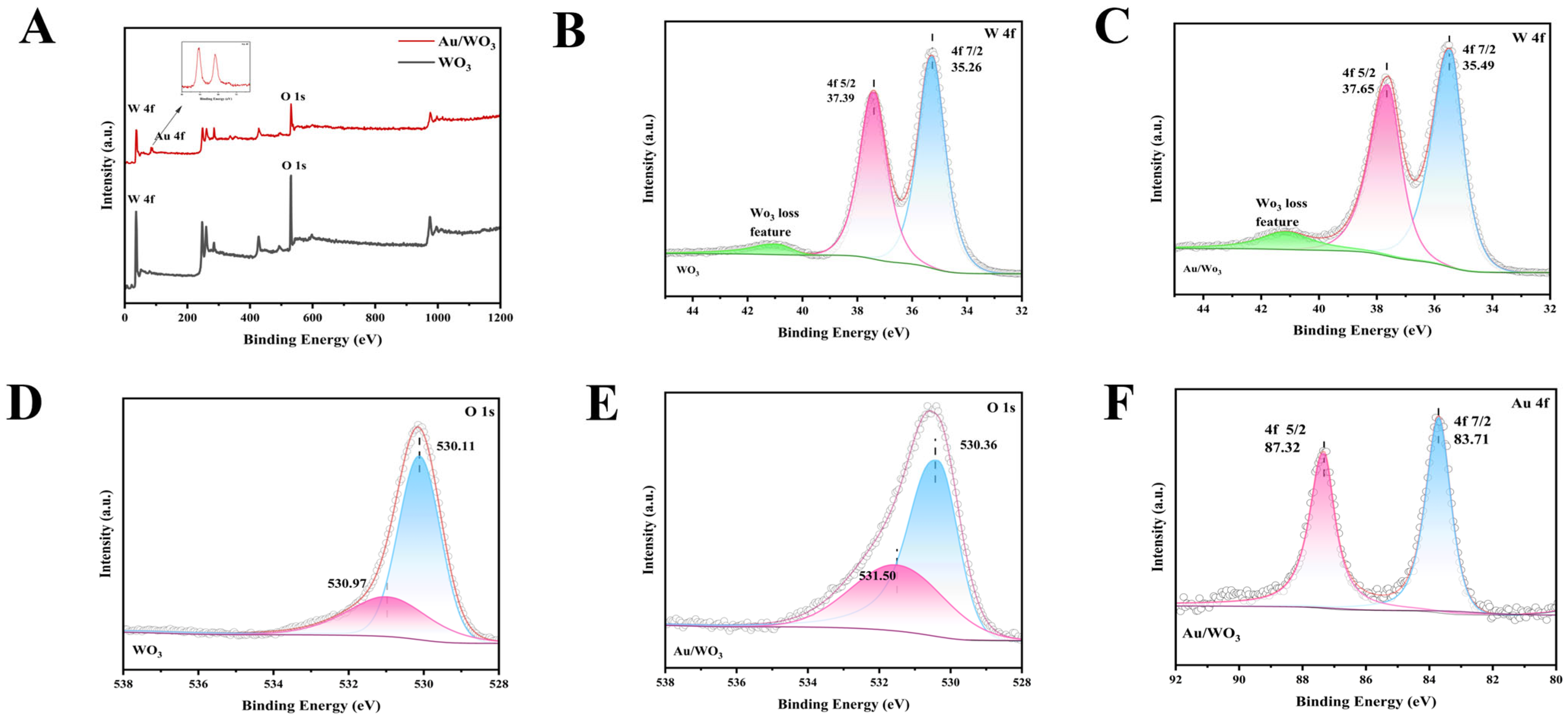
Surface chemical composition and electronic structure of the substrates were investigated by X-ray photoelectron spectroscopy (XPS). All XPS spectra were charge-corrected by referencing the C 1s peak to 284.8 eV, with the corresponding C 1s spectrum provided in Figure S7. Figure 3A presents the survey spectra of both WO3 and Au/WO3, confirming the successful incorporation of Au through the emergence of Au 4f peaks in the composite material. High-resolution spectra of the W 4f region are displayed in Figure 3B,C. For pristine WO3, the spin-orbit doublet corresponding to W 4f7/2 and W 4f5/2 appeared at binding energies of 35.26 eV and 37.39 eV, respectively. After Au deposition, these peaks shifted to 35.49 eV and 37.65 eV, representing a positive shift of approximately 0.25 eV. This increase in binding energy suggests electron transfer from W to Au, resulting in reduced electron density around tungsten nuclei and decreased shielding of core electrons, consistent with the formation of a Schottky junction at the Au/WO3 interface [47]. As further supported by the deconvolution of the high-resolution W 4f spectrum (Figure S7), the appearance of W5+ species following Au deposition indicates an increased presence of oxygen vacancies [48]. Figure 3D,E show the corresponding O 1s spectra, which exhibit noticeable asymmetry and broadening. Each spectrum was fitted with two components corresponding to lattice oxygen (W–O) at lower binding energy and surface-adsorbed oxygen (e.g., –OH or adsorbed H2O) at higher binding energy. The binding energies of the O 1s peaks in WO3 were observed at 530.11 eV and 530.97 eV, while those in Au/WO3 shifted to 530.36 eV and 531.50 eV. Moreover, the relative area of the high-binding-energy component (surface-adsorbed oxygen) increased significantly after Au modification, indicating a rise in surface oxygen vacancy defects. This finding aligns well with the W 4f XPS result. Numerous studies have established that oxygen vacancies can effectively enhance the SERS performance of semiconductor substrates [49,50,51,52]. XPS analysis in this work indicates that, compared to pure WO3, the higher concentration of oxygen defects in Au/WO3 introduces dangling bonds and induces band tailing, driving the system into a metastable state. This electronic configuration facilitates the escape and transfer of surface electrons. The resulting electron delocalization promotes more efficient interfacial charge transfer, thereby significantly improving SERS activity [53,54,55]. As shown in Figure 3F, the Au 4f spectrum displays two well-defined peaks at binding energies of 83.71 eV and 87.32 eV, corresponding to Au0 4f7/2 and 4f5/2, respectively, confirming the metallic nature of the deposited Au nanoparticles on the WO3 nanostructure.
To elucidate the interfacial binding chemistry, particularly in the absence of conventional capping agents, the surface charge of the Au/WO3 composite was investigated. Zeta potential measurement confirms that the substrate possesses a negative surface charge (see Table S5). This inherent negativity is crucial for the initial electrostatic adsorption of cationic probe molecules like R6G. We attribute this negative charge to the adsorption of anionic species, primarily chloride ions (Cl) and possibly gold-chloro complexes, which are inherent by-products from the hydrolysis and photoreduction of the HAuCl4 precursor [56,57]. These species adsorb onto the Au NP surface during the in situ growth, forming a negative electrical layer that functionally mimics the role of citrate in traditional syntheses, thereby facilitating the electrostatic capture of positively charged molecules and contributing to nanoparticle stabilization.
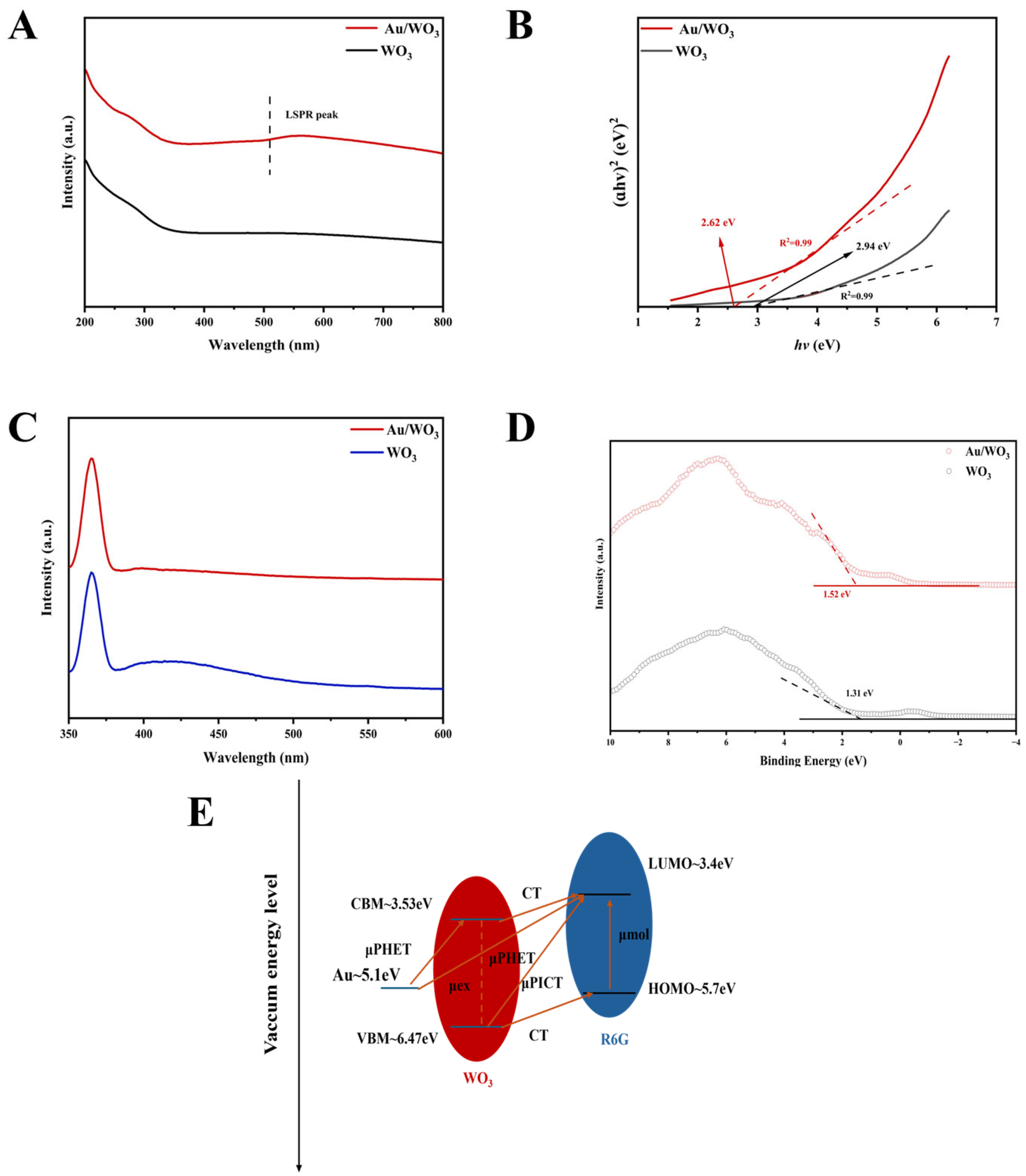
As shown in Figure 4A,B, the experimental results reveal that Au/WO3 exhibits enhanced optical absorption and a reduced bandgap compared to pure WO3, accompanied by a distinct localized surface plasmon resonance (LSPR) absorption feature [58]. To ensure the accuracy and reproducibility of the bandgap determination, the Tauc plots were quantitatively analyzed. The optical bandgap (Eg) was determined by applying a linear fit to the rising edge of the (αhν)2 versus photon energy (hν) plot using a nonlinear fitting tool in Origin Lab software. The fitting interval was carefully selected based on the linearity of the data points, ensuring a high coefficient of determination (R2 > 0.99). The bandgap value was then obtained by extrapolating the fitted line to the intercept with the hν axis, where (αhν)2 = 0. This quantitative fitting method yielded bandgaps of 2.94 eV for pristine WO3 and 2.62 eV for Au/WO3. We note that these values are in close agreement (within 0.01 eV) with our initial estimates, confirming the robustness of the reported bandgap trend and values, while providing a more objective and rigorous foundation. It is well established that a narrower bandgap promotes more efficient visible-light absorption, leading to increased generation of photogenerated charge carriers. This effect facilitates improved charge transfer processes, thereby significantly enhancing the Raman signal intensity. Simultaneously, the capture of photoexcited carriers within the substrate effectively reduces noise arising from resonant Raman scattering [25]. Recent studies have revealed that a portion of the observed SERS signal may originate from photoluminescence (PL) modulated by noble metal nanostructures. However, due to photon scattering mechanisms, PL typically contributes to an unwanted background interference in SERS measurements [59]. Interestingly, Yang et al. elucidated the role of charge transfer in Nd-doped ZnO systems, demonstrating a direct correlation between PL quenching and SERS enhancement [60]. As shown in the PL spectrum in Figure 4C, a significant fluorescence quenching effect is observed in Au/WO3 compared to pure WO3. The quenching peak aligns with the plasmon resonance band of Au NPs, confirming the contribution of an electromagnetic enhancement mechanism. The suppression of PL emission helps mitigate interference from substrate-originated luminescence, which is commonly perceived as Raman noise. The reduced background facilitates the detection of weak molecular Raman signals, which is particularly crucial for trace analyte detection. Therefore, surface modification with Au NPs enhances the detection sensitivity not only by reducing the recombination rate of photogenerated charge carriers but also by minimizing spectral background interference.
The electronic band structures of WO3 and Au/WO3 were further probed using valence band X-ray photoelectron spectroscopy (VB-XPS), as depicted in Figure 4D. The valence band maximum (VBM) was measured at 1.31 eV for pristine WO3 and shifted to 1.52 eV after Au deposition, indicating a modification of the electronic environment. To thermodynamically rationalize the charge transfer process, we evaluated potential charge transfer pathways between the substrate and Rhodamine 6G (R6G) molecules, with a schematic illustration provided in Figure 4E. Based on Tauc plot analysis and VB-XPS results, the conduction band (CB) and valence band (VB) positions of WO3 were determined to be −3.53 eV and −6.47 eV, respectively. The Fermi level (EF) of Au is –5.1 eV [61,62], while the highest occupied molecular orbital (HOMO) and the lowest unoccupied molecular orbital (LUMO) of R6G lie at −5.7 eV and −3.4 eV, respectively. Under 532 nm laser excitation, four distinct resonance mechanisms contribute synergistically to the chemical enhancement: molecular resonance (μmol) within R6G, exciton resonance (μex) in WO3, photoinduced charge transfer (μPICT) between the semiconductor and molecular energy levels, and plasmon-induced hot electron transfer (μPHET). The energy differences from the Fermi level of Au and the VB of WO3 to the LUMO of R6G are 1.7 eV and 3.07 eV, respectively, enabling direct electron transfer from both Au and WO3 to the LUMO of R6G under laser irradiation. This multi-pathway charge injection considerably increases the electron transfer probability and enhances the SERS activity. Additionally, the energy offset between the Fermi level of Au and the CB of WO3 is 1.57 eV, permitting efficient electron transfer from Au to the CB of WO3, thereby further improving charge separation and transfer efficiency. The collective contribution of these interfacial charge transfer processes significantly enhances the SERS performance. Furthermore, the localized electric field generated by Au nanoparticles amplifies the polarization effect resulting from charge transfer, leading to additional SERS enhancement. Through the combined electromagnetic and chemical enhancement mechanisms, the charge transfer between R6G and the Au/WO3 composite is markedly facilitated, resulting in a high enhancement factor of 4.09 × 10⁶ and a detection limit as low as 10−11 M.

4. Conclusions

This work presents a rationally designed Au/WO3 hybrid substrate for surface-enhanced Raman scattering (SERS), fabricated through a green and scalable UV-induced photoreduction strategy. This approach enables the additive-free, in situ deposition of Au nanoparticles onto WO3 nanowires, ensuring an atomically clean interface and offering a viable route toward large-scale production. The exceptional SERS performance of the substrate stems from the synergistic interplay between electromagnetic enhancement from Au nanoparticles and chemical enhancement facilitated by the WO3 support, further amplified by the formation of a Schottky junction and an increased concentration of oxygen vacancies. The substrate demonstrates remarkable operational stability, maintaining 75% of its initial SERS activity after 100 days, along with excellent reproducibility (relative standard deviation of 4.05%). It achieves highly sensitive detection of Rhodamine 6G down to 10−11 M and reliably detects the nerve agent simulant DMMP at 10−4 M, confirming its practical potential for trace analyte sensing. This study not only provides fundamental insights into the charge transfer mechanisms in semiconductor–metal heterostructures but also establishes a robust and eco-friendly fabrication pathway, highlighting a significant step toward the real-world application of SERS technology in environmental and security monitoring.

Supplementary Materials

The following supporting information can be downloaded at: https://www.mdpi.com/article/10.3390/colloids9050070/s1, Figure S1. SEM images of (A) WO3, (B) 150 μL HAuCl4 Au/WO3 and (C) 300 μL HAuCl4 Au/WO3 (D) 450 μL HAuCl4 Au/WO3; Figure S2. Size distribution histograms of Au NPs on WO3; Figure S3. Photos taken by iphone of (A) 150 μL HAuCl4 Au/WO3, (B) 300 μL HAuCl4 Au/WO3 and (C) 450 μL HAuCl4 Au/WO3; Figure S4. SERS performance diagrams with different contents of Au nanoparticles; Figure S5. The uniformity of R6G Raman signals of Au/WO3 at 20 random positions; Figure S6. (A) Raman peak background of WO3 and Au/WO3 at 785 nm, (B) SERS Performance of DMMP under Different Solvent Conditions, (C) (B) SERS Performance of DMMP under Different Solvent Conditions; Figure S7. The XPS W4f spectrum of (A) WO3; (B) Au/WO3, The XPS C1s spectrum of (C) WO3, (D) Au/WO3; Table S1. Experimental reagents; Table S2. Mode assignment of the Raman peaks for R6G; Table S3. Comparison of EFs and limits of detection (LODs) of various semiconductors with SERS activity in the literature (R6G); Table S4. Mode assignment of the Raman peaks for DMMP; Table S5. Zeta potential; Scheme S1. Synthesis diagram of Au/WO3 species [63,64,65,66,67,68].

Author Contributions

Conceptualization, S.M.; methodology, S.M.; validation, C.W. and D.L.; investigation, S.M. and Y.F.; writing—original draft preparation, Q.D. and Y.L.; writing—review and editing, Q.D., L.Z. and L.F.; supervision, C.W.; funding acquisition, C.W. All authors have read and agreed to the published version of the manuscript.

Funding

This work was supported by the National Natural Science Foundation of China (Grant Nos. 12574003, 52471250), Beijing Natural Science Foundation (Grant Nos. L248027 and L245019).

Data Availability Statement

The original contributions presented in this study are included in the article. Further inquiries can be directed to the corresponding authors.

Conflicts of Interest

The authors declare no conflict of interest.

References

  1. Hassanain, W.A.; Izake, E.L. Toward Label-Free SERS Detection of Proteins through Their Disulfide Bond Structure. SLAS Discov. Adv. Sci. Drug Discov. 2020, 25, 87–94. [Google Scholar] [CrossRef] [PubMed]
  2. Feng, R.; Miao, Q.; Zhang, X.; Cui, P.; Wang, C.; Feng, Y.; Gan, L.; Fu, J.; Wang, S.; Dai, Z.; et al. Single-atom sites on perovskite chips for record-high sensitivity and quantification in SERS. Sci. China Mater. 2022, 65, 1601–1614. [Google Scholar] [CrossRef]
  3. Feng, R.; Fu, S.; Liu, H.; Wang, Y.; Liu, S.; Wang, K.; Chen, B.; Zhang, X.; Hu, L.; Chen, Q.; et al. Single-Atom Site SERS Chip for Rapid, Ultrasensitive, and Reproducible Direct-Monitoring of RNA Binding. Adv. Healthc. Mater. 2024, 13, e2301146. [Google Scholar] [CrossRef] [PubMed]
  4. Wang, K.; Gao, Y.; Fang, Z.; Li, H.; Feng, R.; Wang, Y.; Feng, Y.; Li, W.; Zhang, S.; Hu, L.; et al. SERS detection for pesticide residue via a single-atom sites decoration strategy. Appl. Surf. Sci. 2023, 621, 156832. [Google Scholar] [CrossRef]
  5. Dai, X.; Fu, W.; Chi, H.; Mesias, V.S.D.; Zhu, H.; Leung, C.W.; Liu, W.; Huang, J. Optical tweezers-controlled hotspot for sensitive and reproducible surface-enhanced Raman spectroscopy characterization of native protein structures. Nat. Commun. 2021, 12, 1292. [Google Scholar] [CrossRef] [PubMed]
  6. Yang, L.; Kim, T.; Cho, H.; Luo, J.; Lee, J.; Chueng, S.D.; Hou, Y.; Yin, P.T.; Han, J.; Kim, J.H.; et al. Hybrid Graphene-Gold Nanoparticle-Based Nucleic Acid Conjugates for Cancer-Specific Multimodal Imaging and Combined Therapeutics. Adv. Funct. Mater. 2020, 31, 2006918. [Google Scholar] [CrossRef]
  7. Wang, J.; Zhao, J.; Xiao, L.; Zhao, X.; He, S.; Zhou, Y.; Liu, J.; Li, Y.; Peng, L.; Liu, W. Bio-inspired 3D Sub-Microstructures Coupling of Au Nanoparticles for SERS Detection of Trace Fentanyl. Adv. Funct. Mater. 2024, 34, 2411048. [Google Scholar] [CrossRef]
  8. Wu, L.; Tang, X.; Wu, T.; Zeng, W.; Zhu, X.; Hu, B.; Zhang, S. A review on current progress of Raman-based techniques in food safety: From normal Raman spectroscopy to SESORS. Food Res. Int. 2023, 169, 112944. [Google Scholar] [CrossRef]
  9. Afroozeh, A. A Review of Developed Surface-Enhanced Raman Spectroscopy (SERS)-Based Sensors for the Detection of Common Hazardous Substances in the Agricultural Industry. Plasmonics 2024, 20, 63–81. [Google Scholar] [CrossRef]
  10. Li, X.; Xu, C.; Yan, L.; Feng, Y.; Li, H.; Ye, C.; Zhang, M.; Jiang, C.; Li, J.; Wu, Y. A plasmonic AgNP decorated heterostructure substrate for synergetic surface-enhanced Raman scattering identification and quantification of pesticide residues in real samples. Anal. Methods 2022, 14, 3250–3259. [Google Scholar] [CrossRef]
  11. Ustun, O.; Yilmaz, A.; Yilmaz, M. Catalytic and SERS activities of WO3-based nanowires: The effect of oxygen vacancies, silver nanoparticle doping, and the type of organic dye. Phys. Chem. Chem. Phys. 2022, 24, 18615–18626. [Google Scholar] [CrossRef] [PubMed]
  12. Chen, B.; Meng, S.; Liu, D.; Deng, Q.; Wang, C. In Situ SERS Monitoring of Schiff Base Reactions via Nanoparticles on a Mirror Platform. Catalysts 2024, 14, 803. [Google Scholar] [CrossRef]
  13. Han, X.X.; Rodriguez, R.S.; Haynes, C.L.; Ozaki, Y.; Zhao, B. Surface-enhanced Raman spectroscopy. Nat. Rev. Methods Prim. 2021, 1, 87. [Google Scholar] [CrossRef]
  14. Zhan, C.; Chen, X.-J.; Yi, J.; Li, J.-F.; Wu, D.-Y.; Tian, Z.-Q. From plasmon-enhanced molecular spectroscopy to plasmon-mediated chemical reactions. Nat. Rev. Chem. 2018, 2, 216–230. [Google Scholar] [CrossRef]
  15. Ding, S.-Y.; You, E.-M.; Tian, Z.-Q.; Moskovits, M. Electromagnetic theories of surface-enhanced Raman spectroscopy. Chem. Soc. Rev. 2017, 46, 4042–4076. [Google Scholar] [CrossRef] [PubMed]
  16. Alessandri, I.; Lombardi, J.R. Enhanced Raman Scattering with Dielectrics. Chem. Rev. 2016, 116, 14921–14981. [Google Scholar] [CrossRef] [PubMed]
  17. Yang, B.; Shao, X.; Gu, X.; Wang, K.; Ning, X.; Xia, J.; Xie, M.; Tang, Y.; Li, Q.; Tian, S. CuO@AgNPs nanozyme cavity arrays on screen-printed electrodes for ultrasensitive and on-site SERS detection. Chem. Eng. J. 2023, 471, 144522. [Google Scholar] [CrossRef]
  18. Wang, X.; Shi, W.; Jin, Z.; Huang, W.; Lin, J.; Ma, G.; Li, S.; Guo, L. Remarkable SERS Activity Observed from Amorphous ZnO Nanocages. Angew. Chem. Int. Ed. Engl. 2017, 56, 9851–9855. [Google Scholar] [CrossRef]
  19. Estevez, M.; Alvarez, M.; Lechuga, L. Integrated optical devices for lab-on-a-chip biosensing applications. Laser Photon-Rev. 2011, 6, 463–487. [Google Scholar] [CrossRef]
  20. Simas, M.V.; Davis, G.A.; Hati, S.; Pu, J.; Goodpaster, J.V.; Sardar, R. Anisotropically Shaped Plasmonic WO3−x Nanostructure-Driven Ultrasensitive SERS Detection and Machine Learning-Based Differentiation of Nitro-Explosives. ACS Appl. Mater. Interfaces 2025, 17, 11309–11324. [Google Scholar] [CrossRef]
  21. Lombardi, J.R.; Birke, R.L. Theory of Surface-Enhanced Raman Scattering in Semiconductors. J. Phys. Chem. C 2014, 118, 11120–11130. [Google Scholar] [CrossRef]
  22. Park, W.-H.; Kim, Z.H. Charge Transfer Enhancement in the SERS of a Single Molecule. Nano Lett. 2010, 10, 4040–4048. [Google Scholar] [CrossRef]
  23. Wang, X.; Guo, L. SERS Activity of Semiconductors: Crystalline and Amorphous Nanomaterials. Angew. Chem. Int. Ed. Engl. 2019, 59, 4231–4239. [Google Scholar] [CrossRef]
  24. Li, Q.; Wang, J.; Ding, Q.; Chen, M.; Ma, F. Coupling effect on charge-transfer mechanism of surface-enhanced resonance Raman scattering. J. Raman Spectrosc. 2017, 48, 560–569. [Google Scholar] [CrossRef]
  25. Langer, J.; Jimenez de Aberasturi, D.; Aizpurua, J.; Alvarez-Puebla, R.A.; Auguié, B.; Baumberg, J.J.; Bazan, G.C.; Bell, S.E.J.; Boisen, A.; Brolo, A.G.; et al. Present and Future of Surface-Enhanced Raman Scattering. ACS Nano 2020, 14, 28–117. [Google Scholar] [CrossRef]
  26. Yang, B.; Jin, S.; Guo, S.; Park, Y.; Chen, L.; Zhao, B.; Jung, Y.M. Recent Development of SERS Technology: Semiconductor-Based Study. ACS Omega 2019, 4, 20101–20108. [Google Scholar] [CrossRef] [PubMed]
  27. Perala, R.S.; Chandrasekar, N.; Balaji, R.; Alexander, P.S.; Humaidi, N.Z.N.; Hwang, M.T. A comprehensive review on graphene-based materials: From synthesis to contemporary sensor applications. Mater. Sci. Eng. R Rep. 2024, 159, 100805. [Google Scholar] [CrossRef]
  28. Khyzhun, O.; Solonin, Y.; Dobrovolsky, V. Electronic structure of hexagonal tungsten trioxide: XPS, XES, and XAS studies. J. Alloys Compd. 2001, 320, 1–6. [Google Scholar] [CrossRef]
  29. Zou, J.-W.; Li, Z.-D.; Kang, H.-S.; Zhao, W.-Q.; Liu, J.-C.; Chen, Y.-L.; Ma, L.; Hou, H.-Y.; Ding, S.-J. Strong Visible Light Absorption and Abundant Hotspots in Au-Decorated WO3 Nanobricks for Efficient SERS and Photocatalysis. ACS Omega 2021, 6, 28347–28355. [Google Scholar] [CrossRef]
  30. Yu, J.; Chen, C.; Zhang, Q.; Lin, J.; Yang, X.; Gu, L.; Zhang, H.; Liu, Z.; Wang, Y.; Zhang, S.; et al. Au Atoms Anchored on Amorphous C3N4 for Single-Site Raman Enhancement. J. Am. Chem. Soc. 2022, 144, 21908–21915. [Google Scholar] [CrossRef]
  31. Chen, B.; Fan, L.; Li, C.; Xia, L.; Wang, K.; Wang, J.; Pang, D.; Zhu, Z.; Ma, P. Au nanoparticles decorated β-Bi2O3 as highly-sensitive SERS substrate for detection of methylene blue and methyl orange. Analyst 2024, 149, 4283–4294. [Google Scholar] [CrossRef] [PubMed]
  32. Xu, L.; Zhang, H.; Tian, Y.; Jiao, A.; Chen, F.; Chen, M. Photochemical synthesis of ZnO@Au nanorods as an advanced reusable SERS substrate for ultrasensitive detection of light-resistant organic pollutant in wastewater. Talanta 2019, 194, 680–688. [Google Scholar] [CrossRef]
  33. Huang, J.; Ma, D.; Chen, F.; Chen, D.; Bai, M.; Xu, K.; Zhao, Y. Green in Situ Synthesis of Clean 3D Chestnutlike Ag/WO3−x Nanostructures for Highly Efficient, Recyclable and Sensitive SERS Sensing. ACS Appl. Mater. Interfaces 2017, 9, 7436–7446. [Google Scholar] [CrossRef]
  34. Zhai, Y.; DuChene, J.S.; Wang, Y.-C.; Qiu, J.; Johnston-Peck, A.C.; You, B.; Guo, W.; DiCiaccio, B.; Qian, K.; Zhao, E.W.; et al. Polyvinylpyrrolidone-induced anisotropic growth of gold nanoprisms in plasmon-driven synthesis. Nat. Mater. 2016, 15, 889–895. [Google Scholar] [CrossRef]
  35. Uboldi, C.; Bonacchi, D.; Lorenzi, G.; Hermanns, M.I.; Pohl, C.; Baldi, G.; E Unger, R.; Kirkpatrick, C.J. Gold nanoparticles induce cytotoxicity in the alveolar type-II cell lines A549 and NCIH441. Part. Fibre Toxicol. 2009, 6, 18. [Google Scholar] [CrossRef]
  36. Lopez-Sanchez, J.A.; Dimitratos, N.; Hammond, C.; Brett, G.L.; Kesavan, L.; White, S.; Miedziak, P.; Tiruvalam, R.; Jenkins, R.L.; Carley, A.F.; et al. Facile removal of stabilizer-ligands from supported gold nanoparticles. Nat. Chem. 2011, 3, 551–556. [Google Scholar] [CrossRef]
  37. Amendola, V.; Litti, L.; Meneghetti, M. LDI-MS Assisted by Chemical-Free Gold Nanoparticles: Enhanced Sensitivity and Reduced Background in the Low-Mass Region. Anal. Chem. 2013, 85, 11747–11754. [Google Scholar] [CrossRef]
  38. Ma, S.; Xue, J.; Zhou, Y.; Zhang, Z. Photochemical synthesis of ZnO/Ag2O heterostructures with enhanced ultraviolet and visible photocatalytic activity. J. Mater. Chem. A 2014, 2, 7272–7280. [Google Scholar] [CrossRef]
  39. Ameer, F.S.; Pittman, C.U.; Zhang, D. Quantification of Resonance Raman Enhancement Factors for Rhodamine 6G (R6G) in Water and on Gold and Silver Nanoparticles: Implications for Single-Molecule R6G SERS. J. Phys. Chem. C 2013, 117, 27096–27104. [Google Scholar] [CrossRef]
  40. Zheng, Q.; Fu, Y.-C.; Xu, J.-Q. Advances in the chemical sensors for the detection of DMMP—A simulant for nerve agent sarin. Procedia Eng. 2010, 7, 179–184. [Google Scholar] [CrossRef]
  41. Bo, X.; Tang, A.; Dou, M.; Li, Z.; Wang, F. Controllable electrodeposition and mechanism research of nanostructured Bi2Te3 thin films with high thermoelectric properties. Appl. Surf. Sci. 2019, 486, 65–71. [Google Scholar] [CrossRef]
  42. Lin, N.; Lin, Y.; Qian, X.; Wang, X.; Su, W. Construction of a 2D/2D WO3/LaTiO2N Direct Z-Scheme Photocatalyst for Enhanced CO2 Reduction Performance Under Visible Light. ACS Sustain. Chem. Eng. 2021, 9, 13686–13694. [Google Scholar] [CrossRef]
  43. Dokou, E.; Stangland, E.E.; Andres, R.P.; Delgass, W.N.; Barteau, M.A. Comparison of AFM and HRTEM to determine the metal particle morphology and loading of an Au/TiO2 catalyst. Catal. Lett. 2000, 70, 1–7. [Google Scholar] [CrossRef]
  44. Sahu, B.K.; Dwivedi, A.; Pal, K.K.; Pandian, R.; Dhara, S.; Das, A. Optimized Au NRs for efficient SERS and SERRS performances with molecular and longitudinal surface plasmon resonance. Appl. Surf. Sci. 2021, 537, 147615. [Google Scholar] [CrossRef]
  45. Kim, N.H.; Kim, K. Surface-enhanced resonance Raman scattering of rhodamine 6G on Pt nanoaggregates. J. Raman Spectrosc. 2005, 36, 623–628. [Google Scholar] [CrossRef]
  46. Taranenko, N.; Alarie, J.-P.; Stokes, D.L.; Vo-Dinh, T. Surface-Enhanced Raman Detection of Nerve Agent Simulant (DMMP and DIMP) Vapor on Electrochemically Prepared Silver Oxide Substrates. J. Raman Spectrosc. 1996, 27, 379–384. [Google Scholar] [CrossRef]
  47. Kim, S.; Park, S.; Park, S.; Lee, C. Acetone sensing of Au and Pd-decorated WO3 nanorod sensors. Sens. Actuators B Chem. 2015, 209, 180–185. [Google Scholar] [CrossRef]
  48. Foster, A.S.; Gejo, F.L.; Shluger, A.L.; Nieminen, R.M. Vacancy and interstitial defects in hafnia. Phys. Rev. B 2002, 65, 174117. [Google Scholar] [CrossRef]
  49. Cong, S.; Yuan, Y.; Chen, Z.; Hou, J.; Yang, M.; Su, Y.; Zhang, Y.; Li, L.; Li, Q.; Geng, F.; et al. Noble metal-comparable SERS enhancement from semiconducting metal oxides by making oxygen vacancies. Nat. Commun. 2015, 6, 7800. [Google Scholar] [CrossRef]
  50. Liu, H.; Chen, L.; Li, B.; Song, H.; Tan, C.L.; Shi, Y.; Yan, S. Semiconducting Tungsten Trioxide Thin Films for High-Performance SERS Biosensors. Nanomaterials 2025, 15, 1393. [Google Scholar] [CrossRef]
  51. McMillan, K.S.; McCluskey, A.G.; Sorensen, A.; Boyd, M.; Zagnoni, M. Emulsion technologies for multicellular tumour spheroid radiation assays. Analyst 2015, 141, 100–110. [Google Scholar] [CrossRef]
  52. Liang, J.; Zhang, L.; Wang, S.; Yu, Y.; Lei, D. Oxygen Vacancies Enhance SERS Performance of Tungsten-Doped Vanadium Dioxide Nanoparticles. Adv. Mater. Technol. 2024, 10, 2401304. [Google Scholar] [CrossRef]
  53. Sharma, S.; Kumar, R.; Yadav, R.M. Polyacrylonitrile as a versatile matrix for gold nanoparticle-based SERS substrates. Nanoscale Adv. 2024, 6, 1065–1073. [Google Scholar] [CrossRef]
  54. Yu, J.; Chen, C.; Lin, J.; Meng, X.; Qiu, L.; Wang, X. Amorphous Co(OH)2 nanocages achieving efficient photo-induced charge transfer for significant SERS activity. J. Mater. Chem. C 2022, 10, 1632–1637. [Google Scholar] [CrossRef]
  55. Wei, W.; Yao, Y.; Zhao, Q.; Xu, Z.; Wang, Q.; Zhang, Z.; Gao, Y. Oxygen defect-induced localized surface plasmon resonance at the WO3−x quantum dot/silver nanowire interface: SERS and photocatalysis. Nanoscale 2019, 11, 5535–5547. [Google Scholar] [CrossRef]
  56. Sneed, B.T.; Young, A.P.; Tsung, C.-K. Building up strain in colloidal metal nanoparticle catalysts. Nanoscale 2015, 7, 12248–12265. [Google Scholar] [CrossRef]
  57. Ji, X.; Song, X.; Li, J.; Bai, Y.; Yang, W.; Peng, X. Size Control of Gold Nanocrystals in Citrate Reduction: The Third Role of Citrate. J. Am. Chem. Soc. 2007, 129, 13939–13948. [Google Scholar] [CrossRef] [PubMed]
  58. Liu, W.; Bai, H.; Li, X.; Li, W.; Zhai, J.; Li, J.; Xi, G. Improved Surface-Enhanced Raman Spectroscopy Sensitivity on Metallic Tungsten Oxide by the Synergistic Effect of Surface Plasmon Resonance Coupling and Charge Transfer. J. Phys. Chem. Lett. 2018, 9, 4096–4100. [Google Scholar] [CrossRef]
  59. Sun, J.; Hu, H.; Zheng, D.; Zhang, D.; Deng, Q.; Zhang, S.; Xu, H. Light-Emitting Plexciton: Exploiting Plasmon–Exciton Interaction in the Intermediate Coupling Regime. ACS Nano 2018, 12, 10393–10402. [Google Scholar] [CrossRef]
  60. Yang, S.; Yao, J.; Quan, Y.; Hu, M.; Su, R.; Gao, M.; Han, D.; Yang, J. Monitoring the charge-transfer process in a Nd-doped semiconductor based on photoluminescence and SERS technology. Light Sci. Appl. 2020, 9, 117. [Google Scholar] [CrossRef]
  61. Wang, X.-J.; Tian, X.; Sun, Y.-J.; Zhu, J.-Y.; Li, F.-T.; Mu, H.-Y.; Zhao, J. Enhanced Schottky effect of a 2D–2D CoP/g-C3N4 interface for boosting photocatalytic H2 evolution. Nanoscale 2018, 10, 12315–12321. [Google Scholar] [CrossRef] [PubMed]
  62. Handoko, A.D.; Steinmann, S.N.; Seh, Z.W. Theory-guided materials design: Two-dimensional MXenes in electro- and photocatalysis. Nanoscale Horiz. 2019, 4, 809–827. [Google Scholar] [CrossRef]
  63. Hung, S.-F.; Xu, A.; Wang, X.; Li, F.; Hsu, S.-H.; Li, Y.; Wicks, J.; Cervantes, E.G.; Rasouli, A.S.; Li, Y.C.; et al. A Metal-Supported Single-Atom Catalytic Site Enables Carbon Dioxide Hydrogenation. Nat. Commun. 2022, 13, 819. [Google Scholar] [CrossRef]
  64. Lei, Z.; Zhang, X.; Zhao, Y.; Wei, A.; Tao, L.; Yang, Y.; Zheng, Z.; Tao, L.; Yu, P.; Li, J. Enhanced Raman Scattering on Two-Dimensional Palladium Diselenide. Nanoscale 2022, 14, 4181–4187. [Google Scholar] [CrossRef]
  65. Yang, L.; Peng, Y.; Yang, Y.; Liu, J.; Li, Z.; Ma, Y.; Zhang, Z.; Wei, Y.; Li, S.; Huang, Z.; et al. Green and Sensitive Flexible Semiconductor SERS Substrates: Hydrogenated Black TiO2 Nanowires. ACS Appl. Nano Mater. 2018, 1, 4516–4527. [Google Scholar] [CrossRef]
  66. Lin, J.; Shang, Y.; Li, X.; Yu, J.; Wang, X.; Guo, L. Ultrasensitive SERS Detection by Defect Engineering on Single Cu2O Superstructure Particle. Adv. Mater. 2017, 29, 1604797. [Google Scholar] [CrossRef]
  67. Zheng, X.; Zhong, H.; Wang, Z.; Li, J.; Hu, Y.; Li, H.; Jia, J.; Zhang, S.; Ren, F. Fabrication of Stable Substoichiometric WOx Films with High SERS Sensitivity by Thermal Treatment. Vacuum 2022, 198, 110884. [Google Scholar] [CrossRef]
  68. Sun, Z.; Gao, Y.; Ban, C.; Meng, J.; Wang, J.; Wang, K.; Gan, L. 3 Nm-Wide WO3−x Nanorods with Abundant Oxygen Vacancies as Substrates for High-Sensitivity SERS Detection. ACS Appl. Nano Mater. 2023, 6, 8635–8642. [Google Scholar] [CrossRef]
Figure 1. (A) XRD patterns of Au/WO3 and WO3; (B) TEM image of Au/WO3; (C) HAADF-STEM image of Au/WO3; (D) HR-TEM image of Au/WO3; (E) HAADF-STEM image of Au/WO3; STEM-EDS mapping images of W, O, and Au.
Colloids 09 00070 g001
Figure 2. (A,B) show the R6G test SERS spectra on Au/WO3 and WO3 substrates; (C) Uniformity of R6G Raman signals at 20 random positions on Au/WO3; (D) Stability test of Au/WO3; (E) Calibration curves of the logarithmic Raman peak (612 cm−1) versus the logarithmic concentration of R6G on Au/WO3 SERS substrate; (F) DMMP test SERS spectra of Au/WO3 and WO3 substrates.
Colloids 09 00070 g002
Figure 3. (A) Full XPS spectra of Au/WO3 and WO3 substrates; (B,C) High-resolution XPS W4f spectra of WO3 and Au/WO3; (D,E) High-resolution XPS O1s spectra of WO3 and Au/WO3; (F) High-resolution XPS Au4f spectrum of Au/WO3.
Colloids 09 00070 g003
Figure 4. Band structure analysis. (A) UV-Vis absorption spectrum. (B) Tauc plot. (C) Steady-state photoluminescence of Au/WO3 and WO3. (D) XPS-VB mapping. (E) Schematic illustrating the possible charge transfer process between Au/WO3 and R6G upon 532 nm excitation.
Colloids 09 00070 g004
Disclaimer/Publisher’s Note: The statements, opinions and data contained in all publications are solely those of the individual author(s) and contributor(s) and not of MDPI and/or the editor(s). MDPI and/or the editor(s) disclaim responsibility for any injury to people or property resulting from any ideas, methods, instructions or products referred to in the content.