Review

Biomimetic Hierarchical Nanocomposite Hydrogels: From Design to Biomedical Applications

1
Musculoskeletal Research Laboratory of Department of Orthopedics & Traumatology, Innovative Orthopaedic Biomaterial & Drug Translational Research Laboratory, Li Ka Shing Institute of Health Sciences, The Chinese University of Hong Kong, Hong Kong 999077, China
2
Department of Chemistry and Biochemistry, University of California, Los Angeles, CA 90095, USA
3
Weintraub Center for Reconstructive Biotechnology, Division of Advanced Prosthodontics, School of Dentistry, University of California, Los Angeles, CA 90095, USA
*
Authors to whom correspondence should be addressed.
These authors contributed equally to this work.
J. Compos. Sci. 2022, 6(11), 340; https://doi.org/10.3390/jcs6110340
Submission received: 19 October 2022 / Accepted: 21 October 2022 / Published: 4 November 2022

Abstract

:
Natural extracellular matrix (ECM) is highly heterogeneous and anisotropic due to the existence of biomacromolecule bundles and pores. Hydrogels have been proposed as ideal carriers for therapeutic cells and drugs in tissue engineering and regenerative medicine. However, most of the homogeneous and isotropic hydrogels cannot fully emulate the hierarchical properties of natural ECM, including the dynamically spatiotemporal distributions of biochemical and biomechanical signals. Biomimetic hierarchical nanocomposite hydrogels have emerged as potential candidates to better recapitulate natural ECM by introducing various nanostructures, such as nanoparticles, nanorods, and nanofibers. Moreover, the nanostructures in nanocomposite hydrogels can be engineered as stimuli-responsive actuators to realize the desirable control of hydrogel properties, thereby manipulating the behaviors of the encapsulated cells upon appropriate external stimuli. In this review, we present a comprehensive summary of the main strategies to construct biomimetic hierarchical nanocomposite hydrogels with an emphasis on the rational design of local hydrogel properties and their stimuli-responsibility. We then highlight cell fate decisions in engineered nanocomposite niches and their recent development and challenges in biomedical applications.

1. Introduction

The natural ECM network is highly heterogeneous and anisotropic because of the existence of the rigid domains of stiff biomacromolecules and their assemblies, for example, collagen fibers [1,2,3]. The ECM provides a structural scaffold via a network of protein–protein and protein–proteoglycan interactions. Because multicellular cells evolved independently in different multicellular lineages, the composition, as well as the properties of the extracellular matrix, varies with multicellular structures [4]. These interactions are involved in the formation of supramolecular assemblies such as collagen fibrils and elastic fibers, in tissue architecture, and in cell–matrix interactions that regulate cell growth and behavior. The biochemical properties of the ECM allow cells to sense and interact with their extracellular environment using various signal transduction pathways. Meanwhile, the physical properties of the ECM, including its rigidity, density, porosity, insolubility and topography (spatial arrangement and orientation), provide a physical signal to the cells [5]. These interactions are involved in the formation of supramolecular assemblies such as collagen fibrils and elastic fibers, in tissue architecture, and in cell–matrix interactions that regulate cell growth and behavior [6]. The heterogeneity of the natural ECM network also changes in spatiotemporal and mechanical dependence. However, few designs of the existing pure hydrogels can emulate the heterogeneity of the natural ECM network. Trappmann et al. designed a synthetic hydrogel matrix tethered with either stiff or soft ligands to mimic the local mechanics of the natural ECM [7]. They further demonstrated that the local stiffness could be decoupled from the bulk stiffness of the whole hydrogel, where the local stiffness was generated by the doping of collagen fibers. These findings give promise to the use of biomimetic hierarchical nanocomposite hydrogels to recapitulate the heterogeneity of the natural ECM network.
Recent advances in nanobiotechnology, hydrogels, and composition techniques enable the use of well-characterized nanostructures to mimic the heterogeneous topology in 3D [8,9,10]. Therefore, biomimetic hierarchical nanocomposite hydrogels are of great interest in the biomedical fields. The biophysical and biochemical properties of the biomimetic hierarchical nanocomposite hydrogels can be rationally designed and tailored to emulate the cellular microenvironment in natural ECM. Most of the existing nanostructures can be classified into two categories: (i) organic nanostructures, such as polymeric nanoparticles, liposomes, extracellular vesicles, etc. and (ii) inorganic nanostructures, such as silica nanoparticles, gold nanoparticles, and nanorods. Both organic and inorganic nanostructures can be further engineered with various functionalities, such as stimuli-responsive properties, fast in-vivo clearance, and high loading capacity. Biomaterials-based scaffolds, especially hydrogels, hold considerable promise with respect to enhancing the efficacy of tissue engineering and regenerative medicine [11,12,13,14]. Previous studies have shown that hydrogels can be rationally designed to mimic the structures and microenvironments of natural ECM [15,16,17], but they cannot recapitulate the stiff domains due to the soft and deformable nature of polymer networks. Therefore, the combination of nanostructures and hydrogels can maximize the mimicking of the heterogeneous and anisotropic ECM components and organization.
In this review, we first summarize the design principles of biomimetic hierarchical nanocomposite hydrogels with an emphasis on the functionality of the doped nanostructures, including topology manipulation, bioactive reservoir, and ligand presentation. We next classify the various biomedical applications from drug delivery to tissue engineering in cartilage, bone, skin, and nerve fields. This review may shed light on the better design and wilder biomedical applications of biomimetic hierarchical nanocomposite hydrogels in the future.

2. The Functionality of Nanostructures in Biomimetic Hierarchical Nanocomposite Hydrogels

The nanostructures can be engineered with various functionality, including topology manipulation, bioactive reservoir and ligand presentation. The topology of hydrogels influences cell behavior, i.e., —rigid hydrogels promote cell adhesion while soft matrix enables cell spreading. Anisotropic hydrogels lead to focal adhesion formation, while aligned structures guide and organize cell growth. Ligands on hydrogels also affect various cell activities, including adhesion, migration, and differentiation. When nanocomposite hydrogels are used as a bioactive reservoir, they have high loading capacities and are able to release loaded cargo through various stimuli, which further enriched the toolkit of cell regulation. Thus, researchers can pursue one or a combination of those strategies to design hydrogels for different biomedical applications based on their specific demands.

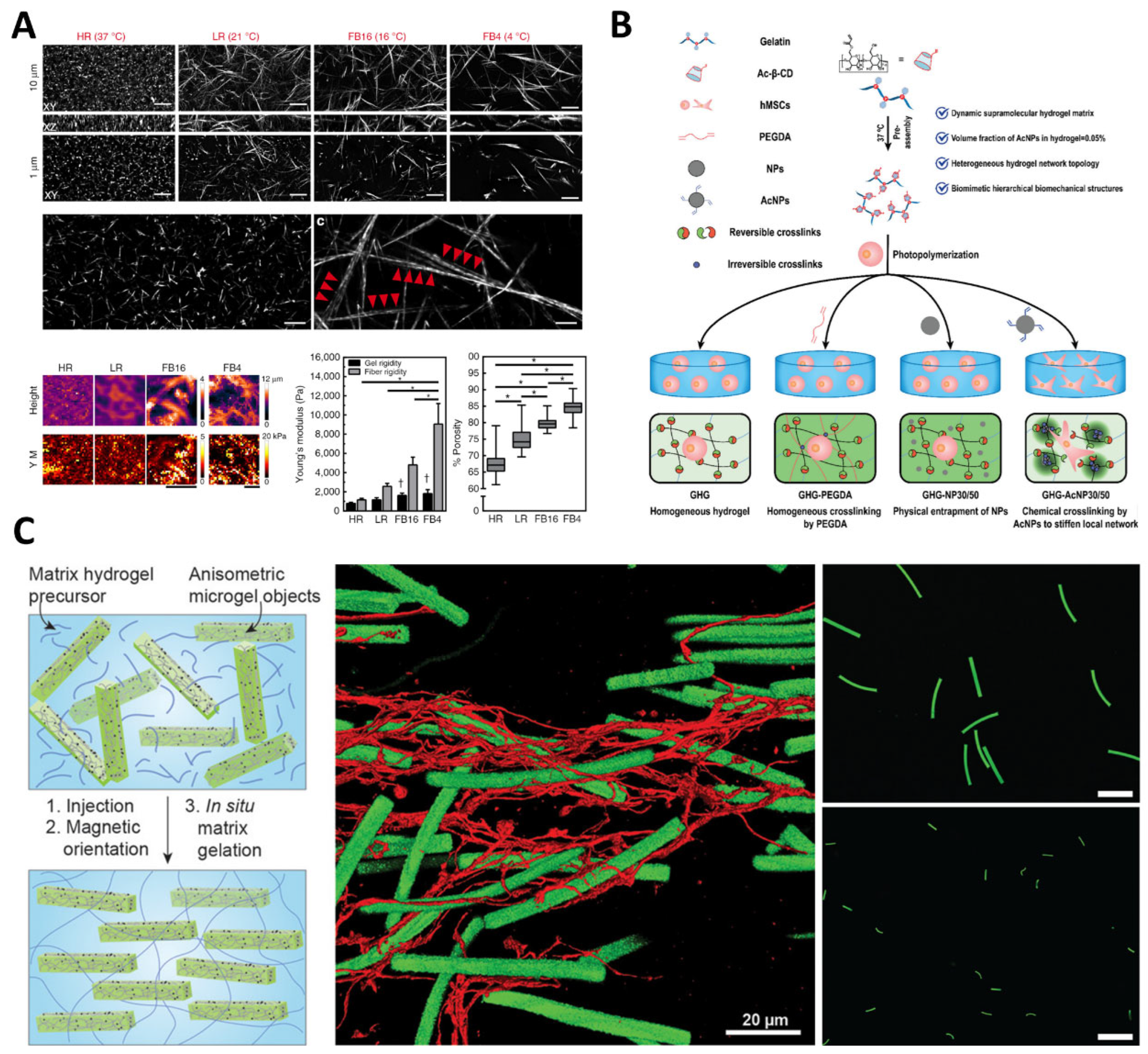
2.1. Topology Manipulation

The biophysical properties of nanocomposite hydrogels are one of the key factors to modulate cell adhesion dynamics. The rigid structures promote the formation and maturation of cell adhesion structures, especially focal adhesions (FAs) [18,19,20]. Kubow et al. reported that the adhesion size in 3D was related to the existence and alignment of collagen fibers or electrospun fibers [21]. Therefore, the nanostructures in nanocomposite hydrogels could be recruited to manipulate the topology of these hydrogels. Doyle et al. reported a local 3D matrix microenvironment in which the local stiffness could be finely tuned through the change of the type of collagen fibers [22]. The microenvironmental ECM was tailored to highly heterogeneous and anisotropic to maximize the focal adhesion formation and maturation (Figure 1A). Yuan et al. developed a dynamic gelatin-based nanocomposite hydrogel providing the local stiffening sites in a soft matrix [23]. The soft matrix enabled the matrix remodeling and cell spreading via the dynamic and reversible host–guest interactions, whereas the stiffened structures strengthened the cell anchoring on the cell adhesive motifs. The encapsulated stem cells exhibited enhanced mechanotransduction and osteogenic differentiation and finally promoted bone regeneration in a bone defect model (Figure 1B). The responsive and on-demand change of hydrogel topology is important to many biomedical applications, especially well-organized and aligned tissues, such as nerves, muscles, and bones. Rose et al. reported an injectable hydrogel doped with magnetoactive objects containing a very small portion of iron oxide nanoparticles [24]. The magnetoactive objects could be induced to aligned status under the external magnetic field. The aligned magnetoactive objects could further induce the aligned growth and organization of neuron promoting localized nerve regeneration (Figure 1C).

2.2. Bioactive Reservoir

The biomimetic hierarchical nanocomposite hydrogels have various type of nanostructures, which has higher loading capacity, more sustained release of loaded cargos, and better manipulation tools compared with those of conventional pure hydrogel networks. Yao et al. reported a bisphosphonate-based hydrogel loaded with various concentrations of magnesium ions [25]. The developed hydrogel exhibited sustained magnesium delivery to the neuron tissues and was able to promote the outgrowth of axons, thereby facilitating peripheral nerve regeneration and functional recovery (Figure 2A). Kang et al. developed a layered double hydroxide-based nanohybrid, where the adenosine was encapsulated inside the interlayer spacing through electrostatic interactions [26]. The sustained release of adenosine acted as a ligand of adenosine A2b receptor (A2bR) and effectively activated the neo-bone formation through various processes, including calcification, mature tissue morphology, and vascularization (Figure 2B). Apart from the free diffusion mechanism, the release of encapsulated cargos in nanocomposite hydrogels can also be triggered and manipulated by internal (pH, redox, enzyme, etc.) and external stimuli (magnetic, light, thermos, etc.). He et al. reported a nanocomposite hydrogel (NH) doped with Pluronic F127 and carbon nanotubes for the treatment of infected wounds [27]. The loading of antibiotic moxifloxacin hydrochloride enabled the pH-responsiveness of the nanocomposite hydrogels. The loaded drugs could be finely delivered and released in an acidic microenvironment in the infected wounds (Figure 2C). Phuong et al. developed a nanocomposite hydrogel embedded with redox-responsive carbon dots [28]. The IR825-loaded carbon dots showed much better fluorescence and photothermal conversion rates upon receiving the stimuli of GSH. The enhanced photothermal properties of IR825@carbon dots under reduction status provided an effective tool for potential cancer treatment (Figure 2D). Qin et al. reported injectable superparamagnetic ferrogels containing iron oxide nanoparticles and Pluronic F127 Indomethacin the iron oxide nanoparticles were loaded into the Pluronic F127 micelles [29]. The release of indomethacin was relatively slow as the diffusion coefficient of hydrophobic drugs in aqueous was quite low when the magnetic field was off. When the magnetic field was switched on, the iron oxide nanoparticles tended to orient and aggregate. Therefore, the micelles were squeezed, and the release of indomethacin was significantly accelerated (Figure 2E). Han et al. developed a light-responsive nanocomposite hydrogel containing PNIPAM backbones and polydopamine nanoparticles [30]. The developed nanocomposite hydrogel showed phase transitions and volume changes under near-infrared (NIR) light. The NIR-induced drug release and NIR-assisted healing could be easily achieved and was able to adapt to various requirements of biomedical applications (Figure 2F).

2.3. Ligand Presentation

The spatiotemporal control of ligand presentation at the nanoscale is highly desirable for the regulation of various cell activities, including adhesion, migration, and differentiation. The nanocomposite hydrogels can either serve as the carrier of ligands or act as the actuator to manipulate the presentation of these ligands. Peng et al. reported a nanocomposite hydrogel carrying a nanoarray of RGD-coated gold nanoparticles [31]. The presentation of the patterned RGD significantly enhanced the adhesion and osteogenic differentiation of mesenchymal stem cells (MSCs) (Figure 3A). Wong et al. developed a soft hydrogel matrix as the cage of RGD-bearing magnetic nanoparticles to reversibly control the presentation of RGD ligands to the seeded stem cells [32]. The “Exposed” and “Hidden” conditions of the RGD ligands can be controlled and switched by applying the “Upward” and “Downward” magnetic fields. The cyclic presentation of RGD ligands maximized the cell adhesion on the hydrogel substrates and significantly promoted the osteogenic differentiation of seeded stem cells (Figure 3B). More recently, Sahar et al. reported an RGD-Modified Alginate−GelMA hydrogel sheet designed for wound healing and soft tissue regeneration. The results confirm that the encapsulated MSCs remain viable within the hydrogel with enhanced collagen deposition. In vivo implantation to excisional wound model in mice confirmed the effectiveness of the GMSC−hydrogel in expediting wound healing via enhancing angiogenesis and suppressing local proinflammatory cytokines [33].

3. The Biomedical Applications of Biomimetic Hierarchical Nanocomposite Hydrogels

Nanocomposite hydrogels with certain formulas are potential materials for various biomedical applications [34]. Customized nanocomposite hydrogels can be used as carriers for cells, drugs, or other bioactive molecules. The incorporation of stimulus-sensitive components with hydrogels allows designed delivery systems to exclusively release specific contents [35]. Although substantial progress in such nanocomposite hydrogels designed for regenerative medicine has been achieved over the past few years, actual clinical uses of nanocomposite hydrogels are rare. From the view of clinical translation, the major concerns could be summarized as stability and biosafety]. Due to the additional interfacial interactions in the nanocomposite hydrogels, the analysis of interactions and the mechanisms of the performance enhancements have become more complicated and fundamental [36]. In some cases, the relatively poor interfacial interactions between the polymer chains and nanomaterials and the uneven dispersion of nanomaterials in the hydrogel matrix dramatically affect the mechanical properties and structural stability of the hydrogels in applications. More importantly, the careful choice of nanomaterials and their concentrations will define the type and intensity of the stimuli that control the release of bioactive molecules from nanocomposite hydrogels. Thus, less or non-toxic nanomaterials should be considered to minimize or eliminate possible side effects of toxic nanomaterials on cells to ensure safe clinical application. Moreover, hydrogels with self-healing and tunable mechanical properties are able to conformally fill irregular injury sites. And the hierarchical 3D structure of hydrogels can provide a suitable microenvironment for cell survival, proliferation, and differentiation. Thus, hydrogels have advantages in tissue regeneration applications [37]. This section summarizes some of the representative biomedical applications of nanocomposite hydrogels, including drug delivery, tissue engineering, and other applications for a comprehensive overview.

3.1. Drug Delivery

With the advantages of good biocompatibility and hydrophilicity, nanocomposite hydrogels can be used to deliver drugs for the treatment of various diseases (Figure 4A). Zhang et al. reported using HA-BP-Mg nanocomposite hydrogel for the controlled and stable release of Mg2+ at bone defect sites to enhance osteogenesis and stimulate bone regeneration [38]. Furthermore, with the combination drug release of Mg2+ and Dexamethasone, the BP-based injectable hydrogel created a positive feedback circuit of drug release regulation, which significantly enhanced bone regeneration at the intended sites. A self-healing hydrogel based on MgSiO3 nanoparticles (NPs) and BP-grafted polymer (HA-BP) has been described by Shi et al. for anti-cancer purposes [39]. Targeted drug delivery was achieved by the protonation of BP and the breaking of chelation with Mg2+ within MgSiO3 NPs. Breast cancer cells (MCF-7) were significantly inhabited by loaded doxorubicin. In terms of breast cancer recurrence prevention, Gao et al. prepared a novel nanocomposite hydrogel functionalized by ferromagnetic vortex-domain iron oxide (FVIOs) with controlled release of the anti-cancer drug doxorubicin]. In vivo postoperative treatment further confirmed significant suppression of the local tumor recurrences with the usage of FVIO-based hydrogels compared to chemotherapy or hyperthermia alone. The development of functionalized nanocomposite hydrogels may help avoid some of the treatment deficiencies in traditional systemic therapies. GO-based nanocomposite has achieved highly targeted synergistic therapy for colorectal cancer. Amini-Fazl et al. reported loading 5-fluorouracil in nanocomposite hydrogel CS/PAA/Fe3O4 enhancing the stability of long-time drug dosing to the colon and rectum [40]. Apart from the application examples listed above, studies on nanocomposite hydrogels are constantly progressing in the prevention [41], diagnosis [42], treatment [43], and prognosis of cancer [44] in recent years.
Also, the incorporation of stimulus-sensitive components into hydrogels has allowed drug release under varied triggers, such as light, temperature, pH, and electric and magnetic fields, which improved the flexibility of the response as well as biomedical performances [45,46]. For example, glucose-sensitive hydrogels allow insulin release in response to the change in glucose concentration, thus maintaining a stable blood glucose level [47]. This type of hydrogel delivery system can be applied in various scenarios in cancer therapy through injections in conjunction with hyperthermia and chemotherapy [48]. Xia et al. presented PSiNPs/PEGDA hybrid hydrogels, which could achieve effective drug release to cancer cells through response to NIR light (Figure 4B) [49]. The light-responsive hydrogels could provide localized inhibition of cancer cells and have shown great potential in localized cancer treatment. pH-responsive hydrogels have also shown good results serving as drug carriers due to the difference in microenvironment between tumors and healthy tissues [50]. Wu et al. prepared an injectable and self-healing hydrogel with pH-responsiveness, which allowed precise drug release control within 0.2 pH change (Figure 4C) [51]. In recent years, magnetically responsive hydrogels have drawn attention due to the non-invasive remote control over internal architecture, actuation, and drug release [45]. By incorporating Fe3O4 magnetic nanoparticle (Fe3O4-MNP) into the CS/GP hydrogel, Zhang et al. reported the application of magnetic thermo-sensitive hydrogel for prolonged delivery of Bacillus Calmette–Guérin in the treatment of bladder cancer (Figure 4D) [52]. In addition to single-responsive nanocomposite hydrogels listed above, multi-responsive nanocomposite hydrogels, such as pH/NIR-controlled hydrogel or magnetic/pH, thermo-responsive hydrogel [53], have complexed the response combination while increased the response flexibility and accuracy, which enhanced its biomedical performance (Figure 4E).

3.2. Regenerative Medicine

In the field of regenerative medicine, the main goals are to provide suitable microenvironments for tissue regeneration and to develop functional biological substitutes that can restore, maintain, or improve tissue function [54,55]. Nanocomposite hydrogels have presented several advantageous properties, including biocompatibility, tunable mechanical properties and porosity, controllable biodegradation, and drug delivery effect, making nanocomposite hydrogels ideal scaffolds for both hard tissue (Figure 5A,B) and soft tissue repair (Figure 5C,D) [56,57].

3.2.1. Cartilage

Cartilage defects and cartilage degeneration are commonly encountered by orthopedic surgeons, especially with the increasing cases of sports traumas and the aging population [62]. The combination of soft polymer chains and stiff nanomaterials in nanocomposite hydrogels can improve their mechanical properties, which allows them to serve as advantageous scaffolds in cartilage regeneration [63]. Shen et al. reported Alg-DA/PDA hydrogel by introducing polydopamine (PDA) NPs into alginate-modified dopamine (Alg-DA) and crosslinking by calcium ions [64]. The Alg-DA/PDA scaffold showed improved mechanical properties, biocompatibility, and appropriate degradation rate, providing an optimized environment for cartilage regeneration. In another study, Susanna Piluso et al. developed 3D nanocomposite hydrogels by embedding starch nanocrystals (SNCs) in a gelatin matrix, which presented increased compressive modulus and good viability of encapsulated chondrogenic progenitor ATDC5 cells, indicating its potential for cartilage tissue engineering [65]. Moreover, hydrogels can be used as biological ink combined with 3D printing technology to realize uniform or gradient pore structures [66]. In the study of Sahar Sultan and coworkers, bio-ink was formed by mixing cellulose nanocrystals (CNCs) into the solution of sodium alginate and gelatin; scaffolds were fabricated by 3D printing with a uniform and porous structure [67]. The printed scaffolds met the requirements in cartilage regeneration, which corroborates that 3D printing is a versatile method to obtain customized structures for cartilage tissue engineering. More recently, Felipe Olate-Moya et al. reported nanocomposite hydrogels based on photo-crosslinkable alginate with conjugation of gelatin and chondroitin sulfate (Figure 5A). A graphene oxide (GO) nanofiller was added to the hydrogels for better printability and cell proliferation [58]. The 3D printed scaffolds showed to be cytocompatible with h-AD-MSCs, making them promising candidates for cartilage regeneration.

3.2.2. Bone

Bone loss caused by trauma, infection, or congenital diseases, has significant effects on the quality of life. Various biomaterial scaffolds have been designed for bone tissue engineering [68]. Compared with hard scaffolds, softer hydrogels have the advantage of injectability and do not require pre-molding, which can fill irregularly shaped defects. Distinctively designed hydrogels with functional nanoparticles have potential in cell differentiation and bone regeneration [69]. Mahmoud Azami et al. prepared a gelatin-amorphous calcium phosphate nanocomposite scaffold with a porous microstructure. When implanted in-vivo, the hydrogel scaffold promoted the mineralization process with good biocompatibility [70]. Yang et al. also reported an injectable hyaluronic acid hydrogel system functionalized by cross-linkable hydrazide groups and bisphosphonate ligands (HA-hy-BP) together with Ca2+ solution (Figure 5B) [71]. Interaction between BP residues and Ca2+ could serve as nuclei for calcium phosphate deposition and further facilitate mineralization.
Composite hydrogels containing inorganic nanoparticles like hydroxyapatite (Ca10(PO4)6(OH)2, HAP), silicate glasses or montmorillonite (MMT) have enhanced mechanical properties and better bone regeneration performances. J Barros et al. studied alginate-nanohydroxyapatite hydrogel systems and reported that 30 wt% nHAP content exhibited the best osteoblastic cell proliferation, trabecular bone formation and matrix mineralization [72]. Meanwhile, Mani Diba and coworkers mixed bioactive glass particles with bisphosphonate-functionalized gelatin to prepare composite colloidal gel for the treatment of osteoporotic bone defects [73]. More recently, Zhong-Kai Cui et al. introduced MMT to the photopolymerizable methacrylated glycol chitosan (MeGC) hydrogel system to fabricate an injectable nanocomposite hydrogel for bone tissue engineering [74]. In-vitro results showed that the hydrogel could promote cell proliferation attachment. Furthermore, they applied the hydrogel in-vivo to a critical-sized mouse calvaria defect model, demonstrating its potential effectiveness for bone regeneration.

3.2.3. Skin

Skin serves as the largest organ of the human body and the first protective line against infections [75]. Wound healing is sometimes challenging because skin defects often vary in terms of shape, size, and depth. While in some cases, when skin defects are combined with systemic diseases, it may lead to serious health problems. Skin tissue regeneration requires scaffolds to be both soft and anti-infective. Nanocomposite hydrogels with different formulas have been widely used to meet such requirements [76]. Shi et al. reported a nanocomposite hydrogel based on pendant bisphosphonate-modified hyaluronan (HA-BP) and AgNO3, which have unique advantages such as great moldability, anti-bacterial properties, and self-healing properties [77]. In vivo results have also confirmed complete epithelium layer regeneration, fewer remaining wounds, and better vascularization with the application of BP-Ag nanocomposite hydrogel, suggesting its promising potential for regenerative wound healing. More recently, Rasul Rakhshaei et al. presented chitosan–gelatin/Zinc oxide nanocomposite hydrogels (CS–GEL/nZnO) with both antibacterial and drug delivery properties, which could be helpful for the wound healing process [78]. Apart from metal ions nanoparticles, graphene-based nanomaterials were also used in the preparation of nanocomposite hydrogels for skin tissue engineering. In a study reported by Huang et al., functionalized graphene oxide (GO) was introduced into the GelMA/ dopamine grafted hyaluronic acid (HA-DA) hydrogel to form the GelMA/HA-DA/GO-βCD-BNN6 nanocomposite hydrogel (Figure 5C) [60]. Both in-vitro and in-vivo results have demonstrated the antibacterial effect of the hydrogel, together with good mechanical properties and biocompatibility. This nanocomposite hydrogel could promote full-thickness wound healing and could be an ideal candidate for skin tissue regeneration.

3.2.4. Nerve

The goal of nerve tissue engineering is to provide a suitable microenvironment for nerve regeneration. Ideally, these nerve grafts should not only meet the mechanical properties and porosity of natural nerves but also have biological effects of promoting neural growth [79,80]. Nanocomposite hydrogels with certain formulas are potential materials. Several studies have focused on graphene-based hybrid hydrogels for nerve regeneration [81]. Huang et al. prepared graphene-based hydrogels by mixing graphene or graphene oxide with polyurethane (Figure 5D) [61]. With its rheological properties suitable for 3D printing and biocompatibility with NSCs, the graphene–polyurethane nanocomposite hydrogel may serve as a viable option to facilitate nerve regeneration. In addition to carbon-based nanomaterials, BP–Metal based hydrogel could also be optimized for nerve regeneration. Our team is currently working on developing BP–Mg nanocomposite hydrogels for peripheral nerve regeneration with good shear-thinning, injectability properties, and sustainable release of Mg2+. We have received optimistic results both in-vitro and in-vivo, suggesting the hydrogels can promote peripheral nerve regeneration and functional recovery.

3.3. Other Applications

In nanocomposite hydrogels, nanoparticles interlink with polymer chains to form strong bonds, resulting in lower adhesion energy and energy dissipation, thus enhancing adhesion (Figure 6A) [82]. Taking advantage of this unique property, adhesive hydrogels have various bio-medicinal applications such as hemostasis [83] and optical lens (Figure 6B). Liang et al. prepared adhesive hemostatic nanocomposite hydrogel based on dopamine-modified hyaluronic acid and reduced GO, which possessed good hemostatic capacity in vivo (Figure 6C) [84]. Li et al. reported tough aluminum hydroxide nanocomposite hydrogels (Al-NC gels) with high transparency (over 95%) for fast-tunable focus lenses (Figure 6D) [85].

4. Summary

Natural ECM consists of a soft matrix and stiff domains to support the various cell activities, such as cell growth, proliferation, differentiation, etc. Nanocomposite hydrogels are highly hydrated soft polymeric networks doped with stiff nanostructures. Therefore, biomimetic hierarchical nanocomposite hydrogels are employed to optimize the biophysical and biochemical regulation of the seeded or encapsulated cells, thereby achieving the manipulation of cell fates. Herein, we give a comprehensive review, from the design principles to the biomedical applications. This review provides various strategies that can be utilized to manipulate the biophysical and biochemical properties of the nanocomposite hydrogels, followed by highlighting their biomedical applications, especially drug delivery and tissue engineering. From the view of clinical translation, the major concerns could be summarized as stability and biosafety. Further studies to broaden the construction tools and expand the application scope still need joint efforts from material and biological scientists.

Author Contributions

Z.Y., J.X., and W.Y. wrote the original manuscript. J.X., J.S., L.Q., and W.Y. proposed the review topic and revised the manuscript. J.X., L.Q., and W.Y. provided the outline and discussion points and supervised the whole drafting processes. All authors have read and agreed to the published version of the manuscript.

Funding

This project was supported by the RGC Areas of Excellence (AoE/M-402/20), RGC Theme-based Research Scheme (T13-402/17-N), Key-Area Research and Development Program of Guangdong Province (2019B010941001), and NSFC/RGC Joint Research Scheme (N_CUHK405/21).

Acknowledgments

Z.Y., J.X. and J.S. contributed equally to this work.

Conflicts of Interest

The authors declare no conflict of interest.

References

  1. Lutolf, M.P.; Hubbell, J.A. Synthetic biomaterials as instructive extracellular microenvironments for morphogenesis in tissue engineering. Nat. Biotechnol. 2005, 23, 47–55. [Google Scholar] [CrossRef] [PubMed]
  2. Klimek, K.; Ginalska, G. Proteins and Peptides as Important Modifiers of the Polymer Scaffolds for Tissue Engineering Applications—A Review. Polymers 2020, 12, 844. [Google Scholar] [CrossRef] [PubMed] [Green Version]
  3. Yoo, K.M.; Murphy, S.V.; Skardal, A. A Rapid Crosslinkable Maleimide-Modified Hyaluronic Acid and Gelatin Hydrogel Delivery System for Regenerative Applications. Gels 2021, 7, 13. [Google Scholar] [CrossRef] [PubMed]
  4. Yue, B. Biology of the extracellular matrix: An overview. J. Glaucoma 2014, 23, S20–S23. [Google Scholar] [CrossRef] [Green Version]
  5. Lu, P.; Takai, K.; Weaver, V.M.; Werb, Z. Extracellular matrix degradation and remodeling in development and disease. Cold Spring Harb. Perspect. Biol. 2011, 3, a005058. [Google Scholar] [CrossRef]
  6. Geiger, B.; Yamada, K.M. Molecular architecture and function of matrix adhesions. Cold Spring Harb. Perspect. Biol. 2011, 3, a005033. [Google Scholar] [CrossRef] [Green Version]
  7. Trappmann, B.; Gautrot, J.E.; Connelly, J.T.; Strange, D.G.T.; Li, Y.; Oyen, M.L.; Stuart, M.A.C.; Boehm, H.; Li, B.J.; Vogel, V.; et al. Extracellular-matrix tethering regulates stem-cell fate. Nat. Mater. 2012, 11, 642–649. [Google Scholar] [CrossRef]
  8. Biondi, M.; Borzacchiello, A.; Mayol, L.; Ambrosio, L. Nanoparticle-Integrated Hydrogels as Multifunctional Composite Materials for Biomedical Applications. Gels 2015, 1, 162–178. [Google Scholar] [CrossRef] [Green Version]
  9. Pomari, A.A.D.; Montanheiro, T.L.D.; de Siqueira, C.P.; Silva, R.S.; Tada, D.B.; Lemes, A.P. Chitosan Hydrogels Crosslinked by Genipin and Reinforced with Cellulose Nanocrystals: Production and Characterization. J. Compos. Sci. 2019, 3, 84. [Google Scholar] [CrossRef] [Green Version]
  10. Deng, J.; Yuk, H.; Wu, J.J.; Varela, C.E.; Chen, X.Y.; Roche, E.T.; Guo, C.F.; Zhao, X.H. Electrical bioadhesive interface for bioelectronics. Nat. Mater. 2021, 20, 229–236. [Google Scholar] [CrossRef]
  11. Courtine, G.; Sofroniew, M.V. Spinal cord repair: Advances in biology and technology. Nat. Med. 2019, 25, 898–908. [Google Scholar] [CrossRef]
  12. Ham, T.R.; Leipzig, N.D. Biomaterial strategies for limiting the impact of secondary events following spinal cord injury. Biomed. Mater. 2018, 13, 024105. [Google Scholar] [CrossRef] [PubMed]
  13. Hong, L.T.A.; Kim, Y.M.; Park, H.H.; Hwang, D.H.; Cui, Y.; Lee, E.M.; Yahn, S.; Lee, J.K.; Song, S.C.; Kim, B.G. An injectable hydrogel enhances tissue repair after spinal cord injury by promoting extracellular matrix remodeling. Nat. Commun. 2017, 8, 533. [Google Scholar] [CrossRef] [PubMed] [Green Version]
  14. Haggerty, A.E.; Maldonado-Lasunción, I.; Oudega, M. Biomaterials for revascularization and immunomodulation after spinal cord injury. Biomed. Mater. 2018, 13, 044105. [Google Scholar] [CrossRef] [PubMed] [Green Version]
  15. Azarfam, M.Y.; Nasirinezhad, M.; Naeim, H.; Zarrintaj, P.; Saeb, M. A Green Composite Based on Gelatin/Agarose/Zeolite as a Potential Scaffold for Tissue Engineering Applications. J. Compos. Sci. 2021, 5, 125. [Google Scholar] [CrossRef]
  16. Kakarla, A.B.; Kong, I.; Nukala, S.G.; Kong, W. Mechanical Behaviour Evaluation of Porous Scaffold for Tissue-Engineering Applications Using Finite Element Analysis. J. Compos. Sci. 2022, 6, 46. [Google Scholar] [CrossRef]
  17. Yin, B.H.; Yang, H.R.; Yang, M. Integrating Soft Hydrogel with Nanostructures Reinforces Stem Cell Adhesion and Differentiation. J. Compos. Sci. 2022, 6, 19. [Google Scholar] [CrossRef]
  18. Chaudhuri, O.; Cooper-White, J.; Janmey, P.A.; Mooney, D.J.; Shenoy, V.B. Effects of extracellular matrix viscoelasticity on cellular behaviour. Nature 2020, 584, 535–546. [Google Scholar] [CrossRef]
  19. Guimaraes, C.F.; Gasperini, L.; Marques, A.P.; Reis, R.L. The stiffness of living tissues and its implications for tissue engineering. Nat. Rev. Mater. 2020, 5, 351–370. [Google Scholar] [CrossRef]
  20. Engler, A.J.; Sen, S.; Sweeney, H.L.; Discher, D.E. Matrix elasticity directs stem cell lineage specification. Cell 2006, 126, 677–689. [Google Scholar] [CrossRef]
  21. Kubow, K.E.; Conrad, S.K.; Horwitz, A.R. Matrix Microarchitecture and Myosin II Determine Adhesion in 3D Matrices. Curr. Biol. 2013, 23, 1607–1619. [Google Scholar] [CrossRef] [PubMed] [Green Version]
  22. Doyle, A.D.; Carvajal, N.; Jin, A.; Matsumoto, K.; Yamada, K.M. Local 3D matrix microenvironment regulates cell migration through spatiotemporal dynamics of contractility-dependent adhesions. Nat. Commun. 2015, 6, 8720. [Google Scholar] [CrossRef] [PubMed] [Green Version]
  23. Yuan, W.H.; Wang, H.X.; Fang, C.; Yang, Y.K.; Xia, X.Y.; Yang, B.G.; Lin, Y.; Li, G.; Bian, L.M. Microscopic local stiffening in a supramolecular hydrogel network expedites stem cell mechanosensing in 3D and bone regeneration. Mater. Horiz. 2021, 8, 1722–1734. [Google Scholar] [CrossRef] [PubMed]
  24. Rose, J.C.; Camara-Torres, M.; Rahimi, K.; Kohler, J.; Moller, M.; De Laporte, L. Nerve Cells Decide to Orient inside an Injectable Hydrogel with Minimal Structural Guidance. Nano Lett. 2017, 17, 3782–3791. [Google Scholar] [CrossRef]
  25. Yao, Z.; Yuan, W.H.; Xu, J.K.; Tong, W.X.; Mi, J.; Ho, P.C.; Chow, D.H.K.; Li, Y.; Yao, H.; Li, X.; et al. Magnesium-Encapsulated Injectable Hydrogel and 3D-Engineered Polycaprolactone Conduit Facilitate Peripheral Nerve Regeneration. Adv. Sci. 2022, 9, 2202102. [Google Scholar] [CrossRef] [PubMed]
  26. Kang, H.M.; Kim, M.; Feng, Q.; Lin, S.; Wei, K.C.; Li, R.; Choi, C.J.; Kim, T.H.; Li, G.; Oh, J.M.; et al. Nanolayered hybrid mediates synergistic co-delivery of ligand and ligation activator for inducing stem cell differentiation and tissue healing. Biomaterials 2017, 149, 12–28. [Google Scholar] [CrossRef]
  27. He, J.H.; Shi, M.T.; Liang, Y.P.; Guo, B.L. Conductive adhesive self-healing nanocomposite hydrogel wound dressing for photothermal therapy of infected full-thickness skin wounds. Chem. Eng. J. 2020, 394, 124888. [Google Scholar] [CrossRef]
  28. Phuong, P.T.M.; Jhon, H.; In, I.; Park, S.Y. Photothermal-modulated reversible volume transition of wireless hydrogels embedded with redox-responsive carbon dots. Biomater. Sci. 2019, 7, 4800–4812. [Google Scholar] [CrossRef]
  29. Qin, J.; Asempah, I.; Laurent, S.; Fornara, A.; Muller, R.N.; Muhammed, M. Injectable Superparamagnetic Ferrogels for Controlled Release of Hydrophobic Drugs. Adv. Mater. 2009, 21, 1354–1357. [Google Scholar] [CrossRef]
  30. Han, L.; Zhang, Y.N.; Lu, X.; Wang, K.F.; Wang, Z.M.; Zhang, H.P. Polydopamine Nanoparticles Modulating Stimuli-Responsive PNIPAM Hydrogels with Cell/Tissue Adhesiveness. ACS Appl. Mater. Interfaces 2016, 8, 29088–29100. [Google Scholar] [CrossRef]
  31. Peng, Y.M.; Liu, Q.J.; He, T.L.; Ye, K.; Yao, X.; Ding, J.D. Degradation rate affords a dynamic cue to regulate stem cells beyond varied matrix stiffness. Biomaterials 2018, 178, 467–480. [Google Scholar] [CrossRef] [PubMed]
  32. Wong, S.H.D.; Wong, W.K.R.; Lai, C.H.N.; Oh, J.; Li, Z.; Chen, X.Y.; Yuan, W.H.; Bian, L.M. Soft Polymeric Matrix as a Macroscopic Cage for Magnetically Modulating Reversible Nanoscale Ligand Presentation. Nano Lett. 2020, 20, 3207–3216. [Google Scholar] [CrossRef] [PubMed]
  33. Ansari, S.; Pouraghaei Sevari, S.; Chen, C.; Sarrion, P.; Moshaverinia, A. RGD-Modified Alginate-GelMA Hydrogel Sheet Containing Gingival Mesenchymal Stem Cells: A Unique Platform for Wound Healing and Soft Tissue Regeneration. ACS Biomater. Sci. Eng. 2021, 7, 3774–3782. [Google Scholar] [CrossRef]
  34. Yuan, W.; Li, Z.; Xie, X.; Zhang, Z.Y.; Bian, L. Bisphosphonate-based nanocomposite hydrogels for biomedical applications. Bioact. Mater. 2020, 5, 819–831. [Google Scholar] [CrossRef] [PubMed]
  35. Chen, T.; Hou, K.; Ren, Q.; Chen, G.; Wei, P.; Zhu, M. Nanoparticle–Polymer Synergies in Nanocomposite Hydrogels: From Design to Application. Macromol. Rapid Commun. 2018, 39, e1800337. [Google Scholar] [CrossRef] [PubMed]
  36. Vashist, A.; Kaushik, A.; Ghosal, A.; Bala, J.; Nikkhah-Moshaie, R.; W, A.W.; Manickam, P.; Nair, M. Nanocomposite Hydrogels: Advances in Nanofillers Used for Nanomedicine. Gels 2018, 4, 75. [Google Scholar] [CrossRef] [Green Version]
  37. Gaharwar, A.K.P.N.A.; Khademhosseini, A. Nanocomposite hydrogels for biomedical applications. Biotechnology and bioengineering. Biotechnol. Bioeng. 2014, 111, 441–453. [Google Scholar] [CrossRef] [Green Version]
  38. Zhang, K.; Lin, S.; Feng, Q.; Dong, C.; Yang, Y.; Li, G.; Bian, L. Nanocomposite hydrogels stabilized by self-assembled multivalent bisphosphonate-magnesium nanoparticles mediate sustained release of magnesium ion and promote in-situ bone regeneration. Acta Biomater. 2017, 64, 389–400. [Google Scholar] [CrossRef]
  39. Shi, L.; Han, Y.; Hilborn, J.; Ossipov, D. “Smart” drug loaded nanoparticle delivery from a self-healing hydrogel enabled by dynamic magnesium-biopolymer chemistry. Chem. Commun. 2016, 52, 11151–11154. [Google Scholar] [CrossRef]
  40. Amini-Fazl, M.S.; Mohammadi, R.; Kheiri, K. 5Fluorouracil loaded chitosan/polyacrylic acid/Fe3O4 magnetic nanocomposite hydrogel as a potential anticancer drug delivery system. Int. J. Biol. Macromol. 2019, 132, 506–513. [Google Scholar] [CrossRef]
  41. Wu, Y.; Wang, H.; Gao, F.; Xu, Z.; Dai, F.; Liu, W. An Injectable Supramolecular Polymer Nanocomposite Hydrogel for Prevention of Breast Cancer Recurrence with Theranostic and Mammoplastic Functions. Adv. Funct. Mater. 2018, 28, 1801000. [Google Scholar] [CrossRef]
  42. Javanmardi, S.; Tamaddon, A.M.; Aghamaali, M.R.; Ghahramani, L.; Abolmaali, S.S. Redox-sensitive, PEG-shielded carboxymethyl PEI nanogels silencing MicroRNA-21, sensitizes resistant ovarian cancer cells to cisplatin. Asian J. Pharm. Sci. 2020, 15, 69–82. [Google Scholar] [CrossRef] [PubMed]
  43. Zhou, L.; Xi, Y.; Xue, Y.; Wang, M.; Liu, Y.; Guo, Y.; Lei, B. Injectable Self-Healing Antibacterial Bioactive Polypeptide-Based Hybrid Nanosystems for Efficiently Treating Multidrug Resistant Infection, Skin-Tumor Therapy, and Enhancing Wound Healing. Adv. Funct. Mater. 2019, 29, 1806883. [Google Scholar] [CrossRef]
  44. Ohta, S.; Hiramoto, S.; Amano, Y.; Sato, M.; Suzuki, Y.; Shinohara, M.; Emoto, S.; Yamaguchi, H.; Ishigami, H.; Sakai, Y.; et al. Production of Cisplatin-Incorporating Hyaluronan Nanogels via Chelating Ligand-Metal Coordination. Bioconjug. Chem. 2016, 27, 504–508. [Google Scholar] [CrossRef]
  45. Pardo, A.; Gómez-Florit, M.; Barbosa, S.; Taboada, P.; Domingues, R.M.A.; Gomes, M.E. Magnetic Nanocomposite Hydrogels for Tissue Engineering: Design Concepts and Remote Actuation Strategies to Control Cell Fate. ACS Nano 2021, 15, 175–209. [Google Scholar] [CrossRef]
  46. Merino, S.; Martín, C.; Kostarelos, K.; Prato, M.; Vázquez, E. Nanocomposite hydrogels: 3D polymer-nanoparticle synergies for on-demand drug delivery. ACS Nano 2015, 9, 4686–4697. [Google Scholar] [CrossRef] [Green Version]
  47. Siegel, R.A.; Gu, Y.; Lei, M.; Baldi, A.; Nuxoll, E.E.; Ziaie, B. Hard and soft micro- and nanofabrication: An integrated approach to hydrogel-based biosensing and drug delivery. J. Control. Release 2010, 141, 303–313. [Google Scholar] [CrossRef] [Green Version]
  48. Du, W.Z.; Zong, Q.D.; Guo, R.R.; Ling, G.X.; Zhang, P. Injectable Nanocomposite Hydrogels for Cancer Therapy. Macromol. Biosci. 2021, 21, 2100186. [Google Scholar] [CrossRef]
  49. Xia, B.; Zhang, W.W.; Shi, J.S.; Li, J.C.; Chen, Z.Y.; Zhang, Q. NIR light-triggered gelling in situ of porous silicon nanoparticles/PEGDA hybrid hydrogels for localized combinatorial therapy of cancer cells. J. Appl. Polym. Sci. 2019, 136, 47443. [Google Scholar] [CrossRef]
  50. Li, W.; Deng, Y.; Chu, Q.; Zhang, P. Gut microbiome and cancer immunotherapy. Cancer Lett. 2019, 447, 41–47. [Google Scholar] [CrossRef]
  51. Wu, M.; Chen, J.S.; Huang, W.J.; Yan, B.; Peng, Q.Y.; Liu, J.F.; Chen, L.Y.; Zeng, H.B. Injectable and Self-Healing Nanocomposite Hydrogels with Ultrasensitive pH-Responsiveness and Tunable Mechanical Properties: Implications for Controlled Drug Delivery. Biomacromolecules 2020, 21, 2409–2420. [Google Scholar] [CrossRef]
  52. Zhang, D.; Sun, P.; Li, P.; Xue, A.B.; Zhang, X.K.; Zhang, H.Y.; Jin, X.B. A magnetic chitosan hydrogel for sustained and prolonged delivery of Bacillus Calmette-Guerin in the treatment of bladder cancer. Biomaterials 2013, 34, 10258–10266. [Google Scholar] [CrossRef]
  53. Xu, X.Y.; Huang, Z.Y.; Huang, Z.Q.; Zhang, X.F.; He, S.Y.; Sun, X.Q.; Shen, Y.F.; Yan, M.N.; Zhao, C.S. Injectable, NIR/pH-Responsive Nanocomposite Hydrogel as Long-Acting Implant for Chemophotothermal Synergistic Cancer Therapy. ACS Appl. Mater. Inter. 2017, 9, 20361–20375. [Google Scholar] [CrossRef] [PubMed]
  54. Yao, Z.; Yan, L.W.; Qiu, S.; He, F.L.; Gu, F.B.; Liu, X.L.; Qi, J.; Zhu, Q.T. Customized Scaffold Design Based on Natural Peripheral Nerve Fascicle Characteristics for Biofabrication in Tissue Regeneration. BioMed Res. Int. 2019, 2019, 3845780. [Google Scholar] [CrossRef] [PubMed] [Green Version]
  55. Langer, R.; Vacanti, J. Advances in tissue engineering. J. Pediatr. Surg. 2016, 51, 8–12. [Google Scholar] [CrossRef] [PubMed] [Green Version]
  56. Zhang, L.; Guo, L.; Wei, G. Recent Advances in the Fabrication and Environmental Science Applications of Cellulose Nanofibril-Based Functional Materials. Materials 2021, 14, 5390. [Google Scholar] [CrossRef]
  57. Lin, K.T.; Wang, A.; Nguyen, A.B.; Iyer, J.; Tran, S.D. Recent Advances in Hydrogels: Ophthalmic Applications in Cell Delivery, Vitreous Substitutes, and Ocular Adhesives. Biomedicines 2021, 9, 1203. [Google Scholar] [CrossRef]
  58. Olate-Moya, F.; Arens, L.; Wilhelm, M.; Mateos-Timoneda, M.A.; Engel, E.; Palza, H. Chondroinductive Alginate-Based Hydrogels Having Graphene Oxide for 3D Printed Scaffold Fabrication. ACS Appl. Mater. Inter. 2020, 12, 4343–4357. [Google Scholar] [CrossRef]
  59. Zhang, K.Y.; Jia, Z.F.; Yang, B.G.; Feng, Q.; Xu, X.; Yuan, W.H.; Li, X.F.; Chen, X.Y.; Duan, L.; Wang, D.P.; et al. Adaptable Hydrogels Mediate Cofactor-Assisted Activation of Biomarker-Responsive Drug Delivery via Positive Feedback for Enhanced Tissue Regeneration. Adv. Sci. 2018, 5, 1800875. [Google Scholar] [CrossRef] [Green Version]
  60. Huang, S.S.; Liu, H.L.; Liao, K.D.; Hu, Q.Q.; Guo, R.; Deng, K.X. Functionalized GO Nanovehicles with Nitric Oxide Release and Photothermal Activity-Based Hydrogels for Bacteria-Infected Wound Healing. ACS Appl. Mater. Inter. 2020, 12, 28952–28964. [Google Scholar] [CrossRef]
  61. Huang, C.T.; Shrestha, L.K.; Ariga, K.; Hsu, S.H. A graphene-polyurethane composite hydrogel as a potential bioink for 3D bioprinting and differentiation of neural stem cells. J. Mater. Chem. B 2017, 5, 8854–8864. [Google Scholar] [CrossRef] [PubMed]
  62. Asadi, N.; Alizadeh, E.; Salehi, R.; Khalandi, B.; Davaran, S.; Akbarzadeh, A. Nanocomposite hydrogels for cartilage tissue engineering: A review. Artif. Cells Nanomed. Biotechnol. 2018, 46, 465–471. [Google Scholar] [CrossRef] [Green Version]
  63. Naranda, J.; Bracic, M.; Vogrin, M.; Maver, U. Recent Advancements in 3D Printing of Polysaccharide Hydrogels in Cartilage Tissue Engineering. Materials 2021, 14, 3977. [Google Scholar] [CrossRef] [PubMed]
  64. Shen, J.; Shi, D.; Dong, L.; Zhang, Z.; Li, X.; Chen, M. Fabrication of polydopamine nanoparticles knotted alginate scaffolds and their properties. J. Biomed. Mater. Res. A 2018, 106, 3255–3266. [Google Scholar] [CrossRef] [PubMed]
  65. Piluso, S.; Labet, M.; Zhou, C.; Seo, J.W.; Thielemans, W.; Patterson, J. Engineered Three-Dimensional Microenvironments with Starch Nanocrystals as Cell-Instructive Materials. Biomacromolecules 2019, 20, 3819–3830. [Google Scholar] [CrossRef] [PubMed]
  66. Huang, J.; Xiong, J.; Wang, D.; Zhang, J.; Yang, L.; Sun, S.; Liang, Y. 3D Bioprinting of Hydrogels for Cartilage Tissue Engineering. Gels 2021, 7, 144. [Google Scholar] [CrossRef]
  67. Sultan, S.; Mathew, A.P. 3D Printed Porous Cellulose Nanocomposite Hydrogel Scaffolds. J. Vis. Exp. 2019, 146, e59401. [Google Scholar] [CrossRef]
  68. Tang, G.; Liu, Z.; Liu, Y.; Yu, J.; Wang, X.; Tan, Z.; Ye, X. Recent Trends in the Development of Bone Regenerative Biomaterials. Front. Cell Dev. Biol. 2021, 9, 665813. [Google Scholar] [CrossRef]
  69. Jiang, S.; Wang, M.; He, J. A review of biomimetic scaffolds for bone regeneration: Toward a cell-free strategy. Bioeng. Transl. Med. 2021, 6, e10206. [Google Scholar] [CrossRef]
  70. Azami, M.; Moosavifar, M.J.; Baheiraei, N.; Moztarzadeh, F.; Ai, J. Preparation of a biomimetic nanocomposite scaffold for bone tissue engineering via mineralization of gelatin hydrogel and study of mineral transformation in simulated body fluid. J. Biomed. Mater. Res.-Part A 2012, 100, 1347–1355. [Google Scholar] [CrossRef]
  71. Yang, X.; Akhtar, S.; Rubino, S.; Leifer, K.; Hilborn, J.; Ossipov, D. Direct “Click” Synthesis of Hybrid Bisphosphonate–Hyaluronic Acid Hydrogel in Aqueous Solution for Biomineralization. Chem. Mater. 2012, 24, 1690–1697. [Google Scholar] [CrossRef]
  72. Barros, J.; Ferraz, M.P.; Azeredo, J.; Fernandes, M.H.; Gomes, P.S.; Monteiro, F.J. Alginate-nanohydroxyapatite hydrogel system: Optimizing the formulation for enhanced bone regeneration. Mater. Sci. Eng. C Mater. Biol. Appl. 2019, 105, 109985. [Google Scholar] [CrossRef] [PubMed] [Green Version]
  73. Diba, M.; Wang, H.; Kodger, T.E.; Parsa, S.; Leeuwenburgh, S.C.G. Highly Elastic and Self-Healing Composite Colloidal Gels. Adv. Mater. 2017, 29, 1604672. [Google Scholar] [CrossRef] [PubMed]
  74. Cui, Z.K.; Kim, S.; Baljon, J.J.; Wu, B.M.; Aghaloo, T.; Lee, M. Microporous methacrylated glycol chitosan-montmorillonite nanocomposite hydrogel for bone tissue engineering. Nat. Commun. 2019, 10, 3523. [Google Scholar] [CrossRef] [PubMed] [Green Version]
  75. Barbu, A.; Neamtu, B.; Zahan, M.; Iancu, G.M.; Bacila, C.; Miresan, V. Current Trends in Advanced Alginate-Based Wound Dressings for Chronic Wounds. J. Pers. Med. 2021, 11, 890. [Google Scholar] [CrossRef]
  76. Liang, Y.; He, J.; Guo, B. Functional Hydrogels as Wound Dressing to Enhance Wound Healing. ACS Nano 2021, 15, 12687–12722. [Google Scholar] [CrossRef]
  77. Shi, L.; Zhao, Y.; Xie, Q.; Fan, C.; Hilborn, J.; Dai, J.; Ossipov, D.A. Moldable Hyaluronan Hydrogel Enabled by Dynamic Metal-Bisphosphonate Coordination Chemistry for Wound Healing. Adv. Healthc. Mater. 2018, 7, 1–9. [Google Scholar] [CrossRef]
  78. Rakhshaei, R.; Namazi, H.; Hamishehkar, H.; Kafil, H.S.; Salehi, R. In situ synthesized chitosan-gelatin/ZnO nanocomposite scaffold with drug delivery properties: Higher antibacterial and lower cytotoxicity effects. J. Appl. Polym. Sci. 2019, 136, 47590. [Google Scholar] [CrossRef]
  79. Boni, R.; Ali, A.; Shavandi, A.; Clarkson, A.N. Current and novel polymeric biomaterials for neural tissue engineering. J. Biomed. Sci. 2018, 25, 90. [Google Scholar] [CrossRef] [Green Version]
  80. Yao, Z.; Yan, L.W.; Wang, T.; Qiu, S.; Lin, T.; He, F.L.; Yuan, R.H.; Liu, X.L.; Qi, J.; Zhu, Q.T. A rapid micro-magnetic resonance imaging scanning for three-dimensional reconstruction of peripheral nerve fascicles. Neural Regen. Res. 2018, 13, 1953–1960. [Google Scholar]
  81. Dalamagkas, K.; Tsintou, M.; Seifalian, A. Advances in peripheral nervous system regenerative therapeutic strategies: A biomaterials approach. Mater. Sci. Eng. C 2016, 65, 425–432. [Google Scholar] [CrossRef] [PubMed]
  82. Zhang, Y.C.; Chen, Q.; Dai, Z.W.; Dai, Y.; Xia, F.; Zhang, X.J. Nanocomposite adhesive hydrogels: From design to application. J. Mater. Chem. B 2021, 9, 585–593. [Google Scholar] [CrossRef] [PubMed]
  83. Liang, Y.P.; Zhao, X.; Hu, T.L.; Chen, B.J.; Yin, Z.H.; Ma, P.X.; Guo, B.L. Adhesive Hemostatic Conducting Injectable Composite Hydrogels with Sustained Drug Release and Photothermal Antibacterial Activity to Promote Full-Thickness Skin Regeneration During Wound Healing. Small 2019, 15, e1900046. [Google Scholar] [CrossRef]
  84. Sun, Z.; Chen, X.Y.; Ma, X.M.; Cui, X.X.; Yi, Z.; Li, X.D. Cellulose/keratin-catechin nanocomposite hydrogel for wound hemostasis. J. Mater. Chem. B 2018, 6, 6133–6141. [Google Scholar] [CrossRef] [PubMed]
  85. Li, F.B.; Zhang, G.Z.; Wang, Z.S.; Jiang, H.Y.; Yan, S.; Zhang, L.; Li, H.J. Strong Wet Adhesion of Tough Transparent Nanocomposite Hydrogels for Fast Tunable Focus Lenses. ACS Appl. Mater. Inter. 2019, 11, 15071–15078. [Google Scholar] [CrossRef] [PubMed]
Figure 1. (A) 3D collagen gel heterogeneity is associated with local ECM stiffening. Red arrows indicate aligned bundled fibrils not found in HR or LR ECMs. * p < 0.05, significantly different. † Significantly different fibre rigidity in the same ECM condition, p < 0.05 (ANOVA) Reproduced with permission from [22], Copyright 2015, Springer Nature. (B) The schematic illustration of local stiffening in dynamic hydrogels by doping silica nanoparticles. Reproduced with permission from [23], Copyright 2021, Royal Society of Chemistry. (C) Soft, heterogeneous, and highly water-swollen anisometric hydrogels doped with magnetic-responsive objects. Reproduced with permission from [24], Copyright 2017, American Chemical Society.
Jcs 06 00340 g001
Figure 2. (A) The nanocomposite hydrogels self-assembled by magnesium ions and bisphosphonates modified hyaluronates. Reproduced with permission from [25], Copyright 2022, John Wiley and Sons. (B) Synergistic nanohybrid hydrogels doped with MgFe-based layered double hydroxide nanocarriers with intercalated adenosine cargo molecule. Reproduced with permission from [26], Copyright 2017, Elsevier. (C) Schematic representation of the pH-responsive CEC/PF/CNT hydrogel preparation. Reproduced with permission from [27], Copyright 2020, Elsevier. (D) The NIPAM-based nanocomposite hydrogels doped with redox-responsive carbon dots. Reproduced with permission from [28], Copyright 2019, Royal Society of Chemistry. (E) The magnetic-responsive disruption of hydrogel network. Reproduced with permission from [29], Copyright 2009, John Wiley and Sons. (F) Schematic illustration of the NIF-light induced drug release and healing of nanocomposite hydrogels. Reproduced with permission from [30], Copyright 2016, American Chemical Society.
Jcs 06 00340 g002
Figure 3. (A) The schematic illustration of gold pattern on biodegradable hydrogels. Reproduced with permission from [31], Copyright 2018, Elsevier. (B) The schematic illustration of soft hydrogel matrix as the cage of RGD ligand-bearing magnetic nanoparticles. Reproduced with permission from [32], Copyright 2020, American Chemical Society.
Jcs 06 00340 g003
Figure 4. Nanocomposite hydrogels for drug delivery applications. (A) Schematic illustration of incorporating stimulus-sensitive components into hydrogels for enhanced biomedical performance. Reproduced with permission from [48], Copyright 2021, John Wiley and Sons. (B) Near-infrared light-response hydrogels for cancer therapy. Reproduced with permission from [49], Copyright 2019, John Wiley and Sons. (C) pH-responsive hydrogels serving as drug carriers for cancer treatment. Reproduced with permission from [51], Copyright 2020, American Chemical Society. (D) Magnetically responsive hydrogels for treatment of bladder cancer. Reproduced with permission from [52], Copyright 2013, Elsevier. (E) Multi-response nanocomposite hydrogels (magnetic/pH, thermo-responsive) for better drug delivery. Reproduced with permission from [53], Copyright 2020, American Chemical Society.
Jcs 06 00340 g004
Figure 5. Nanocomposite hydrogels for regenerative medicine applications: hard tissue and soft tissue repair. (A) Nanocomposite hydrogels based on photo-crosslinkable alginate with conjugation of gelatin and chondroitin sulfate, and graphene oxide (GO) nanofiller for cartilage tissue regeneration. Reproduced with permission from [58], Copyright 2020, American Chemical Society. (B) HA-BP-Mg nanocomposite hydrogel with the combination drug release of Mg2+ and Dexamethasone enhanced bone regeneration. Reproduced with permission from [59], Copyright 2018, John Wiley and Sons. (C) GelMA/HA-DA/GO-βCD-BNN6 nanocomposite hydrogel could promote full thickness wound healing. Reproduced with permission from [60], Copyright 2020, John Wiley and Sons. (D) Graphene–polyurethane nanocomposite hydrogels suitable for 3D printing and showed biocompatibility with NSCs. Reproduced with permission from [61], Copyright 2018, John Wiley and Sons.
Jcs 06 00340 g005
Figure 6. Adhesive hydrogels nanocomposite with various applications. (A) Schematic illustration of adhesion of nanocomposite hydrogels on the substrates. Reproduced with permission from [82], Copyright 2021, Royal Society of Chemistry. (BD) Adhesive hydrogels have various bio-medicinal applications, such as hemostasis and optical lens. Reproduced with permission from [83,84,85], Copyright 2019, 2018, 2019, John Wiley and Sons, Royal Society of Chemistry, American Chemical Society.
Jcs 06 00340 g006
Publisher’s Note: MDPI stays neutral with regard to jurisdictional claims in published maps and institutional affiliations.

