
SEM Evaluation of Endosequence BC Sealer Hiflow in Different Environmental Conditions

 ,
,  , ,
, , 
 and
and
Abstract
:1. Introduction
- Bio-inert, do not interact with tissues.
- Bio-active, interact tissues without creating adverse reactions (osteoinductive and osteoconductive);
- Bio-degradable, absorbable with new bone formation.
2. Materials and Methods
2.1. Sample Preparation
2.1.1. Root Canal Mechanical-Antiseptic Preparation
2.1.2. Root Canal Obturation
2.1.3. Setting Analysis
2.2. SEM Evaluation
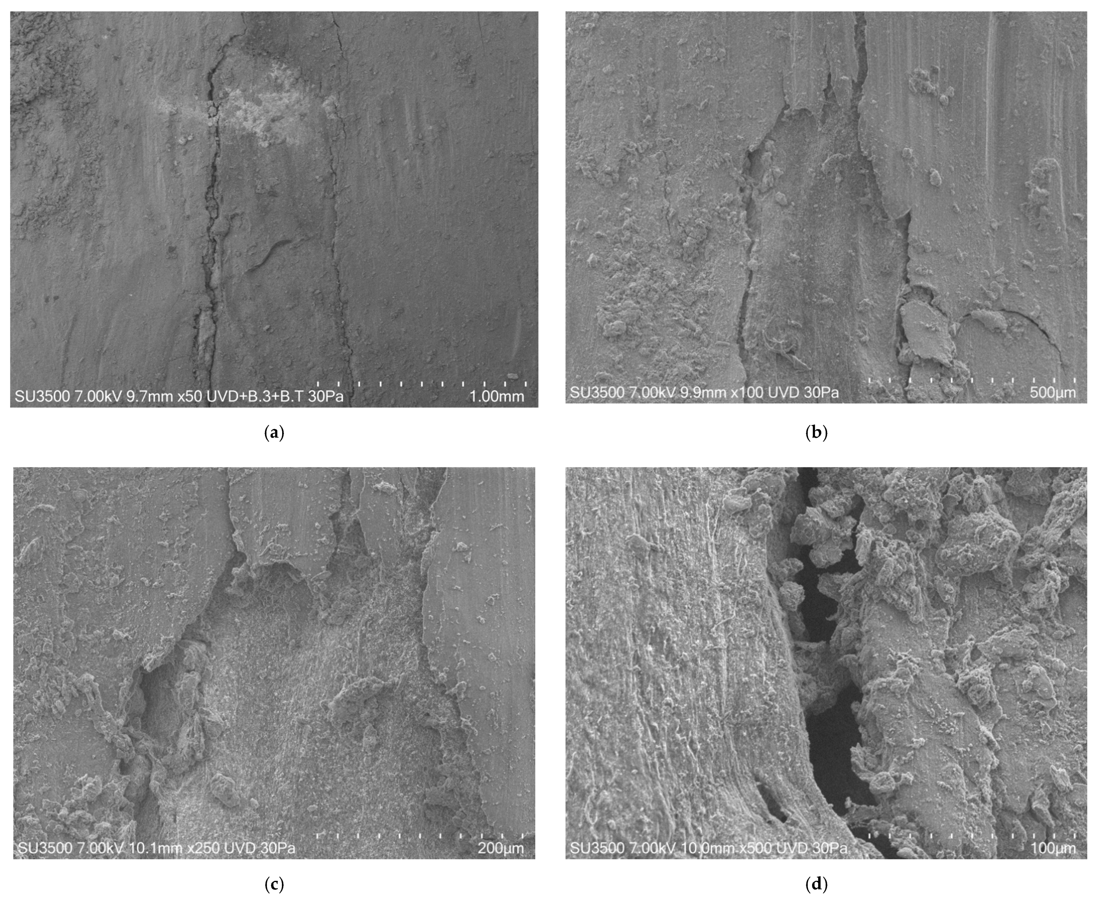
3. Results
4. Discussion
5. Conclusions
Author Contributions
Funding
Conflicts of Interest
References
- Kaur, A.; Shah, N.; Logani, A.; Mishra, N. Biotoxicity of commonly used root canal sealers: A meta-analysis. J. Conserv. Dent. 2015, 18, 83–88. [Google Scholar]
- Staffoli, S.; Plotino, G.; Nunez Torrijos, B.G.; Grande, N.M.; Bossù, M.; Gambarini, G.; Polimeni, A. Regenerative Endodontic Procedures Using Contemporary Endodontic Materials. Materials 2019, 19, 12. [Google Scholar] [CrossRef] [PubMed] [Green Version]
- Jean, A.; Kerebel, B.; Kerebel, L.-M.; Legeros, R.Z.; Hamel, H. Effects of various calcium phosphate biomaterials on reparative dentin bridge formation. J. Endod. 1988, 14, 83–87. [Google Scholar] [CrossRef]
- Pissiotis, E.; Spngberg, L.S.W. Biological evaluation of collagen gels containing calcium hydroxide and hydroxyapatite. J. Endod. 1990, 16, 468–473. [Google Scholar] [CrossRef]
- Gambarini, G.; Seracchiani, M.; Zanza, A.; Miccoli, G.; Del Giudice, A.; Testarelli, L. Influence of shaft length on torsional behavior of endodontic nickel–titanium instruments. Odontology 2020. [Google Scholar] [CrossRef] [PubMed]
- Eymirli, A.; Sungur, D.D.; Uyanik, O.; Purali, N.; Nagas, E.; Cehreli, Z.C. Dentinal Tubule Penetration and Retreatability of a Calcium Silicate-based Sealer Tested in Bulk or with Different Main Core Material. J. Endod. 2019, 45, 1036–1040. [Google Scholar] [CrossRef]
- Chybowski, E.A.; Glickman, G.N.; Patel, Y.; Fleury, A.; Solomon, E.; He, J. Clinical Outcome of Non-Surgical Root Canal Treatment Using a Single-cone Technique with Endosequence Bioceramic Sealer: A Retrospective Analysis. J. Endod. 2018, 44, 941–945. [Google Scholar] [CrossRef] [PubMed]
- Loushine, B.A.; Bryan, T.E.; Looney, S.W.; Gillen, B.M.; Loushine, R.J.; Weller, R.N.; Pashley, D.H.; Tay, F.R. Setting properties and cytotoxicity evaluation of a premixed bioceramic root canal sealer. J. Endod. 2011, 37, 5. [Google Scholar] [CrossRef]
- Miccoli, G.; Seracchiani, M.; Del Giudice, A.; Mazzoni, A.; D’Angelo, M.; Bhandi, S.; Gambarini, G.; Testarelli, L. Fatigue Resistance of Two Nickel-Titanium Rotary Instruments before and after Ex Vivo Root Canal Treatment. J. Contemp Dent. Pract. 2020, 21, 728–732. [Google Scholar]
- Di Nardo, D.; Seracchiani, M.; Mazzoni, A.; Del Giudice, A.; Gambarini, G.; Testarelli, L. Torque Range, a New Parameter to Evaluate New and Used Instrument Safety. Appl. Sci. 2020, 10, 3418. [Google Scholar] [CrossRef]
- Gambarini, G.; Miccoli, G.; Di Nardo, D.; Del Giudice, A.; Mazzoni, A.; Seracchiani, M.; Testarelli, L. Torsional resistance of two new heat treated nickel titanium rotary instruments: An in vitro evaluation. Pesqui. Bras. Odontopediatria Clin. Integr. 2020, 20, 1–7. [Google Scholar] [CrossRef]
- Mazzoni, A.; Pacifici, A.; Zanza, A.; Giudice, A.D.; Reda, R.; Testarelli, L.; Gambarini, G.; Pacifici, L. Assessment of Real-Time Operative Torque during Nickel–Titanium Instrumentation with Different Lubricants. Appl. Sci. 2020, 10, 6201. [Google Scholar] [CrossRef]
- Faraj, S.; Boutsioukis, C. Observer variation in the assessment of root canal curvature. Int. Endod. J. 2017, 50, 167–176. [Google Scholar] [CrossRef]
- Günday, M.; Sazak, H.; Garip, Y. A comparative study of three different root canal curvature measurement techniques and measuring the canal access angle in curved canals. J. Endod. 2005, 31, 796–798. [Google Scholar] [CrossRef] [PubMed]
- Valenti-Obino, F.; Di Nardo, D.; Quero, L.; Miccoli, G.; Gambarini, G.; Testarelli, L.; Galli, M. Symmetry of root and root canal morphology of mandibular incisors: A cone-beam computed tomography study in vivo. J. Clin. Exp. Dent. 2019, 11, 527–533. [Google Scholar] [CrossRef] [PubMed]
- Miccoli, G.; Cicconetti, A.; Gambarini, G.; Giudice, A.D.; Ripanti, F.; Di Nardo, D.; Testarelli, L.; Seracchiani, M. A new device to test the bending resistance of mechanical endodontic instruments. Appl. Sci. 2020, 10, 7215. [Google Scholar] [CrossRef]
- Di Nardo, D.; Gambarini, G.; Seracchiani, M.; Mazzoni, A.; Zanza, A.; Giudice, A.D.; D’Angelo, M.; Testarelli, L. Influence of different cross-section on cyclic fatigue resistance of two nickel-titanium rotary instruments with same heat treatment: An in vitro study. Saudi Endod. J. 2020, 10, 221–225. [Google Scholar]
- Grimaldi, A.; Serpe, C.; Chece, G.; Nigro, V.; Sarra, A.; Ruzicka, B.; Relucenti, M.; Familiari, G.; Ruocco, G.; Pascucci, G.R.; et al. Microglia-Derived Microvesicles Affect Microglia Phenotype in Glioma. Front. Cell. Neurosci. 2019, 13, 41. [Google Scholar] [CrossRef] [Green Version]
- Donfrancesco, O.; Seracchiani, M.; Morese, A.; Ferri, V.; Nottola, S.A.; Relucenti, M.; Gambarini, G.; Testarelli, L. Analysis of Stability in Time of Marginal Adaptation of Endosequence Root Repair Material on Biological Samples. Dent. Hypotheses 2020, 11, 11–15. [Google Scholar]
- Gambarini, G.; Plotino, G.; Grande, N.M.; Testarelli, L.; Prencipe, M.; Messineo, D.; Fratini, L.; D’Ambrosio, F. Differential diagnosis of endodontic-related inferior alveolar nerve paraesthesia with cone beam computed tomography: A case report. Int. Endod. J. 2011, 44, 176–181. [Google Scholar] [CrossRef]
- Gandolfi, M.G.; Iacono, F.; Agee, K.; Siboni, F.; Tay, F.; Pashley, D.H.; Prati, C. Setting time and expansion in different soaking media of experimental accelerated calcium-silicate cements and ProRoot MTA. Oral. Surg. Oral. Med. Oral. Pathol. Oral. Radiol. Endod. 2009, 108, e39–e45. [Google Scholar] [CrossRef] [PubMed]
- Chen, B.; Haapasalo, M.; Mobuchon, C.; Li, X.; Ma, J.; Shen, Y. Cytotoxicity and the Effect of Temperature on Physical Properties and Chemical Composition of a New Calcium Silicate-based Root Canal Sealer. J. Endod. 2020, 46, 531–538. [Google Scholar] [CrossRef] [PubMed]
- Bossù, M.; Saccucci, M.; Salucci, A.; Di Giorgio, G.; Bruni, E.; Uccelletti, D.; Sarto, M.S.; Familiari, G.; Relucenti, M.; Polimeni, A. Enamel remineralization and repair results of Biomimetic Hydroxyapatite toothpaste on deciduous teeth: An effective option to fluoride toothpaste. J. Nanobiotechnol. 2019, 17, 17. [Google Scholar] [CrossRef] [PubMed]
- Relucenti, M.; Miglietta, S.; Bove, G.; Donfrancesco, O.; Battaglione, E.; Familiari, P.; Barbaranelli, C.; Covelli, E.; Barbara, M.; Familiari, G. SEM BSE 3D Image Analysis of Human Incus Bone Affected by Cholesteatoma Ascribes to Osteoclasts the Bone Erosion and VpSEM dEDX Analysis Reveals New Bone Formation. Scanning 2020, 2020, 9371516. [Google Scholar] [CrossRef] [PubMed] [Green Version]


| Measurement | Humid Environment | Wet Environment |
|---|---|---|
| 1 | 7.843 | 14.272 |
| 2 | 14.505 | 13.187 |
| 3 | 19.042 | 9.963 |
| 4 | 1.614 | 9.528 |
| 5 | 2.641 | 7.149 |
| 6 | 4.117 | 26.168 |
| 7 | 4.758 | 20.964 |
| 8 | 4.95 | 11.955 |
| 9 | 5.322 | 10.448 |
| 10 | 7.113 | 11.049 |
| 11 | 5.243 | 11.359 |
| 12 | 7.548 | 12.206 |
| Mean | 6.836 | 13.326 |
| SD | 5.024 | 5.288 |
| Measurement | Humid Environment | Wet Environment |
|---|---|---|
| 1 | 3.638 | 2.388 |
| 2 | 4.63 | 3.875 |
| 3 | 9.468 | 2.691 |
| 4 | 7.057 | 3.61 |
| 5 | 3.884 | 6.381 |
| 6 | 2.111 | 4.464 |
| 7 | 2.273 | 2.267 |
| 8 | 5.277 | 1.786 |
| 9 | 3.983 | 17.057 |
| 10 | 7.881 | 7.761 |
| 11 | 3.823 | 9.273 |
| 12 | 3.481 | 5.672 |
| Mean | 4.792 | 5.602 |
| SD | 2.252 | 4.293 |
Publisher’s Note: MDPI stays neutral with regard to jurisdictional claims in published maps and institutional affiliations. |
© 2021 by the authors. Licensee MDPI, Basel, Switzerland. This article is an open access article distributed under the terms and conditions of the Creative Commons Attribution (CC BY) license (https://creativecommons.org/licenses/by/4.0/).
Share and Cite
Donfrancesco, O.; Del Giudice, A.; Zanza, A.; Relucenti, M.; Petracchiola, S.; Gambarini, G.; Testarelli, L.; Seracchiani, M. SEM Evaluation of Endosequence BC Sealer Hiflow in Different Environmental Conditions. J. Compos. Sci. 2021, 5, 99. https://doi.org/10.3390/jcs5040099
Donfrancesco O, Del Giudice A, Zanza A, Relucenti M, Petracchiola S, Gambarini G, Testarelli L, Seracchiani M. SEM Evaluation of Endosequence BC Sealer Hiflow in Different Environmental Conditions. Journal of Composites Science. 2021; 5(4):99. https://doi.org/10.3390/jcs5040099
Chicago/Turabian StyleDonfrancesco, Orlando, Andrea Del Giudice, Alessio Zanza, Michela Relucenti, Stefano Petracchiola, Gianluca Gambarini, Luca Testarelli, and Marco Seracchiani. 2021. "SEM Evaluation of Endosequence BC Sealer Hiflow in Different Environmental Conditions" Journal of Composites Science 5, no. 4: 99. https://doi.org/10.3390/jcs5040099
APA StyleDonfrancesco, O., Del Giudice, A., Zanza, A., Relucenti, M., Petracchiola, S., Gambarini, G., Testarelli, L., & Seracchiani, M. (2021). SEM Evaluation of Endosequence BC Sealer Hiflow in Different Environmental Conditions. Journal of Composites Science, 5(4), 99. https://doi.org/10.3390/jcs5040099