Case Report

GLI1-Altered Mesenchymal Tumours in the Head and Neck: A Case Report and Literature Review

by
Olivier Janjic
1,*,
Claudio De Vito
2,
Johannes Alexander Lobrinus
2,
Minerva Becker
3 and
Nicolas Dulguerov
1
1
Department of Oto-Rhino-Laryngology-Head and Neck Surgery, Geneva University Hospitals, 1205 Geneva, Switzerland
2
Department of Clinical Pathology, Geneva University Hospitals, 1205 Geneva, Switzerland
3
Department of Imaging and Medical Information Sciences, Geneva University Hospitals, 1205 Geneva, Switzerland
*
Author to whom correspondence should be addressed.
J. Otorhinolaryngol. Hear. Balance Med. 2025, 6(1), 2; https://doi.org/10.3390/ohbm6010002
Submission received: 16 December 2024 / Revised: 25 January 2025 / Accepted: 27 January 2025 / Published: 31 January 2025

Abstract

:
Background and Clinical Significance: GLI1 gene alterations have recently been identified as a pathological phenomenon associated with a distinct novel entity of mesenchymal neoplasms. They have been reported to occur in any soft tissue of the body, with a specific affinity for the head and neck region. The aim of this article is to increase awareness of this entity and provide a detailed summary of the modes of presentation and diagnostic and therapeutic issues surrounding these tumours occurring in the head and neck region. Case Presentation: We report the case of a 39-year-old male patient with ACTB::GLI1 fusion-related mesenchymal tongue tumour who was successfully treated by surgery. Conclusions: GLI1-altered mesenchymal tumours in the head and neck may harbour various clinical presentations. Larger series are needed to better define the clinicopathological range of this novel entity. We suggest a follow-up period of at least 2 years with imaging, followed by a clinical follow-up of 3 years. Certain clinicopathological features may warrant further and more extensive follow-up.

1. Introduction

GLI1 gene alterations have recently been identified as a pathological phenomenon associated with a particular type of soft-tissue neoplasm. These tumours have a wide anatomic distribution and can develop in any part of the body, but a significant proportion are located in the head and neck region. Of these, the large majority (70–80%) is found in the tongue [1]. GLI1 is a member of the GLI gene family, responsible for encoding the GLI1 transcription factor, which is a significant downstream effector within the hedgehog cascade. A multitude of tumours, including glioblastoma multiforme, hepatocellular carcinoma, gastric cancer, and rhabdomyosarcomas, exhibit aberrant hedgehog signalling activation [2]. Reported GLI1 alterations consist in its amplification or its translocation with numerous partners, whose number has constantly been increasing according to recently published works. GLI1-altered mesenchymal tumours are a new diagnostic entity of which all surgeons operating in the head and neck region, radiologists, and pathologists should now be aware, especially when dealing with an atypical presentation of a head and neck mass [3,4]. Until recently, this entity was unknown, and these lesions, once resected and analysed, could easily be misdiagnosed or categorised as “unclassified tumours”. To date, the vast majority of cases described in the literature has been retrieved from past consultations and diagnosed retrospectively. Due to recent work on the subject, we were able to diagnose an ACTB-GLI1 fusion-related tongue tumour in a 39-year-old male patient. Despite the significant attention paid to histopathological and molecular analysis in past research, there are currently limited clinical descriptions available for this uncommon condition. Through a complete literature review, the aim of this article is to increase awareness of this entity and provide a detailed summary of the modes of presentation and the diagnostic and therapeutic issues surrounding these tumours occurring in the head and neck region.

2. Case Report

2.1. Clinical History

In the Department of Otolaryngology of the University Hospital of Geneva, we studied 2020 case of a healthy 39-year-old male patient who did not drink or smoke. He presented to the ENT clinic with a mass at the tip of the tongue that had been growing progressively since the age of 12. Intraoral examination revealed a voluminous tumour measuring approximately 1.5 × 2 cm horizontally and 1 cm deep. It was soft, firm, and covered by tongue mucosa, which did not show any change (Figure 1A). The rest of the physical examination was normal: in particular, no lymphadenopathy was found. The patient stated that he had previously consulted general practitioners who did not conduct any additional investigations due to the lack of symptoms. During our examination, the patient presented with a mild speech impairment and occasional tongue biting. He did not report any pain or weight loss. We conducted a head MRI (details below), which was initially consistent with a benign arterio-venous vascular malformation. No incisional biopsy was undertaken, and the case was consequently presented to the tumour board, who opted to solely treat via surgery. A median glossectomy was performed, and the tumour was successfully removed entirely with negative, although near, margins. A two-year follow-up, including multiple skull MRIs and thoraco-abdominal CT scans, revealed no clinical or radiological evidence of tumour recurrence.

2.2. Imaging

We performed an MRI with a dynamic TWIST sequence after contrast injection (Dotarem). The examination showed a lingual mass, centred on the anterior third of the dorsum of the tongue, measuring 19 × 24 × 20 mm (AP × TR × CC) and spreading the fibres of the intrinsic musculature of the tongue. It showed no infiltration of the extrinsic musculature and extended inferiorly to the genioglossus muscle.
Its signal was heterogeneous with an hypointense signal in T2/STIR and an isosignal in T1. The T1 images showed a fleshy component, with slight diffusion restriction, and homogeneous and strong enhancement. The images exhibited multiple internal serpiginous structures demonstrating flow-voids, which confirmed the vascular origin (some with a calibre up to 2 mm). No significant oedema was found near the lesion. The dynamic TWIST sequences showed early contrast of the lesion in arterial time, with rapid partial venous washout and persistent enhancement of the fleshy component (Figure 1B,C).

2.3. Pathology

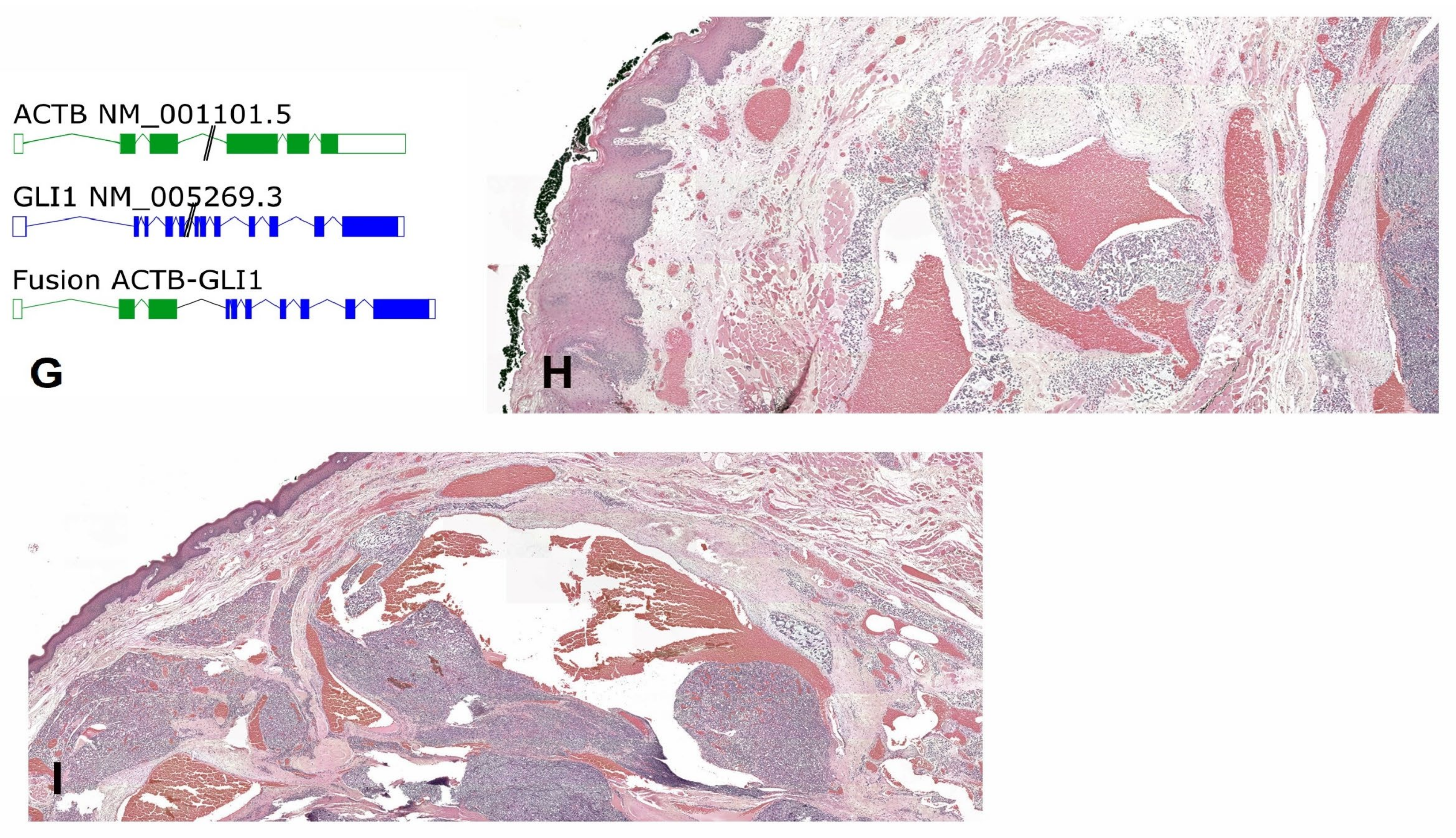
A partial glossectomy was performed, which revealed a 3 cm non-encapsulated whitish soft multinodular tumour (Figure 1D). Histologically, the neoplasm showed a nested architecture with a richly vascular network composed of monomorphic tumoral cells with round-to-oval nuclei, small nucleoli, and a clear cytoplasm (Figure 1E,F). Protrusions of tumour cells into the vascular lumina were noted. No mitosis or necrosis was observed. Immunohistochemistry demonstrated weak and focal S100 staining. SMA, pankeratin, and STAT6 were negative. GLI1 alteration was confirmed using the Illumina TruSight RNA fusion panel (Illumina, San Diego, CA, USA), which demonstrated ACTB-GLI1 fusion with breakpoints of ACTB exon 3 and GLI1 exon 5 (Figure 1G). No evidence of MDM2, CDK4, or GLI1 amplification using Oncoscan CNV assay (ThermoFisher Scientific, Waltham, MA, USA) was noted. Altogether, these findings supported the diagnosis of head and neck mesenchymal neoplasm with GLI1 alterations.
We observed, at the periphery of the neoplasm, below the mucosa, aberrant vascular structures surrounded by neoplastic cells around the vascular wall.

3. Methods

3.1. Search

To conduct the literature review, a computerised literature search was performed through the MEDLINE/PubMed, Embase, and Cochrane databases, applying the following terms: “GLI1” (MeSH term), “mesenchymal tumour” (MeSH term), and “Human Glioma-Associated Oncogene Homolog 1” (free text). These terms were combined using “AND”. Additional articles and abstracts were identified by cross-referencing. Articles published in the English language from the implementation of the above-cited databases until April 2024 were considered for the present review.

3.2. Eligibility Criteria

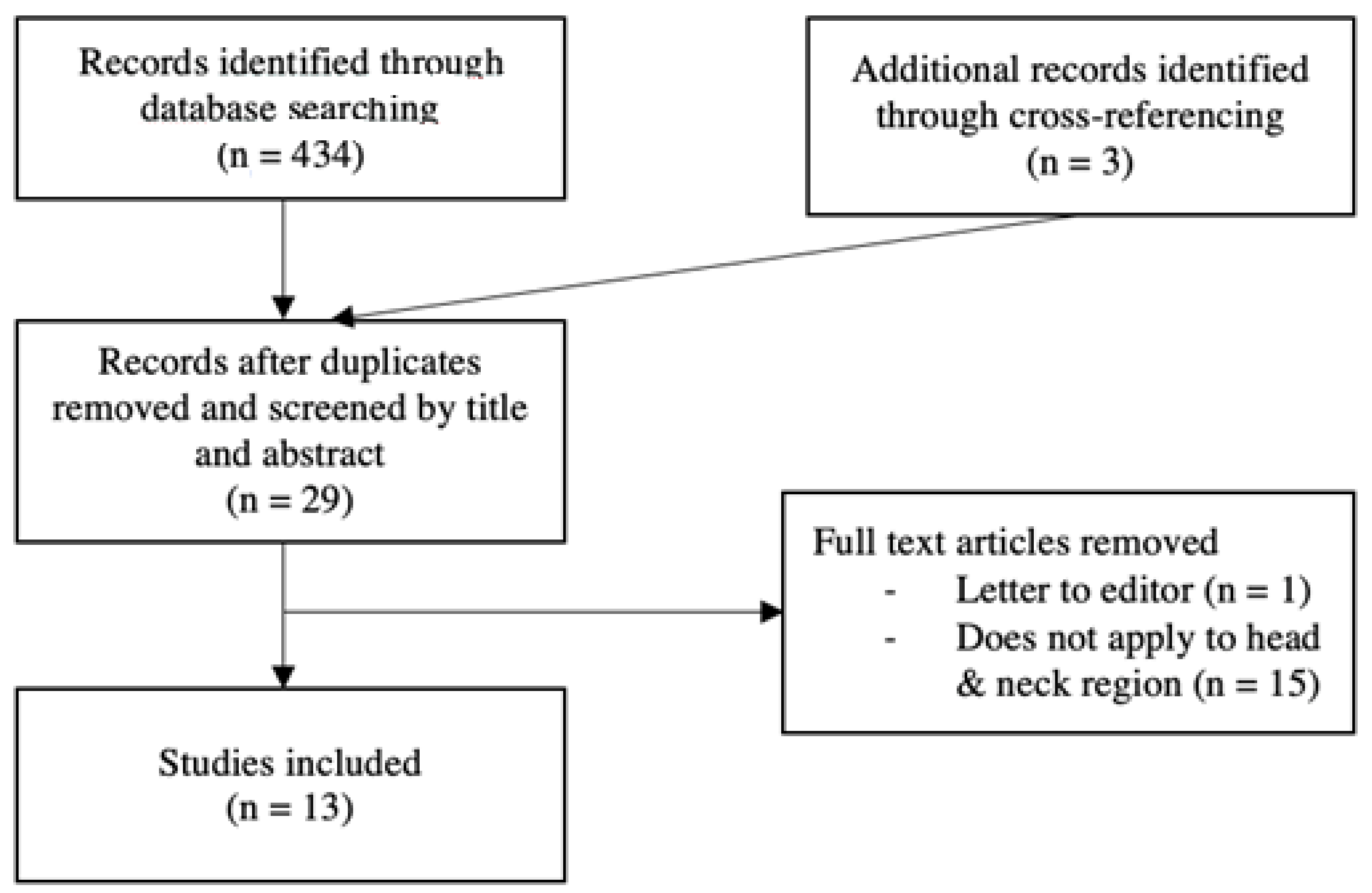
Papers concerning GLI1-altered soft-tissue tumours were evaluated, and head and neck cases were analysed and extracted for the review. Only full-text articles were included. Letters to the editor, posters, and clinical images were excluded. This systematic review was conducted and reported following the PRISMA guidelines (Figure 2).

3.3. Data Extraction

The following data were extracted: sex, age, site of tumour, clinical manifestation and its duration, radiological description, type of treatment, and outcome. Tumour features were collected, such as its size and gross description, the presence of necrosis, and the GLI1 alteration type. Informed consent was obtained to publish the information/image of the case.

4. Results

A total of 13 articles on GLI1-altered soft-tissue tumours were selected, of which 9 reported tumours located in the head and neck region. Including our case, 32 cases have been published so far, with a strong majority of male patients affected (25 male and 7 female patients). The median age was 34 years (IQR 34). Twenty-three tumours occurred in the tongue (of which two at the tongue base and one extending to the oropharynx), three in the neck, one in the submandibular gland, two on the mouth floor, and one each in a palatine tonsil, on the soft palate, and on the oropharynx (not otherwise specified). Out of the 21 cases for which treatment details were specified, tumours were primarily removed through surgery. In two cases, chemotherapy was attempted but had no significant effect, so surgery was carried out instead [5]. Adjuvant radiotherapy and chemotherapy were given to two patients. In one case, this decision was made due to local recurrence [6]. Follow-up was carried out for 17 patients during a period that ranged between 2 and 120 months (mean 31 months). Three patients showed local recurrence with distant and/or regional metastasis, while one patient had only local recurrence [1,7]. The rest showed no evidence of disease at their last follow-up consultation. Tumour size was described in 17 patients and ranged from 0.8 to 5.8 cm (median 2.4 cm, mean 2.87 cm) [5,6,7,8,9,10]. Tumour necrosis was documented for every patient and was found in two cases [1].
Clinical features such as radiological assessment and findings, gross description of the tumour, and patient symptoms were scarcely reported. Essentially, the patients described a growing painless mass causing no other discomfort except the ones created by the mechanical expansion in itself (speech impairment, dysphonia, dysphagia) [6,7,9]. Our case is an exception with respect to the tumour growing time (27 years), as the other reported cases varied from 1 to 6 months [6,7,9]. Macroscopically, the tumours appeared capable of harbouring diverse presentations. The texture varied from fleshy and indurated to spongy, with the most commonly reported colours being brown, white, and grey ([6,7,8,9], our study). Though these tumours were generally considered indolent, some presented with ulceration. Radiological findings were only reported in two patients, and the findings were rather non-specific, describing heterogenous tumours upon injected CT scan [7,9]. The clinicopathological results are partly summarised in Table 1.

5. Discussion

Primary soft-tissue tumours harbouring alterations of the GLI1 gene have been increasingly reported in recent series. In addition to the head and neck region, they can occur in various locations throughout the body, including the kidneys, stomach, duodenum, small intestines, uterine cervix, ovaries, bones, skin, and muscles [1,6,8,10,11,12,13,14,15,16,17,18]. In 2004, Dahlen et al. described tumours with distinctive pericytic features harbouring GLI fusion with the ACTB gene, called “pericytoma with t(7;12)” [5]. Since then, other authors have reported tumours with similar histological and clinical features associated with new mechanisms of GLI alteration such as GLI1 amplification (often combined with co-amplification of DDIT3, CDK4, and MDM2) or fusion with new partners. As progress is made in this field, the list of fusion partners is becoming longer and currently comprises the following genes: MALAT1, PTCH1, DERA, APOD, FOXO4, HNRNPA, TXNIP, NEAT1, and ABCA1 [8,10,19,20,21,22,23].
Various terms have been proposed so far to better nominate this emerging entity, such as “GLI1-altered mesenchymal tumour” by Liu et al., “GLI1-altered epithelioid soft-tissue tumour” by Zhong et al., or “sarcoma of unknown lineage with GLI1 alteration” by Xu et al. [1,6,7]. Histologically, these neoplasms share specific features such as monomorphic and bland cells, prominent capillary networks, and the proliferation of tumour cells around vessels in the soft tissue surrounding the tumour [7]. However, regarding immunohistochemical analysis, GLI-1-altered tumours in different anatomical locations have not yet demonstrated a uniform profile and have proven to be unreliable for diagnostic purposes [7,17,24]. Papke et al. recently reported a series of 20 new cases harbouring GLI1-altered genes that they had named “distinctive nested glomoid neoplasms” and classified as a subset within the larger “GLI1-altered mesenchymal tumour” group [8]. In their view, these nested glomoid neoplasms might represent a benign entity that resembles glomus tumours or well-differentiated neuroendocrine tumours. This subgroup could co-exist on a biological continuum with “pericytoma with t(7;12)”, as they share certain genetical, clinical, and histological characteristics but differ in others: nested glomoid tumours display a nested architecture and a round-to-ovoid cytomorphology while “pericytoma with t(7;12)” shows prominent microcystic spaces and a pseudopapillary architecture [8,12]. As the aggressiveness of GLI1-altered mesenchymal tumours appears to be quite heterogeneous, with some tumours growing slowly for years and others causing distant metastases rapidly, various authors have attempted to identify clinical or pathological features that may be of prognostic value.
Xu et al. suggested that tumour necrosis and an anatomical site outside the oral tongue could be linked to aggressive behaviour. Tumour necrosis was well documented in every reviewed article but was found in only two tumours; those tumours gave rise to metastases [1]. However, two other tumours without necrosis also showed aggressive behaviours (local recurrence and distant metastasis) [6,7]. Regarding prognostic predictions based on anatomical location, our data potentially support the proposition of Xu et al., as only tumours situated outside the oral tongue exhibited aggressive behaviours [1,6,7]. Yet, among cases with reported follow-up, three tumours located outside the oral tongue did not demonstrate any sign of local recurrence or metastasis within a mean follow-up period of 20.3 months. In addition, ten tumours were situated in the oral tongue, and they did not show any relapse after a mean follow-up of 26.5 months. Altogether, subject to clinical gaps, this may suggest that necrosis and an anatomical site outside the oral tongue are not essential factors for aggressive behaviour but may promote it. This might also imply that a location in the oral tongue could be linked to a benign evolution.
Concerning tumour size and ulceration, no correlation between these two characteristics could be established due to the small size of the sample and the lack of data in some cases.
In the reviewed articles, very few clinical descriptions were provided, especially concerning diagnostic procedure and radiological findings. Although mesenchymal tumours with an altered GLI1 form a rare pathological entity, a consistent description of radiological assessment may provide diagnostic and/or prognostic value when reporting such diseases in articles. The role of radiology in diagnostic procedures cannot be disregarded since cases are often ultimately diagnosed by radiologists or based on their recommendations. While diagnostic imaging alone may not be sufficient for the diagnosis of most rare diseases, such assessments serve as critical inputs in the diagnostic procedure. In our case, MRI findings showed multiple internal serpiginous structures demonstrating flow-voids, which made us primarily suspect a vascular origin for the tumour. The identification of aberrant vascular vessels might serve as a radiological feature that could potentially facilitate the challenging process of diagnosing a rare disease such as GLI-altered mesenchymal tumours.
In the present case, there was no evidence of the amplification of MDM2 or CDK4. However, no immunohistochemical analysis of these genes was performed. Such an analysis should have been carried out, given that mutations affecting protein expression may be present even in the absence of a change in the copy number of these genes. It is therefore suggested that immunohistochemical analysis be included in future similar cases, as it may allow a more accurate diagnosis.
As papers on the topic were based on data retrieved from past consultations, the available clinical information was generally incomplete and sometimes entirely lacked follow-up data. In addition, some cases were only recently diagnosed, and there has not been enough time for relevant outcomes to emerge. With recent interest in this field, it is our hope that extensive future studies and larger series are underway. Such analyses will be crucial in defining the clinicopathological range of this novel entity, especially since certain subgroups may exhibit malignant behaviours.
In conclusion, we successfully treated a symptomatic and long-lasting ACTB-GLI1 fusion mesenchymal tumour of the tongue. Our radiological work-up initially indicated a low-flow arterio-venous vascular malformation. Histologically, the neoplasm displayed a nested architecture with a rich vascular network composed of monomorphic tumour cells with round-to-oval nuclei. The described histological features were consistent with the “distinctive nested glomoid neoplasms” group. Furthermore, the benign status of the tumour reinforced this resemblance. We suggested a follow-up period of at least 2 years with imaging followed by a clinical follow-up of 3 years. The longest reported relapse onset was 83 months, hence the recommended follow-up duration [1]. Subject to the lack of strong evidence, features suggestive of malignancy, such as location outside the tongue, presence of necrosis, or a histological description similar to “pericytoma with T(7;12)”, may warrant further and more extensive follow-up.

Author Contributions

O.J. and N.D. wrote the main manuscript text. O.J. prepared Figure 2 and Table 1. C.D.V., J.A.L., and M.B. prepared Figure 1. All authors have read and agreed to the published version of the manuscript.

Funding

This research received no external funding.

Institutional Review Board Statement

Not applicable.

Informed Consent Statement

Written informed consent has been obtained from the patient to publish this paper.

Data Availability Statement

The original contributions presented in this study are included in the article. Further inquiries can be directed to the corresponding author.

Conflicts of Interest

The authors declare no conflicts of interest.

References

  1. Xu, B.; Chang, K.; Folpe, A.L.; Kao, Y.C.; Wey, S.L.; Huang, H.Y.; Gill, A.J.; Rooper, L.; Bishop, J.A.; Dickson, B.C.; et al. Head and Neck Mesenchymal Neoplasms With GLI1 Gene Alterations: A Pathologic Entity With Distinct Histo-logic Features and Potential for Distant Metastasis. Am. J. Surg. Pathol. 2020, 44, 729–737. [Google Scholar] [CrossRef] [PubMed]
  2. Chetty, R. Gene of the month: GLI-1. J. Clin. Pathol. 2020, 73, 228–230. [Google Scholar] [CrossRef] [PubMed]
  3. Yeung, M.C.; Liu, A.P.; Wong, S.-I.; Loong, H.H.; Shek, T.W. GLI1-Altered Mesenchymal Tumor—Multiomic Characterization of a Case Series and Patient-Level Meta-analysis of One Hundred Sixty-Seven Cases for Risk Stratification. Mod. Pathol. 2024, 38, 100635. [Google Scholar] [CrossRef] [PubMed]
  4. Xu, B. Proceedings of the North American Society of Head and Neck Pathology, Los Angeles, CA, March 20, 2022. Emerging Bone and Soft Tissue Neoplasms in the Head and Neck Region. Head Neck Pathol. 2022, 16, 158–167. [Google Scholar] [CrossRef]
  5. Dahlén, A.; Fletcher, C.D.; Mertens, F.; Fletcher, J.A.; Perez-Atayde, A.R.; Hicks, M.J.; Debiec-Rychter, M.; Sciot, R.; Wejde, J.; Wedin, R.; et al. Activation of the GLI Oncogene through Fusion with the β-Actin Gene (ACTB) in a Group of Distinctive Pericytic Neoplasms. Am. J. Pathol. 2004, 164, 1645–1653. [Google Scholar] [CrossRef]
  6. Liu, J.; Mao, R.; Lao, I.W.; Yu, L.; Bai, Q.; Zhou, X.; Wang, J. GLI1-altered mesenchymal tumor: A clinicopathological and molecular analysis of ten additional cases of an emerging entity. Virchows Arch. 2022, 480, 1087–1099. [Google Scholar] [CrossRef]
  7. Zhong, H.; Xu, C.; Chen, X.; Guo, X.; Yang, S. GLI1-altered epithelioid soft tissue tumor: A newly described entity with a predilection for the tongue. Oral Surg. Oral Med. Oral Pathol. Oral Radiol. 2022, 134, e14–e22. [Google Scholar] [CrossRef]
  8. Papke, D.J.J.; Dickson, B.C.M.; Oliveira, A.M.; Sholl, L.M.; Fletcher, C.D.M. Distinctive Nested Glomoid Neoplasm: Clinicopathologic Analysis of 20 Cases of a Mesenchymal Neoplasm With Frequent GLI1 Alterations and Indolent Behavior. Am. J. Surg. Pathol. 2022, 47, 12–24. [Google Scholar] [CrossRef]
  9. Klubíčková, N.; Kinkor, Z.; Michal, M.; Baněčková, M.; Hájková, V.; Michálek, J.; Pink, R.; Dvořák, Z.; Leivo, I.; Skálová, A. Epithelioid Soft Tissue Neoplasm of the Soft Palate with a PTCH1-GLI1 Fusion: A Case Report and Review of the Literature. Head Neck Pathol. 2021, 16, 621–630. [Google Scholar] [CrossRef]
  10. Nitta, Y.; Takeda, M.; Fujii, T.; Itami, H.; Tsukamoto, S.; Honoki, K.; Ohbayashi, C. A case of pericytic neoplasm in the shoulder with a novel DERA–GLI1 gene fusion. Histopathology 2020, 78, 466–469. [Google Scholar] [CrossRef]
  11. Castro, E.; Cortes-Santiago, N.; Ferguson, L.M.S.; Rao, P.H.; Venkatramani, R.; López-Terrada, D. Translocation t(7;12) as the sole chromosomal abnormality resulting in ACTB-GLI1 fusion in pediatric gastric pericytoma. Hum. Pathol. 2016, 53, 137–141. [Google Scholar] [CrossRef] [PubMed]
  12. Kerr, D.A.; Pinto, A.; Subhawong, T.K.; Wilky, B.A.; Schlumbrecht, M.P.; Antonescu, C.R.; Nielsen, G.P.; Rosenberg, A.E. Pericytoma With t(7;12) and ACTB-GLI1 Fusion: Reevaluation of an Unusual Entity and its Relationship to the Spectrum of GLI1 Fusion-related Neoplasms. Am. J. Surg. Pathol. 2019, 43, 1682–1692. [Google Scholar] [CrossRef] [PubMed]
  13. Koh, N.W.C.; Seow, W.Y.; Lee, Y.T.; Lam, J.C.M.; Lian, D.W.Q. Pericytoma With t(7;12): The First Ovarian Case Reported and a Review of the Literature. Int. J. Gynecol. Pathol. 2019, 38, 479–484. [Google Scholar] [CrossRef] [PubMed]
  14. Prall, O.W.J.; McEvoy, C.R.E.; Byrne, D.J.; Iravani, A.; Browning, J.; Choong, D.Y.-H.; Yellapu, B.; O’haire, S.; Smith, K.; Luen, S.J.; et al. A Malignant Neoplasm From the Jejunum With a MALAT1-GLI1 Fusion and 26-Year Survival History. Int. J. Surg. Pathol. 2020, 28, 553–562. [Google Scholar] [CrossRef]
  15. Zeng, Y.; Yao, H.; Jiang, X.; Tang, X.; Wang, X. GLI1-altered Mesenchymal Tumor Involving the Duodenum: Case Report and Literature Review. Int. J. Surg. Pathol. 2023, 31, 1538–1547. [Google Scholar] [CrossRef]
  16. Argani, P.; Boyraz, B.; Oliva, E.; Matoso, A.; Gross, J.; Fridman, E.; Zhang, L.; Dickson, B.C.; Antonescu, C.R. GLI1 Gene Alterations in Neoplasms of the Genitourinary and Gynecologic Tract. Am. J. Surg. Pathol. 2021, 46, 677–687. [Google Scholar] [CrossRef]
  17. Bridge, J.A.; Sanders, K.; Huang, D.; Nelson, M.; Neff, J.R.; Muirhead, D.; Walker, C.; Seemayer, T.A.; Sumegi, J. Pericytoma with t(7;12) and ACTB-GLI1 fusion arising in bone. Hum. Pathol. 2012, 43, 1524–1529. [Google Scholar] [CrossRef]
  18. Rollins, B.T.; Cassarino, D.S.; Lindberg, M. Primary cutaneous epithelioid mesenchymal neoplasm with ACTB-GLI1 fusion: A case report. J. Cutan. Pathol. 2021, 49, 284–287. [Google Scholar] [CrossRef]
  19. Agaram, N.P.; Zhang, L.; Sung, Y.-S.; Singer, S.; Stevens, T.; Prieto-Granada, C.N.; Bishop, J.A.; Wood, B.A.; Swanson, D.; Dickson, B.C.; et al. GLI1-amplifications expand the spectrum of soft tissue neoplasms defined by GLI1 gene fusions. Mod. Pathol. 2019, 32, 1617–1626. [Google Scholar] [CrossRef]
  20. Antonescu, C.R.; Agaram, N.P.; Sung, Y.-S.M.; Zhang, L.; Swanson, D.B.; Dickson, B.C. A Distinct Malignant Epithelioid Neoplasm With GLI1 Gene Rearrangements, Frequent S100 Protein Expression, and Metastatic Potential: Expanding the Spectrum of Pathologic Entities With ACTB/MALAT1/PTCH1-GLI1 Fusions. Am. J. Surg. Pathol. 2018, 42, 553–560. [Google Scholar] [CrossRef]
  21. Pettus, J.R.; Kerr, D.A.; Stan, R.V.; Tse, J.Y.; Sverrisson, E.F.; Bridge, J.A.; Linos, K. Primary myxoid and epithelioid mesenchymal tumor of the kidney with a novel GLI1-FOXO4 fusion. Genes Chromosom. Cancer 2021, 60, 116–122. [Google Scholar] [CrossRef] [PubMed]
  22. Lopez-Nunez, O.; Surrey, L.F.; Alaggio, R.; Herradura, A.; McGough, R.L.; John, I. Novel APODGLI1 rearrangement in a sarcoma of unknown lineage. Histopathology 2021, 78, 338–340. [Google Scholar] [CrossRef] [PubMed]
  23. Agaimy, A. What is new in epithelioid soft tissue tumors? Virchows Arch. 2020, 476, 81–96. [Google Scholar] [CrossRef] [PubMed]
  24. Palsgrove, D.N.; Rooper, L.M.; Stevens, T.M.; Shin, C.; Damm, D.D.; Gagan, J.; Bridge, J.A.; Thompson, L.D.R.; Koduru, P.R.; Bishop, J.A. GLI1-Altered Soft Tissue Tumors of the Head and Neck: Frequent Oropharyngeal Involvement, p16 Immunoreactivity, and Detectable Alterations by DDIT3 Break Apart FISH. Head Neck Pathol. 2022, 16, 1146–1156. [Google Scholar] [CrossRef]
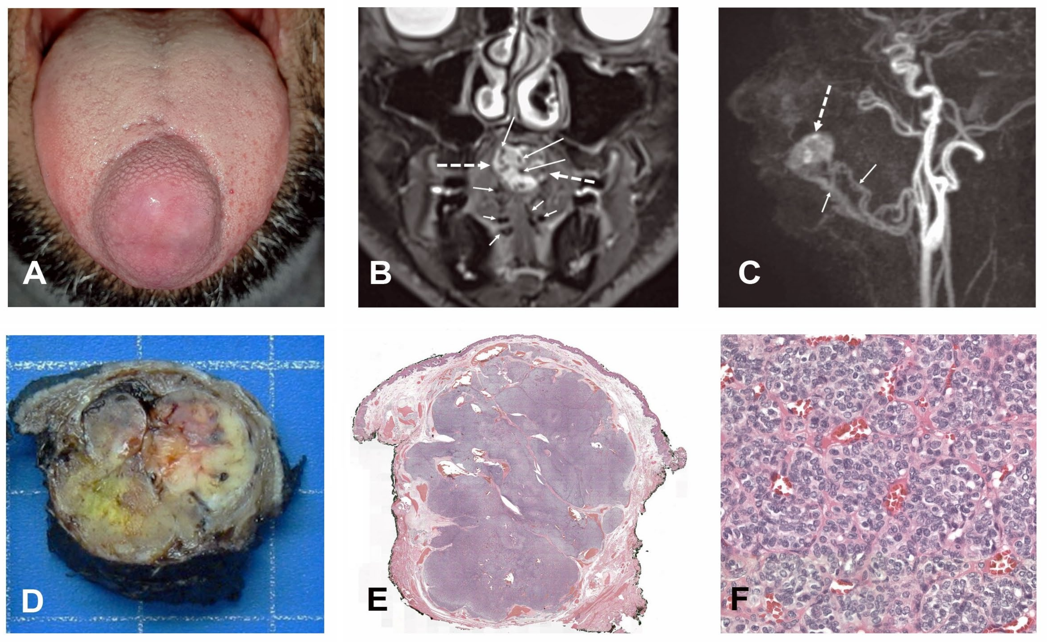
Figure 1. Clinical and pathologic features of the case: “In situ” aspect of the tumour, showing a lump below the mucosa without ulceration (A). Pre-operative MRI showing the typical features of an arterio-venous malformation. Coronal T2 image reveals a 3 cm large hyperintense mass (dashed arrows) in the mobile tongue. Note large, partly serpiginous flow-voids characteristic of vessels with rapid arterial flow (arrows), as well as enlarged lingual arteries (short arrows) (B). The sagittal view of a 3D contrast-enhanced MR angiography sequence shows a very early arterial enhancement pattern of the tongue lesion (dashed arrow). The lesion receives arterial tributaries from the enlarged lingual arteries (short arrows) (C). Macroscopic aspect of the neoplasm showing a non-encapsulated and multinodular tumour (D). Histological findings showing a multilobular and non-encapsulated tumour (E) with a nested architecture composed of monotonous cells with a clear cytoplasm and round-to-oval nuclei (F). ACTB-GLI1 fusion with breakpoints of ACTB exon 3 and GLI1 exon 5 (G). Aberrant vascular vessels surrounded by tumoral cells (H) and dilated vessels with the protrusion of tumoral cells into the lumen (I), located below the mucosa.
Ohbm 06 00002 g001aOhbm 06 00002 g001b
Figure 2. Prisma flowchart.
Ohbm 06 00002 g002
Table 1. Clinicopathologic characteristics of 32 head and neck GLI1-altered tumours.
Study
(N° of Cases)
Body SiteAge/SexMetastasis
(Localization, Months
Before Relapse)
Local Recurrence
(Months Before Relapse)
Outcome (Follow-Up in Month)TreatmentGLI AlterationSize (cm)Necrosis
Xu et al., Antonescu et al., Agaram et al. [11]
Neck39/MY (Lungs, 6)Y (6)AWD (36) Amp. Y
Tongue38/MNNNED (2)PTCH1::GLI1NAN
Tongue46/FNNNED (3)Amp.N
Tongue65/MNA Amp.N
Tongue60/M Amp.N
Subm. gland32/FY (Lymph nodes and lungs, 83)Y (83)AWD (104)NAPTCH1::GLI1Y
Neck37/MNNNED (30)ACTB::GLI1N
Tongue1/MNNNED (2)ACTB::GLI1N
Tongue28/MNN ACTB::GLI1N
Tongue14/MNAACTB::GLI1N
Tongue56/F MALAT1::GLI1N
Dahlen et al. [3]
Tongue27/FNNNED (60)ChT + Surgery *ACTB::GLI10.8N
Tongue11/MNNNED (22)ChT + Surgery *ACTB::GLI15N
Tongue12/MNNNED (120)SurgeryACTB::GLI12.4N
Liu et al. [5]
Tongue base27/MNNNED (16)Surgery + RT/ChTACTB::GLI13.5N
Tongue4/MNNNED (11)SurgeryAmp.3N
Tongue17/MNNNED (5)SurgeryAmp.1N
Mouth floor8/MNY #AWD (40)Surgery + RT/ChTACTB::GLI12N
Mouth floor34/MNASurgeryMALAT1::GLI1NAN
Palsgrove et al. [5]
Palatine tonsil35/MRecent caseSurgeryAmp.1.5N
Tongue base84/MRecent caseSurgeryACTB::GLI1NAN
Oropharynx53/MRecent caseSurgeryACTB::GLI16N
Tongue41/MNASurgeryACTB::GLI1NAN
Tongue65/MSurgeryAmp.2N
Zhong et al. [1]
Tongue base Ω56/MY (Lymph node and bone, 27)NNED (36)Surgery« GLI1 rearrangement »6N
Klubíćková et al. [1]
Soft palate34/FNNNED (4)SurgeryPTCH1::GLI1ca. 3N
Papke et al. [5]
Neck12/MNNNASurgeryACTB::GLI11.5N
Tongue46/FNNNED (18)SurgeryAmp.5.8N
Tongue10/FNASurgeryACTB::GLI12N
Tongue0/MRecent caseSurgeryNA0.9N
Tongue1/MSurgeryAmp.NAN
Current study [1]Tongue39/MNNNED (24)SurgeryACTB::GLI12.5N
Y = yes; N = no; NA = not available; NED = no evidence of disease; AWD = alive with disease; RT = radiotherapy; ChT = chemotherapy; and Amp. = amplification. # Local recurrence occurred twice, once at 27 and a second time at 40 months. * Surgery was performed after an unsuccessful attempt with ChT. Ω Tumour extending to the right oral tongue and lateral pharyngeal wall.
Disclaimer/Publisher’s Note: The statements, opinions and data contained in all publications are solely those of the individual author(s) and contributor(s) and not of MDPI and/or the editor(s). MDPI and/or the editor(s) disclaim responsibility for any injury to people or property resulting from any ideas, methods, instructions or products referred to in the content.