Review

Ag/Au Bimetallic Core–Shell Nanostructures: A Review of Synthesis and Applications

Department of Chemical Engineering and Materials Science, Stevens Institute of Technology, Hoboken, NJ 07030, USA
*
Author to whom correspondence should be addressed.
J. Manuf. Mater. Process. 2025, 9(4), 131; https://doi.org/10.3390/jmmp9040131
Submission received: 20 March 2025 / Revised: 12 April 2025 / Accepted: 14 April 2025 / Published: 15 April 2025

Abstract

:
Silver/gold (Ag/Au) core–shell nanostructures exhibit tunable plasmonic properties and enhanced catalytic performance, enabling applications across sensing, biomedicine, and environmental remediation. This review presents representative synthetic strategies for fabricating Ag/Au bimetallic core–shell nanostructures with three distinct morphologies: nanospheres, nanocubes, and nanowires. For each architecture, we cover the representative synthetic approaches, such as seed-mediated growth, one-pot synthesis, and evaporation deposition methods, along with their corresponding applications. This review provides discussions on the synthesis methods and applications through specific examples, offering researchers guidance for fabricating Ag/Au core–shell nanostructures with tailored morphologies while addressing major challenges in controlling bimetallic formation.

1. Introduction

Nanotechnology, a rapidly developing field, has revolutionized our ability to design and manipulate materials at the nanoscale, unlocking unprecedented properties and functionalities. Nanomaterials, defined by at least one dimension size below 100 nm, are of great interest due to their unique physical and chemical properties, which differ markedly from their bulk counterparts. Metal nanomaterials, particularly noble metals, exhibit a multitude of captivating and distinctive characteristics that enable their exploration for different applications due to their physiochemical properties. Among these nanomaterials, gold (Au) demonstrates fantastic properties that make versatile scaffolds for the fabrication of catalysts [1,2,3,4] photodynamic therapeutic platforms [5], optical applications [6,7] and bio-sensors [8,9,10]. Silver (Ag), another excellent noble metal, exhibits unique characteristics in terms of antimicrobial activity [11], surface-enhanced Raman spectroscopy (SERS) sensitivity [12,13,14,15], chemical sensing capabilities [16,17,18,19,20,21] and biomedical applications [22]. However, monometallic systems face inherent limitations in practical implementation that cannot be ignored, such as lack of enhanced catalytic properties [23,24], synergistic effects [25], chemical stability [26] and functionalization [27].
Compared to monometallic nanostructures, bimetallic nanostructures have become a focus of research due to their unique and diversified properties [28,29,30]. The combination of two different metals in nanostructure synthesis creates opportunities for expanded properties through compositional variations and synergistic interactions between the metals, which produce critical enhancements in surface area, electrocatalytic activity, biocompatibility, electron transfer rates and resistance to intermediate species. Among bimetallic structures, core–shell architectures have emerged as the most widely studied, thanks to established synthetic protocols and the ability to fine-tune their properties by modifying shell thickness and architecture [31].
Among the various core–shell systems, silver/gold (Ag/Au) bimetallic core–shell nanostructures stand out as especially promising candidates. These materials combine the superior electrical conductivity and plasmonic properties of the Ag core with the exceptional chemical stability of the Au shell. The synergistic properties of Ag/Au core–shell nanostructures enable detailed studies of catalytic reaction kinetics and mechanistic pathways [32], while their exceptional biocompatibility makes them ideal for the creation of porous electrodes in bioelectronic devices [33]. Additionally, their corrosion resistance benefits energy applications, showing effectiveness in multiple electrochemical systems [34]. Optimal synergistic effects require sufficiently thin Au shells to maintain the influence of the Ag core at the surface.
Generally, there are three main methods that exist for Ag/Au core–shell nanostructure synthesis: seed-mediated growth, one-pot synthesis and evaporation deposition. Each of these has specific advantages and challenges. Seed-mediated growth offers unmatched control by isolating core formation from shell growth. However, it presents challenges when the seed material undergoes galvanic replacement with the shell metal precursor in solution, leading to unexpected alloy formation at the surface [35]. Specifically, for Ag/Au core–shell nanostructures, the two half-reactions occur through seed-mediated growth [36]:
Ag0 → Ag+(aq) + 1e   ε01 = −0.8 V
AuCl4(aq) + 3e → Au0 + 4Cl(aq)    ε02 = 1.0 V
which leads to the final balanced equation:
AuCl4(aq) + 3Ag0 → 3Ag+(aq) + Au0 + 4Cl(aq)   ε03 = 0.2 V
When a Ag nanoparticle is exposed to a AuCl4 solution, the Ag atoms on its surface are rapidly oxidized and dissolved by AuCl4 ions, releasing Ag+ ions into the reaction mixture. This process results in the creation of small cavities on the surface of the nanoparticle. Concurrently, Au atoms are deposited onto the nanoparticle surface through the reduction of AuCl4. Given that Au and Ag are miscible at the nanoscale, both sharing a face-centered cubic (fcc) crystal structure and having similar lattice constants (αAu = 4.079 Å; αAg = 4.086 Å), the formation of a uniform Ag/Au alloy is generally anticipated. This issue can be effectively managed by employing strong reducing agents that drive the reduction reaction faster than the competing galvanic replacement process [37]. In one-pot synthesis, the careful regulation of reduction kinetics is critical as both metal precursors coexist in the reaction medium. This method enables sequential metal reduction, though concurrent precursor presence frequently causes unwanted surface alloying [38]. Evaporation deposition offers an alternative approach with precise shell formation control through physical vapor deposition [39]. Unlike solution-based methods, this technique produces uniform, dense shells while preventing interfacial core–shell reactions. Although the requirement for substrate fixation limits certain applications, this constraint can be advantageous for developing surface-integrated devices and sensing platforms.
This review provides an overview of the synthesis strategies employed to fabricate Ag/Au bimetallic core–shell nanostructures with diverse morphologies, including nanospheres, nanocubes, and nanowires. We introduce and discuss representative synthetic approaches for each geometry, highlighting successful preparation methods and their corresponding applications. By exploring recent advancements in synthesis techniques, structural variations, and functional applications, this review aims to serve as a practical guide for researchers seeking to synthesize Ag/Au core–shell nanostructures with tailored properties for specific applications. The examination of synthetic strategies and resulting applications in catalysis, sensing, and biomedical sciences highlights the potential of Ag/Au core–shell nanostructures for addressing challenges in scientific and industrial territories.

2. Ag/Au Bimetallic Core–Shell Nanostructures with Diverse Morphologies and Applications
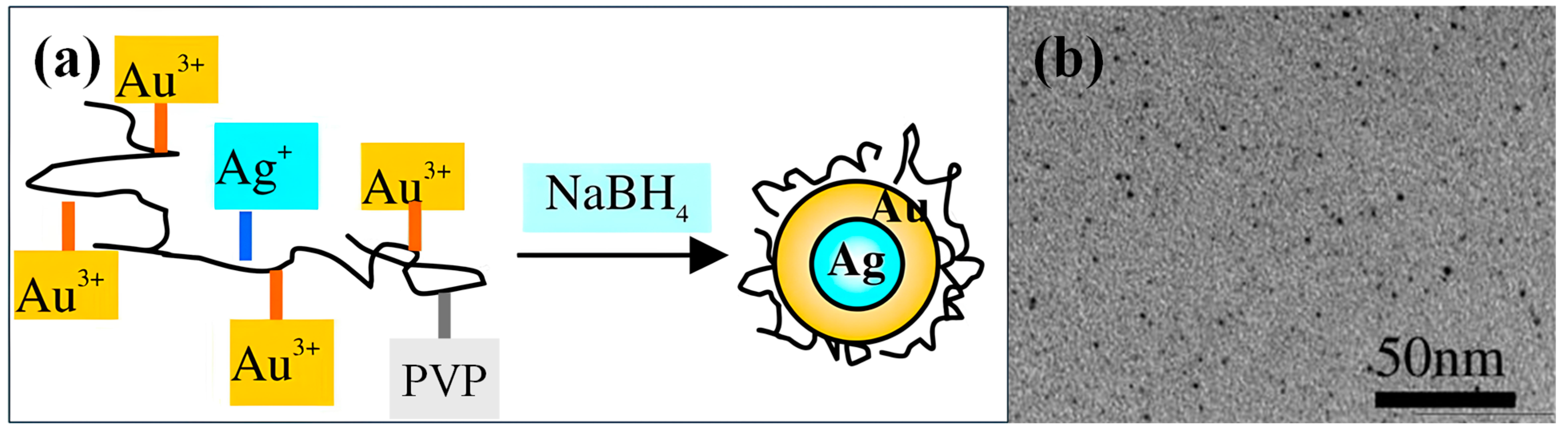
Within the diverse family of bimetallic nanostructures, Ag/Au composites have been engineered in various architectural configurations. This section examines the synthesis and applications of distinct Ag/Au core–shell morphologies, namely, nanospheres, nanocubes and nanowires. Table 1 summarizes these different nanostructures along with their applications. The uniform and isotropic nanospheres provide consistent optical responses and catalytic performance, making them suitable for applications requiring uniformity and reliability [40]. Nanocubes, on the other hand, with flat facets and sharp edges, deliver higher active site densities. Their unique geometry also modulates localized surface plasmon resonance, yielding tunable optical properties [41]. Nanowires, with their high aspect ratio and marked anisotropy, facilitate superior electron transport and directional current conduction, while their distinctive light scattering and absorption behaviors provide significant advantages in optoelectronic applications [42]. For each geometry, we discuss both the synthetic strategies and their potential applications.

2.1. Nanospheres

Ag/Au bimetallic core–shell nanospheres demonstrate significant application potential in catalysis, sensing, environmental monitoring and biomedicine. Synthesis methods including pH-controlled reduction, one-pot synthesis, and thermal evaporation deposition enable precise control over core–shell morphology, shell thickness and interfacial properties, effectively addressing challenges such as galvanic replacement and unwanted alloying.

2.1.1. Synthesis Methods of Nanospheres

Zhao et al. [43] developed a method to synthesize SERS-labeled Ag/Au bimetallic core–shell nanospheres while preventing galvanic replacement. Their approach involved depositing HAuCl4 solution onto Ag nanoparticles under carefully controlled pH conditions. By adjusting the solution pH to 9.5, the galvanic replacement reaction that typically produces hollow Ag/Au structures (E0(Ag+/Ag) = 0.8 V, E0(Au3+/Au) = 1.5 V [49]) was successfully suppressed. Under these basic pH conditions, ascorbic acid becomes a strong reducing agent, driving the rapid reduction of HAuCl4 and preventing its galvanic interaction with Ag nanoparticles [37]. The synthesis procedure involved first preparing a mixture of PVP (poly(N-vinyl-2-pyrrolidone), stabilizer) and ascorbic acid, followed by adding NaOH for pH adjustment. SERS-labeled Ag nanoparticles and HAuCl4 solution were then introduced to this mixture under constant stirring in dark conditions. The resulting nanostructures were isolated via centrifugation and dispersed in ultrapure water for storage. This method produced core–shell particles consisting of 15 ± 3 nm Ag cores encased in Au shells of 4.5 ± 1.0 nm thickness. Figure 1 shows the schematic of the synthesis process of SERS-labeled Ag/Au core–shell nanostructures and the TEM and HAADF-STEM images of the nanostructures.
Zhang et al. [44] described a one-pot synthesis approach for producing small-sized Ag/Au bimetallic core–shell nanoparticles. The method used PVP as a stabilizing agent, which weakly bound to and protected the core–shell nanostructures during the rapid reduction process initiated by NaBH4 injection into AuCl4/Ag+/PVP aqueous solutions. The synthesis procedure involved several sequential steps. First, HAuCl4 and PVP solutions were combined and stirred at low temperature. AgClO4 solution was then introduced to this mixture and stirred under the same temperature conditions. The reduction was initiated when NaBH4 solution was swiftly injected into the vigorously stirred mixture. The resulting colloidal dispersion underwent purification through ultrafiltration, followed by washing with water and ethanol under inert conditions to remove excess reagents and byproducts. To eliminate residual ethanol from the PVP-protected colloid, the mixture was processed using a rotary evaporator at an elevated temperature. The final Ag/Au core–shell nanoparticles, obtained after vacuum drying, exhibited sizes below 2 nm. Figure 2 depicts the synthesis process and the TEM image of PVP-protected Ag/Au core–shell nanoparticles.
He et al. [32] reported a straightforward method for producing Au-coated Ag (AOA) nanospheres using thermal evaporation deposition (TVD). This technique allowed precise control over the Au shell thickness, which is difficult to achieve using traditional solution-based chemical methods due to galvanic replacement. The process involved two main steps. First, silver nanospheres were synthesized by using a modified version of the Lee and Meisel method [50]—sodium citrate mixed with AgNO3 solution, which was then exposed to UV light while stirring gently. This produced uniform Ag nanospheres with a size of 43 ± 6 nm. Second, Ag nanospheres were deposited onto a silicon surface, followed by the use of thermal evaporation to coat them with a 7 nm layer of Au. This was performed by heating a Au source in a thermal evaporator until it vaporized and condensed on the Ag nanospheres, as shown in Figure 3. The resulting AOA nanostructures were then stored in an inert gas environment to prevent degradation.

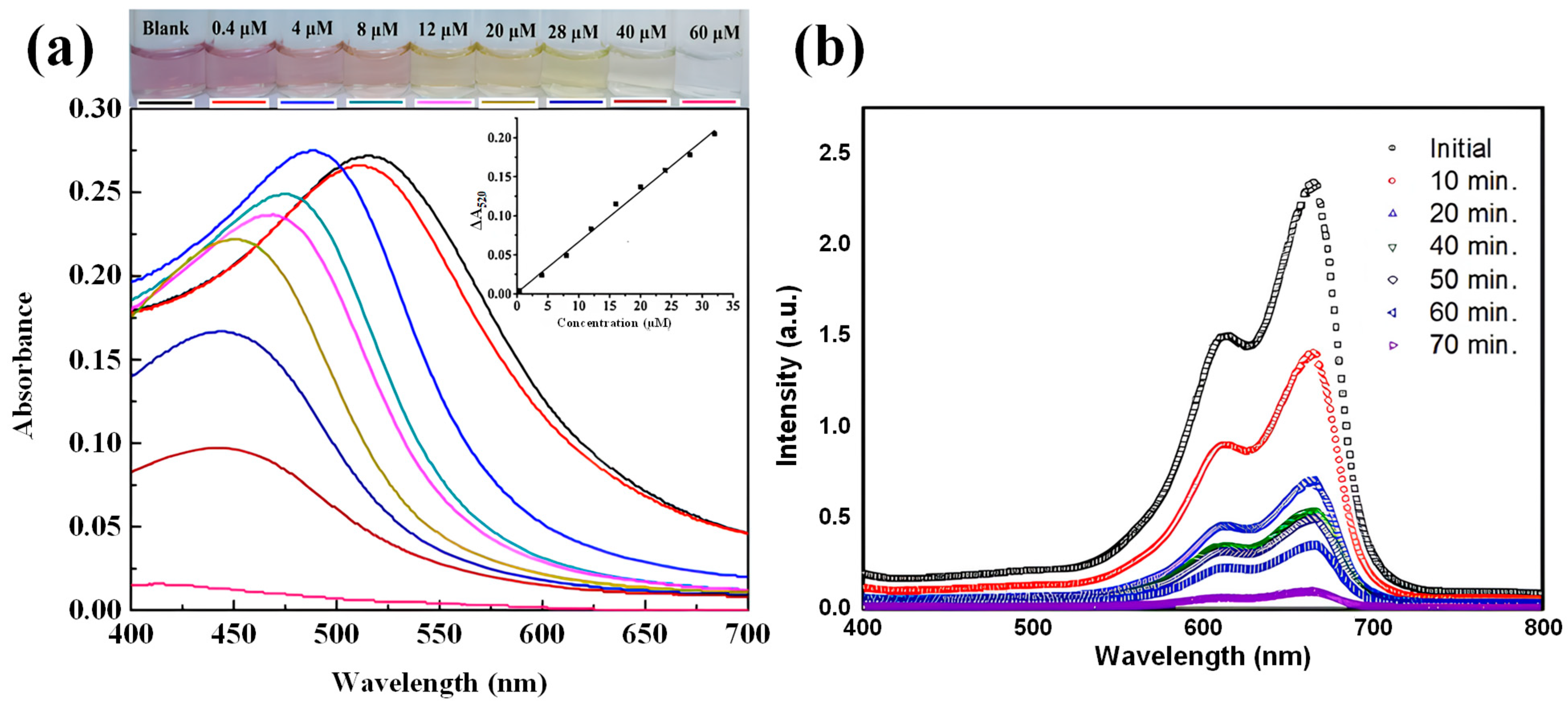
2.1.2. Applications of Nanospheres
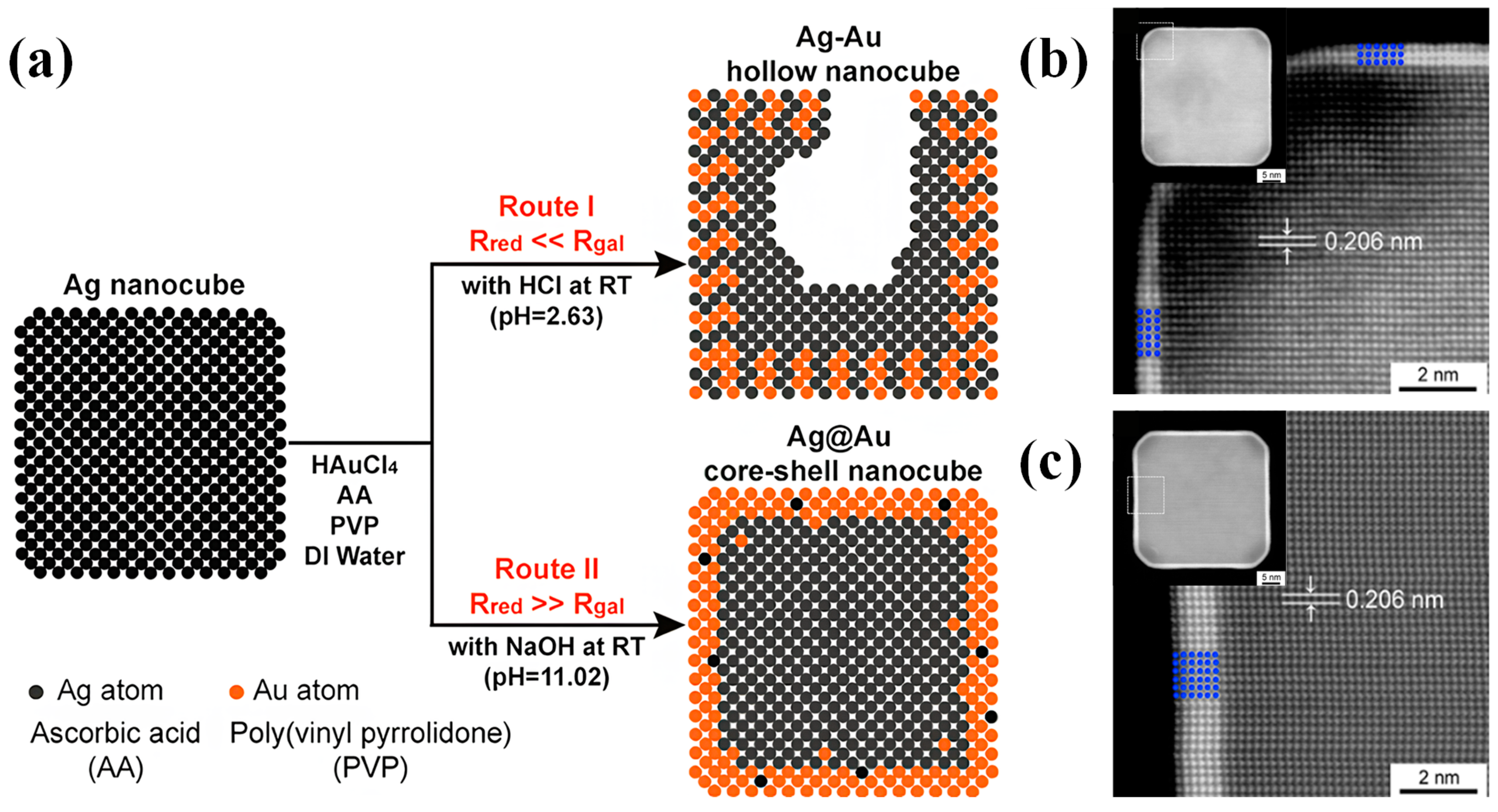
Ag/Au bimetallic core–shell nanospheres demonstrate versatility in their applications, which include water treatment and catalytic applications. In a notable study, Li et al. [45] developed these nanostructures as colorimetric probes for cyanide detection in aqueous solutions. Their research revealed a systematic color transition from purple through orange and yellow to colorless with increasing cyanide concentrations. The underlying detection mechanism exploits cyanide’s ability to progressively etch the Au shell through the formation of [Au(CN)2], a thermodynamically stable complex. This etching process alters the dimensional ratio of the Au shell to the Ag core in the bimetallic nanoparticles, manifesting in distinct spectral and colorimetric changes. In drinking water analysis, the system achieved remarkable sensitivity with a limit of detection of 0.16 µM and a visual detection limit of 0.4 µM, as shown in Figure 4a. Furthermore, by employing polysorbate 40 as a stabilizer, they successfully extended the application to sewage water analysis, achieving a visual detection limit of 4 µM. This achievement demonstrates the robustness of Ag/Au bimetallic core–shell nanoparticles as analytical tools even in complex environmental matrices.
Omar et al. [46] evaluated the photocatalytic performance of Ag/Au bimetallic core–shell nanospheres using methylene blue degradation under solar irradiation. Their UV–Vis spectroscopic analysis revealed a progressive decrease in methylene blue’s absorption peak with increasing exposure time, eventually reaching a minimum value that indicated complete dye degradation, as illustrated in Figure 4b. The core–shell nanostructures achieved a superior degradation efficiency of 96.3%, outperforming pure Ag nanoparticles (88%). This enhanced photocatalytic activity can be attributed to two key mechanisms. First, photo-generated excited electrons interact with oxygen molecules to form oxygen radicals and hydroxyl ions, which facilitate dye molecule degradation. Second, the Au shell exhibits dual absorption capabilities: it absorbs UV light through inter-band electron transitions from 5d to 6sp states, and visible light through the excitation of 6sp electrons, which are redistributed to energy levels above the Fermi level via a continuous electron band. Notably, these excited energetic electrons maintain their state for up to 1 ps, providing sufficient time to interact with reactant molecules and initiate chemical reactions.

2.2. Nanocubes

Ag/Au bimetallic core–shell nanocubes with flat crystal {100} facets and sharp edges provide enhanced catalytic efficiency and tunable optical properties suitable for photothermal therapy, surface-enhanced Raman scattering and biomedical imaging. Synthesis strategies including seed-growth methods enable precise control over shell thickness and uniformity.

2.2.1. Synthesis Methods of Nanocubes

Yang et al. [37] developed a robust method to synthesize Ag/Au bimetallic core–shell nanocubes through direct Au deposition on Ag nanocubes’ surfaces. Their strategy employed a strong reducing agent to suppress galvanic replacement between Ag and HAuCl4. The synthesis involved mixing PVP with ascorbic acid (AA) and NaOH, followed by adding Ag nanocubes and titrating HAuCl4 into the mixture. In this system, HAuCl4 reduction occurs through two competing pathways: galvanic replacement with Ag, which creates hollow structures, and AA-mediated reduction, which forms Au shells on Ag nanocubes. By increasing the pH to enhance AA’s reducing power, HAuCl4 reduction by AA was prioritized over galvanic replacement [51,52], resulting in conformal Au deposition on Ag nanocubes’ surfaces. Figure 5a illustrates these possible reaction pathways, while Figure 5b,c presents the high-angle annular dark-field scanning TEM (HAADF-STEM) images of the core–shell nanocubes with varying Au shell thicknesses.
Despite the successful fabrication of Ag/Au bimetallic core–shell nanocubes, two challenges arise from using NaOH: Ag2O formation at cube corners due to Ag+ reacting with OH and decreasing pH during HAuCl4 titration. Under acidic conditions, these Ag2O patches dissolve, exposing Ag to galvanic replacement with HAuCl4 when using weak reducing agents such as H2Asc [53]. Thus, producing Ag/Au bimetallic core–shell nanocubes with Au shells remains challenging.
Therefore, Zhang et al. [54] developed a defect-assisted approach to synthesize Ag/Au bimetallic core–shell nanocubes with controlled thin shells. Their synthetic strategy employed ethylene glycol (EG) as both solvent and reducing agent, eliminating the need for H2Asc, NaOH, and water, thereby preventing Ag2O formation on Ag nanocubes. The synthesis involved dispersing Ag nanocubes in a heated ethylene glycol solution containing PVP, followed by controlled titration of aqueous HAuCl4. Upon completion of titration, the reaction mixture was rapidly cooled in an ice bath. Figure 6a displays the synthesis process and Figure 6b,c show the HAADF-STEM images of Ag/Au bimetallic core–shell nanocubes. The product was then purified through acetone and water washing steps, with the final core–shell nanostructures being redispersed in water. The synthesis of Ag/Au core–shell nanocubes proceeded through several distinct stages. Initially, galvanic replacement between HAuCl4 and Ag created defects on the nanocubes’ {100} facets while simultaneously depositing Au. These defect sites, possessing high surface energy, became preferential locations for the co-deposition of Ag and Au atoms reduced from their ionic states by ethylene glycol. The defects were subsequently filled, followed by the rapid surface diffusion of additional atoms to form a thin Ag/Au alloy shell. Once galvanic replacement terminated, the continued reduction of HAuCl4 by ethylene glycol deposited pure Au atoms, ultimately producing Ag/Au core–shell nanocubes with thin Au shells.

2.2.2. Applications of Nanocubes
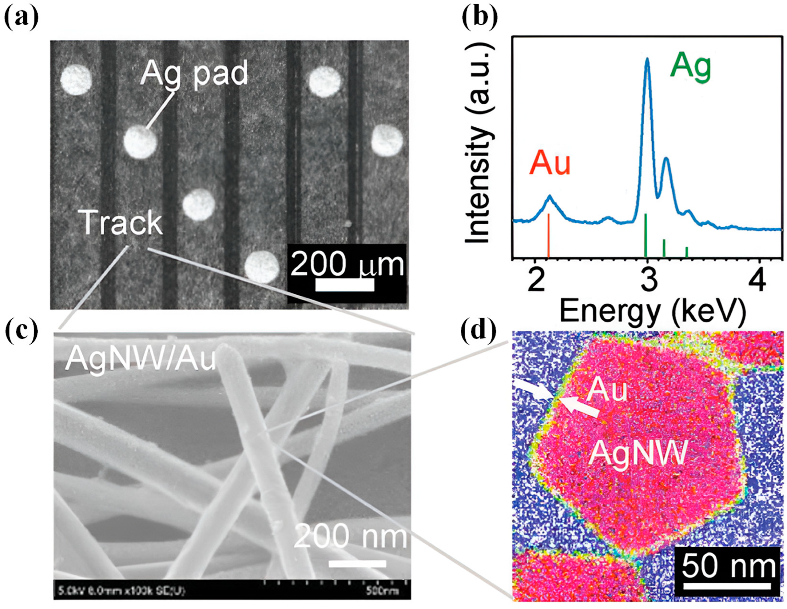
Ag/Au bimetallic core–shell nanocubes demonstrate significant potential in various fields, particularly in biological applications. Liu et al. [47] developed a novel approach using Ag nanocubes as cores with highly branched Au shells, encoded by L-cysteine (L-Cys) and D-cysteine (D-Cys), for photothermal tumor therapy. These three-dimensional bimetallic branched nanostructures exhibited exceptional photothermal conversion efficiency, demonstrating promising results in both in vitro and in vivo tumor therapy. Under laser irradiation, L-CAg/Au and D-CAg/Au nanocubes achieved photothermal conversion efficiencies of 74.11% and 87.28%, respectively. The in vitro photothermal antitumor efficacy of these core–shell nanostructures was evaluated using the MTT method [55,56] on B16, A549 and HeLa cell lines, confirming their effectiveness as photothermal therapy agents. Furthermore, in vivo studies using BALB/c mice implanted with A549 cells demonstrated both positive therapeutic effects and the biosafety of D-CAg/Au nanocubes. Figure 7 illustrates the therapeutic efficacy of these nanostructures: (a) shows the relative viabilities of A549 cells treated with Ag/Au core–shell nanostructures in vitro, while (b) presents the relative tumor volume changes in different treatment groups over a 16-day period.
Beyond biological applications, Ag/Au bimetallic core–shell nanocubes have shown promise in optical applications, particularly in surface-enhanced Raman scattering (SERS). Yang et al. [37] demonstrated enhanced SERS activity in their developed Ag/Au core–shell nanocubes. Using 1,4-benzenedithiol (1,4-BDT) as a probe molecule, they found that the SERS enhancement factor (EF) of Ag/Au nanocubes was 5.4-fold higher than that of pure Ag nanocubes. This superior SERS sensitivity can be attributed to two key factors: first, due to the thin Au shell, this nanostructure maintains electromagnetic enhancement comparable to pure Ag nanocubes, and second, the charge transfer between the Au surface and adsorbed molecules provides additional chemical enhancement, collectively resulting in the increased SERS enhancement factor of Ag/Au nanocubes.

2.3. Nanowires

Ag/Au bimetallic core–shell nanowires with high aspect ratio and anisotropy offer superior electron transport and distinctive optical properties applicable in bioelectrodes, catalysis, energy conversion, microbial fuel cells and CO₂ electrochemical reduction. Synthesis methods including cyan-free electroless plating and conformal epitaxial growth enable precise control over shell thickness and surface smoothness.

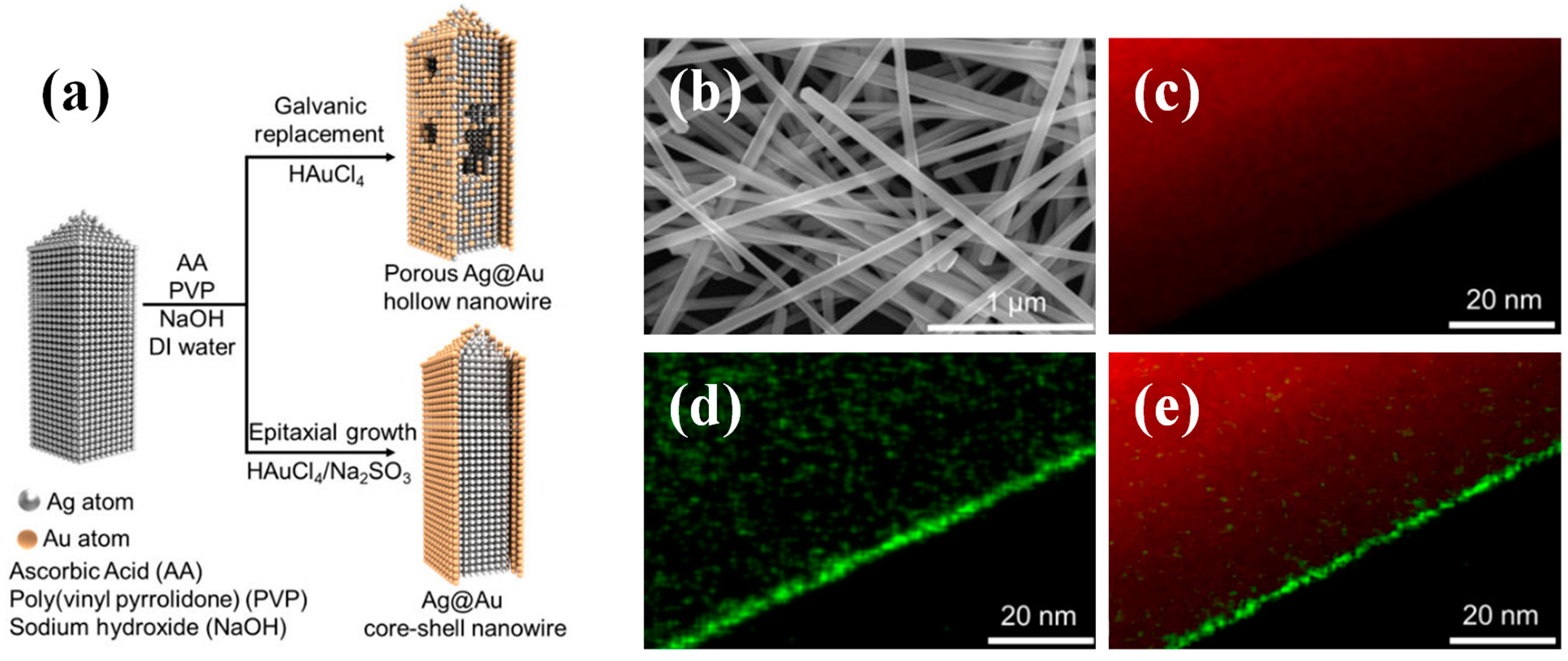
2.3.1. Synthesis Methods of Nanowires

Araki et al. [57] developed a direct method to synthesize Ag/Au nanowires through cyan-free Au electroless plating on Ag nanowire tracks. The process began with spraying Ag nanowire dispersion onto a Parylene substrate, followed by drying and applying a glass plate as a weight to enhance nanowire adhesion. The researchers then patterned the Ag nanowires using laser ablation before performing cyan-free Au electroless plating. This plating process consisted of three steps: dilute sulfuric acid pretreatment, palladium catalyst application, and immersion in Au trisodium disulfite solution at elevated temperature. The final nanostructures were protected with Parylene and PMC3A coatings. Figure 8 presents the structural characterization of the Ag/Au nanowires: SEM images at different magnifications (a,c), the corresponding energy dispersive X-ray spectrometry mapping (b) and a cross-sectional TEM image (d).
Zhu et al. [58] explored a strategy to grow a conformal epitaxial Au shell on Ag nanowires with precisely controlled coating thickness, enhancing both chemical stability and material properties. The resulting Ag/Au bimetallic core–shell nanowires exhibited atomically smooth surfaces, which minimized additional electron scattering and optical losses from the coating, resulting in superior performance with significantly improved lifetime and stability. The synthesis process involved dispersing Ag nanowires in H2O, followed by the addition of PVP, AA and diethylamine solution. The diethylamine played a crucial role by eliminating energy differences between different facets of Ag through non-selective adsorption on the nanowire surface without causing etching. Subsequently, a Au growth solution—prepared from HAuCl4, NaOH, Na2SO3 and H2O—was slowly introduced into the Ag solution. The thickness of the Au shell could be precisely controlled by adjusting the injection time. The final products were isolated through centrifugation, washed with water and stored in aqueous suspension. To prevent galvanic replacement during Au shell deposition, the reduction potential of the Au ion source was decreased by complexing Au3+ ions from the HAuCl4 precursor with SO32– to form the stable complex Au(SO3)22–. Figure 9 illustrates the synthesis process and characterization results: (a) shows a schematic of the two synthetic pathways for Ag/Au bimetallic core–shell nanowires, (b) presents an SEM image of the nanowires and (c–e) display the elemental mapping of Ag and Au in the core–shell structures.

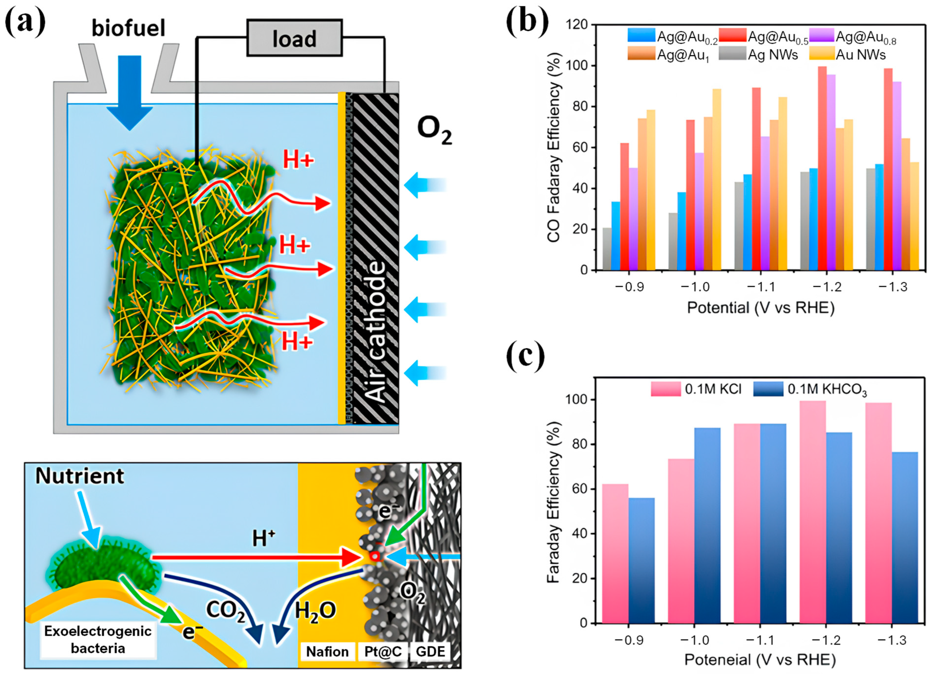
2.3.2. Applications of Nanowires

Ag/Au bimetallic core–shell nanowires exhibit significant application potential, particularly as bioelectrode materials, as explored by Choi et al. [34]. Their investigation yielded three-dimensional Ag/Au bimetallic core–shell nanowire foam combining high porosity, electrical conductivity, biocompatibility, and chemical stability. This structure was designed to enable microbes to harness their electrochemical activity in microbial fuel cells. As Figure 10a depicts, the microbe–nanowire composite served as the anode, facilitating electron emission alongside H+ through biochemical reactions. Meanwhile, a carbon paper–Pt/C Nafion composite functioned as the air cathode, converting H+ to water in the presence of O2. The study demonstrated the exceptional power density performance of bioelectrodes based on Ag/Au core–shell nanowires.
Liu et al. [48] developed Ag/Au bimetallic core–shell nanowires as catalysts for CO2 electrochemical reduction, achieving nearly 100% conversion to CO. The catalyst’s effectiveness resulted from complementary metal properties: the Au shell provided selectivity, while the Ag core facilitated intermediate adsorption. The catalyst efficacy stemmed from metal property complementarity: the Au shell contributed selectivity while the Ag core enhanced intermediate adsorption. Ag demonstrated binding energy for reaction intermediates similar to Au [59], facilitating CO production selectivity and hydrogen evolution suppression. This intermetallic synergy optimized intermediate binding energy, resulting in enhanced Faradaic efficiency. Through precise control of the Ag-to-Au molar ratio, the researchers produced core–shell nanowires with superior CO selectivity in the KCl electrolyte. Figure 10b,c confirmed the catalyst’s electrochemical performance. The Ag/Au bimetallic core–shell nanowires maintained structural and chemical stability throughout the electrochemical reduction process.

3. Concluding Remarks

This review examines Ag/Au bimetallic core–shell nanostructures through three principal architectures: nanospheres, nanocubes and nanowires. The analysis addresses specific synthetic methodologies and applications for each morphology, providing researchers with methodological guidance for this complex field.
For nanospheres, we illustrated three synthetic strategies—seed-mediated growth, one-pot synthesis and evaporation deposition—with demonstrated applications in water treatment and catalysis. Nanocubes synthesized via seed-mediated methods have shown promise in biological applications and optical applications, including surface-enhanced Raman spectroscopy measurement. Nanowires prepared through plating and seed-mediated growth have demonstrated utility as bioelectrode materials and in electrochemical reduction reactions.
These examples highlight the versatility of Ag/Au bimetallic core–shell nanostructures but represent only a limited subset of potential synthetic methodologies and applications. The field’s principal challenge remains in achieving precise core–shell formation control, particularly given the inherent silver–gold reactivity. This review provides strategic methodological approaches for overcoming synthetic obstacles and obtaining morphologically tailored structures for targeted applications.
The discussed applications demonstrate the integration of the chemical stability of Au with the sensing properties of Ag in various cases. Despite synthesis and application advancements, further research remains essential to develop reproducible synthetic protocols. The ongoing investigation of these methodological challenges positions Ag/Au bimetallic core–shell nanostructures as potentially significant contributors to multiple technological areas [60,61].

Author Contributions

S.H.: conceptualization, writing—original draft, writing—review and editing, supervision. Z.T.: writing—review and editing. T.H.: writing—review and editing. D.W.: conceptualization, investigation. J.H.T.: project administration. All authors have read and agreed to the published version of the manuscript.

Funding

This research received no external funding.

Conflicts of Interest

There are no conflicts to declare.

References

  1. Liu, K.; Chen, T.; He, S.; Robbins, J.P.; Podkolzin, S.G.; Tian, F. Observation and Identification of an Atomic Oxygen Structure on Catalytic Gold Nanoparticles. Angew. Chem. Int. Ed. 2017, 56, 12952–12957. [Google Scholar] [CrossRef] [PubMed]
  2. Liu, K.; He, S.; Li, L.; Liu, Y.; Huang, Z.; Liu, T.; Wu, H.; Jiang, X.; Liu, K.; Tian, F. Spectroscopically clean Au nanoparticles for catalytic decomposition of hydrogen peroxide. Sci. Rep. 2021, 11, 9709. [Google Scholar] [CrossRef] [PubMed]
  3. Priecel, P.; Salami, H.A.; Padilla, R.H.; Zhong, Z.; Lopez-Sanchez, J.A. Anisotropic gold nanoparticles: Preparation and applications in catalysis. Chin. J. Catal. 2016, 37, 1619–1650. [Google Scholar] [CrossRef]
  4. Zheng, Y.; Qi, Y.; Tang, Z.; Tan, J.; Koel, B.E.; Podkolzin, S.G. Spectroscopic observation and structure-insensitivity of hydroxyls on gold. Chem. Commun. 2022, 58, 4036–4039. [Google Scholar] [CrossRef] [PubMed]
  5. Hu, Y.; Yang, Y.; Wang, H.; Du, H. Synergistic Integration of Layer-by-Layer Assembly of Photosensitizer and Gold Nanorings for Enhanced Photodynamic Therapy in the Near Infrared. ACS Nano 2015, 9, 8744–8754. [Google Scholar] [CrossRef]
  6. Liu, K.; Wuenschell, J.; Bera, S.; Tang, R.; Ohodnicki, P.R.; Du, H. Nanostructured sapphire optical fiber embedded with Au nanorods for high-temperature plasmonics in harsh environments. Opt. Express 2019, 27, 38125–38133. [Google Scholar] [CrossRef]
  7. Liu, K.; Ohodnicki, P.R.; Kong, X.; Lee, S.S.; Du, H. Plasmonic Au nanorods stabilized within anodic aluminum oxide pore channels against high-temperature treatment. Nanotechnology 2019, 30, 405704. [Google Scholar] [CrossRef] [PubMed]
  8. Lopez-Sanchez, J.A.; Dimitratos, N.; Hammond, C.; Brett, G.L.; Kesavan, L.; White, S.; Miedziak, P.; Tiruvalam, R.; Jenkins, R.L.; Carley, A.F.; et al. Facile removal of stabilizer-ligands from supported gold nanoparticles. Nat. Chem. 2011, 3, 551–556. [Google Scholar] [CrossRef]
  9. Saha, K.; Agasti, S.S.; Kim, C.; Li, X.; Rotello, V.M. Gold nanoparticles in chemical and biological sensing. Chem. Rev. 2012, 112, 2739–2779. [Google Scholar] [CrossRef]
  10. He, S.; Wang, J.; Yang, F.; Chang, T.L.; Tang, Z.; Liu, K.; Liu, S.; Tian, F.; Liang, J.F.; Du, H.; et al. Bacterial Detection and Differentiation of Staphylococcus aureus and Escherichia coli Utilizing Long-Period Fiber Gratings Functionalized with Nanoporous Coated Structures. Coatings 2023, 13, 778. [Google Scholar] [CrossRef]
  11. Franci, G.; Falanga, A.; Galdiero, S.; Palomba, L.; Rai, M.; Morelli, G.; Galdiero, M. Silver Nanoparticles as Potential Antibacterial Agents. Molecules 2015, 20, 8856–8874. [Google Scholar] [CrossRef]
  12. Stiufiuc, R.; Iacovita, C.; Lucaciu, C.M.; Stiufiuc, G.; Dutu, A.G.; Braescu, C.; Leopold, N. SERS-active silver colloids prepared by reduction of silver nitrate with short-chain polyethylene glycol. Nanoscale Res. Lett. 2013, 8, 47. [Google Scholar] [CrossRef] [PubMed]
  13. Li, Y.; Kumar, S.; Huo, T.; Du, H.; Huang, Y.-P. Photon counting Raman spectroscopy: A benchmarking study vs surface plasmon enhancement. Opt. Express 2024, 32, 16657. [Google Scholar] [CrossRef] [PubMed]
  14. Pinkhasova, P.; Chen, H.; Verhoeven, M.W.G.M.; Sukhishvili, S.; Du, H. Thermally annealed Ag nanoparticles on anodized aluminium oxide for SERS sensing. RSC Adv. 2013, 3, 17954. [Google Scholar] [CrossRef]
  15. Kumar, S.; Li, Y.; Huo, T.; Du, H.; Huang, Y. Raman Spectroscopy with Single Photon Counting. In Proceedings of the Frontiers in Optics + Laser Science 2023 (FiO, LS), Tacoma, DC, USA, 9–12 October 2023; p. JM7A.120. [Google Scholar]
  16. Tang, Z.; Chen, T.; Liu, K.; Du, H.; Podkolzin, S.G. Atomic, Molecular and Hybrid Oxygen Structures on Silver. Langmuir 2021, 37, 11603–11610. [Google Scholar] [CrossRef]
  17. Scoullos, E.V.; Hofman, M.S.; Zheng, Y.; Potapenko, D.V.; Tang, Z.; Podkolzin, S.G.; Koel, B.E. Guaiacol Adsorption and Decomposition on Platinum. J. Phys. Chem. C 2018, 122, 29180–29189. [Google Scholar] [CrossRef]
  18. Evtugyn, G.A.; Shamagsumova, R.V.; Padnya, P.V.; Stoikov, I.I.; Antipin, I.S. Cholinesterase sensor based on glassy carbon electrode modified with Ag nanoparticles decorated with macrocyclic ligands. Talanta 2014, 127, 9–17. [Google Scholar] [CrossRef]
  19. Gao, S.; He, S.; Zang, P.; Dang, L.; Shi, F.; Xu, H.; Liu, Z.; Lei, Z. Polyaniline Nanorods Grown on Hollow Carbon Fibers as High-Performance Supercapacitor Electrodes. ChemElectroChem 2016, 3, 1142–1149. [Google Scholar] [CrossRef]
  20. Robbins, J.P.; Ezeonu, L.; Tang, Z.; Yang, X.; Koel, B.E.; Podkolzin, S.G. Propane Dehydrogenation to Propylene and Propylene Adsorption on Ni and Ni-Sn Catalysts. ChemCatChem 2022, 14, e202101546. [Google Scholar] [CrossRef]
  21. Ezeonu, L.; Tang, Z.; Qi, Y.; Huo, F.; Zheng, Y.; Koel, B.E.; Podkolzin, S.G. Adsorption, surface reactions and hydrodeoxygenation of acetic acid on platinum and nickel catalysts. J. Catal. 2023, 418, 190–202. [Google Scholar] [CrossRef]
  22. Thanh, N.T.K.; Green, L.A.W. Functionalisation of nanoparticles for biomedical applications. Nano Today 2010, 5, 213–230. [Google Scholar] [CrossRef]
  23. Redina, E.A.; Kirichenko, O.A. Mono- and Bimetallic Nanoparticles in Catalysis. Catalysts 2024, 14, 68. [Google Scholar] [CrossRef]
  24. Zheng, Y.; Tang, Z.; Podkolzin, S.G. Catalytic Platinum Nanoparticles Decorated with Subnanometer Molybdenum Clusters for Biomass Processing. Chem. A Eur. J. 2020, 26, 5174–5179. [Google Scholar] [CrossRef] [PubMed]
  25. Blosi, M.; Ortelli, S.; Costa, A.L.; Dondi, M.; Lolli, A.; Andreoli, S.; Benito, P.; Albonetti, S. Bimetallic nanoparticles as efficient catalysts: Facile and green microwave synthesis. Materials 2016, 9, 550. [Google Scholar] [CrossRef]
  26. Song, D.; Li, Y.; Lu, X.; Sun, M.; Liu, H.; Yu, G.; Gao, F. Palladium-copper nanowires-based biosensor for the ultrasensitive detection of organophosphate pesticides. Anal. Chim. Acta 2017, 982, 168–175. [Google Scholar] [CrossRef]
  27. Larrañaga-Tapia, M.; Betancourt-Tovar, B.; Videa, M.; Antunes-Ricardo, M.; Cholula-Díaz, J.L. Green synthesis trends and potential applications of bimetallic nanoparticles towards the sustainable development goals 2030. Nanoscale Adv. 2023, 6, 51–71. [Google Scholar] [CrossRef]
  28. Liu, X.; Liang, X.; Yu, J.; Xu, K.; Shen, J.W.; Duan, W.; Zeng, J. Recent development of noble metal-based bimetallic nanoparticles for colorimetric sensing. TrAC Trends Anal. Chem. 2023, 169, 117386. [Google Scholar] [CrossRef]
  29. Dobrynin, D.; Zlotver, I.; Polishchuk, I.; Kauffmann, Y.; Suharenko, S.; Koifman, R.; Kuhrts, L.; Katsman, A.; Sosnik, A.; Pokroy, B. Controlled Synthesis of Bimetallic Gold-Silver Nanostars: Atomic Insights and Predictive Formation Model. Small 2025, 2410152. [Google Scholar] [CrossRef]
  30. Zheng, Y.; Qi, Y.; Tang, Z.; Hanke, F.; Podkolzin, S.G. Kinetics and Reaction Mechanisms of Acetic Acid Hydrodeoxygenation over Pt and Pt-Mo Catalysts. ACS Sustain. Chem. Eng. 2022, 10, 5212–5224. [Google Scholar] [CrossRef]
  31. Wang, C.; Shi, Y.; Qin, D.; Xia, Y. Bimetallic Core−Shell Nanocrystals: Opportunities and Challenges. Nanoscale Horiz. 2023, 8, 1194–1204. [Google Scholar] [CrossRef]
  32. He, S.; Wu, D.; Chen, S.; Liu, K.; Yang, E.-H.; Tian, F.; Du, H. Au-on-Ag nanostructure for in-situ SERS monitoring of catalytic reactions. Nanotechnology 2022, 33, 155701. [Google Scholar] [CrossRef]
  33. He, S.; Tang, Z. Fabrication and Control of Porous Structures Via Layer-By-Layer Assembly on PAH/PAA Polyelectrolyte Coatings. Biomed. J. Sci. Tech. Res. 2023, 51, 43118–43121. [Google Scholar] [CrossRef]
  34. Choi, J.; Min, J.K.; Kim, D.; Kim, J.; Kim, J.; Yoon, H.; Lee, J.; Jeong, Y.; Kim, C.Y.; Ko, S.H. Hierarchical 3D Percolation Network of Ag-Au Core-Shell Nanowire-Hydrogel Composite for Efficient Biohybride Electrodes. ACS Nano 2023, 17, 17966–17978. [Google Scholar] [CrossRef] [PubMed]
  35. Chen, J.; McLellan, J.M.; Siekkinen, A.; Xiong, Y.; Li, Z.Y.; Xia, Y. Facile synthesis of gold-silver nanocages with controllable pores on the surface. J. Am. Chem. Soc. 2006, 128, 14776–14777. [Google Scholar] [CrossRef]
  36. Da Silva, A.G.M.; Rodrigues, T.S.; Haigh, S.J.; Camargo, P.H.C. Galvanic replacement reaction: Recent developments for engineering metal nanostructures towards catalytic applications. Chem. Commun. 2017, 53, 7135–7148. [Google Scholar] [CrossRef] [PubMed]
  37. Yang, Y.; Liu, J.; Fu, Z.W.; Qin, D. Galvanic replacement-free deposition of au on ag for core-shell nanocubes with enhanced chemical stability and SERS activity. J. Am. Chem. Soc. 2014, 136, 8153–8156. [Google Scholar] [CrossRef]
  38. Hou, P.; Liu, H.; Li, J.; Yang, J. One-pot synthesis of noble metal nanoparticles with a core-shell construction. CrystEngComm 2015, 17, 1826–1832. [Google Scholar] [CrossRef]
  39. Sukma, F.O.R.; Hanif, M.A.; Masruroh; Santjojo, D.J.D.H.; Apsari, R.; Susanto, H.; Tazi, I. Effects of thickness and roughness on plasmonic characteristics of gold thin films deposited on polished optical fiber. Mater. Res. Express 2024, 11, 016201. [Google Scholar] [CrossRef]
  40. Wenxin, N.; Ling, Z.; Guobao, X. Seed-mediated growth method for high-quality noble metal nanocrystals. Sci. China Chem. 2012, 55, 2311–2317. [Google Scholar] [CrossRef]
  41. Chen, J.; Wiley, B.; Li, Z.Y.; Campbell, D.; Saeki, F.; Cang, H.; Au, L.; Lee, J.; Li, X.; Xia, Y. Gold nanocages: Engineering their structure for biomedical applications. Adv. Mater. 2005, 17, 2255–2261. [Google Scholar] [CrossRef]
  42. Xia, Y.; Xiong, Y.; Lim, B.; Skrabalak, S.E. Shape-controlled synthesis of metal nanocrystals: Simple chemistry meets complex physics? Angew. Chem. Int. Ed. 2009, 48, 60–103. [Google Scholar] [CrossRef] [PubMed]
  43. Zhao, Y.; Yang, Y.; Luo, Y.; Yang, X.; Li, M.; Song, Q. Double Detection of Mycotoxins Based on SERS Labels Embedded Ag@Au Core-Shell Nanoparticles. ACS Appl. Mater. Interfaces 2015, 7, 21780–21786. [Google Scholar] [CrossRef]
  44. Zhang, H.; Okuni, J.; Toshima, N. One-pot synthesis of Ag-Au bimetallic nanoparticles with Au shell and their high catalytic activity for aerobic glucose oxidation. J. Colloid Interface Sci. 2011, 354, 131–138. [Google Scholar] [CrossRef]
  45. Li, Y.; Wang, Q.; Zhou, X.; Wen, C.Y.; Yu, J.; Han, X.; Li, X.; Yan, Z.F.; Zeng, J. A convenient colorimetric method for sensitive and specific detection of cyanide using Ag@Au core–shell nanoparticles. Sens. Actuators B Chem. 2016, 228, 366–372. [Google Scholar] [CrossRef]
  46. Omar, G.; Abd Ellah, R.G.; Elzayat, M.M.Y.; Afifi, G.; Imam, H. Superior removal of hazardous dye using Ag/Au core–shell nanoparticles prepared by laser ablation. Opt. Laser Technol. 2024, 168, 109868. [Google Scholar] [CrossRef]
  47. Liu, W.; Wang, Y.; Wang, Y.; Li, X.; Qi, K.; Wang, J.; Xu, H. Black Silver Nanocubes@Amino Acid-Encoded Highly Branched Gold Shells with Efficient Photothermal Conversion for Tumor Therapy. ACS Appl. Mater. Interfaces 2023, 15, 236–248. [Google Scholar] [CrossRef]
  48. Liu, J.; Wang, Y.; Jiang, H.; Jiang, H.; Zhou, X.; Li, Y.; Li, C. Ag@Au Core-Shell Nanowires for Nearly 100% CO2-to-CO Electroreduction. Chem. Asian J. 2020, 15, 425–431. [Google Scholar] [CrossRef]
  49. Xia, X.; Wang, Y.; Ruditskiy, A.; Xia, Y. 25th Anniversary Article: Galvanic Replacement: A Simple and Versatile Route to Hollow Nanostructures with Tunable and Well-Controlled Properties. Adv. Mater. 2013, 25, 6313–6333. [Google Scholar] [CrossRef]
  50. Lee, P.C.; Meisel, D. Adsorption and surface-enhanced Raman of dyes on silver and gold sols. J. Phys. Chem. 1982, 86, 3391–3395. [Google Scholar] [CrossRef]
  51. Luty-Błocho, M.; Pacławski, K.; Wojnicki, M.; Fitzner, K. The kinetics of redox reaction of gold(III) chloride complex ions with l-ascorbic acid. Inorganica Chim. Acta 2013, 395, 189–196. [Google Scholar] [CrossRef]
  52. Personick, M.L.; Mirkin, C.A. Making sense of the mayhem behind shape control in the synthesis of gold nanoparticles. J. Am. Chem. Soc. 2013, 135, 18238–18247. [Google Scholar] [CrossRef] [PubMed]
  53. Sun, X.; Kim, J.; Gilroy, K.D.; Liu, J.; König, T.A.F.; Qin, D. Gold-Based Cubic Nanoboxes with Well-Defined Openings at the Corners and Ultrathin Walls Less Than Two Nanometers Thick. ACS Nano 2016, 10, 8019–8025. [Google Scholar] [CrossRef]
  54. Zhang, L.; Zhang, Y.; Ahn, J.; Wang, X.; Qin, D. Defect-Assisted Deposition of Au on Ag for the Fabrication of Core-Shell Nanocubes with Outstanding Chemical and Thermal Stability. Chem. Mater. 2019, 31, 1057–1065. [Google Scholar] [CrossRef]
  55. Ren, W.; Yan, Y.; Zeng, L.; Shi, Z.; Gong, A.; Schaaf, P.; Wang, D.; Zhao, J.; Zou, B.; Yu, H.; et al. A Near Infrared Light Triggered Hydrogenated Black TiO2 for Cancer Photothermal Therapy. Adv. Healthc. Mater. 2015, 4, 1526–1536. [Google Scholar] [CrossRef]
  56. Wang, X.; Sun, X.; Bu, T.; Wang, Q.; Jia, P.; Dong, M.; Wang, L. In situ fabrication of metal-organic framework derived hybrid nanozymes for enhanced nanozyme-photothermal therapy of bacteria-infected wounds. Compos. Part B Eng. 2022, 229, 109465. [Google Scholar] [CrossRef]
  57. Araki, T.; Yoshida, F.; Uemura, T.; Noda, Y.; Yoshimoto, S.; Kaiju, T.; Suzuki, T.; Hamanaka, H.; Baba, K.; Hayakawa, H.; et al. Long-Term Implantable, Flexible, and Transparent Neural Interface Based on Ag/Au Core–Shell Nanowires. Adv. Healthc. Mater. 2019, 8, 1900130. [Google Scholar] [CrossRef] [PubMed]
  58. Zhu, Y.; Kim, S.; Ma, X.; Byrley, P.; Yu, N.; Liu, Q.; Sun, X.; Xu, D.; Peng, S.; Hartel, M.C.; et al. Ultrathin-shell epitaxial Ag@Au core-shell nanowires for high-performance and chemically-stable electronic, optical, and mechanical devices. Nano Res. 2021, 14, 4294–4303. [Google Scholar] [CrossRef]
  59. Zhu, D.D.; Liu, J.L.; Qiao, S.Z. Recent Advances in Inorganic Heterogeneous Electrocatalysts for Reduction of Carbon Dioxide. Adv. Mater. 2016, 28, 3423–3452. [Google Scholar] [CrossRef]
  60. Dey, G.R.; Young, H.L.; Teklu, S.; Soliman, S.S.; Schaak, R.E. Influence of Nanoparticle Seeds on the Formation and Growth of High Entropy Alloys during Core@Shell Nanoparticle Synthesis. ACS Nano 2025, 19, 8826–8841. [Google Scholar] [CrossRef]
  61. Kar, N.; McCoy, M.; Wolfe, J.; Bueno, S.L.A.; Shafei, I.H.; Skrabalak, S.E. Retrosynthetic design of core–shell nanoparticles for thermal conversion to monodisperse high-entropy alloy nanoparticles. Nat. Synth. 2024, 3, 175–184. [Google Scholar] [CrossRef]
Figure 1. (a) Schematic of the synthesis of Ag/Au core–shell nanoparticles with SERS labels. (b) TEM and (c) HAADF-STEM-EDS images of Ag/Au core–shell nanoparticles. Copyright: 2015 American Chemical Society.
Jmmp 09 00131 g001
Figure 2. (a) Schematic of synthesis of Ag/Au core–shell bimetallic nanoparticles using the rapid injection of NaBH4. (b) TEM image of PVP-protected Ag/Au core–shell nanoparticles. Copyright: 2011 Elsevier.
Jmmp 09 00131 g002
Figure 3. (a) Schematic of fabrication steps for the AOA nanostructure. TEM images of (b) Ag nanospheres, (c) Au film and (d) AOA nanostructure; (e) is a zoomed-in image of (d), where the yellow circle shows the Ag nanosphere and the red circle represents the edge of the Au shell. Copyright: 2022 IOP Publishing.
Jmmp 09 00131 g003
Figure 4. (a) Photographs and UV–Vis spectra showing the response of Ag/Au bimetallic core–shell nanospheres to various cyanide concentrations, with the inset displaying the relationship between ∆A520 and cyanide concentration. (b) UV–Vis absorption spectra illustrating the photocatalytic degradation of methylene blue mediated by Ag/Au bimetallic core–shell nanospheres. Copyright: 2016 Elsevier, 2024 Elsevier.
Jmmp 09 00131 g004
Figure 5. (a) Schematic illustration of the two possible routes for Ag/Au bimetallic core–shell nanocube synthesis. (b,c) HAADF-STEM images of Ag/Au core–shell nanocubes with three and six atomic layers, respectively, in thicknesses of Au shells, marked as blue dots. Insets: zoomed-out images. Copyright: 2014 American Chemical Society.
Jmmp 09 00131 g005
Figure 6. (a) Schematic illustration of the synthetic process for transforming Ag nanocubes into Ag/Au bimetallic core–shell nanocubes in EG/PVP solution. (b,c) HAADF-STEM images of samples with three and eight atomic layers, respectively, in different thickness of Au shells, marked with red dots, which were prepared using different volumes of aqueous HAuCl4 in PVP/EG solution. Insets: zoomed-out images, where the dotted black line in (b) represents the identified defects on the side faces of the Ag nanocubes. Copyright: 2019 American Chemical Society.
Jmmp 09 00131 g006
Figure 7. (a) The in vitro results of relative viabilities of A549 cells with Ag/Au bimetallic core–shell nanostructures. (b) The in vivo results of relative tumor volume treated with different Ag/Au bimetallic core–shell nanostructures after 16 days. Copyright: 2023 American Chemical Society.
Jmmp 09 00131 g007
Figure 8. (a,c) SEM images of Ag nanowires with Au layers at different magnifications. (b) The energy dispersive X-ray spectrometry mapping. (d) The TEM image of cross-section of Ag/Au nanowires. Copyright: 2019 Wiley-VCH.
Jmmp 09 00131 g008
Figure 9. (a) The schematic illustration of the two pathways for Ag/Au bimetallic core–shell nanowires synthesis. (b) SEM image of Ag/Au bimetallic core–shell nanowires. Elemental mapping of (c) Ag, (d) Au and (e) Ag/Au nanowires. Copyright: 2021 Springer.
Jmmp 09 00131 g009
Figure 10. (a) The schematic illustration of the operating mechanism of a single–chamber MFC using AACNF as an anode electrode. (b) The CO Faradaic efficiency of Ag/Au core–shell nanowires with different mole ratios at different potentials. (c) The CO Faradaic efficiency of Ag/Au (mole ratio 1:1) core–shell nanowire electrocatalyst in 0.1 M KCl and 0.1 M KHCO3. Copyright: 2023 American Chemical Society, 2020 Wiley-VCH.
Jmmp 09 00131 g010
Table 1. A summary of different nanostructures with their applications and effects.
NanostructureSizeApplicationEffectReference
NanosphereAg Core: 15 ± 3 nm
Au Shell: 4.5 ± 1.0 nm
Detection of Ochratoxin A (OTA) and aflatoxin B1 (AFB1).The LOD for OTA: 0.006 ng/mL.
The LOD for AFB1: 0.03 ng/mL.
[43]
Nanosphere2.0 nmCatalysts of aerobic oxidation of glucose in water.The catalytic activity is 3 times higher than that of pure Au nanoparticles.[44]
NanosphereAg Core: 43 ± 6 nm
Au Shell: 7 nm
Monitoring the 4-NTP to 4-ATP reaction while simultaneously acting as the catalyst to drive this conversion process.The SERS sensitivity is 37 times higher than that of pure Au nanoparticles.[32]
Nanosphere18.0 ± 5.9 nmColorimetric sensing for cyanide detection.The LOD for cyanide: 0.16 ug/mL.[45]
Nanosphere85 nmRemoval of methylene blue (MB) as a model dye pollutant under visible light.The removal efficiency reaches 96.3% for MB, outperforming pure Ag with its 88% efficiency.[46]
NanocubeAg Core: 38 nm in average edge length
Au Shell: 0.6 nm/1.2 nm
Surface-enhanced Raman scattering (SERS) sensitivity enhancement.The SERS sensitivity is 5.4 times higher than pure Ag nanocubes.[37]
NanocubeAg Core: 100 ± 10 nm
Au Shell: branched Au nanorods
Photothermal tumor therapy.The photothermal conversion efficiency reaches 87.28%.[47]
NanowirePorous surface (pore size ranging from 1 to 10 μm)3D porous electrodes.High electrical conductivity (99.33–753.04 S/m) and mechanical stability. [34]
NanowireDiameter of 130 nm and length of 2~3 umCatalysts for CO2 to CO electrochemical reduction reaction.Nearly 100% Faraday efficiency in 0.1 M KCl electrolyte at an overpotential of ca. −1.0 V.[48]
Disclaimer/Publisher’s Note: The statements, opinions and data contained in all publications are solely those of the individual author(s) and contributor(s) and not of MDPI and/or the editor(s). MDPI and/or the editor(s) disclaim responsibility for any injury to people or property resulting from any ideas, methods, instructions or products referred to in the content.

