Abstract
The synergetic effects of electromagnetic and chemical enhancements via the combination of semiconductor nanomaterials with noble metal nanoparticles is crucial to the performance of surface-enhanced Raman scattering (SERS). Here, WO3/TiO2 photonic crystal films in the form of three-dimensional inverse opals were fabricated via the co-assembly of polymer colloidal templates with water-soluble precursors in order to simultaneously grow both constituent metal oxides with tailored electronic properties and photonic band gaps. The surface modification of compositionally tuned WO3/TiO2 inverse opals by Ag nanoparticles is demonstrated to be an efficient method to boost SERS efficiency in the detection of 4−mercaptobenzoic acid via the synergy of plasmonic effects with charge transfer and slow-light trapping.
1. Introduction
Heterostructured metal oxide photonic crystals (PCs) have been attracting particular interest as an advanced approach for the fabrication of efficient dielectric SERS substrates that combine slow-photon effects with chemical enhancement [1,2]. In this work, composite WO3/TiO2 photonic crystal films in the form of three-dimensional (3D) macroporous inverse opals are surface modified by Ag nanoparticles in order to exploit the synergy of light trapping by 3D periodic structures and chemical enhancement with plasmonic amplification on the overall SERS effect.
2. Materials and Methods
WO3/TiO2 photonic crystal films were deposited via the co-assembly of monodisperse 287 nm polymer spheres with Ti(IV) bis(ammonium lactato) dihydroxide [2] and ammonium metatungstate [3] aqueous precursors on glass substrates at nominal W/Ti molar ratios of 1:0.25 and 1:2. After self-assembly, the films were calcined at 430 °C to remove the polymer matrix and crystallize the metal oxides (MOs) in the inverse opal structure. Silver nanoparticles of 10 nm diameter were deposited on the PC films, which were labelled as Ag−PC287 Y:Y, with Y:Y being the W/Ti ratio.
3. Discussion
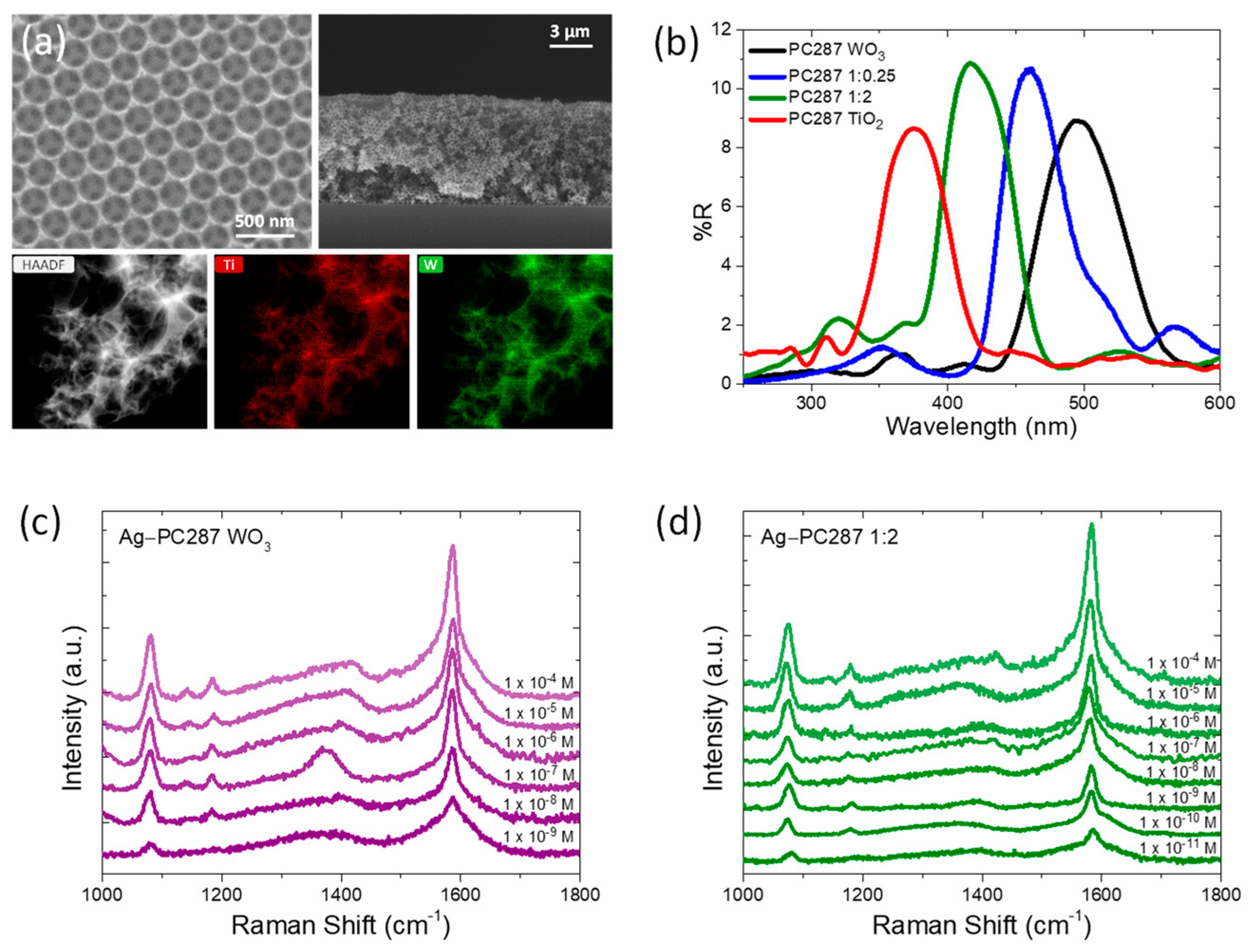
3.1. Structural and Compositional Characterization
SEM and TEM images of the PC films display a 3D network of uniform interconnected void macropores consisting of both metal oxides according to Ti and W EDX elemental maps (Figure 1a). The photonic band gap position of the WO3/TiO2 PC films shifted to higher wavelengths as the WO3 content increased, as shown by the specular reflectance (R%) spectra for the PC287 films in Figure 1b, allowing fine tuning of the photonic properties by altering the films’ composition.
Figure 1.
(a) (Upper) Top view and cross-section SEM images and (Lower) TEM image and elemental EDX maps of Ti and W for PC287 1:0.25; (b) specular reflectance (R%) spectra for PC287 films; (c,d) SERS spectra of 4−MBA on Ag−PC287 WO3 and 1:2 substrates.
3.2. Ag−WO3/TiO2 SERS Substrates
The SERS activity of inverse opal films was examined using 4−MBA (4−mercaptobenzoic acid) as the probe molecule. The characteristic peaks of 4−MBA were detected on the Ag−WO3/TiO2 substrates (Figure 1c,d), where the dominant peaks at 1078 cm−1 and 1585 cm−1 can be assigned to the in-plane ring breathing (C−S) and the aromatic ring (C−C) vibration modes. The amplification of SERS intensity can be attributed to the enhanced electromagnetic field at the interface of the Ag and WO3/TiO2 inverse opal skeleton, as well as the interfacial charge transfer from MOs’ conduction band to the lowest unoccupied molecular orbital (LUMO) of 4−MBA molecules. In Ag−WO3/TiO2 films, electron transfer between TiO2 and WO3 can improve charge separation and moderate recombination enabling the detection of smaller 4−MBA concentrations. The minimum analyte detection can reach very low values down to 10−11 mol/L for the Ag−PC287 1:2 substrate, which can also be related to the overlap of blue-edge slow photons with the Ag nanoparticles’ localized surface plasmon resonance. Ag−WO3/TiO2 nanocomposites are proposed as highly efficient SERS substrates for the sensitive detection of biological analytes.
Author Contributions
Conceptualization, V.L.; methodology, M.-A.A. and V.L.; validation, S.G.; formal analysis, M.-A.A.; investigation, M.-A.A., E.S., P.T. and S.G.; resources, V.L.; writing—original draft preparation, M.-A.A.; writing—review and editing, M.-A.A. and V.L.; supervision, V.L.; project administration, V.L.; funding acquisition, V.L. All authors have read and agreed to the published version of the manuscript.
Funding
This research received no external funding.
Institutional Review Board Statement
Not applicable.
Informed Consent Statement
Not applicable.
Data Availability Statement
The data presented in this study are available on request from the corresponding author. The data are not publicly available due to privacy issues.
Acknowledgments
The research work was supported by the Hellenic Foundation for Research and Innovation (HFRI) under the 3rd Call for HFRI PhD Fellowships (fellowship number: 5570).
Conflicts of Interest
The authors declare no conflicts of interest.
References
- Papadakis, D.; Diamantopoulou, A.; Pantazopoulos, P.A.; Palles, D.; Sakellis, E.; Boukos, N.; Stefanou, N.; Likodimos, V. Nanographene oxide−TiO2 photonic films as plasmon-free substrates for surface-enhanced Raman scattering. Nanoscale 2019, 11, 21542–21553. [Google Scholar] [CrossRef] [PubMed]
- Alessandri, I.; Lombardi, J.R. Enhanced Raman Scattering with Dielectrics. Chem. Rev. 2016, 116, 14921–14981. [Google Scholar] [CrossRef] [PubMed]
- Apostolaki, M.A.; Sakellis, E.; Tsipas, P.; Giannouri, M.; Gardelis, S.; Boukos, N.; Dimoulas, A.; Likodimos, V. Three-phase co-assembly of compositionally tunable WO3/TiO2 inverse opal photoelectrodes. Appl. Surf. Sci. 2023, 613, 155919. [Google Scholar] [CrossRef]
Disclaimer/Publisher’s Note: The statements, opinions and data contained in all publications are solely those of the individual author(s) and contributor(s) and not of MDPI and/or the editor(s). MDPI and/or the editor(s) disclaim responsibility for any injury to people or property resulting from any ideas, methods, instructions or products referred to in the content. |
© 2024 by the authors. Licensee MDPI, Basel, Switzerland. This article is an open access article distributed under the terms and conditions of the Creative Commons Attribution (CC BY) license (https://creativecommons.org/licenses/by/4.0/).