Proceeding Paper

Design, Fabrication and Characterization of PVA/PLGA Electrospun Nanofibers Carriers for Improvement of Drug Delivery of Gliclazide in Type-2 Diabetes †

by
Bibhu Prasad Panda
1,*,
Mok Xiu Wei
1,
Naveen Kumar Hawala Shivashekaregowda
2 and
Sujata Patnaik
3
1
Department of Pharmaceutical Technology, School of Pharmacy, Lakeside Campus, Taylor’s University, No 1, Jalan Taylor’s, Subang Jaya 47500, Selangor, Malaysia
2
Department of Pharmaceutical Chemistry, School of Pharmacy, Lakeside Campus, Taylor’s University, No 1, Jalan Taylor’s, Subang Jaya 47500, Selangor, Malaysia
3
University College of Pharmaceutical Sciences, Kakatiya University, Warangal 506009, Telangana, India
*
Author to whom correspondence should be addressed.
Presented at the 1st International Electronic Conference on Pharmaceutics, 1–15 December 2020; Available online: https://iecp2020.sciforum.net/.
Proceedings 2021, 78(1), 14; https://doi.org/10.3390/IECP2020-08689
Published: 1 December 2020
(This article belongs to the Proceedings of The 1st International Electronic Conference on Pharmaceutics)

Abstract

:
Poor solubility, erratic bioavailability and delivery challenges associated with gliclazide, which is commonly used in type 2 diabetes mellitus (T2DM) treatment, are overcome by exploring electrospun nanofibers technology. Employing emulsion electrospinning method with polyvinyl alcohol (PVA) alone and in combination with poly(d,l-lactide-co-glycolide) (PLGA), nanofibers were fabricated. Different concentrations of PLGA at 0.05%, 0.10% and 0.15% w/v were added to PVA to achieve a modified drug release profile to meet the typical physiological needs of T2DM, such as a faster drug release at meals followed by prolonged release to maintain constant plasma glucose level, which is highly desirable in T2DM management. Fabricated gliclazide-nanofibers were characterized by various studies, such as solubility, in-vitro drug release, drug release kinetic, scanning electron microscopy (SEM), differential scanning calorimetric (DSC), and Fourier transform infrared (FTIR) spectroscopy. GLZNF2, formulation of Drug:PVA:PLGA 0.1:10:0.05% w/v produced optimized gliclazide nanofibers. The optimized GLZNF2 nanofibers were incorporated into gelatin capsule for oral administration. SEM image of optimized formulation (GLZNF2) shows cylindrical shaped fiber, indicating gliclazide incorporated homogeneously in polymers with average fiber diameter 4.357 ± 0.83 µm. The solubility and dissolution rate of gliclazide nanofibers significantly improved compared to pure gliclazide. The gliclazide nanofibers produce a biphasic drug release profile, initial fast release, followed by prolonged release. Oral fabricated gliclazide fibers have tremendous potential as a drug carrier, and alternative technology for the improvement of solubility, dissolution rate, reduction in the dosing frequency and better blood glucose control could be explored in T2DM management.

1. Introduction

The electrospun method of making electrostatic fiber is one of the advanced and straightforward techniques of nanotechnology, and has tremendous potential as a drug carrier for the delivery of therapeutics. Polymeric electrospun fibers of diameters ranging from several nanometers to several micrometres are developed as drug carriers employing polymer solutions under the influence of an electrostatic field [1]. Polymeric nanofiber is a good candidate to develop as a specialized carrier system for the delivery of various therapeutics due to its unique characteristics, which include high surface area, high porosity, ability to load a high amount of the drug and greater flexibility in the modulation of drug release. Fabricated nanofibers based on nanotechnology are being extensively investigated for various applications, such as drug delivery system, gene delivery system, surgical intervention, wound dressing, catalysts and sensor [2].
Gliclazide is a second generation of sulphonylurea commonly used in the treatment of Type 2 Diabetes Mellitus. It promotes the production of insulin from the beta cells in the pancreas and stimulates insulin sensitivity. Gliclazide belongs to Biopharmaceutical Classification Systems (BCS) class II drugs, with low solubility and high membrane permeability and lipophilicity. The low solubility and dissolution rate of Gliclazide result in variable gastrointestinal absorption following oral administration cause erratic bioavailability [3]. Hence, to improve the drug delivery challenges and therapeutic efficacy of gliclazide in type 2 diabetes mellitus (T2DM), a polymeric nanofiber specialized carrier system for oral delivery of gliclazide was explored in this study. Moreover, long term therapy management of T2DM with conventional formulation required multiple dosing to maintain fasting and postprandial plasma glucose level. There is a huge demand to design and develop a suitable delivery system which meets the typical physiological needs of T2DM. The specialized oral delivery system, which modifies the drug release as per plasma glucose requirement and improves patient compliance, is of great importance in T2DM [3,4]. A few studies reported that incorporation of gliclazide into polymer matrices such as solid dispersion and nanocrystal formulation improved solubility, dissolution rate and achieve sustained release of the drug. However, the technique of electrospinning in the making of electrostatic fiber as a carrier for delivery of gliclazide with polyvinyl alcohol (PVA) and poly(d,l-lactide-co-glycolide) (PLGA) polymers was not reported and explored. In this research, gliclazide loaded PVA/PLGA electrostatic fibers were fabricated to improve the drug delivery challenges with enhanced drug dissolution and modified drug release profile through the electrospinning approach.
In this investigation, employing the emulsion electrospinning method and polyvinyl alcohol (PVA) alone and in combination with poly(d,l-lactide-co-glycolide) (PLGA) as core polymers, gliclazide nanofibers were fabricated. Both PVA and PLGA are synthetic, biocompatible polymers approved by the United States Food and Drug Administration for drug product development, used as the main polymer for nanofiber fabrication. PVA is a good candidate to promote drug dissolution of a poorly soluble drug due to its unique characteristics, which include very high water solubility and being s non-toxic, highly bio-compatible and orally safe polymer [5]. PLGA was also selected as a secondary polymer to modulate the drug release of gliclazide loaded PVA nanofibers. PLGA has been commonly applied as a biocompatible polymer in achieving sustained drug delivery over a prolonged period of time of many therapeutics reported [3,6,7]. In this study, different concentrations of PLGA at 0.05%, 0.10% and 0.15% w/v were added to PVA to achieve a modified drug release profile to meet the typical physiological needs of T2DM, such as a faster drug release at the time of meals followed by prolonged drug release profile over an extended period to maintain constant plasma glucose level, highly desirable for better T2DM management [8]. In this study, we have designed, developed and characterized gliclazide loaded PVA/PLGA electrospun nanofibers as potential carriers for improvement of oral drug delivery of gliclazide in Type-2 Diabetes. The optimized formulation of gliclazide loaded nanofibers was incorporated into an empty gelatin capsule for oral administration [9]. The morphological and physicochemical characterizations of the gliclazide-loaded PVA/PLGA nanofibers were performed. The fabricated gliclazide nanofibers were extensively investigated for solubility studies, in vitro drug release studies, drug release kinetic studies, scanning electron microscopy studies (SEM), differential scanning calorimetric (DSC) studies and Fourier transform infrared (FTIR) spectroscopy studies and discussed in detail [10,11,12,13].

2. Methods

2.1. Materials

Gliclazide was purchased from Toronto Research Chemical Inc. Labchem Sdn. Bhd., (Petaling Jaya, Malaysia).Polyvinyl alcohol was supplied by R&M Chemicals ((Petaling Jaya, Malaysia). Poly (d,l-lactide-co-glycolide) (MW 12,000) was purchased from Sigma-Alrich, supplied by Labchem Sdn. Bhd., (Petaling Jaya, Malaysia). All other chemicals and reagents used in this research work were analytical grade.

2.2. Preparation of Spinning Solution for Gliclazide Electrospun Nanofibers

Polymer PVA was dissolved in distilled water to form 10% (w/v) PVA solution. The polymeric PVA mixture was then stirred for 4 h at a temperature of 80 °C followed by cooling at room temperature. Then, gliclazide, 0.1% w/v and PLGA in different concentration 0.05%, 0.10%, 0.15%, were pre-dissolved in 1 mL of acetone and added in to PVA polymer solution. The mixture was then stirred for at least 20 min at room temperature to form homogenous solution before performing the electrospinning process. The design of different batches of formulations and their compositions used in the preparation of the spinning solution for gliclazide loaded PVA/PLGA electrospun nanofibers is shown in Table 1 [6].

2.3. Electrospinning for Gliclazide Nanofibers

The electrospinning process was performed under ambient condition (22 ± 1 °C temperature). The spinning solution was loaded into a 5 mL syringe pump with a metallic needle (inner diameter was 0.33 mm). The tip of the syringe’s needle was connected with a positive electrode of the high voltage power supply, while the collector that was wrapped with aluminium foil was connected with the grounded electrode. An electrical potential of 19 kV was applied. The distance between the tip of the needle and collector was 18 cm and the flow rate was 1 mL/h. Fabrication parameters of electrostatic fiber used in the development of gliclazide nanofiber are presented in Table 1. A series of drug-loaded spinning solutions of gliclazide in PVA/PLGA were exposed to the electrospinning process to develop gliclazide nanofibers. The fabricated gliclazide nanofibers were stored in a desiccator with silica gel beads and characterized subsequently for physicochemical studies [12].

2.4. Characterizations of Gliclazide Nanofibers

2.4.1. Drug Content Study

The UV spectrophotometric method was used for quantification of gliclazide in drug-loaded nanofibers. Electrospun nanofiber (equivalent to 5 mg gliclazide) was accurately weighed and dissolved in 10 mL of spinning solution (a solvent that was used in the preparation of the spinning solution). Then, the resultant solution was further added to the PBS solution and analyzed for drug contents using the UV spectrophotometer by PerkinElmer Lambda XLS, at 228 nm, against the blank solutions. Each experiment was carried out in triplicate [3].

2.4.2. Solubility Test

The solubility of pure gliclazide and gliclazide nanofibers was performed using an orbital shaker (by Heidolph Rotamax 120). 10mg of pure gliclazide and 10 mg equivalent of gliclazide nanofibers were added into the medium (water) and shaken with shaker for 48 h at 25 ± 0.5 °C under constant vibration. Samples withdrawn were filtered, diluted, and analyzed using the UV-spectrophotometer by PerkinElmer Lambda XLS at a wavelength of 228 nm. Each experiment was carried out in triplicate [3].

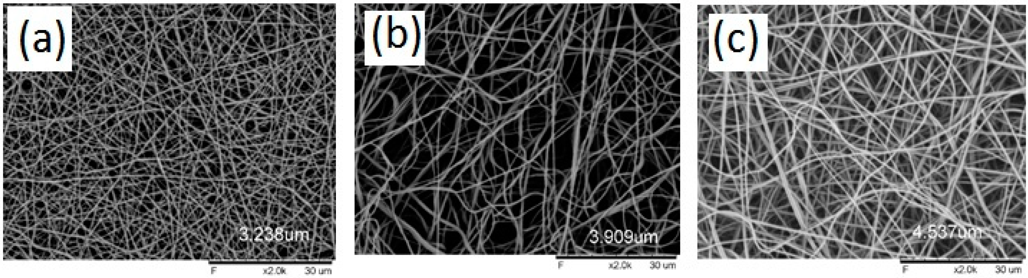
2.4.3. Scanning Electron Microscopy (SEM) Studies

The morphology of blank PVA nanofibers, BNF0 formulation, gliclazide nanofibers, GLZNF1 formulation and optimized gliclazide nanofibers, GLZNF2 formulation were observed using a tabletop scanning electron microscope (Hitachi TM3000) with 20 kV. The metal carriers of copper stubs with double-sided conductive tape were used to fix the electrospun nanofibers samples. Before an examination, a thin layer of gold was coated on the sample using an ion sputtering device. The diameter of fibers from the SEM image was measured using ImageJ software. Different parts of each nanofiber sample was selected for measurement and the average fiber diameter was calculated [14].

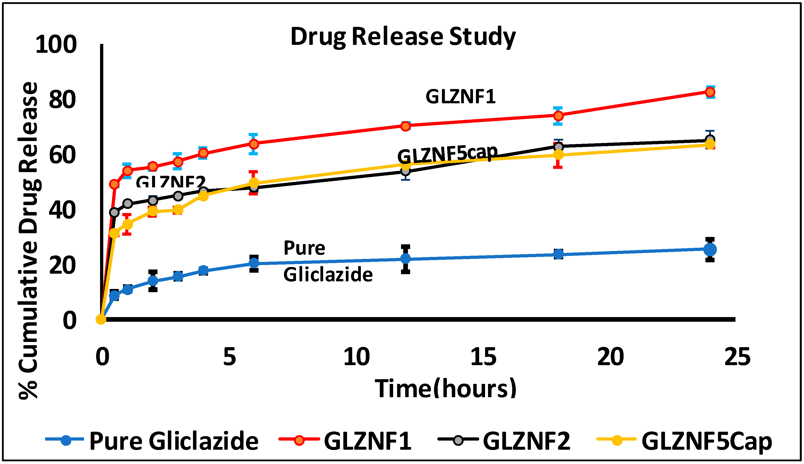
2.4.4. In Vitro Drug Release Studies and Drug Release Kinetic Studies

The in vitro drug release studies were performed for pure gliclazide, nanofiber formulations, optimized formulation and optimized formulation in capsule, and were determined by USP dissolution apparatus II. Electrospun nanofiber (equivalent to 5 mg gliclazide) was accurately weighed and the nanofibers sample was gently rolled over a glass carrier and inserted into the dissolution apparatus. The samples were then put into vessels containing 900 mL phosphate buffer (pH 7.4) under the stirring rate 50 rpm, maintained at 37 ± 0.5 °C. Aliquots of 5 mL were withdrawn at fixed time intervals (0.5, 1, 2, 3, 4, 6, 12, 18 and 24 h) and replaced with fresh phosphate buffer solution. The sample solution was filtered with 0.45 µm and then analyzed using a UV-spectrophotometer by Perkin Elmer Lambda XLS with a wavelength of 228 nm. Then, the cumulative percentage drug release was calculated and plotted against time. Data derived from the in-vitro release were fitted into five common kinetic models including zero-order kinetics, first-order kinetics, Higuchi’s plots and Korsmeyer–Peppas plots [12] to predict the mechanism of drug release from the formulated nanofiber. The correlation coefficient (R2) value was calculated for each model and each experiment was carried out in triplicate [3,15,16].

2.4.5. Differential Scanning Calorimeter (DSC)

The drug excipient compatibility and thermal behavior of pure gliclazide and polymers of nanofiber formulations were evaluated using a PerkinElmer Differential Scanning Calorimetry with Hyper DSC8500. The electrospun nanofibers were accurately weighed in aluminium pans and sealed with a press. The DSC thermograms of pure gliclazide, gliclazide nanofibers GLZNF1 formulation, and optimized gliclazide nanofibers GLZNF2 formulation were obtained at a heating rate of 10 °C/min from 30 °C up to 200 °C. The thermal scanning of pure gliclazide and gliclazide nanofibers samples was carried out in the oven by purging nitrogen gas at a flow rate of 20 mL/min. The aluminum oxide and indium powders were employed as reference and standard, respectively, during the study [3,17,18].

2.4.6. Fourier Transform Infrared Spectroscopy (FT-IR)

The FT-IR spectra of pure gliclazide, GLZNF1 gliclazide nanofibers formulation and GLZNF2 optimized gliclazide nanofiber formulation were recorded using a PerkinElmer Spectrum 100 FT-IR spectrometer (Perkin Elmer Inc., Wellesley, MA, USA). The interaction and compatibility between drug and polymer were characterized in these studies. The scanning range of the spectral analysis was carried out between 400 to 4000 cm−1 and with a resolution of 4 cm−1, respectively. The mean of 32 scans was used to form the spectra [3,19].

3. Results and Discussion

The multiple dosing of oral hypoglycemic agents and chronic therapy has been a common practice in T2DM management. Frequent administration of oral hypoglycemic agents, side effects and poor patient compliance are the major challenges associated with gliclazide, a clinically safe and approved first line of drug commonly recommended in T2DM management.
Conventional gliclazide oral dosage forms produce poor bioavailability, erratic therapeutic response, required large dose with frequent oral administration in T2DM. A conventional gliclazide oral treatment failed to meet the typical physiological goal of T2DM, such as basic needs between meals and during the night, which demands a faster drug release followed by prolonged drug release profile to maintain a constant plasma glucose level over an extended period of time. To overcome drug delivery challenges associated with gliclazide oral delivery, an innovative functional polymeric electrospun nanofibers technology was adopted as a carrier for the delivery of gliclazide. The main objective of this research study was to explore the advanced concept and technology of electrospun nanofibers in design, development and delivery of oral gliclazide dosage form in T2DM management.
Gliclazide nanofibers were successfully fabricated by the emulsion electrospinning method with polyvinyl alcohol (PVA) alone and in combination with poly(d,l-lactide-co-glycolide) (PLGA) used as the functional polymer in the fabrication of nanofibers as shown in Table 1. In this study, different concentrations of PLGA at 0.05%, 0.10% and 0.15% w/v were added to PVA to achieve a modified drug release profile for better T2DM management. All gliclazide nanofibers formulations passed the pharmacopeial requirement for the drug content assay test, which showed the drug content assay in the range of 96.13 ± 1.14% to 98.36 ± 0.87% presented in Table 2. The solubility of all gliclazide nanofibers formulations compared with pure gliclazide increase in solubility data were shown in Table 2. The gliclazide nanofibers formulations GLZNF1, containing 10% PVA, showed a 4.17 fold increase in the solubility, whereas optimized GLZNF2 containing 10% PVA and 0.05% PLGA formulation showed a 2.84 fold increase in solubility compared to the pure drug gliclazide, respectively. The decrease in solubility of optimized GLZNF2 nanofibers formulations compared to GLZNF1 nanofibers formulations, containing 10% PVA, may be contributed to by the incorporation of PLGA into nanofibers. It is also evident from the solubility study that as the PLGA concentration was increased in the formulation of PVA based nanofibers, the gliclazide solubility decreased, as shown in Table 2. The shape and morphology of gliclazide loaded nanofibers formulations and blank PVA nanofibers were characterized using a scanning electron microscope (SEM), shown in Table 2 and depicted in Figure 1. The SEM studies of all gliclazide nanofibers formulations revealed that nanofibers were successfully fabricated, which were able to produce an average fiber diameter in the range of 3.238 ± 0.47 µm to 5.537 ± 2.73 µm. The morphological characterization of SEM images analysis of gliclazide nanofibers formulations showed an increase in the average fiber diameter with an increase in polymer concentration in the formulation. SEM images of gliclazide nanofibers of the optimized formulation GLZNF2 shows that the cylindrical shape of fiber indicates that gliclazide was incorporated homogeneously in the polymer with the average fiber diameter of 4.357 ± 0.83 µm.
In in vitro drug release studies for all gliclazide nanofibers, formulations along with pure gliclazide were performed and are reported in Table 3 and Figure 2. All gliclazide loaded nanofibers formulations have shown significant enhancement in dissolution rate compared to pure gliclazide in PBS dissolution media at pH 7.4. Percentage of drug release for optimized gliclazide nanofibers formulations GLZNF2 and pure gliclazide at 0.5 h and 24 h were compared using PBS dissolution media at pH 7.4. The percentage of drug release for GLZNF2 was 38.35 ± 0.49 at 0.5 h and 65.08 ± 3.08 at 24 h, whereas pure gliclazide was able to release 8.62 ± 1.2 at 0.5 h and 25.18 ± 3.8 at 24 h, respectively. These drug release studies also revealed that with the addition of PLGA with an increase in concentration, the drug release from PVA based nanofibers was decreased and modulated to a modified drug release. The findings of drug release studies of gliclazide nanofibers formulations emphasize gliclazide nanofibers produced a biphasic drug release profile, an initial fast burst release, followed by a prolonged drug release. The initial rapid release of gliclazide from the nanofibers can be due to the high surface area of the nanofibers, surface deposition of the drug and numerous porosities of the electrospun nanofiber mats. Moreover, conversion into an amorphous state of gliclazide drug upon nanofiber formulation increases the dissolution rate [9]. In general, the excellent wettability of PVA can promote the dissolution rate and solubility of gliclazide nanofiber formulation, whereas with the addition of PLGA polymer into drug-loaded PVA, nanofiber provide a delayed release profile over extended periods. The release can be delayed to timescales of over 24 h, indicating a formulation significantly suitable to achieve a modified release profile for gliclazide. However, to make oral delivery of GLZNF2, optimized nanofibers formulation were encapsulated into an empty gelatin capsule (number 1 size). The optimized nanofibers formulation and gelatin capsule encapsulated optimized nanofibers formulation comparative to drug release studies showed the time of gliclazide release from the capsule dosage was found to be longer during the first hours, whereas in 24 h, the dissolution rate of both depicted a similar drug release profile.
The in vitro drug release profiles of all fabricated gliclazide nanofiber formulations were applied in different drug release kinetic models and interpreted in the form of graphical presentation and evaluated by correlation coefficient (R2), represented in Table 4. The highest degree of correlation coefficient determines the suitable mathematical model that follows gliclazide nanofiber drug release kinetics. All the gliclazide nanofibers formulations release kinetics data show a good fit to the Higuchi square root model, which implies the release of gliclazide from the nanofiber matrix as a square root of time-dependent process and diffusion controlled. The optimized gliclazide nanofibers formulation GLZNF2 release kinetics data were found to be best fitted by the Higuchi square root model and showed a higher degree of correlation coefficient (R2) than other models, which can be confirmed by comparing the values for the regression coefficient of the zero-order (R02 = 0.523), first-order (R12 = 0.7114), Higuchi’s plots (R2 = 0.727) and Korsmeyer–Peppas plots (Rk2 = 0.2233) equations. To further confirm the diffusion mechanism of optimized gliclazide nanofibers formulation GLZNF2, the data were then fitted to the Korsmeyer–Peppas equation. The release exponent value (n) of optimized gliclazide nanofibers formulation GLZNF2 is 0.4498, which is less than (n) 0.5, which implies that the drug release from the system follows a fickian diffusion release process.
The differential scanning calorimetry thermograms of pure gliclazide, gliclazide nanofibers, GLZNF1 formulation and optimized gliclazide nanofibers, GLZNF2 formulation are shown in Figure 3. The DSC thermogram of pure gliclazide showed a sharp endothermic peak at 172.13 °C, indicating the melting point (transition temperature) of gliclazide. The presence of the endothermic peak demonstrated that pure gliclazide is in the crystalline state. In DSC thermograms of both optimized nanofibers formulation GLZNF2 and gliclazide nanofibers formulation, GLZNF1 did not show any sharp melting peak analogous to the endothermic peaks of gliclazide. The observation indicates that gliclazide had been highly dispersed in the electrospun polymeric nanofibers samples at a 10%:0.05%:0.1% of the PVA:PLGA:Gliclazide ratio, converted to an amorphous state during the process of electrospinning.
Fourier transform infrared spectroscopic analysis was conducted for pure gliclazide, gliclazide nanofibers, GLZNF1 formulation and optimized gliclazide nanofibers, GLZNF2 formulation, and is depicted in Figure 4. The FTIR spectrum of pure gliclazide exhibited characteristic peaks at 1164.06 cm−1 (Sulphonyl S=O stretching), 1354.05 cm−1 (SO2NH stretching), 1597.09 cm−1 (Secondary amine N-H bending), 1709.92 cm−1 (Acyclic ketone carbonyl (C=O) stretching), 3113.16 cm−1 (=CH stretching) and 3275.19 cm−1 (Secondary amine N-H stretching), respectively. Spectral comparative analysis of gliclazide nanofibers, GLZNF1 formulation and optimized gliclazide nanofibers, GLZNF2 formulation with pure gliclazide, the spectrum shows all these gliclazide characteristic peaks range were observed in all gliclazide loaded nanofiber formulation presented in Figure 4; this indicates the presence of gliclazide in the nanofiber formulation. In GLZNF2, optimized gliclazide nanofibers showed most of the characteristic peaks of gliclazide such as a peak at 1098 cm−1 (S=O stretching), 1341.6 (SO2NH stretching), 1708.8 cm−1 (C=O stretching) and 3266 cm−1 (N-H stretching). The intensity of some peaks in formulated nanofiber is decreased, indicating the entrapment of gliclazide in the formulated nanofiber. Furthermore, in the fingerprint region of gliclazide spectra, the intensities of several sharp peaks were significantly decreased, or some disappeared totally, indicating the formation of amorphous composites between gliclazide and polymer in the nanofiber formulation.

4. Conclusions

The purpose of this study was to fabricate drug-loaded fibers and establish a proof of concept for the electrospun method of making electrostatic fiber as a functional specialized carrier system for oral delivery of gliclazide in type 2 diabetes mellitus (T2DM). The drug delivery challenges associated with oral gliclazide delivery are poor solubility, low dissolution rate, variable gastrointestinal absorption and erratic bioavailability. In this research, gliclazide loaded PVA/PLGA electrostatic fibers were successfully fabricated to improve the drug delivery challenges with enhanced drug dissolution and a modified drug release profile employing the emulsion electrospinning method. The formulation composed of Drug:PVA:PLGA in a 0.1:10:0.05% w/v ratio produced optimized and desired gliclazide nanofibers. The optimized formulation of gliclazide loaded nanofibers was incorporated into an empty gelatin capsule for oral administration. To establish electrostatic fibers as a functional specialized carrier system for oral delivery, the developed gliclazide nanofibers were extensively investigated for morphological and physicochemical characterizations such as solubility studies, in vitro drug release studies, drug release kinetic studies, scanning electron microscopy studies (SEM), differential scanning calorimetric (DSC) studies and Fourier transform infrared (FTIR) spectroscopy studies. The SEM image of optimized gliclazide nanofibers formulation shows the cylindrical shape of fiber indicates gliclazide was incorporated homogeneously in the polymer. The solubility and dissolution rate of gliclazide nanofibers were significantly improved compared to pure gliclazide. This study also highlights that optimized gliclazide nanofibers formulation, developed with PVA/PLGA, successfully achieved a modified drug release to meet the typical physiological needs of T2DM, such as a faster drug release at the time of meals followed by prolonged drug release profile over an extended period to maintain constant plasma glucose level, highly desirable in T2DM management. Fabricated gliclazide fibers in oral dosage form have tremendous potential as a drug carrier and alternative technology for the improvement of solubility, dissolution rate, reduction in the dosing frequency and better blood glucose control, and could be explored in T2DM management.

Author Contributions

Investigation, methodology, validation, writing—original draft, B.P.P., investigation, data curation, M.X.W.; writing—review and editing, N.K.H.S.; writing—review and editing S.P. All authors have read and agreed to the published version of the manuscript.

Institutional Review Board Statement

Not applicable for studies not involving humans or animals.

Acknowledgments

This study was supported by the School of Pharmacy, Taylor’s University, Malaysia.

Conflicts of Interest

The authors declare no conflict of interest.

References

  1. Balogh, A.; Farkas, B.; Faragó, K.; Farkas, A.; Wagner, I.; Verreck, G.; Nagy, Z.K.; Marosi, G. Melt-blown and electrospun drug-loaded polymer fiber mats for dissolution enhancement: A comparative study. J. Pharm. Sci. 2015, 104, 1767–1776. [Google Scholar] [CrossRef] [PubMed]
  2. Payab, S.; Davaran, S.; Tanhaei, A.; Fayyazi, B.; Jahangiri, A.; Farzaneh, A.; Adibkia, K. Triamcinolone acetonide-Eudragit® RS100 nanofibers and nanobeads: Morphological and physicochemical characterization. Artif. Cells Nanomed. Biotechnol. 2016, 44, 362–369. [Google Scholar] [CrossRef]
  3. Panda, B.P.; Krishnamoorthy, R.; Bhattamisra, S.K.; Shivashekaregowda, N.K.H.; Seng LBin Patnaik, S. Fabrication of Second Generation Smarter PLGA Based Nanocrystal Carriers for Improvement of Drug Delivery and Therapeutic Efficacy of Gliclazide in Type-2 Diabetes Rat Model. Sci. Rep. 2019, 9, 17331. [Google Scholar] [CrossRef]
  4. Sarkar, A.; Tiwari, A.; Bhasin, P.S.; Mitra, M. Pharmacological and pharmaceutical profile of gliclazide: A review. J. Appl. Pharm. Sci. 2011, 1, 11–19. [Google Scholar]
  5. Huang, W.; Yang, Y.; Zhao, B.; Liang, G.; Liu, S.; Liu, X.L.; Yu, D.G. Fast dissolving of ferulic acid via electrospun ternary amorphous composites produced by a coaxial process. Pharmaceutics 2018, 10, 115. [Google Scholar] [CrossRef] [PubMed]
  6. Ghafoor, B.; Aleem, A.; Najabat Ali, M.; Mir, M. Review of the fabrication techniques and applications of polymeric electrospun nanofibers for drug delivery systems. J. Drug Deliv. Sci. Technol. 2018, 48, 82–87. [Google Scholar] [CrossRef]
  7. Vashisth, P.; Raghuwanshi, N.; Srivastava, A.K.; Singh, H.; Nagar, H.; Pruthi, V. Ofloxacin loaded gellan/PVA nanofibers-Synthesis, characterization and evaluation of their gastroretentive/mucoadhesive drug delivery potential. Mater. Sci. Eng. C 2017, 71, 611–619. [Google Scholar] [CrossRef] [PubMed]
  8. Jahangiri, A.; Davaran, S.; Fayyazi, B.; Tanhaei, A.; Payab, S.; Adibkia, K. Application of electrospraying as a one-step method for the fabrication of triamcinolone acetonide-PLGA nanofibers and nanobeads. Colloids Surf. B Biointerfaces 2014, 123, 219–224. [Google Scholar] [CrossRef] [PubMed]
  9. Poller, B.; Strachan, C.; Broadbent, R.; Walker, G.F. A minitablet formulation made from electrospun nanofibers. Eur. J. Pharm. Biopharm. 2017, 114, 213–220. [Google Scholar] [CrossRef] [PubMed]
  10. Illangakoon, U.E.; Nazir, T.; Williams, G.R.; Chatterton, N.P. Mebeverine-loaded electrospun nanofibers: Physicochemical characterization and dissolution studies. J. Pharm. Sci. 2014, 103, 283–292. [Google Scholar] [CrossRef] [PubMed]
  11. Zhang, Z.; Li, W.; Wang, G.; Qu, Y.L.; Yu, D.G. Electrospun 4th-Generation Solid Dispersions of Poorly Water-Soluble Drug Utilizing Two Different Processes. J. Nanomater. 2018, 2018, 2012140. [Google Scholar] [CrossRef]
  12. Jahangiri, A.; Barzegar-Jalali, M.; Javadzadeh, Y.; Hamishehkar, H.; Adibkia, K. Physicochemical characterization of atorvastatin calcium/ezetimibe amorphous nano-solid dispersions prepared by electrospraying method. Artif. Cells Nanomed. Biotechnol. 2017, 45, 1138–1145. [Google Scholar] [CrossRef] [PubMed]
  13. Panda, B.P.; Krishnamoorthy, R.; Shivashekaregowda, N.K.H.; Patnaik, S. Influence of Poloxamer-188 on design and development of second generation PLGA Nanocrystals of Metformin Hydrochloride. Nano Biomed. Eng. 2018, 10, 334–343. [Google Scholar] [CrossRef]
  14. Baishya, H. Application of Mathematical Models in Drug Release Kinetics of Carbidopa and Levodopa ER Tablets. J. Dev. Drugs 2017, 6, 1–8. [Google Scholar] [CrossRef]
  15. Wójcik-Pastuszka, D.; Krzak, J.; Macikowski, B.; Berkowski, R.; Osiński, B.; Musiał, W. Evaluation of the release kinetics of a pharmacologically active substance from model intra-articular implants replacing the cruciate ligaments of the knee. Materials 2019, 12, 1202. [Google Scholar] [CrossRef]
  16. Javadzadeh, Y.; Ahadi, F.; Davaran, S.; Mohammadi, G.; Sabzevari, A.; Adibkia, K. Preparation and physicochemical characterization of naproxen-PLGA nanoparticles. Colloids Surf. B Biointerfaces 2010, 81, 498–502. [Google Scholar] [CrossRef] [PubMed]
  17. Panda, B.P. Impact of statistical central composite face centered design approach on method and process optimization of metformin hydrochloride loaded plga nanoformulation. Micro Nanosyst. 2017, 9, 55–71. [Google Scholar] [CrossRef]
  18. Fonseca, C.; Simões, S.; Gaspar, R. Paclitaxel-loaded PLGA nanoparticles: Preparation, physicochemical characterization and in vitro anti-tumoral activity. J. Control. Release 2002, 83, 273–286. [Google Scholar] [CrossRef]
  19. Panda, B.P.; Jessica, G. Extraction and Performance Evaluation of Salvia hispanica Mucilage as Natural Disintegrants for Optimization of Pyrilamine Maleate Fast Dissolving Tablets. Nat. Prod. J. 2015, 1, 288–298. [Google Scholar] [CrossRef]
Figure 1. Scanning electron microscopy (SEM) images of (a) blank polyvinyl alcohol (PVA) nanofibers, BNF0 formulation, (b) gliclazide nanofibers, GLZNF1 formulation, and (c) optimized gliclazide nanofibers, GLZNF2 formulation.
Proceedings 78 00014 g001
Figure 2. Represents dissolution profiles of pure gliclazide, nanofiber formulation (GLZNF1), optimized formulation (GLZNF2) and optimized formulation in capsule (GLZNF5Cap), at phosphate buffer, pH 7.4. All data points plotted as the mean ± SD (n = 3).
Proceedings 78 00014 g002
Figure 3. DSC thermograms of pure gliclazide, gliclazide nanofibers GLZNF1 formulation and optimized gliclazide nanofibers GLZNF2 formulation.
Proceedings 78 00014 g003
Figure 4. FTIR spectra of pure gliclazide, GLZNF1, gliclazide nanofibers formulation and GLZNF2, optimized gliclazide nanofibers formulation.
Proceedings 78 00014 g004
Table 1. Design of different batches of formulations and their composition used in gliclazide loaded polyvinyl alcohol (PVA)/poly(d,l-lactide-co-glycolide) (PLGA) electrospun nanofibers.
Formulation Code% w/vNanofiber Fabrication Parameters
GliclazidePVAPLGAMetallic Needle (Inner Diameter)
in mm
Flow Rate in
mL/h
Voltage Applied
in kV
Distance between Needle’s Tip and Collector
in cm
Ambient Condition
(Temperature °C)
BNF0
(BlankPVA Nanofiber)
01000.331191822 ± 1
GLZNF10.11000.331191822 ± 1
GLZNF20.1100.050.331191822 ± 1
SGNCF30.1100.100.331191822 ± 1
GLZNF40.1100.150.331191822 ± 1
GLZNF5Caps
(GLZNF2 in capsule)
0.1100.050.331191822 ± 1
GLZNF: Gliclazide nanofiber; PLGA: poly(d,l-lactide-co-glycolide); PVA: polyvinyl alcohol; Caps: Capsule.
Table 2. Electrospun nanofibers formulations and their physicochemical characterizations.
Formulation CodeDrug Content (%)Folds Increase in Solubility ± SD (Compared with Pure Gliclazide)Average Fiber Diameter ± SD (µm) (from SEM Studies)
BNF0 (Blank PVA Nanofiber)--3.238 ± 0.47
GLZNF196.82 ± 1.694.17 ± 1.043.909 ± 1.53
GLZNF298.36 ± 0.872.84 ± 1.754.537 ± 1.88
SGNCF396.24 ± 2.502.25 ± 0.285.261 ± 1.45
GLZNF496.13 ± 1.141.84 ± 0.175.537 ± 2.73
GLZNF5Caps97.86 ± 1.362.84 ± 1.944.537 ± 1.88
SD: standard deviation; (n = 3).
Table 3. Drug release studies for pure gliclazide and different gliclazide loaded electrospun nanofibers.
Time
(h)
% Cumulative Drug Release ± SD
Pure GliclazideGLZNF1
(10% PVA)
GLZNF2
(Optimized Formulation)
GLZNF3GLZNF4GLZNF5Cap
(Optimized Formulation in Capsule)
0.58.62 ± 1.248.87 ± 0.7938.35 ± 0.4927.306 ± 2.914.130 ± 0.7931.16 ± 0.94
110.79 ± 0.8253.84 ± 2.5841.72 ± 0.7530.690 ± 1.516.254 ± 1.4234.37 ± 3.45
213.714 ± 3.455.12 ± 1.0243.00 ± 1.3831.968 ± 2.217.964 ± 1.7438.96 ± 1.62
315.40 ± 1.357.25 ± 2.944.69 ± 0.4634.092 ± 1.419.638 ± 0.5639.6 ± 1.1
417.53 ± 0.860.22 ± 1.8646.40 ± 0.935.370 ± 2.4620.934 ± 2.844.69 ± 0.67
620.08 ± 2.663.62 ± 3.4347.68 ± 1.4137.908 ± 3.0222.194 ± 2.4149.37 ± 3.93
1221.77 ± 4.5769.99 ± 1.0653.62 ± 3.2841.724 ± 1.9824.750 ± 0.9756.16 ± 0.47
1823.47 ± 1.2573.81 ± 2.9362.53 ± 2.3245.972 ± 2.4528.152 ± 0.2559.56 ± 4.24
2425.18 ± 3.882.47 ± 2.0165.08 ± 3.0849.806 ± 2.8531.536 ± 1.4763.37 ± 1.12
SD: standard deviation; (n = 3).
Table 4. Drug release kinetics models of gliclazide electrospun nanofibers.
Gliclazide Nanofiber FormulationZero-Order PlotsFirst-Order PlotsHiguchi’s PlotsKorsmeyer–Peppas Plots
Correlation Coefficient (R02)Correlation Coefficient (R12)Correlation Coefficient (R2)Correlation Coefficient (Rk2)Diffusional Exponent (n)Type of Release
GLZNF10.4780.76270.7120.20410.453Fickian diffusion
GLZNF20.5230.71140.7270.22330.4498Fickian diffusion
SGNCF30.5340.65540.76810.23810.4292Fickian diffusion
GLZNF40.50050.54990.79140.28040.3938Fickian diffusion
GLZNF5Cap0.59960.75860.84990.2660.4834Fickian diffusion
Publisher’s Note: MDPI stays neutral with regard to jurisdictional claims in published maps and institutional affiliations.

