Proceeding Paper

Sub-100 nm Chitosan-Triphosphate-DNA Nanoparticles for Delivery of DNA Vaccines †

1
CICS-UBI—Health Sciences Research Center, University of Beira Interior, Avenida Infante D Henrique, 6200-506 Covilhã, Portugal
2
InoCure s.r.o, R&D Lab, Prumyslová 1960, 250 88 Celákovice, Czech Republic
*
Author to whom correspondence should be addressed.
Presented at the 1st International Electronic Conference on Pharmaceutics, 1–15 December 2020; Available online: https://iecp2020.sciforum.net/.
Proceedings 2021, 78(1), 12; https://doi.org/10.3390/IECP2020-08653
Published: 1 December 2020
(This article belongs to the Proceedings of The 1st International Electronic Conference on Pharmaceutics)

Abstract

:
Intramuscular delivery is one of the main routes for DNA vaccines administration. However, it requires large amounts of the DNA to be administered and external stimulation to encourage the internalization of the DNA. In this work, we consider alternative routes for less invasive administration and develop drug delivery systems (DDS) for intranasal administration. Chitosan polyplexes using sodium tripolyphosphate (TPP) as a crosslinker were prepared using the ionic gelation method. Our method allowed preparation of nanoparticles with the size below 50 nm, which is at least two times lower than previously reported sizes. Moreover, despite the small sizes, we obtained DNA encapsulation efficiencies of about 70%. Parameters that may affect the encapsulation efficiency were investigated, including different TPP-chitosan ratios and concentrations of DNA. We found that encapsulation efficiency of DNA inside the particles decreases with the increasing TPP-chitosan ratio. Moreover, increasing the DNA concentration leads to a higher encapsulation efficiency. Small (<50 nm) chitosan nanoparticles hold enormous potential as DNA carriers due to their physiological barriers and subsequent internalization.

1. Introduction

Cancer is the second most prevalent cause of death in the world that does not have a universal cure. Human papilloma virus (HPV) is among the main carcinogenic pathogens and its infection is related to several cancers, such as cervical and oropharyngeal [1,2]. Cervical cancer is the 4th largest cause of cancer in women worldwide [3]. The prophylactic vaccination against the human papilloma virus (HPV) infection that is already commercially available, Gardasil®, from Merck, prevents infection from HPV-16 and 18, which are responsible for around 70% of cervical cancers [4]. However, the current vaccine does not have a therapeutic effect against the already infected cells. That is, the vaccine can prevent the infection by HPV, but cannot prevent the development of cancers from pre-existing infections [5]. Therapeutic cancer vaccines are still a major area of ongoing research.
One of the promising approaches for therapeutic vaccines is DNA vaccination. DNA vaccination is applicable against a great range of viral, bacterial, and parasitic diseases, including HPV [6,7]. In comparison with traditional vaccines, DNA vaccines have numerous promising advantages. They are simpler to synthesize and can be produced on a large scale. In general, they are considered to be safer, since the pathogen is not required in the vaccine production. Additionally, DNA vaccines do not require refrigeration for storage, transport, and distribution, which may allow for easier distribution worldwide [8,9]. DNA vaccines are particularly appropriate for antitumor and anticancer treatment due to the fact that their encoded antigen can be expressed inside the antigen presenting cells (APCs). These cells can then activate the needed immune response for the dissolution or destruction of a recognized infected cell [10,11]. However, the delivery of the DNA vaccines to the APCs remains challenging. In this work, we evaluate ionotropic chitosan gelation as a potential method for the encapsulation and delivery of DNA vaccines.
Successful drug delivery systems (DDS) should provide a number of features, such as the ability to penetrate through several anatomical barriers, sustained and controlled release of their active pharmaceutical ingredients (APIs) as well as locally, stable and deep tissue penetration. These features can be enabled by nanometer-sized carriers made of synthetic and natural polymers. Nanocarriers can also be used for drug delivery through mucosal layers, and help the cellular internalization of the API [12,13]. Ionotropic gelation is one of the most common methods used for the encapsulation and delivery of DNA. The nanoparticles are produced by mixing the ionic crosslinker, tripolyphosphate (TPP), with the cationic polymer, chitosan (CS), under constant magnetic stirring. Chitosan is known as a good carrier for the delivery and internalization of non-viral vectors for gene delivery. The polycationic nature of CS enables electrostatic complexation with the DNA, which under controlled conditions would lead to the formation of DNA-CS complexes. With the optimum concentrations and other conditions, nanoparticles with sizes below 100 nm can be formed [14,15,16,17]. However, such small particles are rarely reported.
In this work, we developed and optimized the ionotropic gelation method for the encapsulation of DNA in order to produce nanoparticles with sizes ranging from 30 to 60 nm. We demonstrated that the method is robust and reproducible and, moreover, leads to high DNA encapsulation efficiency.

2. Materials and Methods

2.1. Materials

Medical grade chitosan 95/1000 was purchased from Heppe Medical, sodium tripolyphosphate (TPP) was obtained from Across Organics, deoxyribonucleic acid sodium salt (DNA) was acquired from MPBIO, 35% hydrochloric acid (HCl), while sodium hydroxide palettes (NaOH) and glacial acetic acid (AA) were all acquired from VWR. The following solutions were freshly prepared by using deionized water from VWR: 2M HCl, 10M NaOH, 1% and 2% (v/v) acetic acid.

2.2. Methods

2.2.1. Ionotropic Gelation

Ionotropic gelation using chitosan as a polymer and TPP as a crosslinker was used to prepare the nanoparticles. Particles with and without DNA were prepared using the same procedure adopted from [18] with slight modifications. Particles without DNA were used as a negative control. The process of ionic gelation is depicted in Figure 1. Next, 0.1% (w/v) chitosan (CS) in 1% (v/v) acetic acid and 0.1% (w/v) of TPP solution in water were prepared as stock solutions. The pH level of the chitosan and TPP solutions were adjusted to 5.2–5.5 and pH 2 by the addition of NaOH and HCl, respectively. For the experiments with DNA encapsulation, the DNA was dissolved in the TPP solution to reach the needed concentration. Then, both chitosan and TPP solutions were filtered using a 0.45 μm polyethersulfone syringe filter (from VWR). A syringe pump (New Era Pump System, Inc., Farmingdale, NY, USA) was used to add TPP solution to the chitosan solution dropwise with the flow rate of 0.25 mL/min. To ensure there was a consistent drop size, needle size 20 was used in all experiments, leading to an addition rate of about 15–16 drops/min. During the addition, the chitosan solution was stirred vigorously (600 rpm) using a magnetic stirrer. The solution was mixed for an additional 30 min after all of the TPP was consumed. All experiments were done at room temperature. As a positive control, 0.1% (w/v) deoxyribonucleic acid sodium salt (DNA) in water was prepared and mixed with the blank particles without DNA in a 1:1 ratio (by volume). A combined solution was incubated for 2 h at room temperature for DNA adsorption to chitosan-TPP nanoparticles to take place.
All experiments were performed in triplicates and the results are presented as means ± standard deviation (SD).

2.2.2. Encapsulation Efficiency

To estimate the amount of the DNA encapsulated in the chitosan NPs, the NPs solution was spun down using Centurion Scientific Benchtop Centrifuge for 20 min at 22.000 RCF and the DNA concentration in the supernatant was measured. The indirect encapsulation efficiency (iEE) was calculated as in (1), where c(total) is the theoretical DNA concentration in the solution and c(sup) is the measured DNA concentration after the encapsulation.
i E . E .   %   = c ( t o t a l ) c ( sup ) c ( t o t a l ) × 100 %
To verify the accuracy of the method, the amount of the encapsulated DNA was also estimated in the particles (dEE). NPs with and without DNA were spun down for 20 min at 22.000 RCF. The supernatant was removed for iEE measurement and the equivalent volume (1 mL) of 2% acetic acid and an additional 100 μL of 2M HCl were added. The solution was vortexed for one minute and then sonicated using the ultrasound homogenizer (from Qsonica sonicators) for 30 s at 40% amplitude. The solution was spun down for 20 min at 22.000 RCF mode to make sure that no pellet was formed and the DNA concentration was measured. The dEE was calculated as a ratio of measured and theoretical DNA concentrations.
In both cases, DNA concentration was used spectrophotometrically. That is, supernatant or dissolved nanoparticle solution (1 mL) was added to the quartz cuvette. The solution was diluted 2 to 3 times with 1% acetic acid, and the absorbance was measured using a cuvette reader from SpectraMax. The absorbance value at 260 nm was recorded.

2.2.3. Particle Size Determination

The size of the prepared CS-TPP-DNA-polyplexes was also analyzed using the Nanophox Dynamic Light Scattering (DLS) with photon cross-correlation spectroscopy from Sympatec. The particles were analyzed immediately after the preparation and after 72 h to evaluate the stability of the particle suspension. All DLS experiments were carried out at a temperature of 25 °C.

3. Results

Ionotropic gelation is a popular method for DNA encapsulation. Multiple authors have demonstrated the ability to prepared nanoparticles with DNA that are below 300 nm [18,19,20]. Liping et al. has shown that particles smaller than 200 nm can be prepared by using a solution with low chitosan and TPP concentrations of 0.1% [19]. However, to the best of our knowledge, nanoparticles with a size below 100 nm have not yet been prepared. In this work, we have used low solution concentrations and slow controlled drop-wise addition in order to make nanoparticles with a size of around 30–60 nm. We investigated the influence of the TPP/CS ratio and DNA concentration on the size and EE of formed nanoparticles.

3.1. Influence of Changing the TPP/CS Ratio on NPs Size and dEE

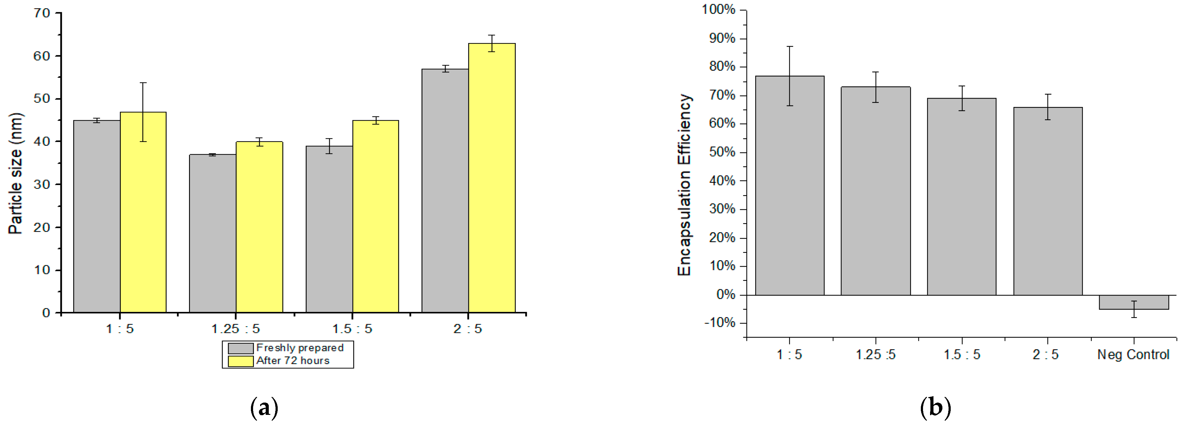
To investigate the influence of the TPP/CS ratio on nanoparticle size and DNA encapsulation efficiency, four NPs solutions with TPP/CS ratios of 1:5, 1.25:5, 1.5:5, and 2:5 were prepared. In these experiments, 35 μg/mL DNA was used. The formulation of prepared samples as well as their physicochemical characteristics are summarized in Table 1. As shown in Figure 2a, using the ionotropic gelation with controlled addition speed, very small nanoparticles of 30–50 nm were obtained. Only the particles with a high TPP/CS ratio led to the formation of larger particles of about 57 nm. In all cases, the particle size did not change significantly after storing them in the fridge for 72 h. Only a slight increase of about 2–6 nm was detected. However, as shown in Table 1, the formed particles were not monodispersed and had polydispersity index results of >0.5.
The amount of the DNA encapsulated in the microparticles was measured using the direct method after the extraction of the DNA from the collected nanoparticles. Prior to the measurement, the amount of the DNA that was adsorbed on the surface of the NPs was evaluated by mixing the prepared blank NPs with the DNA solution and measuring the concentration of the DNA in the supernatant. A total of 35 μg/mL of DNA was added, and −0.98 ± 1.21 μg/mL was measured (not shown), indicating that only about 3% of the DNA may be adsorbed on the NP’s surface. As shown in Figure 2b, the negative control showed that there is no interference between the excipients used in particle preparation. As shown in Figure 2b, all formulations showed a good efficiency of encapsulation with measured dEE of 77%, 73%, 69%, and 66% for samples 1 to 4, respectively. Increasing the TPP/CS ratio led to a small decrease in the encapsulation efficiency but only of about 10%.

3.2. Influence of Changing the DNA Concentration in NP Size and dEE

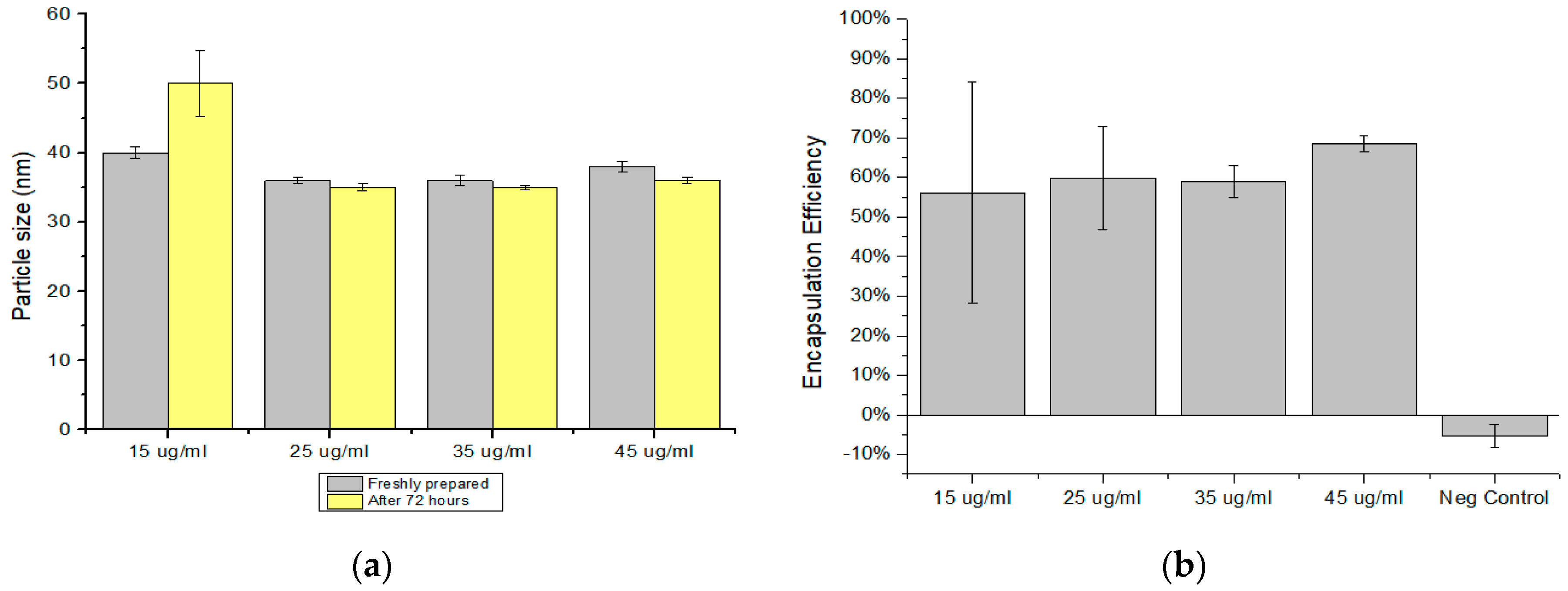
To investigate the influence of the DNA concentration on the nanoparticles size and DNA encapsulation efficiency, four formulations of NPs with DNA concentrations of 15, 25, 35, and 45 µg/mL were prepared. As mentioned before, particle size was measured immediately after the preparation and after 72 h to evaluate the NPs stability. The results are summarized in Table 2 and depicted in Figure 3a,b. In all cases, very small nanoparticles, below 40 nm were formed. The particle size increased slightly after storage, yet the solution could be considered stable. A small increase of about 10 nm could be detected in the sample with 15 μg/mL.
The encapsulation efficiency measurements of nanoparticles with different amounts of DNA added were evaluated as described before. As shown in Figure 3b, in all cases good encapsulation efficiencies were reached, with dEE results of 56%, 60%, 59%, and 69% for samples 5 to 8, respectively. Overall, our results suggest that higher encapsulation efficiency can be obtained with the increasing concentration of DNA.

4. Discussion and Future Work

In this work, we found that increasing the TPP/CS ratio does not influence the particle size and encapsulation efficiency. This result had also been reported previously by Turan and Akbuğa [18]. Similar results were observed by Liping et al. [19]. Increasing the polymer concentration leads to an increase in particle size, while the influence of the polymer concentration was noted by Liping et al. [19]. The effect of chitosan molecular weight was studied by Huang et al. [20]. They have shown that the low molecular weight of chitosan may lead to nanoparticles that are smaller than 70 nm. In this work, we used low molecular weighted medical purity chitosan, explored low solution concentrations, and obtained particles that are mostly below 50 nm. The reasons behind such small particle sizes are still under investigation. We believe that one of the factors influencing the size is the polymer mixing rate, which is controlled by both the flow rate of the dropwise addition and the stirring rate. Indeed, our preliminary experiments suggest that a faster addition or larger droplet size may lead to larger nanoparticles. The influence of the addition rate on the ionotropic CS-DNA-TPP gelation has not yet been reported.
Compared to the other reported studies, we have measured slightly lower DNA encapsulation efficiencies—usually below 80%, while Turan and Akbuğa reported around a 90% encapsulation efficiency [18]. This may be one of the drawbacks of extremely small nanoparticles. Our future work will focus on the optimization of the CS-DNA-TPP nanoparticles for the DNA internalization in eukaryotic cells. Depending on the successful results, low EE can be compensated for by the easier NP uptake caused by targeted cells. Moreover, the influence of the nanoparticle size on the mucopenetration and mucoadhesion will be tested to evaluate the feasibility of the intranasal administration.

5. Conclusions

Ionic gelation of CS/TPP nanoparticles is a promising delivery system for nucleic acids such as plasmid DNA. We have shown that these carriers can be downscaled below 50 nm, which may enable more efficient internalization of such NPs by relevant cells. Moreover, we have shown that good quantities of DNA can be encapsulated using the CS/TPP nanoparticles. This is especially promising for the delivery of the DNA vaccines, as high quantities of the DNA are needed to achieve effective immunization and therapeutic effects. In further studies, we will evaluate the cellular uptake and the mucopenetration/mucoadhesion of these particles.

Author Contributions

R.N. designed the study, performed the experiments, conducted data processing, conducted analysis of the results and wrote the paper; Â.S. helped with designing the study, provided supporting material and reviewed the paper; A.S. guided the design of work and experiments, helped with the data analysis and writing of the manuscript; A.A. helped with method development, supervised the work and contributed with insights and discussions; M.B. helped with the development of the experiments, as well as with discussions and ideas. All authors have read and agreed to the published version of the manuscript.

Institutional Review Board Statement

Not applicable.

Informed Consent Statement

Not applicable.

Data Availability Statement

The data presented in this study are available on request from the corresponding author.

Acknowledgments

R.N. has received support from the Erasmus+ traineeship program for his research activities at InoCure s.r.o. The project was supported by TACR project number FW01010445.

Conflicts of Interest

The authors declare no conflict of interest. The founding sponsors had no role in the design of the study; in the collection, analyses, or interpretation of data; in the writing of the manuscript, and in the decision to publish the results.

Abbreviations

The following abbreviations are used in this manuscript:
AA: Acetic acid; API: Active pharmaceutical ingredients; APC: Antigen presenting cells; CS: Chitosan; DDS: Drug delivery systems; dEE: Direct encapsulation efficiency; DLS: Dynamic Light Scattering; DNA: Deoxyribonucleic acid; EE: Encapsulation efficiency; HCl: Hydrochloric acid; HPV: Human papilloma virus; iEE: Indirect encapsulation efficiency; MHC: Major histocompatibility complex; MRT: Mean residence time; NaOH: Sodium hydroxide; NPs: Nanoparticles; pDNA: plasmid DNA; Rpm: Revolutions per minute; SD: Standard deviation; TPP: Sodium tripolyphosphate.

References

  1. Nagai, H.; Kim, Y.H. Cancer prevention from the perspective of global cancer burden patterns. J. Thorac. Dis. 2017, 9, 448–451. [Google Scholar] [CrossRef] [PubMed]
  2. Doorbar, J.; Egawa, N.; Griffin, H.; Kranjec, C.; Murakami, I. Human papillomavirus molecular biology and disease association. Rev. Med. Virol. 2015, 25, 2–23. [Google Scholar] [CrossRef] [PubMed]
  3. Arbyn, M.; Weiderpass, E.; Bruni, L.; de Sanjosé, S.; Saraiya, M.; Ferlay, J.; Bray, F. Estimates of incidence and mortality of cervical cancer in 2018: A worldwide analysis. Lancet Glob. Health 2020, 8, e191–e203. [Google Scholar] [CrossRef] [PubMed]
  4. Padmanabhan, S.; Amin, T.; Sampat, B.; Cook-Deegan, R.; Chandrasekharan, S. Intellectual property, technology transfer and manufacture of low-cost HPV vaccines in India [published correction appears in Nat Biotechnol. 2012 Feb;30(2):193]. Nat. Biotechnol. 2010, 28, 671–678. [Google Scholar] [CrossRef] [PubMed]
  5. Almeida, A.M.; Queiroz, J.A.; Sousa, F.; Sousa, A. Cervical cancer and HPV-Infection: Ongoing therapeutic research to counteract the action of E6 and E7 oncoproteins. Drug Discov. Today 2019, 24, 2044–2057. [Google Scholar] [CrossRef] [PubMed]
  6. Ha, S.; Jeon, B.; Youn, J.; Kim, S.; Cho, S.; Sung, Y. Protective effect of DNA vaccine during chemotherapy on reactivation and reinfection of Mycobacterium tuberculosis. Gene Ther. 2005, 12, 634–638. [Google Scholar] [CrossRef] [PubMed]
  7. Okuda, K.; Xin, K.Q.; Haruki, A.; Kawamoto, S.; Kojima, Y.; Hirahara, F.; Okada, H.; Klinman, D.; Hamajima, K. Transplacental genetic immunization after intravenous delivery of plasmid DNA to pregnant mice. J. Immunol. 2001, 167, 5478–5484. [Google Scholar] [CrossRef] [PubMed]
  8. Smith, H.A.; Klinman, D.M. The regulation of DNA vaccines. Curr. Opin. Biotechnol. 2001, 12, 299–303. [Google Scholar] [CrossRef]
  9. Kutzler, M.A.; Weiner, D.B. DNAvaccines: Ready for prime time? Nat. Rev. Genet. 2008, 9, 776–788. [Google Scholar] [CrossRef] [PubMed]
  10. Anderson, R.J.; Schneider, J. Plasmid DNA and viral vector-based vaccines for the treatment of cancer. Vaccine 2007, 25 (Suppl. 2), B24–B34. [Google Scholar] [CrossRef] [PubMed]
  11. Lee, J.; Arun Kumar, S.; Jhan, Y.Y.; Bishop, C.J. Engineering DNA vaccines against infectious diseases. Acta Biomater. 2018, 80, 31. [Google Scholar] [CrossRef]
  12. Mukherjee, S.; Ray, S.; Thakur, R.S. Solid lipid nanoparticles: A modern formulation approach in drug delivery system. Indian J. Pharm. Sci. 2009, 71, 349–358. [Google Scholar] [CrossRef] [PubMed]
  13. Panyam, J.; Labhasetwar, V. Biodegradable nanoparticles for drug and gene delivery to cells and tissue. Adv. Drug Deliv. Rev. 2003, 55, 329–347. [Google Scholar] [CrossRef]
  14. López-León, T.; Carvalho, E.L.; Seijo, B.; Ortega-Vinuesa, J.L.; Bastos-González, D. Physicochemical characterization of chitosan nanoparticles: Electrokinetic and stability behavior. J. Colloid Interface Sci. 2005, 283, 344–351. [Google Scholar] [CrossRef] [PubMed]
  15. Morimoto, M.; Saimoto, H.; Usui, H.; Okamoto, Y.; Minami, S.; Shigemasa, Y. Biological activities of carbohydrate-branched chitosan derivatives. Biomacromolecules 2001, 2, 1133–1136. [Google Scholar] [CrossRef] [PubMed]
  16. Li, X.B.; Tsushima, Y.; Morimoto, M.; Saimoto, H.; Okamoto, Y.; Minami, S.; Shigemasa, Y. Biological activity of chitosan-sugar hybrids: Specific interaction with lectin. Polym. Adv. Technol. 2000, 11, 176–179. [Google Scholar] [CrossRef]
  17. Di Martino, A.; Sittinger, M.; Risbud, M.V. Chitosan: A versatile biopolymer for orthopaedic tissue-engineering. Biomaterials 2005, 26, 5983–5990. [Google Scholar] [CrossRef] [PubMed]
  18. Özbaş-Turan, S.; Akbuğa, J. Plasmid DNA-loaded chitosan/TPP nanoparticles for topical gene delivery. Drug Deliv. 2011, 18, 215–222. [Google Scholar] [CrossRef] [PubMed]
  19. Wang, L.; Zhang, W.; Zhou, Q.; Liu, S.; Xu, W.; Sun, T.; Liang, Y. Establishing Gene Delivery Systems Based on Small-Sized Chitosan Nanoparticles. J. Ocean Univ. China 2018, 17, 1253–1260. [Google Scholar] [CrossRef]
  20. Huang, K.S.; Sheu, Y.R.; Chao, I.C. Preparation and Properties of Nanochitosan. Polym. Plast. Technol. Eng. 2009, 48, 1239–1243. [Google Scholar] [CrossRef]
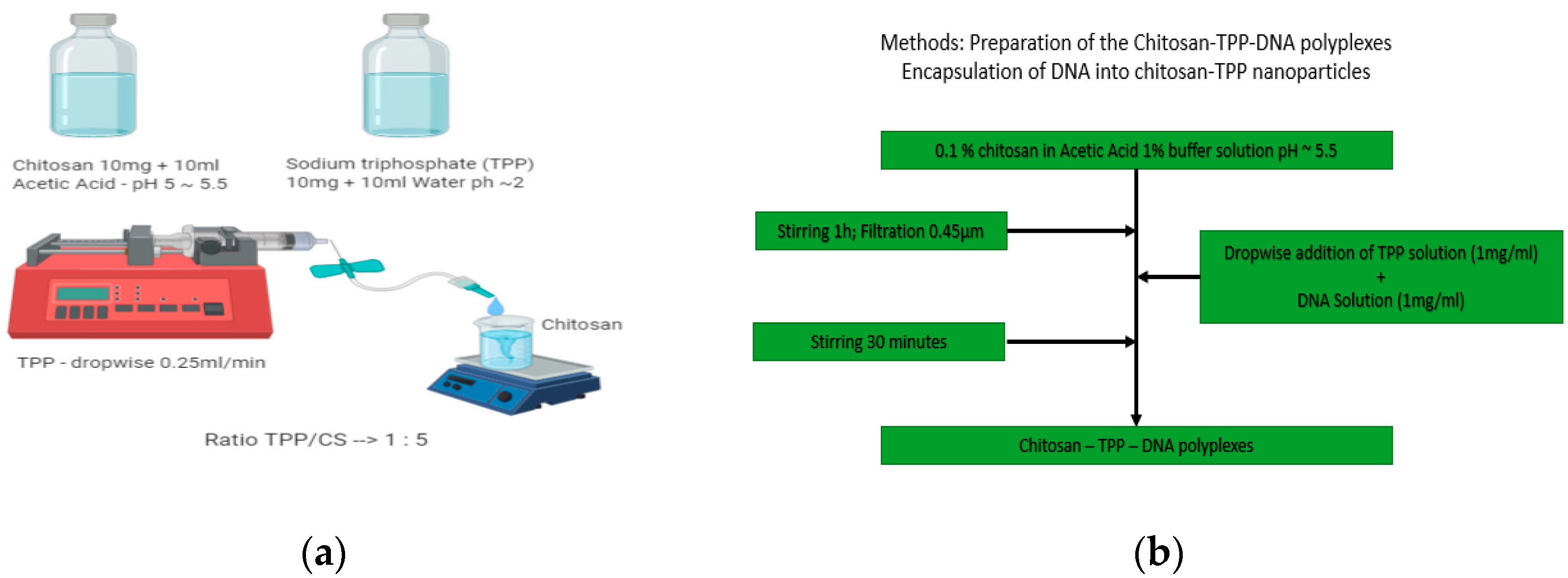
Figure 1. (a) Schematic illustration of chitosan-tripolyphosphate (TPP) nanoparticles (b) Flow chart of the nanoparticle with DNA preparation. First, solutions with optimized concentrations were prepared. Then, TPP with DNA was added to the chitosan solution dropwise while stirring.
Proceedings 78 00012 g001
Figure 2. (a) Particle size (nm) freshly prepared and after 72 h with different TPP/CS ratios; (b) Results of the encapsulation efficiency with different TPP/CS ratios.
Proceedings 78 00012 g002
Figure 3. (a) Particle sizes (nm) freshly prepared and after 72 h with different DNA concentrations; (b) Results of the encapsulation efficiency with different DNA concentrations.
Proceedings 78 00012 g003
Table 1. Average particle size, Polydispersity Index (PDI), and encapsulation efficiency of chitosan-TPP-DNA polyplexes with different TPP/CS ratios.
TPP/CS RatioZ-Average Size (nm)Polydispersity Index (PDI)Z—Average Size after 72 h (nm)(PDI) after 72 hEncapsulation Efficiency (%)
1:545 ± 0.50.55 ± 0.0147 ± 6.90.51 ± 0.00677 ± 10
1.25:537 ± 0.30.50 ± 0.0340 ± 0.90.48 ± 0.00473 ± 5
1.5:539 ± 1.70.46 ± 0.0445 ± 0.90.40 ± 0.00969 ± 4
2:557 ± 0.80.50 ± 0.0263 ± 1.90.46 ± 0.01066 ± 4
Table 2. Average particle size, PDI, and encapsulation efficiency of chitosan-TPP-DNA polyplexes with different DNA concentrations.
DNA Concentration (μg/mL)Z-Average Size (nm)Polydispersity Index (PDI)Z—Average Size after 72 h (nm)(PDI) after 72 hEncapsulation Efficiency (%)
15 ± 540 ± 0.80.59 ± 0.0150 ± 4.80.39 ± 0.2656 ± 28
25 ± 535 ± 0.50.48 ± 0.0535 ± 0.50.53 ± 0.00660 ± 13
35 ± 536 ± 0.80.47 ± 0.0535 ± 0.30.56 ± 0.00659 ± 4
45 ± 538 ± 0.70.47 ± 0.0436 ± 0.40.55 ± 0.0169 ± 2
Publisher’s Note: MDPI stays neutral with regard to jurisdictional claims in published maps and institutional affiliations.