Proceeding Paper

Cytomorphological, Molecular Diagnosis and Evaluation of Insertion of the LINE-1 Element in the C-MYC Gene in Canine Transmissible Venereal Tumor: Applicability in Veterinary Clinical Routine †

by
Faro Thamirys Aline Silva
1,2,*,
Ferreira Wallax Augusto Silva
2 and
De Oliveira Edivaldo Herculano Correa
2,3
1
Postgraduate Program in Genetics and Molecular Biology (PPGBM), Institute of Biological Sciences, Universidade Federal do Pará, Belém 66075-110, Pará, Brazil
2
Laboratory of Tissue Culture and Cytogenetics, Evandro Chagas Institute, Ananindeua 66093-020, Pará, Brazil
3
Institute of Exact and Natural Sciences, Faculty of Natural Sciences, Universidade Federal do Pará (UFPA), Belém 66075-110, Pará, Brazil
*
Author to whom correspondence should be addressed.
Presented at the First International Electronic Conference on Genes: Theoretical and Applied Genomics, 2–30 November 2020; Available online: https://iecge.sciforum.net/.
Proceedings 2021, 76(1), 4; https://doi.org/10.3390/IECGE-07156
Published: 2 November 2020

Abstract

:
Canine transmissible venereal tumor (CTVT) is the oldest known cancer in the world. Cytomorphologically, the cases are classified into three types. Previous studies have shown that the insertion of the transposable element LINE-1 in the MYC gene has diagnostic importance. Therefore, we characterized this insertion in samples of the three different cytomorphological types of CTVT, in order to verify whether there were any differences concerning the presence or location of the insertion and the size of the fragment. Our results showed that LINE-1 was inserted in all the samples analyzed, in the same position and presenting the same size (400 bp). We conclude that even though it is important for the oncogenesis process, this insertion has no influence on the cytomorphological type and the observed clinical differences.

Published: 2 November 2020

1. Introduction

Canine transmissible venereal tumor (CTVT), also known as infectious sarcoma, venereal granuloma or Sticker tumor [1], is a cancer that occurs naturally and spreads directly through the transfer of cells between individuals [2]. It is the oldest known transmissible cancer in the world, since the tumor cells found in the animals affected today are correspondents of an animal from approximately 11 thousand years ago, probably when the dogs were first domesticated [3,4].
The disease manifests frequently in the external genitalia of animals, being transmitted by direct contact through coitus, and due to its anatomical location, is usually called genital CTVT [5]. Secondarily, because of the species communication and licking habits, the tumor can occur in the areas around the eyes, mouth and nasal cavity, being called extragenital CTVT [5,6]. This type, due to its atypical location, is difficult to diagnose with histiocytes, mastocytomas, amelanotic melanomas, lymphomas and poorly differentiated carcinomas as differential diagnoses [7].
Cytological and histopathological analyses in some cases are not sufficient to conclude the diagnosis of CTVT, as well as immunohistochemistry, since CTVT has the same origin as histiocytes [8]. Hence, the molecular method can be used for the definitive diagnosis, in which the insertion of the transposable element LINE-1 in the MYC gene, which is characteristic of CTVT tumor cells, is identified [9].
Morphologically, CTVT is classified in three cytomorphological types: (1) lymphocytic, characterized by more than 60% of round cells, with finely granular cytoplasm, central nucleus and a few intracytoplasmic vacuoles; (2) plasmacytic, characterized by containing more than 60% of cells with a broad cytoplasm, eccentric nuclei and a large amount of vacuoles; and (3) mixed, presenting both lymphocytoid and plasmacytoid cells, neither of which exceed 59% [10,11]. Of them, the plasmacytic type is the most common cytomorphological type, and is related to chemoresistance [10,12,13]. Hence, due to its prognostic and predictive value, the determination of the cytomorphological classification has gained importance in veterinary clinical practice.
Considering that the insertion of the transposable element LINE-1 in the MYC gene has been reported in every sample so far, the purpose of this work was to analyze if there are any differences concerning the size and insertion site between different cytomorphlogical types, which could be associated to their distinct clinical characteristics.

2. Materials and Methods

2.1. Ethical Aspects of the Research

This study was approved by the Ethics Committee on the Use of Animals (Protocol nº 2530101017—UFPA) and that the owners of the animals signed a Free and Informed Consent Term for donation of the samples.

2.2. Sample Collection

Biopsies were collected from 36 animals. Fragments of 1 cm from the affected genital region were collected and stored in RNAlaterTM (Thermo Fisher). In addition, the anamnesis and clinical examination of the animal were carried out and data concerning the past and current information about the animal, time of neoplasia evolution, location, previous therapy, breeding and reproduction habits were obtained. Two samples of normal tissue from the penis and vulva of animals were also collected for necropsy.

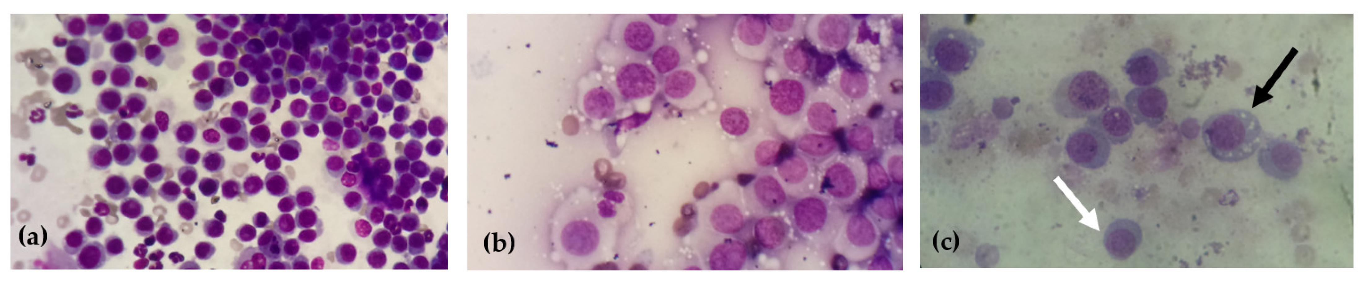
2.3. Cytological Diagnosis and Cytomorphological Classification

For cytological diagnosis and classification, the slides were prepared and fixed in methanol for five minutes and after fixation they were stained using Giemsa stain in a 1: 1 dilution, for 15 min. After a detailed scan of the slides, areas with the best pattern of cell distribution and staining were selected, and then at least ten fields were observed at 40× magnification, for CTVT cytomorphological classification according to the predominant cell type—plasmacytoid, lymphocytoid or mixed [10].

2.4. DNA Extraction, Amplification, Sequencing, and Alignment

For molecular analysis, genomic DNA (gDNA) from all tissue samples was isolated using the ReliaPrepTM gDNA Tissue MiniPrep System (Promega Corporation) following the manufacturer’s instructions. The DNA samples were purified using EZ-10 Spin Column PCR Product Purification kit (Bio Basic/Ludwig Biotec) and quantified with Nano-Drop-2000 spectrophotometer (ThermoFisher Scientific, Waltham, MA, USA) and with with a Qubit™ 4 Fluorometer (ThermoFisher Scientific, Waltham, MA, USA) and stored at −80 °C. Genomic DNA was used as a template for polymerase chain reaction (PCR) in order to amplify the LINE-1/MYC insertion. The amplification conditions and the primers used were detailed in studies [9,14]. Then, PCR products were analyzed by electrophoresis in 3% agarose and subsequently sequenced with the BigDye Terminator Cycle Sequencing Standard kit (Applied Biosystems, Foster City, CA, USA) version 3.1, purified using a BigDye X Terminator Purification Kit (Applied Biosystems), and analyzed on an ABI 3130 automated sequencer (Applied Biosystems). All sequence chromatograms were edited and aligned in BioEdit 7.2.6.1 software, using the ClustalW algorithm.

3. Results and Discussion

Thirty-six samples were collected from animals presenting different ages, sex and races: 12% of these animals were males and 67% females; 33% of the animals were elderly aged (7 years or older) and 67% were young animals (up to 6 years old). The cytological diagnosis as well as the cytomorphological classification showed that four samples were plasmocytic, five mixed type and 27 lymphocytic (Figure 1). Regarding breeds, 83% were mixed-breed animals, 14% Poodle and 3% Dachshund.
The molecular diagnosis, based on the identification of the insertion of the transposon LINE-1 in the MYC gene proved to be an efficient and definitive diagnosis, found in all analyzed tumor samples, but not in the control samples (Figure 2), as a fragment of approximately 400 bp, as later confirmed by sequencing samples.
Precisely because there are different cell lines with different biological behaviors, cytomorphological diagnosis in cases of CTVT has gained in importance in veterinary oncology in terms of therapeutics and prognosis [15]. The percentage of cytomorphological types found in our study was different from those mentioned in the literature, as reported in Amaral et al. (2007) [10], in which the most frequent type was the plasmacytic type (52.53%), followed by the mixed type (29.11%) and finally, the lymphocytic type (18.36%) as well as in Gaspar’s et al. (2010) [16], in which the frequency of the plasmacytic type was 50%, lymphocytic 19% and mixed 31% and in the research by Lima et al. (2016) the plasmacytic type represented 45% of the cases, followed by the lymphocytic type (30%) and lastly the mixed type (25%) [12].
Some researchers have already reported different subtypes according to the Brazilian regions in which the plasmacytic type was more frequent in the southeast compared to the south [10]. A study carried out in the city of Mato-Grosso, the plasmacytic type was also the most prevalent (81.8%) and the lymphocytic type was the least found cytomorphological type (6.8%) [17], differing again from our data.
PCR results proved to be a highly sensitive test for the detection of the rearrangement of the LINE-1 element in the MYC gene in CTVT, since all the samples of the transmissible tumors studied amplified a fragment of 400 pb, which characterizes such rearrangement, corroborating the observations by O’Neill (2011) [18]. There was no difference in the insertion site in the three cytomorphological types evaluated. Some studies have shown different insertion sizes in CTVT samples, but generally close to the 400 bp size [7,9,12,14].

4. Conclusions

We can conclude that even though it is important for the oncogenesis process, the rearrangement of the LINE-1 element in the MYC gene has no influence in the cytomorphological type and its clinical differences.

Institutional Review Board Statement

The study was approved by Committee on Animal Use of the Federal University of Para (CEUA/UFPA) in the meeting of 11/24/2017 (protocol code CEUA 2530101017).

Acknowledgments

This study was financed in part by the Coordenação de Aperfeiçoamento de Pessoal de Nível Superior—Brasil (CAPES)—Finance Code 001.

References

  1. Goldschmidt, M.H.; Hendrick, M.J. Tumors of the skin and soft tissues. In Tumors in Domestic Animals, 4th ed.; Meuten, D.J., Ed.; Iowa State: Ames, IA, USA, 2002; Chapter 2; pp. 45–118. [Google Scholar]
  2. Ostrander, E.A.; Davis, B.W.; Ostrander, G.K. Transmissible Tumors: Breaking the Cancer Paradigm. Trends Genet. 2016, 32, 1–15. [Google Scholar] [CrossRef] [PubMed]
  3. Murgia, C.; Pritchard, J.K.; Kim, S.Y.; Fassati, A.; Weiss, R.A. Clonal Origin and Evolution of a Transmissible Cancer. Cell 2006, 126, 477–487. [Google Scholar] [CrossRef] [PubMed]
  4. Rebbeck, C.A.; Thomas, R.; Breen, M.; Leroi, A.M.; Burt, A. Origins and evolution of a transmissible cancer. Evolution 2009, 63, 2340–2349. [Google Scholar] [CrossRef] [PubMed]
  5. Setthawongsin, C.; Tangkawattana, S.; Rungsipipat, A.; Techangamsuwan, S. Computerized Cytomorphometric and Cytomorphological Analysis of Canine Transmissible Venereal Tumours. J. Comp. Pathol. 2018, 163, 18–22. [Google Scholar] [CrossRef] [PubMed]
  6. Strakova, A.; Murchison, E.P. The cancer which survived: Insights from the genome of an 11,000 year-old cancer. Curr. Opin. Genet. 2015, 30, 49–55. [Google Scholar] [CrossRef] [PubMed]
  7. Park, M.S.; Kim, Y.; Kang, M.S.; Oh, S.Y.; Cho, D.Y.; Shin, N.S.; Kim, D.Y. Disseminated transmissible venereal tumor in a dog. J. Vet. Diagn. Investig. 2006, 18, 130–133. [Google Scholar] [CrossRef] [PubMed]
  8. Das, U.; Das, A.K. Review of canine transmissible venereal sarcoma. Vet. Res. Commun. 2000, 24, 545–556. [Google Scholar] [CrossRef] [PubMed]
  9. Fonseca, L.S.; Mota, L.S.; Colodel, M.M.; Ferreira, I.; Brandão, C.V.S.; Rocha, N.S. Spontaneous canine transmissible venereal tumor: Association between different phenotypes and the insertion LINE-1/c-myc. Rev. Colomb. Cienc. Pecu. 2012, 25, 402–408. [Google Scholar]
  10. Amaral, A.S.; Bassani-Silva, S.; Ferreira, I.; Fonseca, L.S.; Andrade, F.H.E.; Gaspar, L.F.J.; Rocha, N.S. Cytomorphological characterization of transmissible canine venereal tumor. Rev. Port. Cienc. Vet. 2007, 102, 253–260. [Google Scholar]
  11. Florez, M.M.; Feo, H.; Yamatogi, R.; Yamatogi, R.S.; Aguiar, A.J.; Araújo, J.P., Jr.; Rocha, N.S. Cell cycle kinetics, apoptosis rates and gene expressions of MDR-1, TP53, Bcl-2 and BAX in transmissible venereal tumor cells and their association with therapy response. Vet. Comparat. Oncol. 2016, 14, 1–15. [Google Scholar]
  12. Lima, C.R.O.; Faleiro, M.B.R.; Rabelo, R.E.; Vulcani, V.A.S.; Rubini, M.R.; Torres, F.A.G.; Moura, V.M.B.D. Insertion of the LINE-1 element in the C-MYC gene and immunoreactivity of C-MYC, p53, p21 and p27 proteins in different morphological patterns of the canine TVT. Arq. Bras. Med. Vet. Zootec. 2016, 68, 658–666. [Google Scholar] [CrossRef]
  13. Flórez, L.M.M. Expressão dos Genes MDR-1, TP53, BCL-2 E BAX em Tumor Venéreo Transmissível Canino e sua Relação com a Agressividade e Resposta à Terapia. Ph.D. Thesis, Unesp, Botucatu, 2014; p. 81. [Google Scholar]
  14. Spin, J.S.F.; Da Mota, L.S.; DA Castelli, L.S.L.S.; Silva, E.C.; Ferreira, S.B.; Rocha, I.; Sousa, N. Detecção molecular do rearranjo Line-1/c-MYC em tumores venéreos transmissíveis caninos espontâneos. Clín. Vet. 2010, 15, 64–68. [Google Scholar]
  15. Silva, J.S.; Da Silva, D.M.M.; Silva, V.B.; Da Silva, D.A.; Silva, R.S.; De Lucena, R.B. Classificação citopatológica e epidemiologia do TVT nos cães atendidos no Hospital Veterinário da UFPB. In Proceedings of the 38º Congresso Brasileiro Da Anclivepa 2017, Anais do 38º CBA, Recife, Brazil, 2017; p. 0808. [Google Scholar]
  16. Gaspar, L.F.; Ferreira, I.; Colodel, M.M.; Brandão, C.V.; Rocha, N.S. Spontaneus canine transmissible venereal tumor: Cell morphology and influence on p-glycoprotein expression. Turk. J. Vet. Anim. Sci. 2010, 34, 447–454. [Google Scholar]
  17. Valençola, R.A.; Antunes, T.R.; Sorgatto, S.; Oliveira, B.B.; Godoy, K.C.S.; De Souza, A.I. Aspectos citomorfológicos e frequência dos subtipos do tumor venéreo transmissível canino no município de Campo Grande, Mato Grosso do Sul, Brasil. Acta Vet. Bras. 2015, 9, 82–86. [Google Scholar]
  18. O’neill, I.D. Concise review: Transmissible animal tumors as models of the cancer stem-cell process. Cancer Stem. Cells 2011, 29, 1909–1914. [Google Scholar] [CrossRef] [PubMed]
Figure 1. Cytology images of canine transmissible venereal tumor (CTVT) of different cytomorphological types: (a) lymphocytic type—predominance of more than 60% of round cells, with little presence of cytoplasmic vacuoles and centralized nuclei; (b) plasmacytic type—predominance of more than 60% of ovoid cells with more abundant cytoplasms and eccentric nuclei; (c) mixed type—there is the presence of both cell subtypes; however, neither type exceeds 59% of the total cells analyzed in the slide. Black arrow—plasmacytoid cell; White arrow—lymphocytoid cell.
Proceedings 76 00004 g001
Figure 2. Molecular diagnosis of CTVT. Yellow arrow—400 bp; red arrow—500 bp; orange arrow—900 bp; 1 and 13—molecular weight of 1 kb; 2—dog testicle DNA as the negative control of CTVT. There are two fragments of different sizes (900 bp and ~400 bp), respectively, transposon and MYC gene; 3—sample of positive CTVT according to the histopathological report; 4 to 7—CTVT samples showing the insertion of the transposon Line-1 (900 bp) in the MYC gene (~400 bp), in which there is only 1 fragment; 8 to 12—positive internal sample controls (~480 pb).
Proceedings 76 00004 g002
Publisher’s Note: MDPI stays neutral with regard to jurisdictional claims in published maps and institutional affiliations.

