Identification and Characterization of Metabolic Potential of Different Strains from Genus Rhizobium †


Abstract
:1. Introduction
2. Material and Methods
2.1. Bacterial Strains
2.2. PCR and Sequencing
2.3. Biolog GEN III
2.4. Data Analysis and Visualisation
3. Results
3.1. Bacterial Species
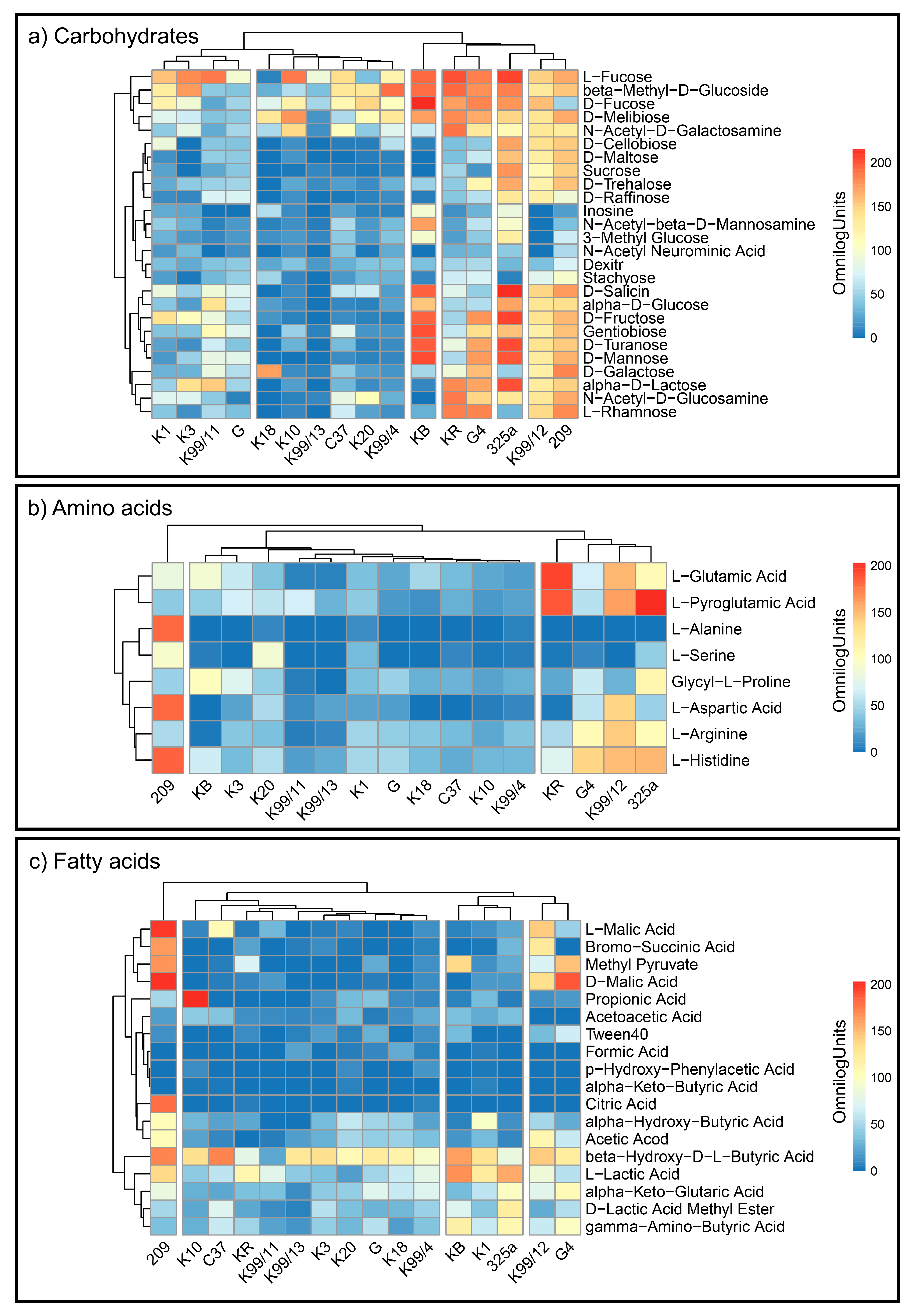
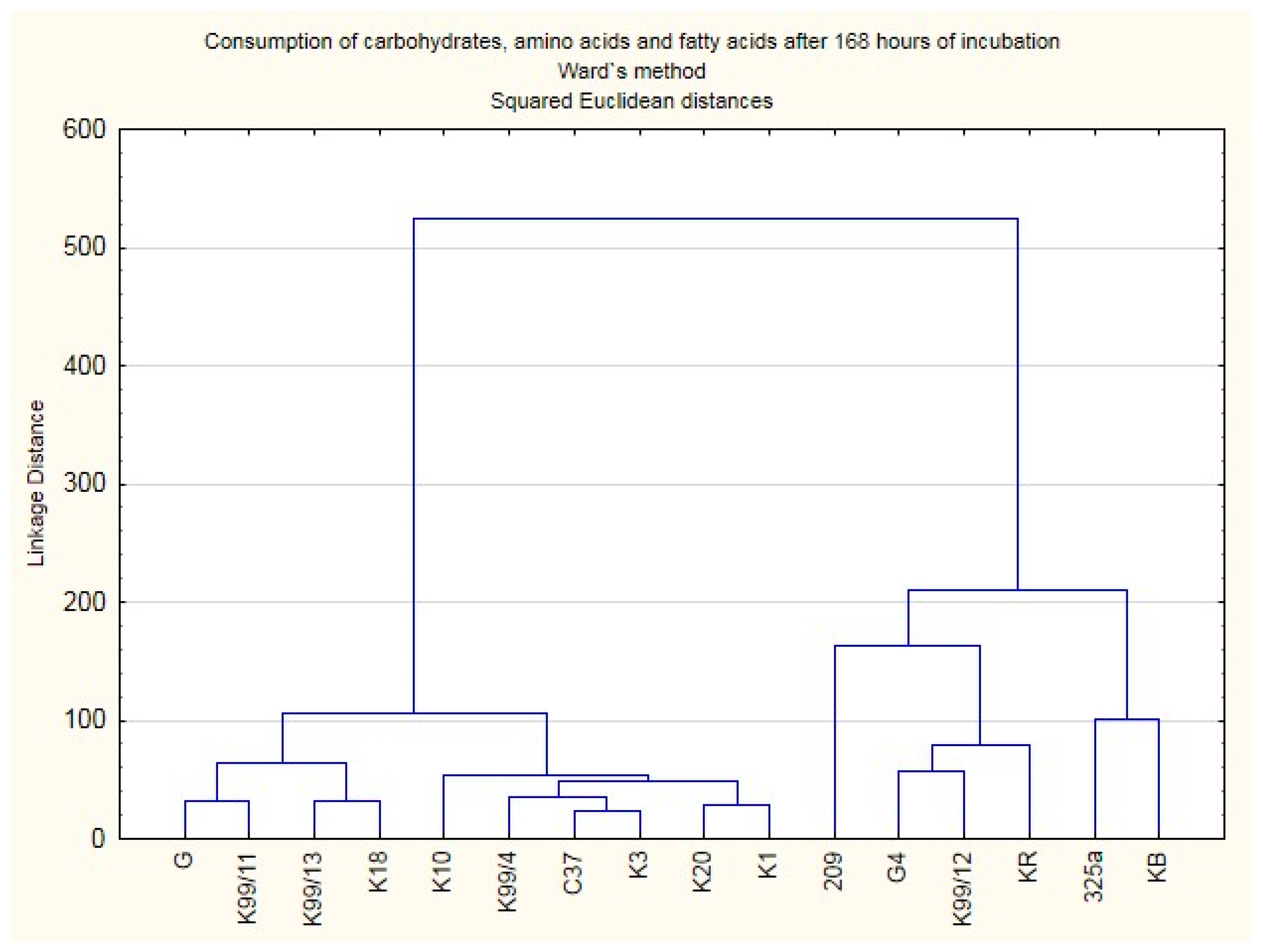
3.2. Metabolic Activity of Rhizobium Leguminosarum Bacteria
4. Conclusions
Author Contributions
Funding
Conflicts of Interest
References
- Stasiak, G.; Mazur, A.; Koper, P.; Żebracki, K.; Skorupska, A. Symbiosis of rhizobia with legume plants (Fabaceae). Postep. Mikrobiol. 2016, 55, 289–299. [Google Scholar]
- Kereszt, A.; Mergaert, P.; Kondorosi, E. Bacteroid development in legume nodules: Evolution of mutual benefit or of sacrificial victims? Mol. Plant-Microbe Interact. 2011, 24, 1300–1309. [Google Scholar] [CrossRef]
- Oke, V.; Long, S.R. Bacteroid formation in the Rhizobium-legume symbiosis. Curr. Opin. Microbiol. 1999, 2, 641–646. [Google Scholar] [CrossRef] [PubMed]
- Weisburg, W.G.; Barns, S.M.; Pelletier, D.A.; Lane, D.J. 16S Ribosomal DNA Amplification for Phylogenetic Study. J. Bacteriol. 1991, 173, 697–703. [Google Scholar] [CrossRef] [PubMed]
- Okonechnikov, K.; Golosova, O.; Fursov, M. UGENE team Unipro UGENE: A unified bioinformatics toolkit. Bioinformatics 2012, 28, 1166–1167. [Google Scholar] [CrossRef] [PubMed]
- Blast NCBI. Available online: https://blast.ncbi.nlm.nih.gov/Blast.cgi (accessed on 10 October 2019).
- McMurdie, P.J.; Holmes, S. phyloseq: An R Package for Reproducible Interactive Analysis and Graphics of Microbiome Census Data. PLoS ONE 2013, 8, e61217. [Google Scholar] [CrossRef] [PubMed]
- Ward, J.H. Hierarchical Grouping to Optimize an Objective Function. J. Am. Stat. Assoc. 1963, 58, 236–244. [Google Scholar] [CrossRef]


| Strain Symbol | Location | Plant | Year |
|---|---|---|---|
| C37 | Poland, Lublin | Trifolium sp. | 1960 |
| 209 | USA, Madison | Trifolium sp. | 1960 |
| 325a | USSR, Leningrad (now Russia, Petersburg) | Trifolium sp. | 1957 |
| G | Poland, Gnojno | Trifolium sp. | 1994 |
| G4 | Poland, Grabów | White clover (T. repens L.) | 1995 |
| KB | Poland, Grabów | White clover (T. repens L.) | 1995 |
| KR | Poland, Puławy | Red clover (T. pratense L.), var. “Raba” | 1996 |
| K1 | Poland, Stare Pole | Trifolium sp. | 2000 |
| K3 | Poland, Łabunie | Trifolium sp. | 2000 |
| K10 | Poland, Opatów | Trifolium sp. | 2000 |
| K18 | Poland, Puławy | White clover (T. repens L.) | 2004 |
| K20 | Poland, Puławy | White clover (T. repens L.) | 2004 |
| K 99/4 | Poland, Puławy | Trifolium sp. * | 1998 |
| K 99/11 | Poland, Wielichowo | Trifolium sp. | 1999 |
| K 99/12 | Poland, Wielichowo | Trifolium sp. | 1999 |
| K 99/13 | Poland, Wielichowo | Trifolium sp. | 1999 |
| Strain Symbol | Closest Species | Identity |
|---|---|---|
| 209 | Rhizobium legiuminosarum | 100% |
| G | Rhizobium legiuminosarum | 100% |
| K10 | Rhizobium legiuminosarum | 100% |
| K99/12 | Rhizobium legiuminosarum | 100% |
| K99/4 | Rhizobium legiuminosarum | 100% |
| KR | Rhizobium legiuminosarum | 100% |
| C37 | Rhizobium legiuminosarum | 99% |
| 325a | Rhizobium legiuminosarum | 99% |
| G4 | Rhizobium legiuminosarum | 99% |
| K3 | Rhizobium legiuminosarum | 99% |
| K99/11 | Rhizobium legiuminosarum | 99% |
| K99/13 | Rhizobium legiuminosarum | 99% |
| KB | Rhizobium legiuminosarum | 99% |
| K1 | Rhizobium legiuminosarum | 98% |
| K20 | Rhizobium legiuminosarum | 97% |
| K18 | Rhizobium legiuminosarum | 97% |
Publisher’s Note: MDPI stays neutral with regard to jurisdictional claims in published maps and institutional affiliations. |
© 2021 by the authors. Licensee MDPI, Basel, Switzerland. This article is an open access article distributed under the terms and conditions of the Creative Commons Attribution (CC BY) license (http://creativecommons.org/licenses/by/4.0/).
Share and Cite
Gawryjołek, K.; Furtak, K.; Grządziel, J.; Gałązka, A. Identification and Characterization of Metabolic Potential of Different Strains from Genus Rhizobium. Proceedings 2020, 66, 19. https://doi.org/10.3390/proceedings2020066019
Gawryjołek K, Furtak K, Grządziel J, Gałązka A. Identification and Characterization of Metabolic Potential of Different Strains from Genus Rhizobium. Proceedings. 2020; 66(1):19. https://doi.org/10.3390/proceedings2020066019
Chicago/Turabian StyleGawryjołek, Karolina, Karolina Furtak, Jarosław Grządziel, and Anna Gałązka. 2020. "Identification and Characterization of Metabolic Potential of Different Strains from Genus Rhizobium" Proceedings 66, no. 1: 19. https://doi.org/10.3390/proceedings2020066019
APA StyleGawryjołek, K., Furtak, K., Grządziel, J., & Gałązka, A. (2020). Identification and Characterization of Metabolic Potential of Different Strains from Genus Rhizobium. Proceedings, 66(1), 19. https://doi.org/10.3390/proceedings2020066019