Proceeding Paper

Test for the Production and Assay of the Proteolytic Activities of Halophilic Bacteria and Archaea Isolated from Algerian Hypersaline Environments †

by
Hania Benmebarek
1,*,
Juan-José Escuder-Rodríguez
2,
María-Isabel González-Siso
2 and
Karima Karroub
1
1
Equipe Métabolites des Extrêmophiles, Laboratoire de Recherche Biotechnologie et Qualité des Aliments (BIOQUAL), Institut de la Nutrition, de l’Alimentation et des Technologies Agro- Alimentaires (INATAA), Université Frères Mentouri Constantine 1 (UFMC1), Route de Ain El Bey, 25000 Constantine, Algeria
2
Grupo EXPRELA, Centro de Investigacións Científicas Avanzadas (CICA), Facultade de Ciencias, Universidade da Coruña, 15071 A Coruña, Spain
*
Author to whom correspondence should be addressed.
Presented at the 1st International Electronic Conference on Microbiology, 2–30 November 2020; Available online: https://ecm2020.sciforum.net/.
Proceedings 2020, 66(1), 12; https://doi.org/10.3390/proceedings2020066012
Published: 24 December 2020
(This article belongs to the Proceedings of The 1st International Electronic Conference on Microbiology)

Abstract

:
The present work was carried out on 133 halophilic strains isolated on MGM (Modified Growth Medium) medium with 12 and 23% (w/v) of salt. A screening of the extracellular proteolytic activities, carried out on the same medium supplemented with casein or gelatin at 1% (w/v), allowed us to select 24 bacterial and 21 archaeal strains presenting a precipitate around the colonies for casein and/or a translucent halo (after addition of Frazier’s reagent) for gelatin. The enzymatic test was performed on liquid medium in microculture in a 2 mL Eppendorf tube. The assay of the proteolytic activity, using Azocasein as substrate, followed two protocols—the first with PBS and the second with Tris HCl, with positive and negative controls—and demonstrated interesting results for 10 strains among the 45 tested including five bacteria and five archaea. These underwent morphological, physiological and molecular characterizations based on amplification and sequencing of the 16S ribosomal RNA gene.

1. Introduction

Microbial life can be found over a wide range of extreme conditions (salinity, pH, temperature, pressure, light intensity, oxygen and nutrient conditions). Hypersaline environments constitute typical examples of environments with extreme conditions due to their high salinity, exposure to high and low temperatures, low oxygen conditions and, in some cases, high pH values. Bacteria and Archaea are the most widely distributed organisms in these environments [1]. These unconventional conditions for life suggest that such microbes use original strategies (specific enzymes) to adapt to physicochemical constraints that they have to face. Several of these enzymatic activities are nowadays good candidates to be used in biotechnological processes [2]. However, in comparison to other groups of extremophilic microorganisms such as the thermophiles and the alkaliphiles, the halophiles of all three domains have been relatively little exploited [3].
Proteases represent the largest and most important segment in the industrial enzyme market, used in detergents, food processing and the leather industry, as biocatalysts in organic synthesis, and, among many other applications, as therapeutics because their roles are involved in key decisions throughout an organism in several physiological and metabolic processes [4]. Therefore, obtaining and preparing pure enzymes which are stable and active under multiple extreme conditions (alkaline pH, high salt concentrations and wide temperature) are scientifically and industrially significant [5].

2. Materiel and Methods

2.1. Samples and Strain Isolation

Brine samples used in the present study were collected from two solar saltern on 2016—namely, Ezzemoul, Ain M’lila, east of Algeria and Betioua, Aarzew, west of Algeria. Samples were spread on solid Modified Growth Medium [6], containing 5 g peptone and 1 g yeast extract with 12.5 and 23% (w/v) salt concentration at pH 7.5. Prokaryotic isolates were picked after 7–15 days of incubation at 37 °C, based on colony pigmentation, size, and margin and immediately streak-plated at least 3–4 times on fresh agar plates with the appropriate medium until purity was confirmed [7]. Additionally, 41 strains were recovered from previous work. A total of 133 strains (68 bacteria and 65 Archaea) were preserved on plats at 4 °C.

2.2. Morphology and Physiology

The isolates were submitted to morphology examination using cells from exponentially growing cultures. The colonies’ aspect was examined. Cell morphology and Gram were observed. The proteolytic activity of the isolates was screened on solid MGM (Modified Growth Medium) media (0.3% peptone and 0.1% yeast extract) supplemented with 1% (w/v) casein [8], or 1% (w/v) gelatin [9]; positive results were detected after 5 days for bacteria and 10 days for archaea, which were incubated at 37 °C by the presence of a precipitate around the colonies for casein and/or a translucent halo (after addition of Frazier’s reagent) for gelatin. Growth rates were estimated on MGM liquid medium at different salt concentrations (0, 2.5, 5, 7.5, 10, 12.5, 15, 17.5, 20, 22.5, 25 and 27%). The culture was incubated in aerobic conditions at 37 °C over 24 h for bacteria and 4 h for archaea. The optical density at 660 nm was measured with a Synergy H1 Hybrid Multi-Mode Microplate Reader [10].

2.3. DNA Extraction, 16S rRNA Gene Amplification and Strain Identification

The total genomic DNA of 10 proteolytic selected strains was prepared from colonies resuspended in MilliQ water by heating at 98 °C/10 min for bacteria and at 72 °C/10 min for Archaea, followed by centrifugation at 13,000 rpm for 5 min [6]; the tubes were immediately cooled on ice.
Bacterial and archaeal 16S rRNA genes were amplified using primer sets 27F/1492R [11] and 21F/1492R [12], respectively. PCR was performed using a 50 µL reaction mixture containing the following (per reaction): 5 µL of PCR buffer 10×, 4 µL of deoxyribonucleotide triphosphate mixture (10 mM each), 0.5 µL of each primer (10 µM), 0.25 µL (1.25 U) DreamTaq and 1µL of template DNA. The following PCR conditions were used: 95 °C for 30 s followed by 40 cycles of 95 °C for 30 s, 45.2 °C for 30 s and 72 °C for 1 min, and finally an extension step of 8 min at 72 °C. The amplified DNA supplied with gel-Green was subjected to electrophoresis on 0.7% (w/v) agarose gels in (TE) buffer and then visualized under UV light. Identities with described taxa were investigated using the nBLAST tool against the EzBioCloud database of cultured organisms.

2.4. Enzymatic Assay

The enzymatic assay was carried out using two methods based on azocasein (Sigma, St. Louis, MO, USA) as substrate. Absorbance was performed using a plate reader (using a Synergy H1 Hybrid Multi-Mode Microplate Reader).
Method 1: Cell-free culture supernatants were prepared by centrifugation for 5 min at 12,000 g. The reaction was performed in 50 mM Phosphate-buffered saline (PBS) solution pH 7.5 with 50 µL of azocasein 3% (w/v) and with 50 µL of culture supernatant for a final volume of 750 µL. The reaction was incubated at 37 °C for 1 h and stopped by adding 125 µL of 20% (w/v) trichloroacetic acid. The positive control was prepared by using a solution of proteinase K, and the blank assay was realized using the culture medium. After centrifugation at 15,000 g for 10 min, the absorbance of the supernatant was measured at OD366 nm. One unit of protease activity was defined as the amount required to produce enough acid-soluble material from azocasein to yield an absorbance of 0.01 at 366 nm, following 1 h of incubation [10,13].
Method 2: In this assay, 0.5 mL of reaction mixture consisting of 0.25 mL of crude enzyme (cell-free supernatant) and 0.25 mL of 0.8% (w/v) azocasein in 0.1 M Tris-HCl buffer (pH 8.0) containing NaCl at 12.5% (w/v) for bacteria and 23% (w/v) for archaea were used. The reaction mixture was incubated at 37 °C for 2 h with mild shaking. The reaction was terminated by adding 0.5 mL of 10% (w/v) trichloroacetic acid and the mixture was allowed to stand at room temperature for 30 min. The precipitate was removed by centrifugation at 10,000 rpm for 10 min. Subsequently, a volume of 300 mL of the initial supernatant fluid was transferred into a microcentrifuge tube and then mixed with 350 mL of 1.0 N NaOH. The absorbance was measured at 440 nm. One unit (U) of halophilic protease activity was defined as the amount of enzyme activity that produces a change in absorbance of 0.01 at 440 nm in 2 h at 37 °C under the standard assay conditions. The halophilic protease activity was calculated according to the following equation:
Halophilic protease activity   ( U / µ L ) = ( A B ) V t V e 0.01
where A and B are the optical densities of the crude enzyme and the control, respectively, Vt is the total reaction volume and Ve is the volume of crude enzyme [14,15].

3. Results

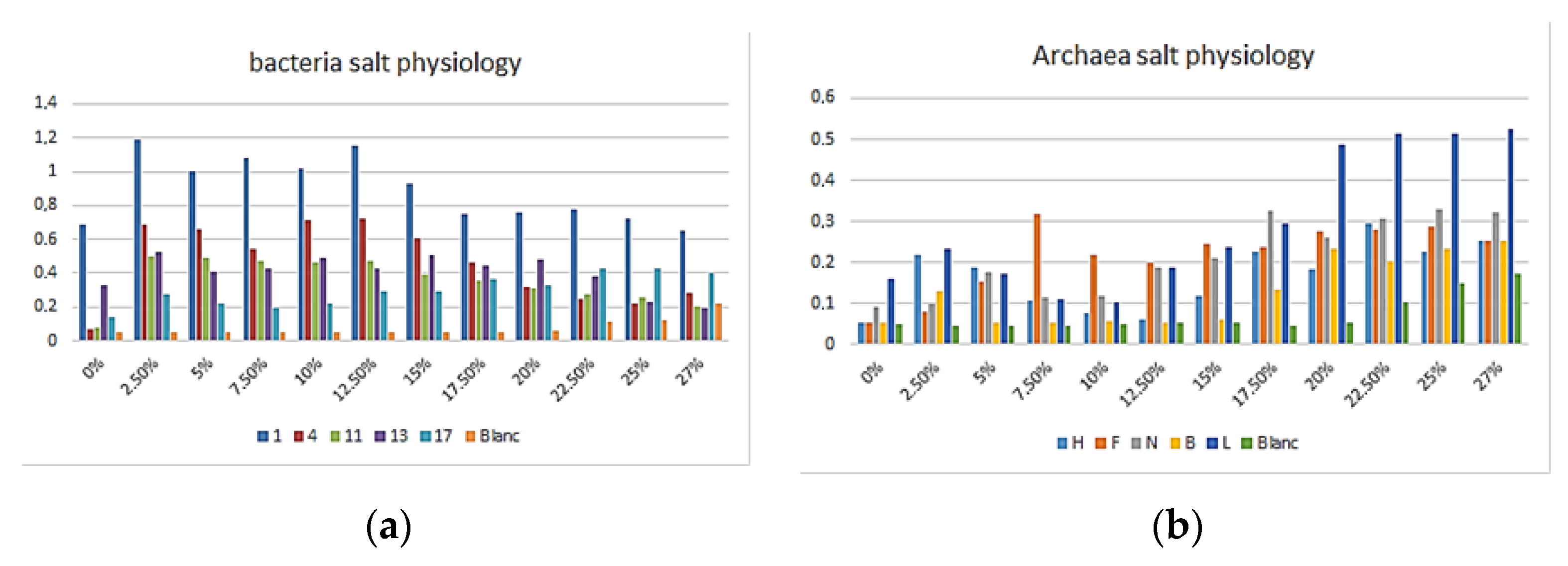
3.1. Morphology and Physiology

The morphological analysis of cells for 45 proteolytic strains showed that most of archaea were Gram-positive with cocci dominant, and for bacteria we found the two types of Gram with rod- shaped ones being dominant, as shown in Table 1.
The growth rates of selected strains were estimated as described before. The results in Figure 1 demonstrate that bacteria strain number 4 have a large salinity growth interval (2.5–17.5% w/v), which means that it is a moderately halophilic bacteria; in addition, the archaea strain N has a growth salt interval from 15 to 27% (w/v), suggesting that it is an extreme halophile [16].

3.2. Identification by 16S rRNA Gene Analysis

The 16S rRNA gene analysis related their grouping in seven genera: Virgibacillus, Idiomarina, Halobacillus for bacteria; Halorubrum, Halobacterium, Halogeometricum, and Natrinema for archaea. Bacteria Gram-negative isolates of the Idiomarina genus belonged to the Gammaproteobacteria Class, Idiomarinacaea family and Gram-positive of Virgibacillus and Halobacillus belonged to the Bacilli Class, Bacillaceae family [16,17]. The archaea genera all belonged to the Halobacteria Class, Halorubrum: Halorubraceae family; Natrinema: Natrialbaceae family; Halobacterium: Halobacteriaceae family; Halogeometricum: Haloferacaceae; they all have high salt tolerance and general pigment production [16].

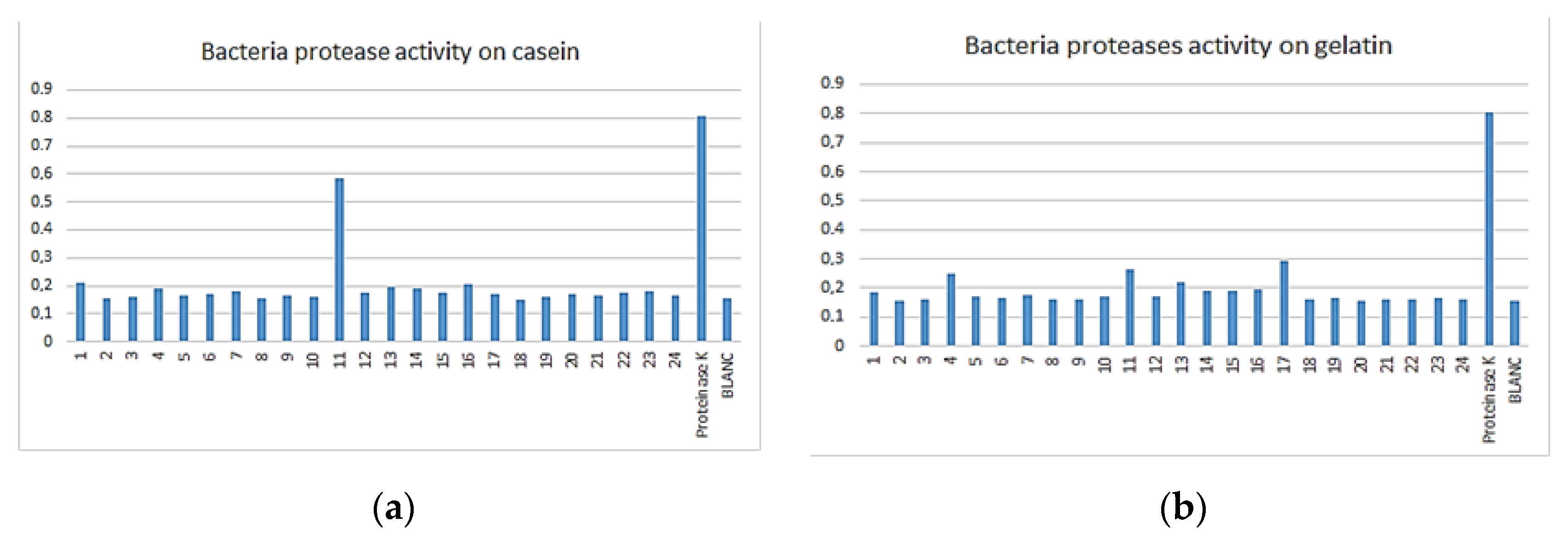
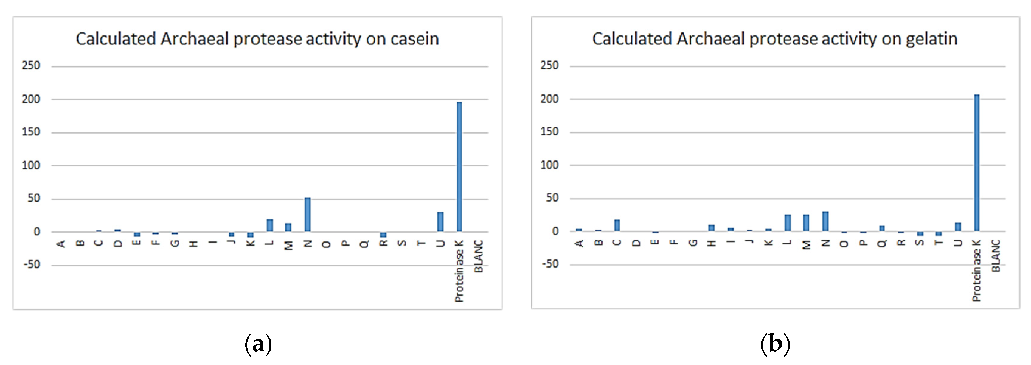
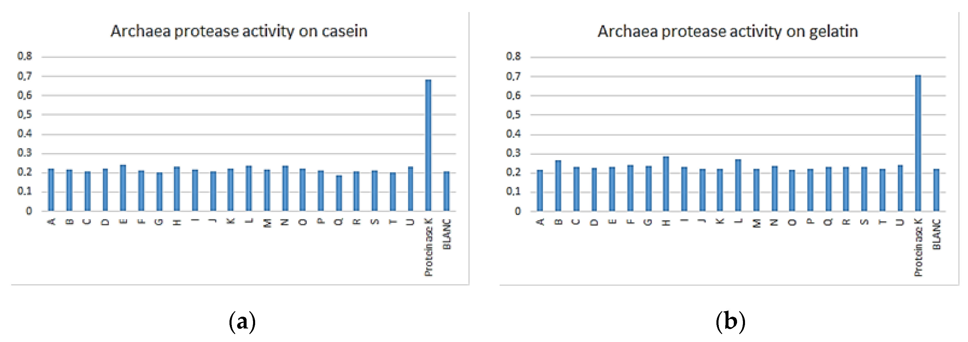
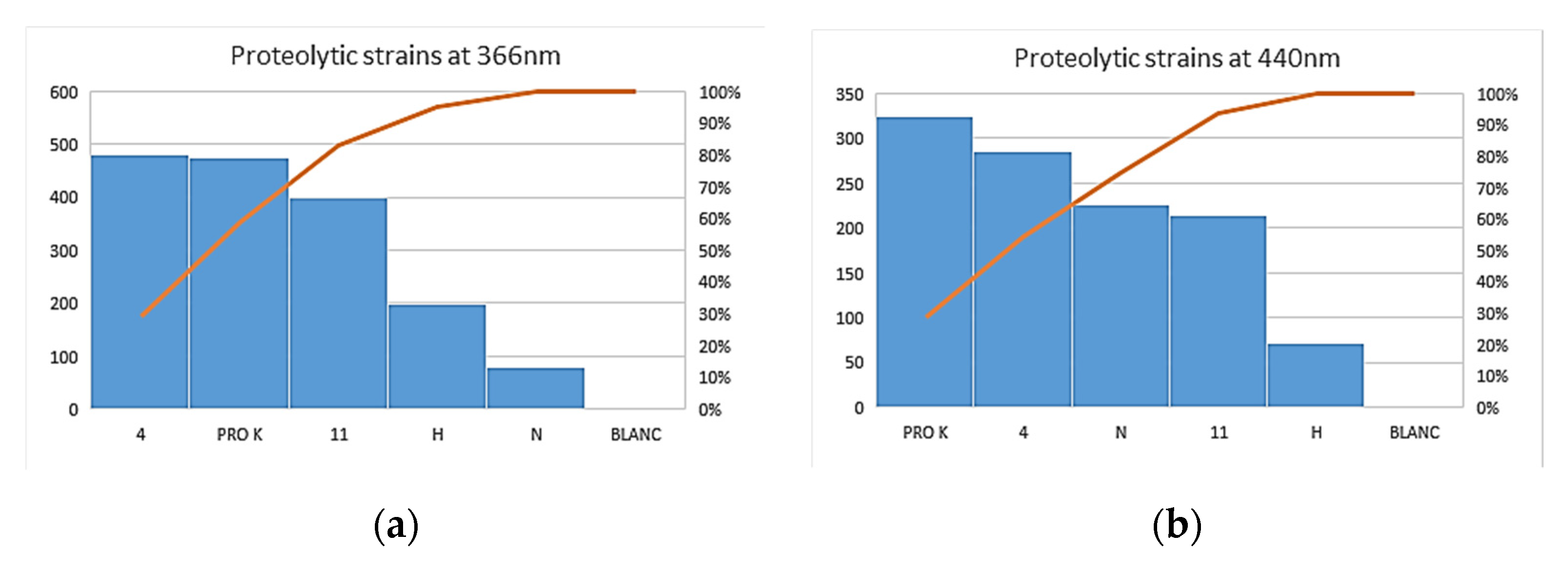
3.3. Enzymatic Assay

The enzymatic assay was first performed for the 45 proteolytic selected strains with azocasein using PBS buffer [10,13] after production on casein and gelatin media. Results presented on Figure 2 and Figure 3 allowed us to select five bacteria and five archaea that showed interesting enzymatic activity: 1, 4, 11, 13 and 17 for bacteria; B, F, H, L and N for archaea.
The enzymatic assay was confirmed for archaeal strains using a second method also based on azocasein, but in Tris- HCl buffer and including NaCl 23% (w/v), as described above [15]. This time, results presented in Figure 4 confirm a good proteolytic activity for only four strains: L, M, N and U.
For further tests, we combined the results of two methods and selected only four strains: 4 and 11 for bacteria, H and N for archaea.
The enzymatic activity (Figure 5) for selected proteolytic bacteria and archaea isolates, estimated with the two methods, showed to be very good in comparison with the positive control, especially with bacteria strain 4 (Idiomarina sp.) which conserves its proteolytic properties with the two methods. This means that in the absence (0%, w/v) or presence (12.5%, w/v) of NaCl, the proteolysis is well performed.
For archaea, strain N (Natrinema sp.) showed a good proteolytic activity with the second method in the presence of NaCl (23%, w/v), which is reported by other studies [8,18]; however, the activity is completely lost in the first one probably because salt is missing.
If we combine this results with those of salt physiology, demonstrated in Figure 1, we can easily see that Idiomarina sp. conserve its proteolytic activity due to the large salinity growth interval (2.5–17.5%, w/v); at the same time, the archaea Nitronema sp. require a high salt concentration, which explains the proteolytic activity lost in the first enzymatic assay.

4. Conclusions

The proteolytic activity production from archaeal and bacterial microorganisms isolated from Algerian sites was investigated in this study. In total, 45 Halophilic isolates were able to produce protease. Extremely halophilic archaeal isolates were affiliated to Natrinema, Halorubrum, Halobacterium, halogeomatricum and bacterial isolates to Virgibacillus, Idiomarina, and Halobacillus genera, of which several strains could produce hydrolytic enzymes. This study found that the genera Idiomarina and Natrinema showed the most important proteolytic activity (Figure 5).
According to the results, it is suggested that Natrinema and Idiomarina genera are excellent candidates for the production of proteolytic enzymes. Proteolytic activity produced by Natrinema under high salinity conditions could make this strain an interesting candidate for future investigation. These extremely halophilic isolates were selected for further studies for their great biotechnological applications with respect to their capacity to produce different hydrolases. It would be more constructive if these enzymes are purified from the isolates and then characterized, which is the next step in the current work.

Acknowledgments

We wish to acknowledge the following organizations for providing financial support for this study: the Algerian Ministry of Higher Education and Scientific Research and the laboratory of the group EXPRELA (Universidade da Coruña, Spain).

Conflicts of Interest

The authors declare no conflict of interest.

References

  1. Moreno, M.D.L.; Pérez, D.; García, M.T.; Mellado, E. Halophilic Bacteria as a Source of Novel Hydrolytic Enzymes. Life 2013, 3, 38–51. [Google Scholar] [CrossRef] [PubMed]
  2. Grégoire, P.; Fardeau, M.; Gauasco, S.; Bouanane, A.; Michotey, V.; Bonin, P.; Dubourg, K.; Cambard, J.; Ollivier, B. Les micro-organismes de l’extrême. Press. Therm. Climat. 2009, 146, 49–61. [Google Scholar]
  3. Ma, Y.; Galinski, E.A.; Grant, W.D.; Oren, A.; Ventosa, A. Halophiles 2010: Life in Saline Environments. Appl. Environ. Microbiol. 2010, 76, 6971–6981. [Google Scholar] [CrossRef] [PubMed]
  4. Coêlho, D.F.; Saturnino, T.P.; Fernandes, F.F.; Mazzola, P.G.; Silveira, E.; Tambourgi, E.B. Azocasein Substrate for Determination of Proteolytic Activity: Reexamining a Traditional Method Using Bromelain Samples. BioMed Res. Int. 2016, 1–6. [Google Scholar] [CrossRef] [PubMed]
  5. Ghafouri, H.; Askari, M.; Sarikhan, S. Purification and characterization of an extracellular haloalkaline serine protease from the moderately halophilic bacterium, Bacillus iranensis (X5B). Extremophiles 2015, 20, 115–123. [Google Scholar] [CrossRef] [PubMed]
  6. Nuttall, S.; Bath, C.; Pfeiffer, M.; Santos, F.; Eichler, J.; Mcalpine, T. The Halohandbook. Media, March, 1–144. 2008. Available online: http://scholar.google.com/scholar?hl=en&btnG=Search&q=intitle:The+Halohandbook#0 (accessed on 24 April 2017).
  7. Dammak, D.F.; Smaoui, S.M.; Ghanmi, F.; Boujelben, I.; Maalej, S. Characterization of halo-alkaline and thermostable protease from Halorubrum ezzemoulense strain ETR14 isolated from Sfax solar saltern in Tunisia. J. Basic Microbiol. 2016, 56, 337–346. [Google Scholar] [CrossRef] [PubMed]
  8. Cojoc, R.; Merciu, S.; Popescu, G.; Dumitru, L.; Enache, M.; Kamekura, M.; Enache, M. Extracellular hydrolytic enzymes of halophilic bacteria isolated from a subterranean rock salt crystal. Rom. Biotechnol. Lett. 2009, 14, 4658–4664. [Google Scholar]
  9. Gutiérrez, C.; González, C. Method for Simultaneous Detection of Proteinase and Esterase Activities in Extremely Halophilic Bacteria. Appl. Microbiol. 1972, 24, 516–517. [Google Scholar] [CrossRef] [PubMed]
  10. Gomri, M.A.; Rico-Díaz, A.; Escuder-Rodríguez, J.-J.; Khaldi, T.E.M.; González-Siso, M.-I.; Kharroub, K. Production and Characterization of an Extracellular Acid Protease from Thermophilic Brevibacillus sp. OA30 Isolated from an Algerian Hot Spring. Microorganisms 2018, 6, 31. [Google Scholar] [CrossRef] [PubMed]
  11. Siroosi, M.; Amoozegar, M.A.; Khajeh, K.; Fazeli, M.; Rezaei, M.H. Purification and characterization of a novel extracellular halophilic and organic solvent-tolerant amylopullulanase from the haloarchaeon, Halorubrum sp. strain Ha25. Extremophiles 2014, 18, 25–33. [Google Scholar] [CrossRef] [PubMed]
  12. Galkiewicz, J.P.; Kellogg, C.A. Cross-Kingdom Amplification Using Bacteria-Specific Primers: Complications for Studies of Coral Microbial Ecology. Appl. Environ. Microbiol. 2008, 74, 7828–7831. [Google Scholar] [CrossRef]
  13. Guendouze, A.; Plener, L.; Bzdrenga, J.; Jacquet, P.; Rémy, B.; Elias, M.; Lavigne, J.-P.; Daudé, D.; Chabrière, E. Effect of Quorum Quenching Lactonase in Clinical Isolates of Pseudomonas aeruginosa and Comparison with Quorum Sensing Inhibitors. Front. Microbiol. 2017, 8, 227. [Google Scholar] [CrossRef] [PubMed]
  14. Brock, F.M.; Forsberg, C.W.; Buchanan-Smith, J.G. Proteolytic activity of rumen microorganisms and effects of proteinase inhibitors. Appl. Environ. Microbiol. 1982, 44, 561–569. [Google Scholar] [CrossRef] [PubMed]
  15. Chuprom, J.; Bovornreungroj, P.; Ahmad, M.; Kantachote, D.; Dueramae, S. Approach toward enhancement of halophilic protease production by Halobacterium sp. strain LBU50301 using statistical design response surface methodology. Biotechnol. Rep. 2016, 10, 17–28. [Google Scholar] [CrossRef]
  16. Kanekar, P.P.; Kanekar, S.P.; Kelkar, A.S.; Dhakephalkar, P.K. Halophiles—Taxonomy, Diversity, Physiology and Applications. In Microorganisms in Environmental Management: Microbes and Environment; Springer Science+Business Media: New York, NY, USA, 2012; Volume 9789400722, pp. 1–819. [Google Scholar]
  17. De la Halba, R.R.; Sánchez-porro, C.; Marquez, M.C.; Ventosa, A. Extremophiles Halophiles. In Extremophiles Handbook; Horikochi, K., Ed.; Springer Science+Business Media: New York, NY, USA, 2011. [Google Scholar]
  18. Karray, F.; Ben Abdallah, M.; Kallel, N.; Hamza, M.; Fakhfakh, M.; Sayadi, S. Extracellular hydrolytic enzymes produced by halophilic bacteria and archaea isolated from hypersaline lake. Mol. Biol. Rep. 2018, 45, 1297–1309. [Google Scholar] [CrossRef] [PubMed]
Figure 1. Salt optima for selected bacteria and archaea strains: (a) growth rates OD600 nm of bacteria on different salt concentration liquid MGM medium; (b) growth rates OD600 nm of archaea on different salt concentration liquid MGM medium.
Proceedings 66 00012 g001
Figure 2. Bacterial proteasic activity assay tested on azocasein at 366 nm: (a) OD366nm for bacterial activities on casein; (b) OD366 nm for bacterial activities on gelatin.
Proceedings 66 00012 g002
Figure 3. Archaeal proteasic activity assay tested on azocasein at 366 nm: (a) OD366nm for archaeal activities on casein; (b) OD366 nm for archaeal activities on casein.
Proceedings 66 00012 g003
Figure 4. Archaeal proteasic activity assay tested on azocasein at 440 nm: (a) OD440 nm for archaeal activities on casein; (b) OD440 nm for archaeal activities on gelatine.
Proceedings 66 00012 g004
Figure 5. Determination of proteolytic activity for selected strains. (a) at OD366 nm; (b) at OD440 nm.
Proceedings 66 00012 g005
Table 1. Morphological aspect of proteolytic bacteria and archaea isolates.
GramCell formProtease
PositiveNegativeCocciRodCoccobacilliCaseinGelatin
Bacteria81631651623
Archaea1171233021
Publisher’s Note: MDPI stays neutral with regard to jurisdictional claims in published maps and institutional affiliations.

