Proceeding Paper

Functional Nano-Hydroxyapatite for Applications in Conservation of Stony Monuments of Cultural Heritage †

1
Istituto di Cristallografia, IC-CNR, Via Salaria Km 29.300, 00016 Rome, Italy
2
FIMIF, University Polytechnic of Tirana, 1001 Tirana, Albania
3
Dipartimento di Scienze, Università Roma Tre, Largo S. L. Murialdo 1, 00146 Rome, Italy
4
Istituto di Cristallografia, IC-CNR, Via G. Amendola, 122/O, 70126 Bari, Italy
5
Istituto per la Scienza e Tecnologia dei Plasmi, ISTP-CNR, Via G. Amendola, 122/O, 70126 Bari, Italy
6
Ivodent Academy, Center of Techniques Studies, 1010 Tirana, Albania
*
Author to whom correspondence should be addressed.
Presented at the 2nd International Electronic Conference on Crystals, 10–20 November 2020; Available online: https://iocc_2020.sciforum.net/.
Proceedings 2020, 62(1), 11; https://doi.org/10.3390/proceedings2020062011
Published: 4 January 2021
(This article belongs to the Proceedings of The 2nd International Online Conference on Crystals)

Abstract

:
Stony monuments must continuously be safeguarded from damage caused over time, in particular from the detrimental effects of weathering. One of the new environmentally-friendly (nano) materials for stone reinforcement, particularly suitable for marble and calcareous (limestone, sandstone) artifacts, is Ca10(PO4)6(OH)2 hydroxyapatite (HAp), which has a considerably lower dissolution rate and solubility compared to CaCO3 calcite (the building block of marble materials): thus, HAp has been proposed for the protection of calcareous monuments against acidic rain corrosion. Promising results have been obtained, but further optimization is necessary as the treated layer is often incomplete, cracked and/or porous. Several parameters need to be optimized, in this way a homogeneous layer can be obtained, and consequently the formation of metastable can be avoided, soluble phases instead of HAp. These include: the pH of the starting solution; the effect of organic and inorganic additions in particular, that of ethanol, which is known to adsorb calcite, thus possibly favoring the growth of the HAp layer. The formation of HAp nanoparticles and their application on stony substrates has been investigated by means of a multi-methodological approach based on scanning electron microscopy, x-ray diffraction, small- and/or wide-angle x-ray scattering, Fourier-transform infrared spectroscopy, and finally, in situ measurements of laser-induced breakdown spectroscopy and acid attack preliminary tests on stony substrates.

1. Introduction

Rocks are among the most durable building materials used in monuments belonging to all ancient and modern civilizations. However, over time, the stone can be subject to various degradation processes leading to physical and chemical modifications. Although these effects may be limited to the surface and be negligible to the structural stability of the affected buildings, they can represent a major problem in decorative elements of artistic value, where all details should be preserved [1]. The challenge for conservators and material scientists involved in stone conservation has always been to find a way to stop or delay the effects of these degradation processes. The basic principle of the patrimony is that the cultural heritage is an incalculable and integral legacy to our future: observing and knowing the past, will help future generations to better tackle the future. Thus, the conservation of stone heritage is always a delicate and complex task, due to the multiple variables that have to be taken into account in order to identify problems, and to define the necessary conservation actions, in addition to selecting materials and the best procedures to be used. The variety of factors to be analyzed include the intrinsic stone properties (from geological features up to mechanical behavior), the state of conservation, the degradation mechanisms and the environmental factors.
One of the most promising technologies employed for lowering the previously described degradation processes, is that of nanomaterials, nowadays commonly applied in the maintenance of world cultural heritage, with the aim of improving the consolidation and protection treatments of the damaged stone materials that they are made of [2]. Such nanomaterials display important advantages that could solve many of the problems associated with the traditional interventions that often have serious issues such as a lack of the necessary compatibility with the original substrate and a durable performance. The application of nanotechnology in cultural heritage conservation is characterized by the possibility of designing consolidating products that are strongly compatible with the original stone substrate. Furthermore, the properties of the material change significantly when its particles are about 100 nm in size. The nanoparticles must show: stability and sustained photoactivity; biological and chemical inactivity; nontoxicity; antimicrobial properties for lowering the ecotoxicological impact on animals and plants [3]; low cost; suitability towards visible or near UV light; a high conversion efficiency and a high quantum yield. In addition, these treatments should also possess water repellent properties which favor self-cleaning actions and prevent the generation of damage caused by water. The most commonly used inorganic consolidating agents are products based on Ca(OH)2 calcium hydroxide nanoparticles [4], due to their compatibility with a large proportion of the built and sculptural heritage. As well as other hydroxides (Mg(OH)2, Sr(OH)2), metal oxides (TiO2, ZnO), and metal nanoparticles (Au, Ag, Pt)) have been reported in the literature, focusing on their potential as consolidants on different artifacts of cultural heritage [2,5]. However, one of the most challenging nanomaterials is Ca10(PO4)6(OH)2 hydroxyapatite (HAp), already applied in a large variety of technological and biomedical applications, mainly due to its close relationship with the mineral component of hard human tissues [6,7]. In cultural heritage conservation it is used for carbonate stone consolidation [2]. HAp can be applied for the consolidation of limestone, marble and sandstone with different carbonate contents. This product is not introduced directly into the stone material, but as an aqueous solution of diammonium hydrogen phosphate (DAP) with calcium ions coming from the CaCO3 calcite mineral—the main constituents of marble and limestone (carbonate substrates), which can be derived from the partial dissolution of the same substrate. Thanks to its low viscous nature, this aqueous consolidant product is able to penetrate deeply into the stone, generating a significant improvement in the mechanical properties of the same stone. HAp is tested as an additional protective treatment for the corrosion protection of marble from acid rain [8]. The study of compatibility and adaptability requires that the physical and chemical properties of both consolidator products and stone substrate are well known. Such knowledge is crucial to ensure the outcome of the present project. Materials of interest, synthesized in our labs have been analyzed by using: (1) X-ray diffraction (XRD), which is effective on crystalline materials and able to carry out information on chemical composition, size, shape and atomic structure, (2) small- and/or wide-angle scattering (SAXS/WAXS), a powerful tool to investigate the domain of phosphate particles as a function of their optical properties; in the case of SAXS the technique can be applied to HAp nanoparticle characterization; (3) Fourier-transform infrared (FTIR) spectroscopy, a reliable technique for investigating hydroxyl anions and variations within anionic and cationic groups in the obtained materials; (4) scanning electron microscopy for checking the morphology of nanoparticles; (5) biological evaluation of the antimicrobial properties of obtained HAp materials, through direct contact and disc diffusion methods versus most common Gram-positive and Gram-negative bacteria, present in human or animal biosystems; (6) Laser-induced breakdown spectroscopy (LIBS), which is a micro-destructive technique able to obtain quali-quantitative information on museums artifacts.

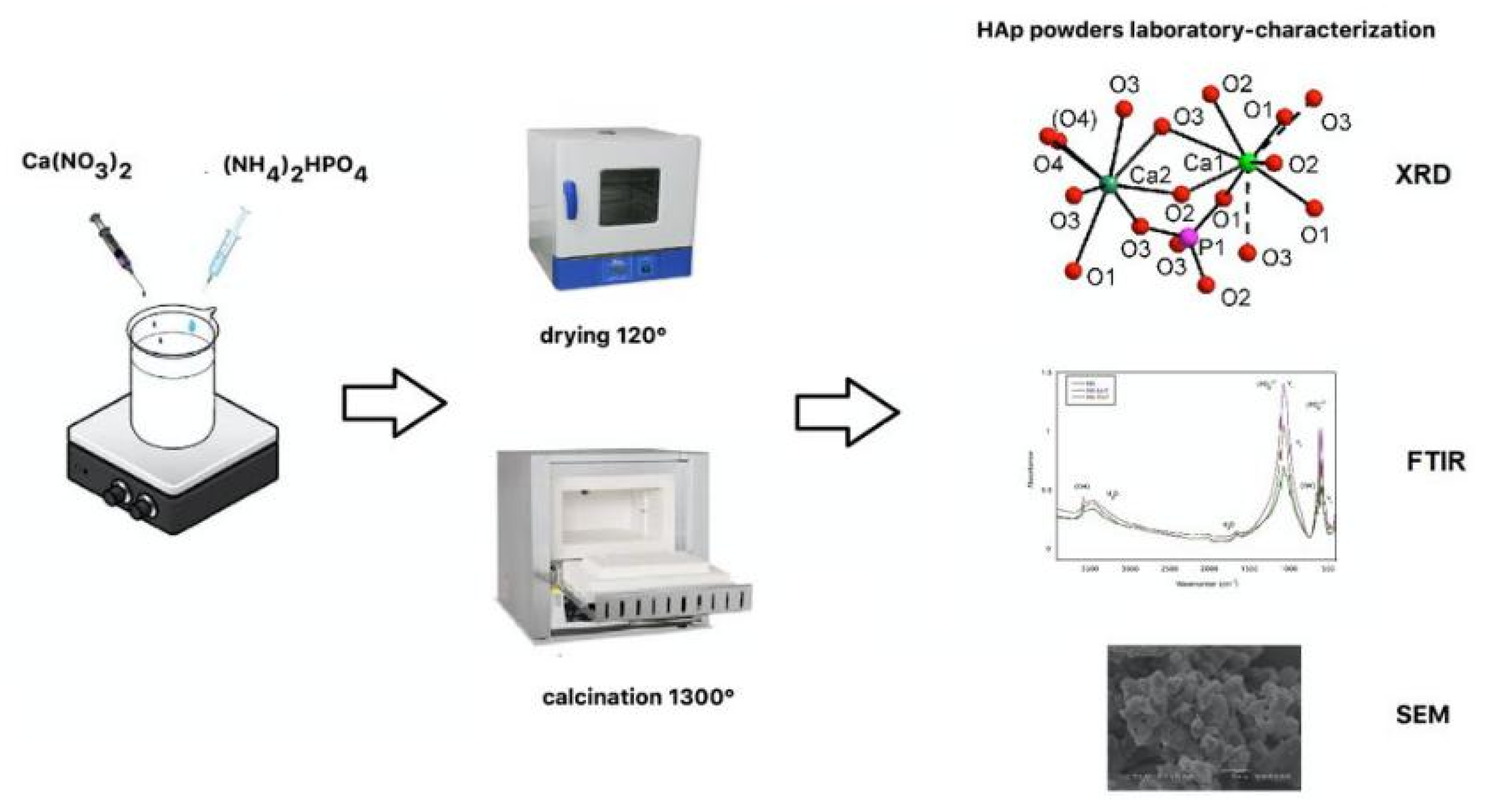
2. Experimental Methods

The schematic presentation for the synthesis and the first laboratory characterization is shown in Figure 1. The synthesis of the hydroxyapatite powder can be obtained by a chemical-precipitation method at room temperature and pH = 10, using Ca(NO3)2⋅4H2O and (NH4)2HPO4 as reactants; more details are reported in [7].
X-ray diffraction (XRD) is typically employed to investigate the changes of crystalline phases in phosphate samples [7,9,10]. Powder XRD data were collected at room temperature by using an automated Rigaku RINT2500 diffractometer (50 KV, 200 mA) equipped with the silicon strip Rigaku D/teX Ultra detector [7]. An asymmetric Johansson Ge (111) crystal was used to select the monochromatic Cu Kα1 radiation (λ = 1.54056 Å). The angular range 8–100° (2θ) was scanned with a step size of 0.02° (2θ) and counting time of 4 s/step. Measurements were executed in transmission mode by introducing the sample within a glass capillary with a 0.3 mm of internal diameter and mounted on the axis of the goniometer. Lattice parameters and space group were carried out using EXPO2013 software [11].
Small angle X-ray scattering (SAXS) is the most commonly used technique to determine the morphology features of a set of nanoparticles. The technique takes advantage of the electron density function difference between the studied objects and surrounding medium. As a technique, Small-angle X-ray scattering (SAXS), consists of recording at very low angles from the beam axes (typically 0.1–10°) of the elastic scattering of X-rays by a sample. This angular range contains information regarding the structure of scatterer entities, such as nanoparticles and micro- and macromolecules, among others. Depending on the studied systems, the SAXS technique could provide information of the distances between partially ordered materials and pore sizes, as well as other data [12]. Depending on the experimental setup, SAXS is capable of delivering structural information of objects whose size ranges between 0.5 and 100 nm.
Wide angle X-ray scattering (WAXS) is the technique that is most commonly identified with molecular structures at the resolution of atomic positions and is sometimes used synonymously with X-ray diffraction. As one can guess from the names, the important difference between WAXS and SAXS is that the WAXS measures the X-rays scattered into wide angles. These angles should be large enough to define probe lengths that are comparable to atom–atom distances and/or atomic planes in a crystal. Due to the intensity of the scattered X-rays is measured at higher angles, the collimation and flight path requirements should be more compact for a dedicated WAXS instrument than to instruments optimized for SAXS. A higher level of morphological and structural information can be gained by collecting SAXS and WAXS data, individually or in combination (SWAXS) [13].
Fourier transform infrared (FTIR) spectroscopy is one of the techniques most widely used for molecular characterization. Powder spectra were achieved on a Nicolet iS50 FTIR spectrometer equipped with a DTGS detector and a KBr beam splitter; nominal resolution was 4 cm−1 and 64 scans were averaged for specimen and background. The specimen was prepared by mixing 1 mg of HAp with 200 mg of KBr.
Scanning electron microscopy (SEM) observations were conducted by a high-resolution FE-SEM Zeiss Gemini at LIME (Laboratorio Interdipartimentale di Microscopia Elettronica), University Roma Tre, Rome, Italy. The instrument was equipped with two SEs (secondary electrons), one 5-sector BSE (back scattered electron) and a CL (cathodo-luminescence) detector, in addition to a high-resolution EDS 60 mm2 detector for elemental analysis.
Biological evaluation of the antimicrobial properties of obtained HAp materials, through direct contact and disc diffusion methods versus most common Gram-positive and Gram-negative bacteria present in animal or plant biosystems. These two techniques allow the observation of the antimicrobial activities of the HAp sample. Briefly, in the direct contact method a bacterial suspension is transferred to a vial containing the material and then the mixture is spread onto agar plates and incubated for several hours. After this period, the number of bacterial colonies on each plate is counted and compared against a growth-positive control in the absence of materials [14]. The disc diffusion test was performed on the agar plate containing sterilized filter paper discs, which had been previously treated and soaked with HAp nanoparticles. Upon the spread onto the plate of the bacterial suspension and a period of incubation, the diameters (mm) of the inhibition zone can be measured and compared with both positive and negative controls [15].
Laser-induced breakdown spectroscopy (LIBS) is a versatile technique that provides nearly instant multi-elemental analysis including low atomic number elements e.g., Be, C, Li, F, Na, Mg, Al, and Si of materials, both in the laboratory and in the field. This is achieved by focusing a short laser pulse on the surface of the sample, and by analyzing the resulting spectrum from the laser-induced plasma. LIBS has been employed in the analysis of archaeological sites, historical monuments and ancient artifacts for assessing the qualitative, semi-quantitative and quantitative elemental content of materials such as pigments, pottery, glass, stones, metals, minerals, meteorites, gems and fossils [16,17,18]. A simple scheme of a working LIBS set-up is depicted in Figure 2.

3. Characterization

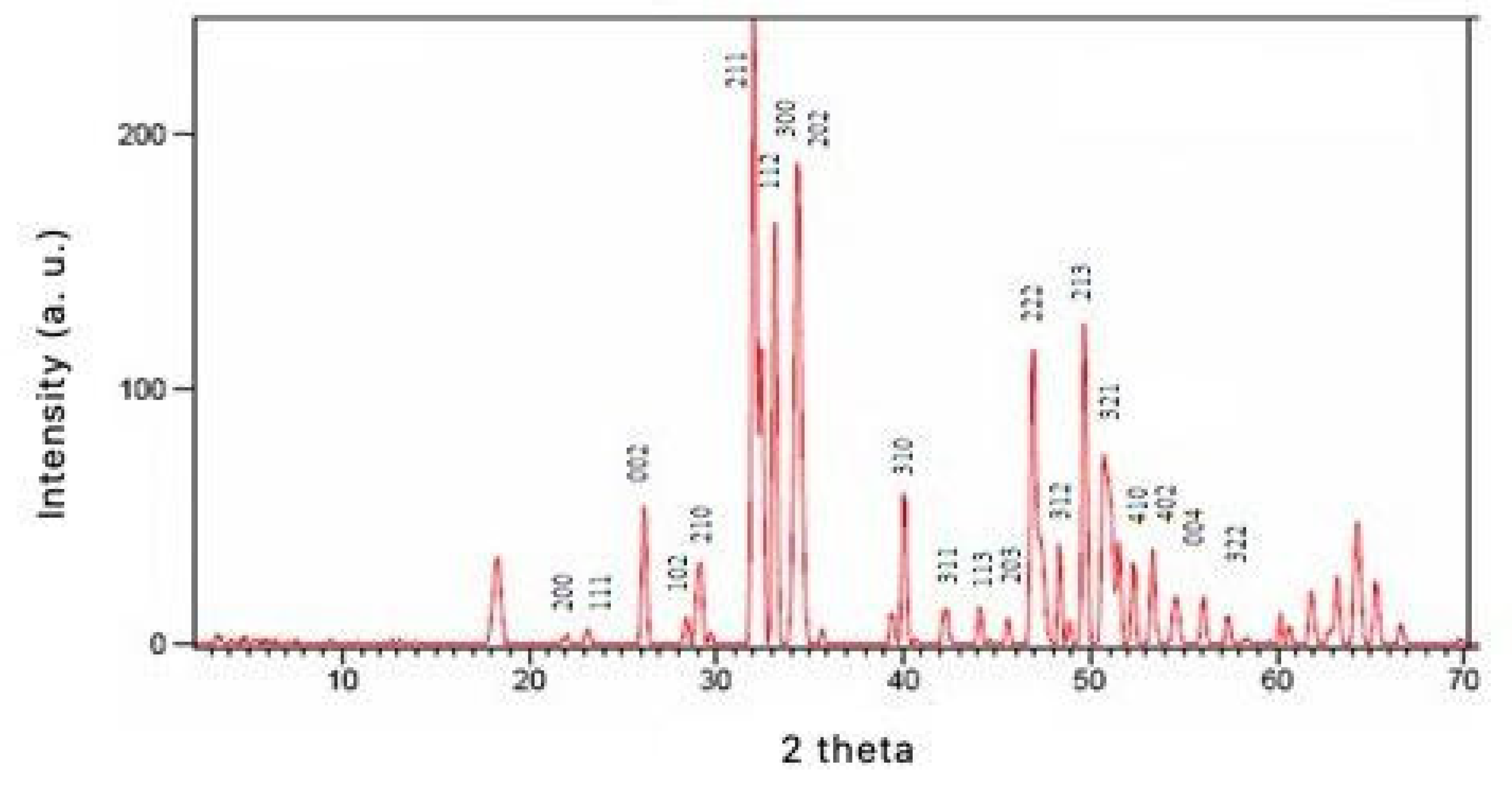
3.1. X-ray Diffraction

The indexing of the HAp powder diffraction pattern was achieved by using EXPO2013 software [11]. Crystallinity = 81% was achieved according to [19]. Lattice parameters, a = b = 9.4145(4) Å, c = 6.8758(5) Å, V = 6.8758(5) Å3, are similar to those of the most common HAp phases [20,21], while the space group determination gave the most common hexagonal P63/m space group of apatite [20,21]. In the powder pattern of HAp, some unindexed peaks were recognized as belonging to the Ca3(PO4)2 tricalcium phosphate (TCP) specie [22], according to a quantitative ratio of HAp = 88.9% and TCP = 11.1% in the specimen; such outcome confirm those of [23,24], on partial transformation of HAp in TCP (T = ca. 900 °C), and with recent studies on TCP [25,26,27]. Figure 3 reports a typical XRD spectrum for HAp calcined at 900°, together with most important reflections.

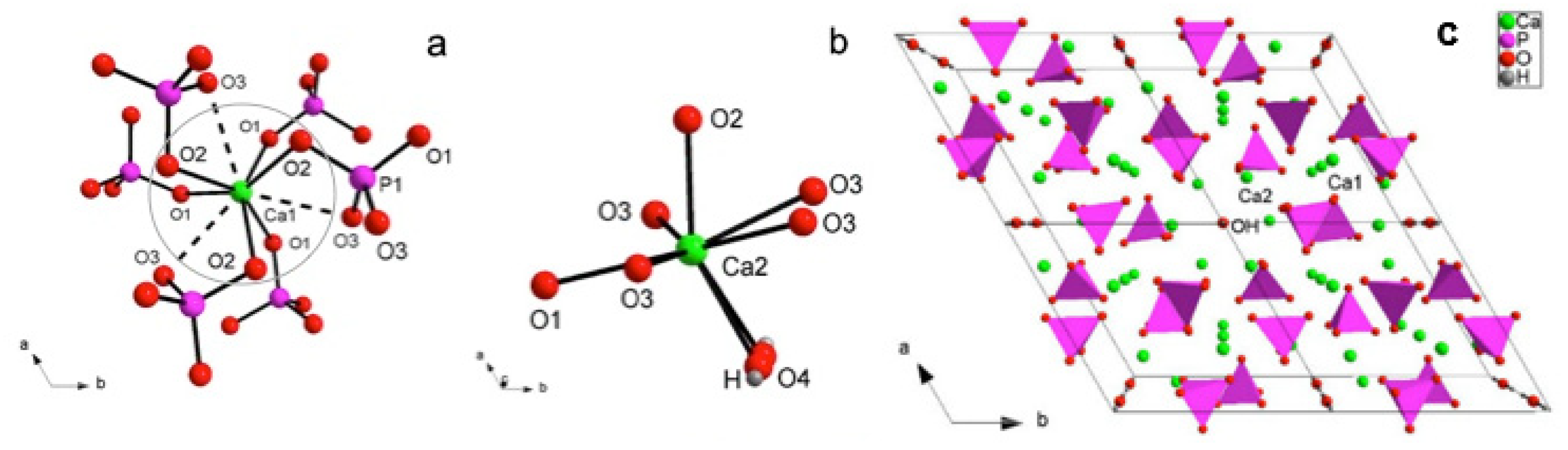
3.2. X-ray Crystal Structure
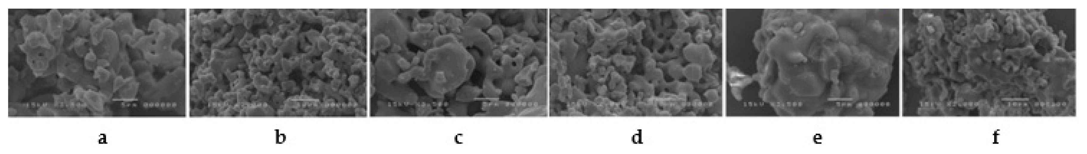
P63/m hydroxyapatite displays two structural sites, both occupied by Ca atoms engaged in a complex coordinations: according to the usual notations for site apatite site labelling [21], Ca1 displays a CaO6 metaprism geometry (metaprism: a polyhedron intermediate between an octahedron and a trigonal prism) (detail in Figure 4a), while Ca2 shows a CaO6(OH) coordination (Figure 4b), being quite similar to a distorted CaO7 pentagonal bipyramid, whose ideal equatorial plane is characterized by O1 and two couples of symmetry-related O3 atoms and, and whose ideal vertices are represented by an O2 atom and a O4OH hydroxyl group [7]. Worthy of note is the wide range of cationic substitutions within the HAp structure, from Mn, Na, Fe, Zn for different biomedical applications [7], up to rare earth elements for luminescent imaging applications [7,28].
In order to explain the P63/m HAp structure, we can improve the crystal formula by writing it as (Ca(1)4Ca(2)6)(PO4)6(OH)2, better highlighting the Ca1 and Ca2 positions; a further examination, in fcat, highlights the zeolitic character of HAp, where the framework is made up of arrays of face-sharing Ca(1)O6 polyhedra corner-connected to six different PO4 tetrahedra down c making up (Ca(1)4(PO4)6)10− anionic moieties (Figure 4a); the present assemblage results in one-dimensional hexagonal tunnels occupied by (Ca(2)6(OH)2)10+ counter-ions (Figure 4c), whereas the strong Ca1…O3 interactions (Figure 4a) contribute to stabilize the whole structure [7].

3.3. SEM Microscopy

High-resolution SEM investigation of HAp samples show the presence of different morphologies, such as compact and porous aggregates, and massive assemblages. The porous aggregates are the most common, with the largest crystallite size of ~10 μm (Figure 5a–c). The massive fragments and compact aggregates (Figure 5d–f) have the largest dimensions ~5 μm (Figure 5e) and, respectively, ~35 μm (Figure 5f). In all cases, the crystallites show a rounded morphology, from elongated to sub-spherical, and the most common dimension is ~5 μm. SEM investigations also suggested the growth of porous morphologies on massive fragments (Figure 5d,f).

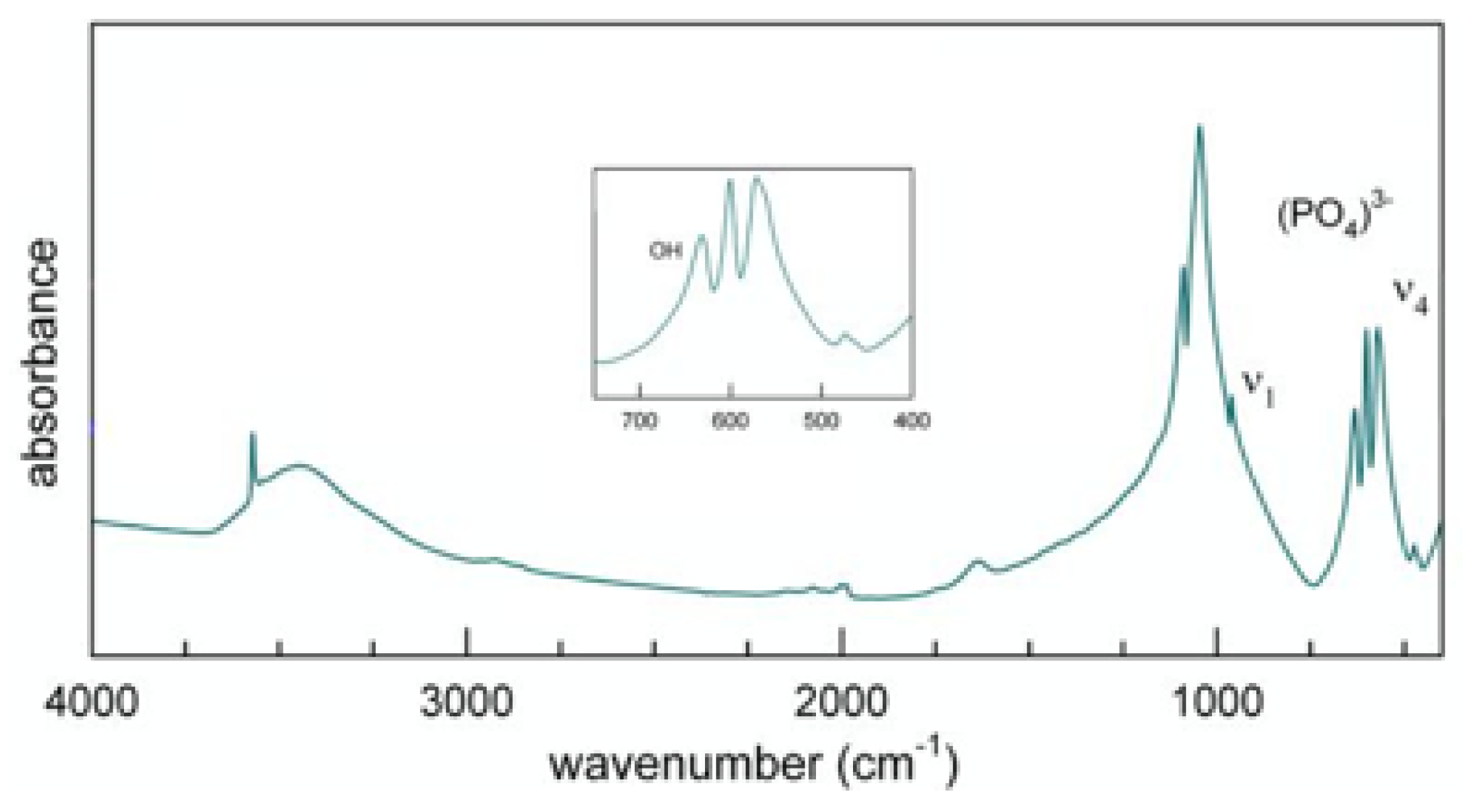
3.4. FTIR Spectroscopy

The literature on the interpretation of the FTIR spectra of hydroxyapatite is quite exhaustive [28,29,30] as it is a well-established technique, but at the same time, it is particularly useful due to it being able to detect (OH) groups in HAp with accuracy [7,28]. Phosphorous compounds typically show a strong molecular character with respect to their vibrational properties, therefore FTIR spectra are dominated by internal (PO4)3− modes. The spectrum of HAp, presents a strong doublet at 1091–1039 cm−1 assigned to the asymmetric stretching (ν3), while the weaker peak at 962 cm−1 can be assigned to the symmetric stretching mode of the (PO4) tetrahedron [31]. A couple of medium intense and very sharp peaks at 602 and 564 cm−1 can be assigned to the triple degenerate antisymmetric bending mode of the (PO4) tetrahedron [32]. The sharpness of these bands, according to [33], is usually an indicator of a well-crystallized HAp, moreover confirming the result of crystallinity from XRD analysis. Worthy of note, the spectrum of HAp displays a finely resolved sharp peak at 3570 cm−1, pertinent to the stretching mode of the hydroxyl group [29]. Such peak is superimposed with a broad absorption due to water (moisture) adsorbed on the KBr disk [34]; the bending mode of this moisture is also appreciable as a broad component at 1600 cm−1. Investigation of deuterated samples [35] highlights that the relatively sharp peak at 663 cm−1 is caused by the O-H libration (as reported within the inset in Figure 6). In Figure 6, an FTIR spectrum of a synthetic HAp (900 °C) is reported, while Table 1 reports the measured absorption in the infrared spectrum and relative assignments for hydroxyapatite sample.

3.5. SAXS/WAXS

SAXS can be used to study the morphological properties of HAp nanoparticles and in this way, to follow the crystallization process and the role and influence of various experimental setup parameters (temperature, speed, time etc) and/or additives to accelerate/inhibit the process. In this way it would be possible to control the size and shape of the HAp nanoparticles. Once SAXS experimental curves have been obtained, software such as SasView (www.sasview.org) can be used to estimate/refine the shape and average size of the particles, as well as their polydispersity [36].

3.6. Biological Evaluation of the Antimicrobial Properties of Obtained Nanophases
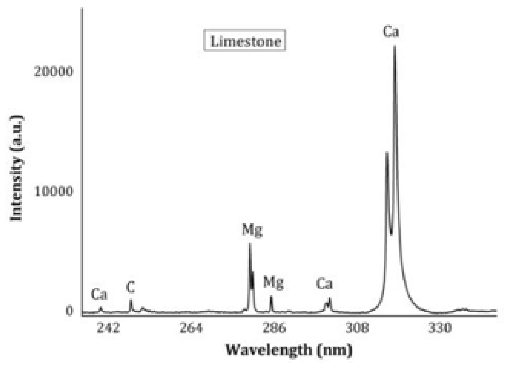
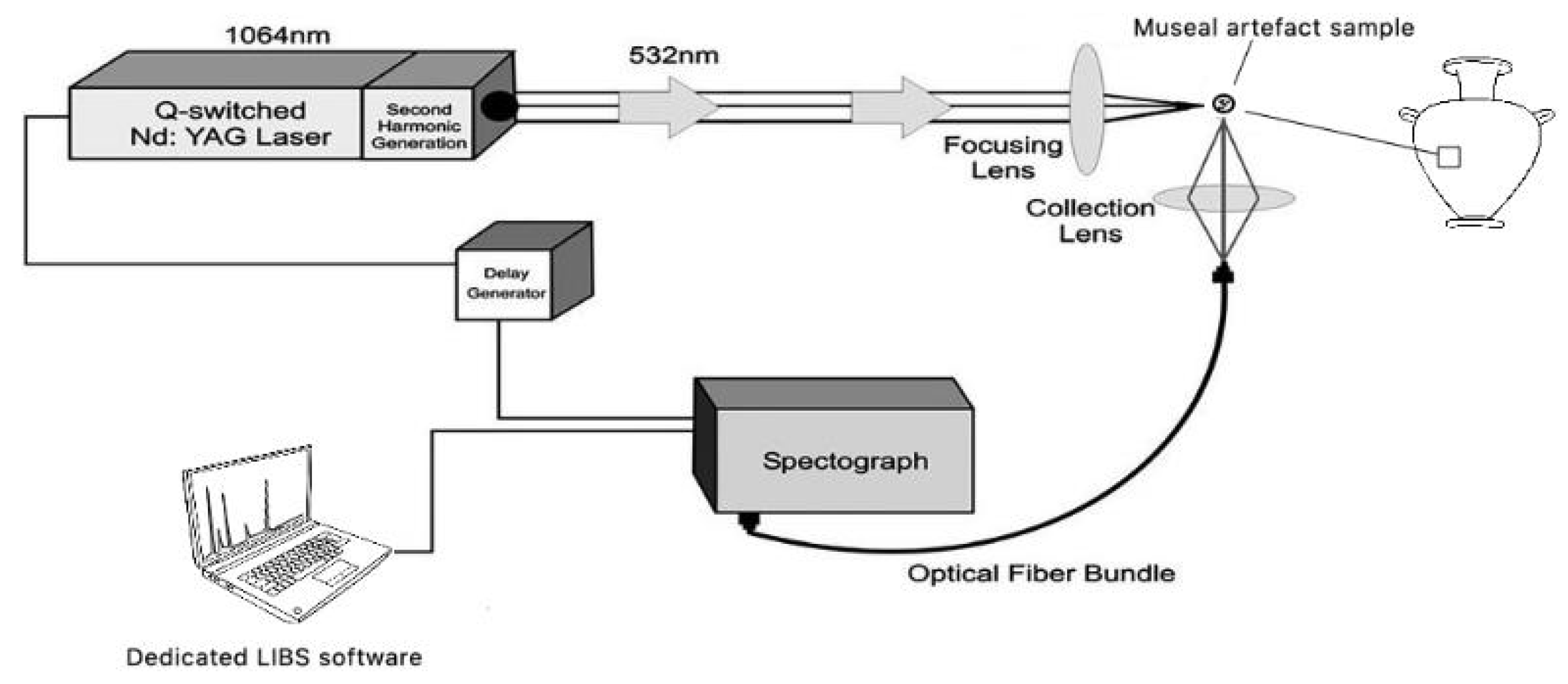
As with all surface treatments, HAp nanoparticles can be leached into the surrounding environment leading to potential ecotoxicity in the soil and water, with adverse effects on the associated biota and the bacteria-dependent processes [3]. Even though there is limited understanding of the environmental fate of engineered nanoparticles after release from treated surfaces, ecotoxicological studies reported different results of bacterial inhibition, stimulation, survival and death, which depend on dose, species and test procedures [37].
HAp nanoparticles may enter the soil and water systems pertinent the archeological sites directly, by leaching from building surfaces, or indirectly, via wastewater sludges used as fertilizers. Nano HAp tend to partition into the sludge in wastewater treatment plants and consequently the soil can be resulted contaminated by HAp particles and/or their transformation products [3]. For this reason, careful interest is devoted to the topic of biological evaluation of the HAp nanophases, although the need of a standard test for antimicrobial efficacy to determine their impact on ecological processes still exists.
The anti-microbial activity and properties of HAp nanoparticles can be assessed in vitro by direct contact and disc diffusion methods in a solid medium, using common Gram-positive and Gram-negative bacteria, abundant in animal and plant biosystems. A variety of methods for determining antimicrobial activity have been previously studied, showing that the results obtained are profoundly influenced by the method selected, the microorganisms used to carry out the test and the degree of solubility of each test compound [38].

3.7. LIBS

In recent decades, LIBS has been widely applied in the field of cultural heritage protection. Nowadays, the use of handheld LIBS instrumentation (hLIBS) is considered a future prospective for this field. Senesi et al. (2018) [39] was one of the first to study to the application of hLIBS in archeological geological applications. A limestone fragment was analyzed from a masonry block at the Castello Svevo in Bari (Italy) that exhibited surface degradation in the form of a black crust. Both the unaltered limestone core and the surface alteration crust were analyzed at ten different positions. The single, averaged LIBS spectrum provided the sample’s geochemical fingerprint based on eight elements—C, Na, Mg, Al, Si, Ca, Fe, and K. As expected, C, Mg and Ca were the major constituents of the unaltered limestone (Figure 7), whereas the altered surface layer was characterized by the presence of Al, Si, and Fe plus lesser amounts of Na and K derived from the marine aerosol.

3.8. Carbonatic Substrate Treatment

HAp coating is usually formed by mixing aqueous solutions at various concentrations of DAP with CaCl2 calcium chloride and making them react with calcitic powders. CaCl2 is added to different formulations, so that the Ca2+ ions necessary for the reaction can be provided without dissolution of the carbonate substrate. (CaCl2:DAP = 1:1000). The (0.1 M DAP + 0.1 mM CaCl2) solution was the most promising [8]. The concentration was chosen on the basis of preliminary experiments to avoid an excessive thickness of the treated layer that would lead to drying cracks, and the solution was modified to study the influence of pH, cationic additions, ethanol additions, etc. [8].

3.9. Acid Attack Preliminary Test

The main mechanism that leads to carbonate surface recession is dissolution in CO2 saturated water at pH 5.6, while the contribution of acid rain attack is lower, with the pH range of rain generally being between 4 and 5.5; for this reason, no significant variations are expected by lowering the pH inside that range, while a further decrease would result in switching to a more acidic regime, controlled by different kinetics. Moreover, HAp is known to be the most stable calcium phosphate in a pH range spanning from 4 to 4.5. For this reason, in case soluble phases were formed from the reaction, the behavior of samples at very low pH (below 4) would not be representative of their behavior at pH > 4 [8].
The optimization of HAp treatment for carbonate surfaces protection toward acid rain was investigated [8], highlighting that samples treated at pH 11—despite the initial assumption that the higher amount of (PO4)3− forming as a result of DAP dissociation would enhance HAp growth—are less acid resistant. This is probably due to higher layer porosity and to the formation of soluble phases together with HAp. Moreover, the fast deprotonation of (HPO4)2− makes the practical advantage negligible. None of the tested cationic additions at pH 11 provided a better efficacy of treatment. Besides, the addition of ethanol at very low concentrations remarkably increased the resistance of HAp treated samples and the uniformity of the treated layer, thus making it possible to reduce the concentration of diammonium hydrogen phosphate (DAP) used and control the thickness of the treated layer.

Funding

this research received no external funding.

Acknowledgments

B.D., F.B., and D.S. thank the Bilateral Scientific Cooperation Project 2018-2019 between CNR (Consiglio Nazionale delle Ricerche) and MoES (Ministry of Education and Sport of the Republic of Albania) “New nanomaterials for applications in conservation and consolidation of stony materials part of Culture Heritage in Albania”.

References

  1. Pesce, C.; Moretto, L.M.; Orsega, E.F.; Pesce, G.L.; Corradi, M.; Weber, J. Effectiveness and Compatibility of a Novel Sustainable Method for Stone Consolidation Based on Di-Ammonium Phosphate and Calcium-Based Nanomaterials. Materials 2019, 12, 3025. [Google Scholar] [CrossRef] [PubMed]
  2. David, M.E.; Ion, R.-M.; Grigorescu, R.M.; Iancu, L.; Andrei, E.R. Nanomaterials Used in Conservation and Restoration of Cultural Heritage: An Up-to-Date Overview. Materials 2020, 13, 2064. [Google Scholar] [CrossRef] [PubMed]
  3. Reyes-Estebanez, M.; Ortega-Morales, B.O.; Chan-Bacab, M.; Granados-Echegoyen, C.; Camacho-Chab, J.C.; Pereanez-Sacarias, J.E.; Gaylarde, C. Antimicrobial engineered nanoparticles in the built cultural heritage context and their ecotoxicological impact on animals and plants: A brief review. Herit. Sci. 2018, 6, 52. [Google Scholar] [CrossRef]
  4. El Bakkari, M.; Bindiganavile, V.; Boluk, Y. Facile Synthesis of Calcium Hydroxide Nanoparticles onto TEMPO-Oxidized Cellulose Nanofibers for Heritage Conservation. ACS Omega 2019, 4, 20606–20611. [Google Scholar] [CrossRef]
  5. Dida, B.; Siliqi, D.; Baldassarre, F.; Karaj, D.; Hasimi, A.; Kasemi, V.; Nika, V.; Vozga, I. Nanomaterialet për Konservimin e Trashëgimisë Kulturore; SHLBSH: Tirana, Albania, 2020; ISBN 978-99943-2-468-2. [Google Scholar]
  6. Rakovan, J.R.; Pasteris, J.D. A technological gem: Materials, Medical, and Environmental Mineralogy of Apatite. Elements 2015, 11, 195–200. [Google Scholar] [CrossRef]
  7. Baldassarre, F.; Altomare, A.; Corriero, N.; Mesto, E.; Lacalamita, M.; Bruno, G.; Sacchetti, A.; Dida, B.; Karaj, D.; Della Ventura, G.D.; et al. Crystal Chemistry and Luminescence Properties of Eu-Doped Polycrystalline Hydroxyapatite Synthesized by Chemical Precipitation at Room Temperature. Crystals 2020, 10, 250. [Google Scholar] [CrossRef]
  8. Graziani, G.; Sassoni, E.; Franzoni, E.; Scherer, G.W. Hydroxyapatite Coatings for Marble Protection: Optimization of Calcite Covering and Acid Resistance. Appl. Surf. Sci. 2016, 368, 241–257. [Google Scholar] [CrossRef]
  9. Angenault, J.; Couturier, J.C.; Souron, J.P.; Siliqi, D.; Quarton, M. The martensitic nature of the transition monoclinic ⇌ rhombohedral of LiSn2(PO4)3. J. Mater. Sci. Lett. 1992, 11, 1705–1707. [Google Scholar] [CrossRef]
  10. Capitelli, F.; Harcharras, M.; Assaaoudi, H.; Ennaciri, A.; Moliterni, A.G.G.; Bertolasi, V. Crystal structure of new hexahydrate dicobalt pyrophosphate Co2P2O7·6H2O: comparison with Co2P2O7·2H2O, α-, β- and γ- Co2P2O7. Z. Kristallogr. 2003, 218, 345–350. [Google Scholar] [CrossRef]
  11. Altomare, A.; Cuocci, C.; Giacovazzo, C.; Moliterni, A.; Rizzi, R.; Corriero, N.; Falcicchio, A. EXPO2013: A kit of tools for phasing crystal structures from powder data. J. Appl. Crystallogr. 2013, 46, 1231–1235. [Google Scholar] [CrossRef]
  12. Glatter, O.; Kratky, O. Small-Angle X-Ray Scattering; Academic: London, UK, 1982. [Google Scholar]
  13. Altamura, D.; Lassandro, R.; Vittoria, F.A.; De Caro, L.; Siliqi, D.; Ladisa, M.; Giannini, C. X-ray microimaging laboratory (XMI-LAB). J. Appl. Crystallogr. 2012, 45, 869–873. [Google Scholar] [CrossRef]
  14. Resmin, C.M.; Dalpasquale, M.; Vielmo, N.I.C.; Mariani, F.Q.; Villalba, J.C.; Anaissi, F.J.; Caetano, M.M.; Tusi, M.M. Study of physico-chemical properties and in vitro antimicrobial activity of hydroxyapatites obtained from bone calcination. Prog. Biomater. 2019, 8, 1–9. [Google Scholar] [CrossRef] [PubMed]
  15. Phatai, P.; Futalan, C.M.; Kamonwannasit, S.; Khemthong, P. Structural characterization and antibacterial activity of hydroxyapatite synthesized via sol-gel method using glutinous rice as a template. J. Sol-Gel. Sci. Technol. 2019, 89, 764–775. [Google Scholar] [CrossRef]
  16. Senesi, G.S. Laser-Induced Breakdown Spectroscopy (LIBS) applied to terrestrial and extraterrestrial analogue geomaterials with emphasis to minerals and rocks. Earth-Sci. Rev. 2014, 139, 231–267. [Google Scholar] [CrossRef]
  17. Senesi, G.S.; Capitelli, F. Compositional, mineralogical and structural investigation of meteorites by XRD and LIBS. In Hypersonic Meteoroid Entry Physics; Colonna, G., Capitelli, M., Laricchiuta, A., Eds.; IOP Series in Plasma Physics; IOP Publishing: Bristol, UK, 2019; pp. 5-1–5-39. [Google Scholar]
  18. Anglos, D. Laser-induced breakdown spectroscopy in heritage science. Phys. Sci. Rev. 2019, 4, 20180005. [Google Scholar] [CrossRef]
  19. Landi, E.; Tampieri, A.; Celotti, G.; Sprio, S. Densification behavior and mechanisms of synthetic hydroxyapatites. J. Eur. Ceram. Soc. 2000, 20, 2377–2387. [Google Scholar] [CrossRef]
  20. Wilson, R.M.; Elliott, J.C.; Dowker, S.E.P. Rietveld refinement of the crystallographic structure of human dental enamel apatites. Am. Mineral. 1999, 84, 1406–1414. [Google Scholar] [CrossRef]
  21. Rossi, M.; Ghiara, M.R.; Chita, G.; Capitelli, F. Crystal-chemical and structural characterization of fluorapatites in ejecta from Somma-Vesuvius volcanic complex. Am. Mineral. 2011, 96, 1828–1837. [Google Scholar] [CrossRef]
  22. Yashima, M.; Sakai, A.; Kamiyama, T.; Hoshikawa, A. Crystal structure analysis of β-tricalcium phosphate Ca3(PO4)2 by neutron powder diffraction. J. Sol. State Chem. 2003, 175, 272–277. [Google Scholar] [CrossRef]
  23. Kong, L.B.; Ma, J.; Boey, F. Nanosized hydroxyapatite powders derived from coprecipitation process. J. Mater. Sci. 2002, 37, 1131–1134. [Google Scholar] [CrossRef]
  24. Skorokhod, V.V.; Solonin, S.M.; Dubok, V.A.; Kolomiets, L.L.; Permyakova, T.V.; Shinkaruk, A.V.; Solonin, S.M.; Dubok, V.A.; Kolomiets, L.L.; Permyakova, T.V.; et al. Decomposition activation of hydroxyapatite in contact with β-tricalcium phosphate. Powder Metall. Met. C+ 2010, 49, 5–6. [Google Scholar] [CrossRef]
  25. El Khouri, A.; Elaatmani, M.; Della Ventura, G.; Sodo, A.; Rizzi, R.; Rossi, M.; Capitelli, F. Synthesis, structure refinement and vibrational spectroscopy of new rare-earth tricalcium phosphates Ca9RE(PO4)7 (RE = La, Pr, Nd, Eu, Gd, Dy, Tm, Yb). Ceram. Int. 2017, 43, 15645–15653. [Google Scholar] [CrossRef]
  26. Capitelli, F.; Rossi, M.; El Khouri, A.; Elaatmani, M.; Corriero, N.; Sodo, A.; Della Ventura, G. Synthesis, structural model and vibrational spectroscopy of lutetium tricalcium phosphate Ca9Lu(PO4)7. J. Rare Earth 2018, 36, 1162–1168. [Google Scholar] [CrossRef]
  27. Altomare, A.; Rizzi, R.; Rossi, M.; El Khouri, A.; Elaatmani, M.; Paterlini, V.; Della Ventura, G.; Capitelli, F. New Ca2.90(Me2+)0.10(PO4)2 β-tricalcium Phosphates with Me2+ = Mn, Ni, Cu: Synthesis, Crystal-Chemistry, and Luminescence Properties. Crystals 2019, 9, 288. [Google Scholar] [CrossRef]
  28. Paterlini, V.; Bettinelli, M.; Rizzi, R.; El Khouri, A.; Rossi, M.; Della Ventura, G.; Capitelli, F. Characterization and Luminescence of Eu3+- and Gd3+-Doped Hydroxyapatite Ca10(PO4)6(OH)2. Crystals 2020, 10, 806. [Google Scholar] [CrossRef]
  29. Koutsopoulos, S. Synthesis and characterization of hydroxyapatite crystals: A review study on the analytical methods. J. Biomed. Mater. Res. 2002, 62, 600–612. [Google Scholar] [CrossRef]
  30. Jastrzębski, W.; Sitarz, M.; Rokita, M.; Bułat, K. Infrared spectroscopy of different phosphates structures. Spectrochim. Acta A 2011, 79, 722–727. [Google Scholar] [CrossRef]
  31. El Khouri, A.; Zegzouti, A.; Elaatmani, M.; Capitelli, F. Bismuth-substituted hydroxyapatite ceramics synthesis: Morphological, structural, vibrational and dielectric properties. Inorg. Chem. Comm. 2019, 110, 107568. [Google Scholar] [CrossRef]
  32. Iconaru, S.-L.; Motelica-Heino, M.; Predoi, D. Study on Europium-Doped Hydroxyapatite Nanoparticles by Fourier Transform Infrared Spectroscopy and Their Antimicrobial Properties. J. Spectrosc. 2013, 2013, 1–10. [Google Scholar] [CrossRef]
  33. Markovic, M.; Fowler, B.O.; Tung, M.S. Preparation and comprehensive characterization of a calcium hydroxyapatite reference material. J. Res. Natl. Inst. Stand. Technol. 2004, 109, 553. [Google Scholar] [CrossRef]
  34. Predoi, D.; Barsan, M.; Andronescu, E.; Vatasescu-Baltan, R.A.; Costache, M. Hydroxyapatite-iron oxide bioceramic prepared using nano-size powders. J. Optoelectron. A. Mat. 2007, 9, 3609–3613. [Google Scholar]
  35. Fowler, B.O. Infrared studies of apatites. I. Vibrational assignments for calcium, strontium, and barium hydroxyapatites utilizing isotopic substitution. Inorg. Chem. 1974, 13, 194–207. [Google Scholar] [CrossRef]
  36. Degli Esposti, L.; Adamiano, A.; Tampieri, A.; Ramire-Rodriguez, G.B.; Siliqi, D.; Giannini, C.; Ivachenko, P.; Martra, G.; Lin, F.H.; Delgado-Lopez, J.M.; et al. Combined Effect of Citrate and Fluoride Ions on Hydroxyapatite Nanoparticles. Cryst. Growth Des. 2020, 20, 3163–3172. [Google Scholar] [CrossRef]
  37. Eduok, S.; Coulon, F. Engineered nanoparticles in the environments: Interactions with microbial systems and microbial activity. In Microbial Ecotoxicology; Cravo-Laureau, C., Ed.; Springer: Cham, Switzerland, 2017; pp. 63–107. [Google Scholar]
  38. Valgas, C.; Machado de Souza, S.; Smânia, E.F.A.; Smânia, A., Jr. Screening methods to determine antibacterial activity of natural products. Braz. J. Microb. 2007, 38, 369–380. [Google Scholar] [CrossRef]
  39. Senesi, G.S.; Manzini, D.; De Pascale, O. Application of a laser-induced breakdown spectroscopy handheld instrument to the diagnostic analysis of stone monuments. Appl. Geochem. 2018, 96, 87–91. [Google Scholar] [CrossRef]
Figure 1. Hydroxyapatite (HAp) synthesis, and first characterization in laboratory.
Proceedings 62 00011 g001
Figure 2. Typical Laser-induced breakdown spectroscopy (LIBS) set-up.
Proceedings 62 00011 g002
Figure 3. Typical X-ray diffraction (XRD) spectrum of HAp calcinated at 900°.
Proceedings 62 00011 g003
Figure 4. (Ca(1)4(PO4)6)10− anionic moiety, and (grey inset) detail of CaO6 polyhedron (dashed lines: Ca-O interactions with bonding character) (a); Ca2 coordination environment (b); representation, down crystallographic axis c, of the HAp framework (c).
Proceedings 62 00011 g004
Figure 5. SEM images of synthetic HAp samples with different morphologies: porous aggregates (ac); massive fragments and compact aggregates (df).
Proceedings 62 00011 g005
Figure 6. FTIR spectrum of synthetic HAp (900 °C).
Proceedings 62 00011 g006
Figure 7. LIBS spectrum acquired in the range 240–335 nm showing the C line at 247.85 nm, Ca lines at 239.86, 300.68, 300.94, 315.88, 317.93 nm and Mg lines at 285.21, 279.55, 280.27 nm in the unaltered limestone sample.
Proceedings 62 00011 g007
Table 1. Experimental absorption in the infrared spectrum and relative assignments for hydroxyapatite sample.
HApAssignment
3571νs(OH)
1637δ(H2O)
1089
1046ν3(PO4)3−
962ν1(PO4)3−
632δ(OH)
601
570ν4(PO4)3−
473ν2(PO4)3−
Disclaimer/Publisher’s Note: The statements, opinions and data contained in all publications are solely those of the individual author(s) and contributor(s) and not of MDPI and/or the editor(s). MDPI and/or the editor(s) disclaim responsibility for any injury to people or property resulting from any ideas, methods, instructions or products referred to in the content.

