Proceeding Paper

Dose-Dependent Cytotoxicity of the Origanum vulgare and Carvacrol on Triple Negative Breast Cancer Cell Line †

by
Brian Enrique Rojo-Ruvalcaba
1,
Teresa Arcelia García-Cobián
2,
Sara Pascoe-González
2,
Tannia Isabel Campos-Bayardo
2,
Luz María Guzmán-García
1,
María Cristina Gil-Gálvez
1,
Zyanya Escobar-Millán
1,
Eduardo Huerta-García
1 and
Trinidad García-Iglesias
1,*
1
Laboratorio de Inmunología, Dpto. Fisiologia del Centro Universitario de Ciencias de la Salud, Universidad de Guadalajara, Sierra Mojada 950, Col. Independencia, P.C. 44340 Guadalajara, Jalisco, Mexico
2
Instituto de Terapeutica Experimental y Clinica, Dpto. Fisiologia del Centro Universitario de Ciencias de la Salud, Universidad de Guadalajara, Sierra Mojada 950, Col. Independencia, P.C. 44340 Guadalajara, Jalisco, Mexico
*
Author to whom correspondence should be addressed.
Presented at the 1st International Electronic Conference on Nutrients—Nutritional and Microbiota Effects on Chronic Disease, 2–15 November 2020; Available online: https://iecn2020.sciforum.net/.
Proceedings 2020, 61(1), 6; https://doi.org/10.3390/IECN2020-07000
Published: 30 October 2020

Abstract

:
Uncontrolled proliferation and death resistance are two hallmarks of cancer cells. Breast cancer (BC) has the highest incidence and mortality in women worldwide and national; subtype triple negative (TN) is the most aggressive type because it is not susceptible to conventional therapy. Since 2004, the World Health Organization (WHO) approved use of alternative treatments as adjuvants based on evidence of its benefits. Since then, different natural alternatives to treat cancer have been studied, including Origanum vulgare (Ov) and carvacrol (Crv), one of its main compounds. However, cytotoxic potential of these products has not been studied in this subtype of BC. The objective of this study was to evaluate the cytotoxic effect of Ov and Crv on TN BC cell line (HCC-70). Lethal dose 50 was determined on control cell line HaCaT through 3-(4,5-dimethylthiazol-2-yl)-2,5-diphenyltetrazolium bromide (MTT) assays with Ov and Crv stimulus in different doses and concentrations; the found dose was used on the HCC-70 cell line. The results were evaluated by ANOVA, finding that Ov reduced the proliferation by 94.05% ± 0.11 (p < 0.001) and Crv by 93.43% ± 0.21 (p < 0.001) compared to untreated cells. This suggests that both Ov and Crv present a powerful cytotoxic effect against the HCC-70 cell line and are promising options that should receive further study.

1. Introduction

Cancer is defined by the WHO as a cellular uncontrolled process of growth and dissemination that can appear anywhere on the body [1]. Breast cancer (BC) is a type that originates in breast tissues, and it can be spread through blood and lymphatic vessels [2,3].
Perou proposed a molecular classification of breast cancer in 2000, identifying four subtypes based on expression or absence of a series of receptors necessary for the efficacy of conventional treatment: Luminal A is the most common and least aggressive (positive for progesterone receptor and estrogen receptor). Luminal B has a higher proliferation index and a least response to conventional treatment (positive for progesterone receptor, estrogen receptor and Her2/neu). HER2/neu is the least common subtype, and highly aggressive due to increased expression of genes related with cellular proliferation (Her2/neu positive). The last basal subtype (triple negative, TN): does not express any of the receptors named above; this subtype is not susceptible to conventional treatments [4,5,6].
Regarding epidemiology, cancer is a great public health problem worldwide. The WHO suggests there are 10,000,000 cases of cancer per year worldwide, and 6,000,000 deaths because of cancer every year. It has also announced that the cancer population will increase up to 20 million cases/year in this year [7]. Talking about breast cancer, the last report of Global Cancer Observatory shows that worldwide incidence of BC is 2,088,849 cases, and in 2018 a total of 626,679 deaths were recorded because this type of cancer. Based on these data, BC leads in incidence and mortality in women worldwide [8]. Furthermore, cases diagnosed in late stages decrease the survival probabilities due to low effectiveness of conventional treatment in advanced stages of cancer. This diagnosis happens mainly in underdeveloped and developing countries where the costs for the patient can exceed their income [9,10].
Natural products have been used throughout human history in traditional medicine, and in later years, it has become evident that the development of novel treatments based on bioactive natural products would define the future of global healthcare, with natural products leading the way to the discovery of new therapeutic compounds for the treatment of human diseases [11,12,13,14]. Further, since 2004, the WHO approved the use of alternative treatments as adjuvants based on evidence of its benefits, showing a clear revival of interest in herbal medicine, and development of novel phytochemical anticancer agents have gained significant recognition in the field of cancer therapy [15,16]. In this field, medicinal plants represent one of the main sources of active compounds with antiproliferative activities, having been shown to be an excellent source of new drugs that, in addition to their effectiveness, do not have large side-effect consequences compared to synthetic drugs [16,17,18].
The infusions are products with various biological properties, and for centuries they have been used in traditional medicine. In recent decades, with the resurgence of interest in traditional medicine, herbalism has acquired great interest around the world due to the anticancer activity and the possible use as an adjuvant treatment of certain plants [19,20]. Here is highlighted Origanum vulgare (Ov); commonly called oregano, it is a perennial herbaceous plant (living more than two years), aromatic of the genus Origanum belonging to the Lamiaceae family. In addition to its uses in the kitchen, it is used in home remedies and as complementary medicine. Around 64 compounds have been described in this plant, and it has been found that carvacrol (Crv) is the one found in the highest proportion, a highly bioactive monoterpenoid phenol [19,21,22,23,24,25,26,27]. Both oregano and Crv have been studied for their cytotoxic potential in different subtypes of breast cancer; however, cytotoxic potential of these products has not been studied in this subtype of breast cancer.
The objective of this study was to evaluate the cytotoxic effect of the Ov and Crv on the TN BC cell line (HCC-70) and compare if all the compounds present in the Ov infusion can exert a synergistic effect that enhances the cytotoxic activity, or if Crv has the highest cytotoxic activity against this type of breast cancer.

2. Methods

2.1. Chemicals

Dulbecco’s Modified Eagle Medium (DMEM) Roswell Park Memorial Institute (RPMI-1640), dimethyl sulfoxide (DMSO), 3-(4,5-dimethylthiazol-2-yl)-2,5-diphenyltetrazolium bromide (MTT), phosphate buffered saline (PBS), and Crv (≥98% purity) were purchased from Sigma Aldrich (St Louis, MO, USA). Fetal bovine serum (FBS), extraction buffer (EB), trypan blue (TB), and Antibiotic-Antimycotic (AA) were purchased from Thermo Fischer ScientificTM (Waltham, MA, USA). All solvents and chemicals were of an analytical grade.

2.2. Plant Material

The Ov used in this study is an endemic species from the region of Guadalajara, Mexico.

2.3. Preparation of Ov Infusion

A total of 250 mg of dry leaves were ground to a fine powder using a porcelain mortar. The powder was suspended in 10 mL of sterile boiling water for 5 min. The resulting infusion was filtered with a 0.22 µM sterile filter into a laminar flow hood (Figure 1A).

2.4. Preparation of Crv Solution

A total of 20 µL of Crv was mixed with 80 µL of DMSO (allowing Crv to solubilize in an aqueous medium) and 900 µL of culture medium to obtain a 2% solution. Then the solution was mixed for 10 second in a vortex (Figure 1B).

2.5. Cell Lines and Cell Cultures

The cells lines were purchased from American Type Culture Collection (ATCC; Manassas, VA, USA). Human keratinocyte transformed and immortalized HaCaT cells were maintained in RPMI supplemented with 10% of FBS and 1% of AA. This cell line was used as control for this study. Human TN BC cells HCC-70 were maintained in DMEM supplemented with 10% of FBS and 1% of AA. The cell cultures were kept in an incubator under physiological conditions (37 °C, 95% humidity and 5% CO2 saturation). All cells were cultures and received the necessary culture media changes until reaching a confluence of 100%. Then cells were recovered by trypsinization technique (1–2 mL of trypsin in the culture flask without culture medium and incubate for 5 min), trypsin was inactivated with culture medium and the cells were centrifuged (1500 RPM for 5 min) to obtain a pellet. This was resuspended with 1 mL of culture medium.

2.6. Cell Viability Assay and Cell Count

The cell viability was evaluated through TB assay, and the cell count was performed with a Neubauer chamber. TB assay is based on the principle that live cells possess intact cell membranes that exclude certain dyes, such as trypan blue. A total of 1 µL of cells was mixed with 9 µL of TB to create a 1:10 dilution, then placed in the Neubauer chamber. Viable cells were counted when the chamber was analyzed in the microscope.

2.7. Cytotoxicity Test (MTT Test)

The MTT assay is based on the capability of viable cells to convert this yellow water-soluble tetrazolium salt into insoluble purple formazan crystals by cleavage of the tetrazolium ring by dehydrogenase enzymes. This water insoluble product can be solubilized using organic solvents and the resulting colored solutions are spectrophotometrically measured, getting an absorbance directly proportional to the percentage of viable cells [28]. Cells were seeded in triplicate in 96-well plates at a density of 1 × 10⁴ cells/well. After 24 h of culture, cells were treated with or without increasing concentrations of Ov infusion and Crv solution (culture medium was used for untreated groups) and incubated for 24 h. Then MTT (5 mg/1 mL PBS) was added to the cells, incubating for 18–24 h. The formazan crystals were dissolved with EB. The absorbance was measured at a wavelength of 570 nm. The untreated cells were considered 100% viable. The concentration that reduces the cell viability to half is observed as IC50, a measure of the effectiveness of a substance in inhibiting cell multiplication. The lethal dose 50 was found for both the Ov infusion and the Crv in the control cell line used in TN BC cells (HCC-70). The experiments were carried out in triplicate at independent times.

2.8. Statistical Analysis

Results were expressed as means ± SD. An ANOVA was performed using IBM SPSS Statistics 24, and a p value of <0.05 was considered statistically significant.

3. Results and Discussion

3.1. Cell Viability

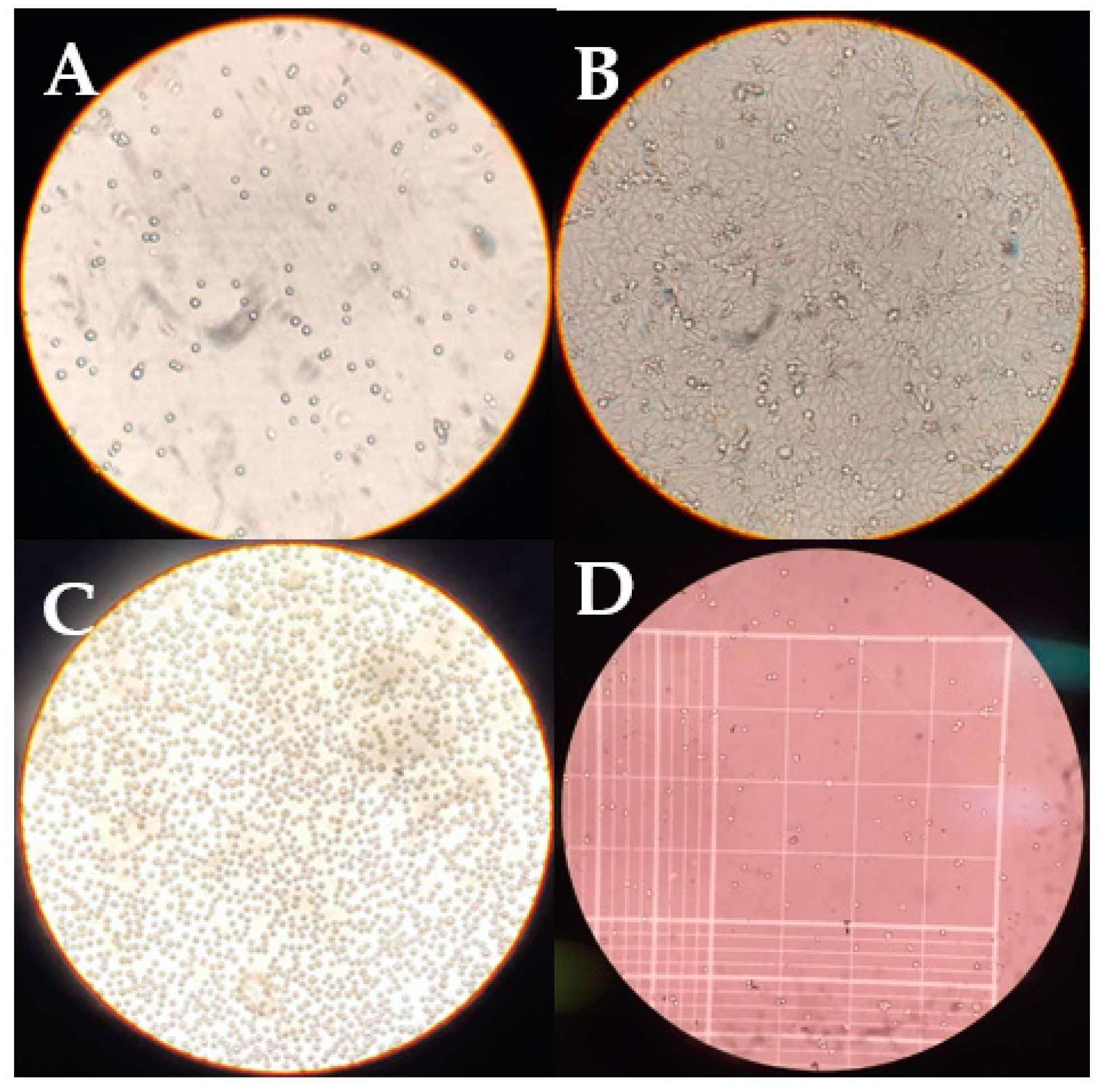
Figure 2A,B shows the difference in the proliferation of the HaCaT cell line culture. Figure 2C shows a culture with 90% confluence after the trypsinization process, indicating successful cell recovery. Percentages exceeding 80% were found in the cell viability tests, indicating the good state of the cell culture. An example of this assay in the HCC-70 cell line is shown in the Figure 2D.

3.2. Cytotoxicity

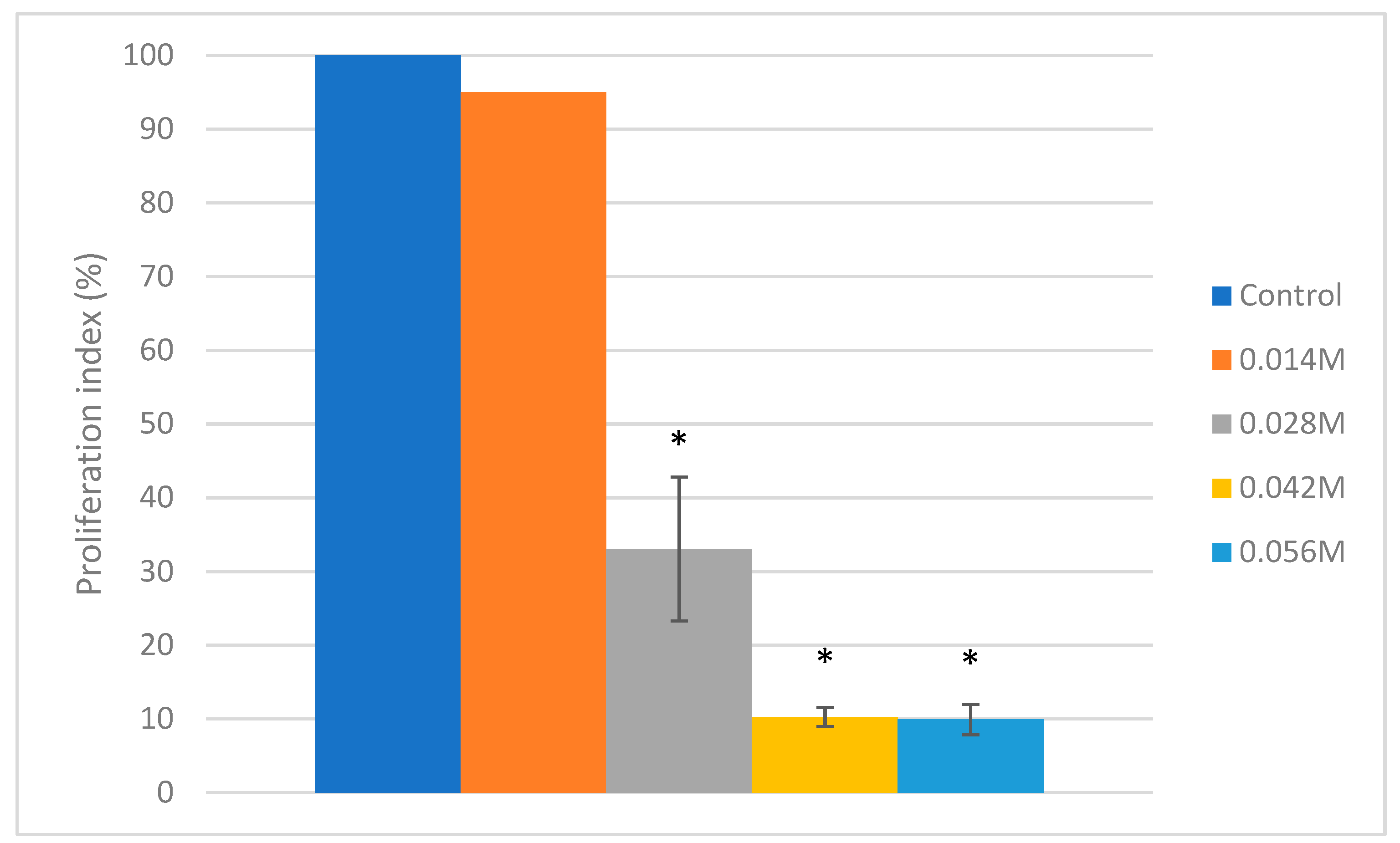
In the first trial, a dose response curve was performed in the HaCaT cell line, exposing it to four ascending doses of 2% Crv (20 µL, 40 µL, 60 µL and 80 µL) to standardize the IC50. The results showed that the proliferation indices decreased in a dose-dependent manner with significant reductions in proliferation index values from the second dose, which had a concentration of 0.028 M (33.06% ± 5.35), and it was further potentiated in the third (10.28% ± 1.29) and fourth doses (9.95% ± 2.06) with concentrations of 0.042 M and 0.056 M, respectively (Figure 3).
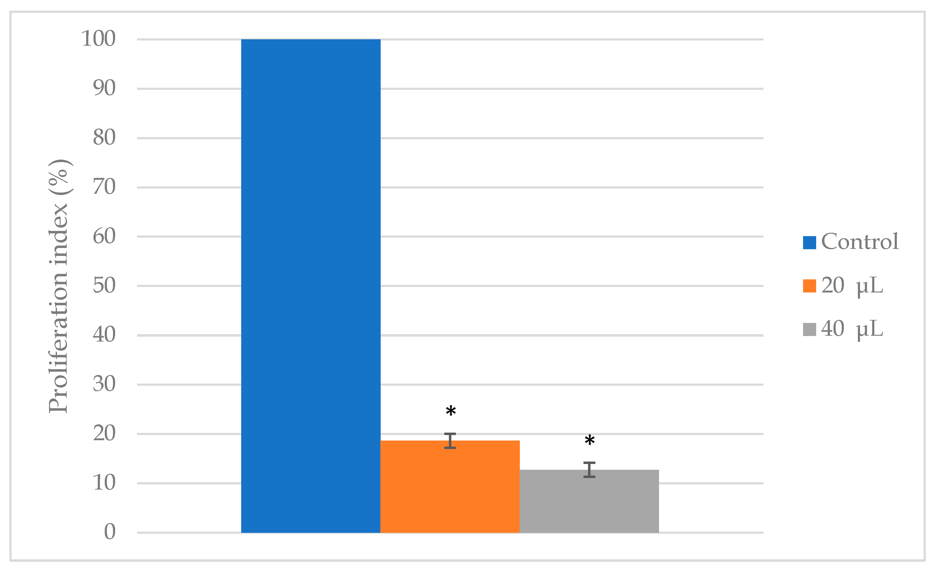
After that, the control cell line HaCaT was exposed to two ascending doses of the Ov infusion (20 µL and 40 µL) at a concentration of 250 g/10 mL, finding that the proliferation indices decreased to 18.62% ± 1.43 with the first dose and 12.75% ± 1.41 with the second (Figure 4).
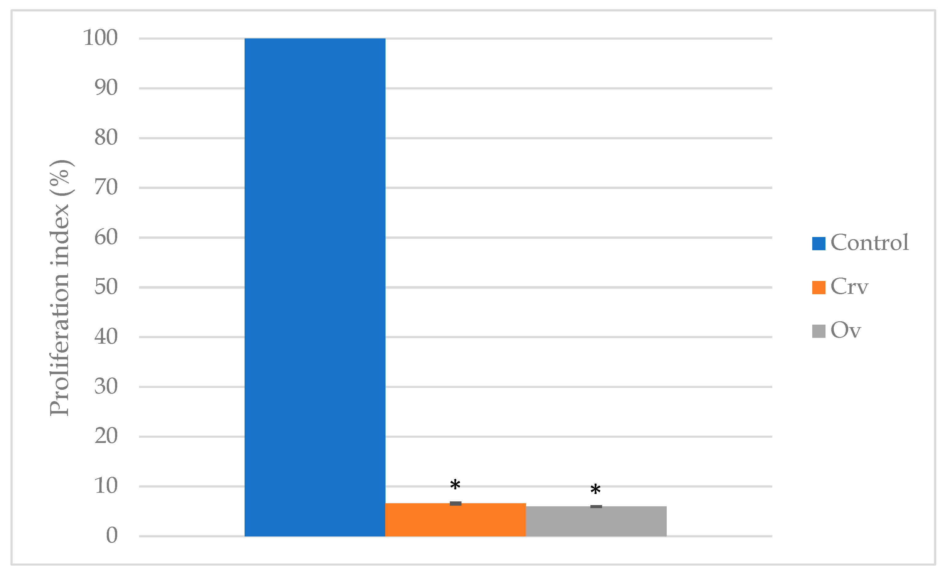
Once the lethal dose 50 for the Ov infusion and Crv had been standardized, the cytotoxicity test was carried out in the TN BC cell line (HCC-70) which was exposed to dose 2 of Crv (0.028 M) and dose 2 of the Ov infusion (40 µL). The results showed that the Ov infusion reduced the proliferation index of these cells to 5.95% ± 0.11 (p < 0.0001) while the Crv reduced it to 6.57 ± 0.21 (p <0.001) (Figure 5).
Similar results on the cytotoxicity of carvacrol against different cancer cell lines have been reported. For example, Fahad Khan et al. in 2017 found in their study that carvacrol doses of 25, 50, 100, 250 and 500 µM reduce the in vitro cell viability of prostate cancer cells (DU145) by 79.42%, 59.48%, 46.85%, 15.63% and 11.70%, respectively, after a 24-hour stimulus exposure. The lethal dose 50 for this cell line was found with a concentration of approximately 84.39 µM [29]. Moreover, Hazem Elshafie et al. in 2017 analyzed the in vitro cytotoxic potential of the compounds present in a higher proportion in oregano against hepatocarcinoma cells (HepG2), reporting that carvacrol was the compound with the greatest potential to inhibit the proliferation of this cell line. The lethal dose 50 found in this study was 48 mg/L [19]. These results, among some other studies that have been carried out on this compound and its properties against cancer cells, are consistent with the fact that carvacrol has a potent in vitro cytotoxic effect against different cancer cell lines like MDA-231 and MCF-7 [29,30,31,32,33,34,35]. However, the present study provides the first evidence on the effectiveness of this compound against TN BC cells (HCC-70), demonstrating the potency of its cytotoxic activity against aggressive subtypes of BC.
Talking about Ov, studies of this plant have been carried out with different cancer cell lines, as in colon cancer (HT-29), melanoma (A375) and hepatocarcinoma (HepG2), where a significant reduction in cell viability has been found in a dose-dependent manner [19,36,37]. Specifically, about breast cancer, Makrane et al. reported in 2018 that MDA-MB-231 BC cells were more susceptible to the cytotoxic effect of Ov compared to colon cancer cells (HT-29), as it was observed that cell viability decreased drastically in BC cells as the dose of Ov increased, finding the IC50 at a dose of 87.09 µg/mL [37].
Moreover, Katerina Spyridopoulou et al. reported that Ov showed anti-proliferative activity against MCF-7 (luminal A) BC cells in a dose-dependent manner, in their study, different cancer cell lines were subjected to Ov and the BC cell line was the third in which a stronger anti-proliferative effect was observed, with a lethal dose 50 of 10.0 ± 1.7 µg/mL [38]. Although there are few studies on the cytotoxicity of Ov in cancer cell lines the most relevant ones agree that it has a powerful cytotoxic potential.Specifically, in TN-BC cells, the effect of the infusion of Ov or Crv has not been studied, since most of the studies that have been published on the subject are in cell lines such as MCF-7 (luminal A) and MDA-MB-231 (breast adenocarcinoma) [22,23,31,33,38,39,40,41,42].
However, analyzing the results of studies on the cytotoxic effect of Ov and Crv on cancer cell lines, it can be inferred that both have a powerful cytotoxic activity against these cells. Some studies have even carried out tests in in vivo models with Ov, finding that it has the ability to significantly slow tumor growth of colon carcinoma in mice, among other effects [36]. Based on all the above and on the results of the present study, we can say that both the infusion of Ov and Crv show a powerful cytotoxic effect against TN BC cells (HCC-70), and these results represent the precedent for further studies to continue analyzing the efficacy of this effect because the results are promising so that in the near future, alternatives of adjuvant treatment can be offered in the management of aggressive types of BC.

4. Conclusions

In conclusion, the present study provides insight into adjuvant treatments efficacy for breast cancer, especially the Ov infusion and Crv for treatment of aggressive BC subtypes like TN. Moreover, this information lays the groundwork so that in the near future effective and economical adjuvant treatments can be promoted, that are beneficial for the patient and society at different levels including patient health, and individual and collective economy, because Ov is a plant that is easily accessible to the general population. Furthermore, this study opens the way for future studies, with the aim of further analyzing the efficacy of Ov infusion and Crv in this BC subtype, performing in vivo preclinical tests showing the forcefulness of its use, with the sole purpose of evaluating the biosecurity of its use, and finally performing tests of its cytotoxic activity as adjuvant and support to the conventional treatments in patients with TN breast cancer.

Acknowledgments

The authors thank Universidad de Guadalajara for providing the facilities to carry out this study.

Conflicts of Interest

The authors declare no conflict of interest.

References

  1. Hanahan, D.; Weinberg, R.A. Hallmarks of cancer: The next generation. Cell 2011, 144, 646–674. [Google Scholar] [CrossRef]
  2. GLOBOCAN. All Cancers. 2018. Available online: http://gco.iarc.fr/today (accessed on 26 February 2020).
  3. GLOBOCAN. Breast. 2018. Available online: http://gco.iarc.fr/today (accessed on 26 February 2020).
  4. Perou, C.M.; Sørile, T.; Eisen, M.B.; Van De Rijn, M.; Jeffrey, S.S.; Ress, C.A.; Pollack, J.R.; Ross, D.T.; Johnsen, H.; Akslen, L.A.; et al. Molecular portraits of human breast tumours. Nature 2000, 406, 747–752. [Google Scholar] [CrossRef]
  5. OMS. OMS|Nuevas Directrices de la OMS para Fomentar el uso Adecuado de las Medicinas Tradicionales. WHO. 2013. Available online: https://www.who.int/mediacentre/news/releases/2004/pr44/es/ (accessed on 26 February 2020).
  6. Oniga, I.; Pușcaș, C.; Silaghi-Dumitrescu, R.; Olah, N.K.; Sevastre, B.; Marica, R.; Marcus, I.; Sevastre-Berghian, A.C.; Benedec, D.; Pop, C.E.; et al. Origanum vulgare ssp. vulgare: Chemical composition and biological studies. Molecules 2018, 23, 2077. [Google Scholar] [CrossRef]
  7. Sharifi-Rad, M.; Varoni, E.M.; Iriti, M.; Martorell, M.; Setzer, W.N.; del Mar Contreras, M.; Salehi, B.; Soltani-Nejad, A.; Rajabi, S.; Tajbakhsh, M.; et al. Carvacrol and human health: A comprehensive review. Phyther. Res. 2018, 32, 1675–1687. [Google Scholar] [CrossRef]
  8. OMS. OMS|Cáncer. Available online: https://www.who.int/topics/cancer/es/ (accessed on 26 February 2020).
  9. CDC. ¿Qué es el cáncer de mama?|CDC. Available online: https://www.cdc.gov/spanish/cancer/breast/basic_info/what-is-breast-cancer.htm (accessed on 26 February 2020).
  10. ACS. ¿Qué es el Cáncer de Seno? Available online: https://www.cancer.org/es/cancer/cancer-de-seno/acerca/que-es-el-cancer-de-seno.html (accessed on 26 February 2020).
  11. Imigo, G.F.; Mansilla, S.E.; Delama, G.I.; Poblete, S.M.T.; Fonfach, Z.C. Clasificación molecular del cáncer de mama. Cuad. Cir. 2011, 25, 67–74. [Google Scholar] [CrossRef]
  12. CDC. Cáncer de Mama Triple Negativo|CDC. Available online: https://www.cdc.gov/spanish/cancer/breast/triple-negative.htm (accessed on 26 February 2020).
  13. Amin, A.R.M.R.; Kucuk, O.; Khuri, F.R.; Shin, D.M. Perspectives for cancer prevention with natural compounds. J Clin Oncol. 2009, 27, 2712–2725. [Google Scholar] [CrossRef]
  14. Reynoso-Noverón, N.; Mohar, A. El cáncer en México: Propuestas para su control. Salud Publica Mex. 2014, 56, S27–S32. [Google Scholar] [CrossRef]
  15. Fitzmaurice, C.; Dicker, D.; Pain, A.; Hamavid, H.; Moradi-Lakeh, M.M.M. The Global Burden of Cancer 2013. JAMA Oncol. 2015, 1, 505–527. [Google Scholar] [CrossRef]
  16. Dias, D.A.; Urban, S.; Roessner, U. A Historical overview of natural products in drug discovery. Metabolites 2012, 2, 303–336. [Google Scholar] [CrossRef]
  17. Cordell, G.; Colvard, M. Natural products and traditional medicine. J. Nat. Prod. 2012, 75, 514–525. [Google Scholar] [CrossRef]
  18. Atanasov, A.G.; Waltenberger, B.; Pferschy-Wenzig, E.-M.; Linder, T.; Wawrosch, C.; Uhrin, P.; Temml, V.; Wang, L.; Schwaiger, S.; Heiss, E. Discovery and resupply of pharmacologically active plant- derived natural products: A review. Biotechnol. Adv. 2015, 33, 1–18. [Google Scholar] [CrossRef]
  19. Thomford, N.E.; Senthebane, D.A.; Rowe, A.; Munro, D.; Seele, P.; Maroyi, A.; Dzobo, K. Natural products for drug discovery in the 21st century: Innovations for novel drug discovery. Int. J. Mol. Sci. 2018, 19, 1578. [Google Scholar] [CrossRef]
  20. Khazir, J.; Mir, B.A.; Pilcher, L.; Riley, D.L. Role of plants in anticancer drug discovery. Phytochem Lett. 2014, 7, 173–181. [Google Scholar] [CrossRef]
  21. Llana-Ruiz-Cabello, M.; Maisanaba, S.; Puerto, M.; Pichardo, S.; Jos, A.; Moyano, R.; Cameán, A.M. A subchronic 90-day oral toxicity study of Origanum vulgare essential oil in rats. Food Chem. Toxicol. 2017, 101, 36–47. [Google Scholar] [CrossRef]
  22. Elshafie, H.S.; Armentano, M.F.; Carmosino, M.; Bufo, S.A. Cytotoxic Activity of Origanum Vulgare L. on Hepatocellular Carcinoma cell Line HepG2 and Evaluation of its Biological Activity. Molecules 2017, 22, 1435. [Google Scholar] [CrossRef]
  23. Renukadevi, S.; Perumalsamy, H. Anti-proliferative activity of Origanum vulgare inhibited lipogenesis and induced mitochondrial mediated apoptosis in human stomach cancer cell lines. Biomed. Pharmacother. 2018, 108, 1835–1844. [Google Scholar] [CrossRef]
  24. Dragoeva, A.P.; Koleva, V.P.; Nanova, Z.D. Allelopathy of Cold Water Extracts from Origanum vulgare ssp. vulgare L. Sci. Res. 2014, 3, 144–150. [Google Scholar] [CrossRef]
  25. Brđanin, S.; Bogdanović, N.; Kolundžić, M.; Milenković, M. Antimicrobial activity of oregano (Origanum vulgare L.) and basil (Ocimum basilicum L.) Extracts. Adv. Technol. 2015, 4, 5–10. [Google Scholar] [CrossRef]
  26. Zhang, X.; Guo, Y.; Wang, C.; Li, G.; Xu, J. Phenolic compounds from Origanum vulgare and their antioxidant and antiviral activities. Food Chem. 2014, 152, 300–306. [Google Scholar] [CrossRef]
  27. Gottumukkala Venkateswara Rao TMukhopadhyay TAnnamalai NRadhakrishnan, M.R.S. Chemical constituents and biological studies of Origanum vulgare Linn. Pharm. Res. 2011, 3, 143–146. [Google Scholar]
  28. Criollo-mendoza, M.S.; Id, G.V.; Heredia, J.B. Flavonoids and Phenolic Acids from Oregano: Occurrence, Biological Activity and Health Benefits. Plants 2017, 7, 1–23. [Google Scholar]
  29. Arigesavan, K.; Sudhandiran, G. Carvacrol exhibits anti-oxidant and anti-inflammatory effects against 1, 2-dimethyl hydrazine plus dextran sodium sulfate Induced inflammation associated carcinogenicity in the Colon of Fischer 344 rats. Biochem. Biophys. Res. Commun. 2015. [CrossRef]
  30. Mosmann, T. Rapid colorimetric assay for cellular growth and survival: Application to proliferation and cytotoxicity assays. J. Immunol. Methods 1983, 65, 55–63. [Google Scholar] [CrossRef]
  31. Khan, F.; Khan, I.; Farooqui, A.; Ansari, I.A. Carvacrol Induces Reactive Carvacrol Induces Reactive Oxygen Species (ROS)-mediated Apoptosis Along with Cell Cycle Arrest at G0/G1 in Human Prostate Cancer Cells. Nutr. Cancer 2017, 69, 1075–1087. [Google Scholar] [CrossRef]
  32. Han, X.; Parker, T.L. Anti-inflammatory, tissue remodeling, immunomodulatory, and anticancer activities of oregano (Origanum vulgare) essential oil in a human skin disease model. Biochim. Open 2017, 4, 73–77. [Google Scholar] [CrossRef]
  33. Lim, W.; Ham, J. Carvacrol induces mitochondria-mediated apoptosis via disruption of calcium homeostasis in human choriocarcinoma cells. Cell Physiol. 2018, June, 1–13. [Google Scholar] [CrossRef]
  34. Khan, I.; Bahuguna, A.; Kumar, P.; Bajpai, V.K.; Kang, S.C. In vitro and in vivo antitumor potential of carvacrol nanoemulsion against human lung adenocarcinoma A549 cells via mitochondrial mediated apoptosis. Sci. Rep. 2018, 8, 712–714. [Google Scholar] [CrossRef]
  35. Elbe, H.; Yigitturk, G.; Cavusoglu, T.; Baygar, T.; Ozgul Onal, M.; Ozturk, F. Comparison of ultrastructural changes and the anticarcinogenic effects of thymol and carvacrol on ovarian cancer cells: Which is more effective? Ultrastruct Pathol. 2020, 44, 193–202. [Google Scholar] [CrossRef]
  36. Arunasree, K.M. Anti-proliferative effects of carvacrol on a human metastatic BCcell line, MDA-MB 231. Phytomedicine 2010, 17, 581–588. [Google Scholar] [CrossRef]
  37. Vokou, D.; Kokkinit, S.; Bessiere, J. Geographic Variation of Greek Oregano (Origanum vulgare ssp. hirtum) Essential Oils. Biochem. Syst. Ecol. 1993, 21, 287–295. [Google Scholar] [CrossRef]
  38. Spyridopoulou, K.; Fitsiou, E.; Bouloukosta, E.; Tiptiri-Kourpeti, A.; Vamvakias, M.; Oreopoulou, A.; Papavassilopoulou, E.; Pappa, A.; Chlichlia, K. Extraction, Chemical Composition, and Anticancer Potential of Origanum onites L. Essential Oil. Molecules 2019, 24, 2612. [Google Scholar] [CrossRef]
  39. Makrane, H.; El Messaoudi, M.; Melhaoui, A.; El Mzibri, M.; Benbacer, L.; Aziz, M. Cytotoxicity of the Aqueous Extract and Organic Fractions from Origanum majorana on Human Breast Cell Line MDA-MB-231 and Human Colon Cell Line HT-29. Adv. Pharm. Sci. 2018, 2018, 9. [Google Scholar] [CrossRef]
  40. Dhaheri YAl Attoub, S.; Arafat, K.; Abuqamar, S.; Viallet, J.; Saleh, A.; Agha, H.A.; Eid, A.; Iratni, R. Anti-Metastatic and Anti-Tumor Growth Effects of Origanum majorana on Highly Metastatic Human BCCells: Inhibition of NFκB Signaling and Reduction of Nitric Oxide Production. PLoS ONE 2013, 8, 1–17. [Google Scholar]
  41. Cittera, A.; Cazzola, R.; Cestaro, B.; Precliniche, S. Antioxidant Properties of oregano (ohganum vulgare) leaf extracts. J. Food Biochem. 2000, 24, 453–465. [Google Scholar]
  42. Dhaheri YAl Eid, A.; Abuqamar, S.; Attoub, S.; Khasawneh, M. Mitotic Arrest and Apoptosis in BCCells Induced by Origanummajorana Extract: Upregulation of TNF-a and Downregulation of Survivin and Mutant p53. PLoS ONE 2013, 8, 1–14. [Google Scholar]
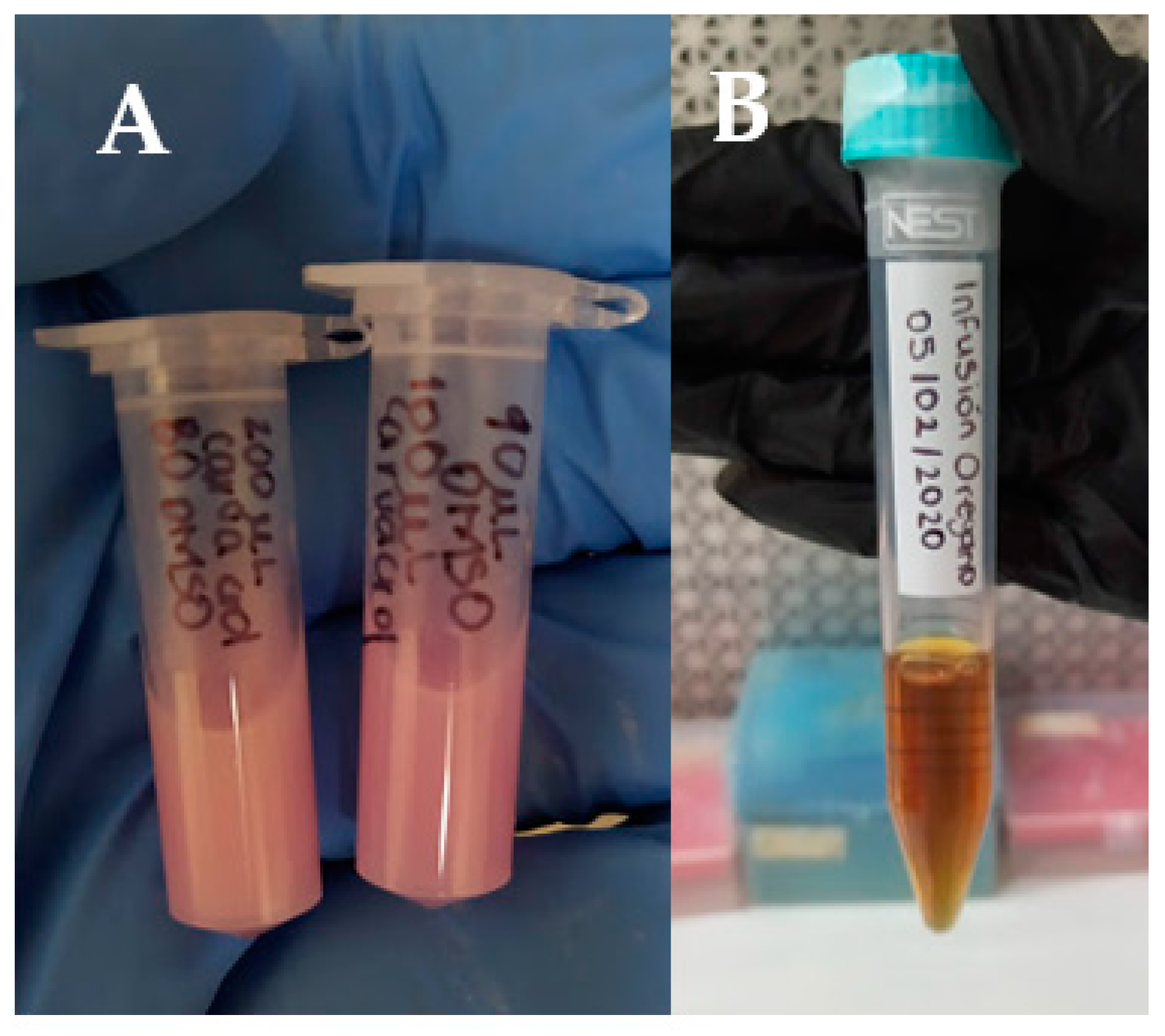
Figure 1. Photographs of the carvacrol (Crv) solution (A) and Origanum vulgare (Ov) infusion (B). Both stimuli were prepared the same day that were used.
Proceedings 61 00006 g001
Figure 2. (A) HaCaT cell culture at day 1. (B) HaCaT cell culture upon reaching >90% confluence; in addition to cell proliferation it is appreciated how the cells are adhered to the base of the culture flask. (C) HaCaT cell culture after the recovery process by trypsinization; a more rounded shape is observed in cultured cells after the adherent cell recovery process. (D) Viable cells appear white and those that are not are stained blue. Practically no non-viable cells can be seen. Photographs of cell cultures were taken by observing them under an inverted microscope while the photograph of Neubauer’s chamber was taken by observing it under a microscope.
Proceedings 61 00006 g002
Figure 3. Graphic representation of the proliferation index of HaCaT cells exposed to Crv. Significance * p < 0.0001. Data are expressed as mean of three replicates.
Proceedings 61 00006 g003
Figure 4. Graphic representation of the proliferation index of HaCaT cells exposed to the Ov infusion. Significance * p < 0.0001. Data are expressed as mean of three replicates.
Proceedings 61 00006 g004
Figure 5. Graphic representation of the proliferation index of triple negative breast cancer (TN BC) cells exposed to Ov infusion and Crv. Significance * p < 0.0001 Data are expressed as mean of three replicates.
Proceedings 61 00006 g005
Publisher’s Note: MDPI stays neutral with regard to jurisdictional claims in published maps and institutional affiliations.