Proceeding Paper

Peptide-Based Biosensor for Express Diagnostics of Coronavirus Respiratory Infections †

1
St. Petersburg Electrotechnical University “LETI”, Center of Microtechnology and Diagnostics, Chair of Micro- and nanoelectronics, Faculty of Electronics, 5, Professor Popov str., 197376 St. Petersburg, Russia
2
Institute of Highly Pure Biopreparations, 7, Pudozhsakya str., 197110 St Petersburg, Russia
3
St. Petersburg Pasteur Institute, 14, Mira str., 197101 St Petersburg, Russia
4
Institute of Macromolecular Compounds, RAS, 31, Bol’shoy Prospekt V.O., 199004 St Petersburg, Russia
*
Author to whom correspondence should be addressed.
Presented at the 1st International Electronic Conference on Biosensors, 2–17 November 2020; Available online: https://iecb2020.sciforum.net/.
Proceedings 2020, 60(1), 52; https://doi.org/10.3390/IECB2020-07059
Published: 2 November 2020
(This article belongs to the Proceedings of The 1st International Electronic Conference on Biosensors)

Abstract

:
At the end of year 2019 the first reports appeared of a new coronavirus and on 31st December 2019 WHO declared a public health emergency of international concern. To date (as of 6:08 pm CET, 24th November 2020) according to WHO the new coronavirus, now called severe acute respiratory syndrome (SARS)-CoV-2, has infected 58,900,547 people and killed 1,393,305 people worldwide. It is extremely important to develop means for express diagnostics to ensure prompt action to limit the spread of infection. One of the diagnostic approaches, is the detection of viral particles in swabs. This approach can be realized using a biosensor with specific ligands, based on peptide molecules complementary to surface viral proteins. The concept of the so-called Systems of Conjugated Ionic-Hydrogen Bonds (abbreviated—SSIVS, CIHBS) implemented in the Protein-3D computer program, was applied to analyze the spatial structures of the bonds between the SARS-CoV-2 spike protein and the ACE-2 (Angiotensin converting enzyme 2) receptor, in order to reveal the perspective peptide sequences. There are two clearly marked areas of contact of the spike with the cell receptor—upper and lower, which are visualized in the SSIVS form, and the complex formed at this site is strong enough to ensure its attachment to the coronavirus spike and can compete for binding with the ACE-2 receptor. Two peptides were developed that form a spatial structure complementary to the coronavirus spike: of eight (No. one) and of 15 (No. two) amino acid residues. The peptides were covalently bound to biochip platforms via neutral linkers to form sites with peptides No. one and No. two. The third site has a neutral hydrophilic surface to serve as a reference. The platform was integrated with a microfluidic channel and was used as a flow through device. The detection of bound viral particles was carried out using UV excitation and direct registration of viral proteins fluorescence. The preliminary laboratory tests demonstrated the efficiency of the biosensor.

1. Introduction

At the end of 2019, the first reports were documented of a new coronavirus, similar to those that caused the outbreaks of severe acute respiratory syndrome (SARS) in 2002–2004 and Middle East Respiratory Syndrome (MERS) in 2012. The WHO later announced a pandemic caused by the new COVID-19 virus.
To date, the new coronavirus (Figure 1), now called SARS-CoV-2, has infected about 60,000,000 people and killed about 1,400,000 people worldwide [1,2].
The virus is adsorbed on the cell surface in the area of ACE-2 (Angiotensin converting enzyme 2) receptors using spikes (Figure 1b), formed of trimer fusion S-protein, and then penetrates into the cell and starts the process of replication with the help of the cell’s apparatus. To inactivate the coronavirus, and for its rapid analysis, molecules with spatial structures complementary to the spikes of the coronavirus are required. Thus, development of 3D molecular structures that are spatially complementary to the S-protein seems to offer a promising tool for the inactivation and identification of SARS-Cov-2.
The aim of this work is to search for short peptides that could compete with the ACE-2 receptor for recognizing fragments of the coronavirus spike.

2. Materials and Methods

The in silico development of peptides was carried out using Protein 3D software [3], developed at the Centre of Microtechology and Diagnostics of St. Petersburg Electrotechnical University “LETI” [4].
The basic 3D structures of target proteins were obtained from the protein data bank [5].
The synthesis of peptides was carried out using a standard automatic procedure.

3. Results and Discussion

Currently, two studies are known (6LZG.pdb and 6m0j.pdb, both unpublished, with a resolution of 2.5 and 2.45 A) that are devoted to the study of the complexes of the SARS-CoV-2 coronavirus spikes with a protein. Due to different resolutions, they slightly differ in Systems of Conjugated Ionic-Hydrogen Bonds (SSVIS), which will be seen from the further presentation.
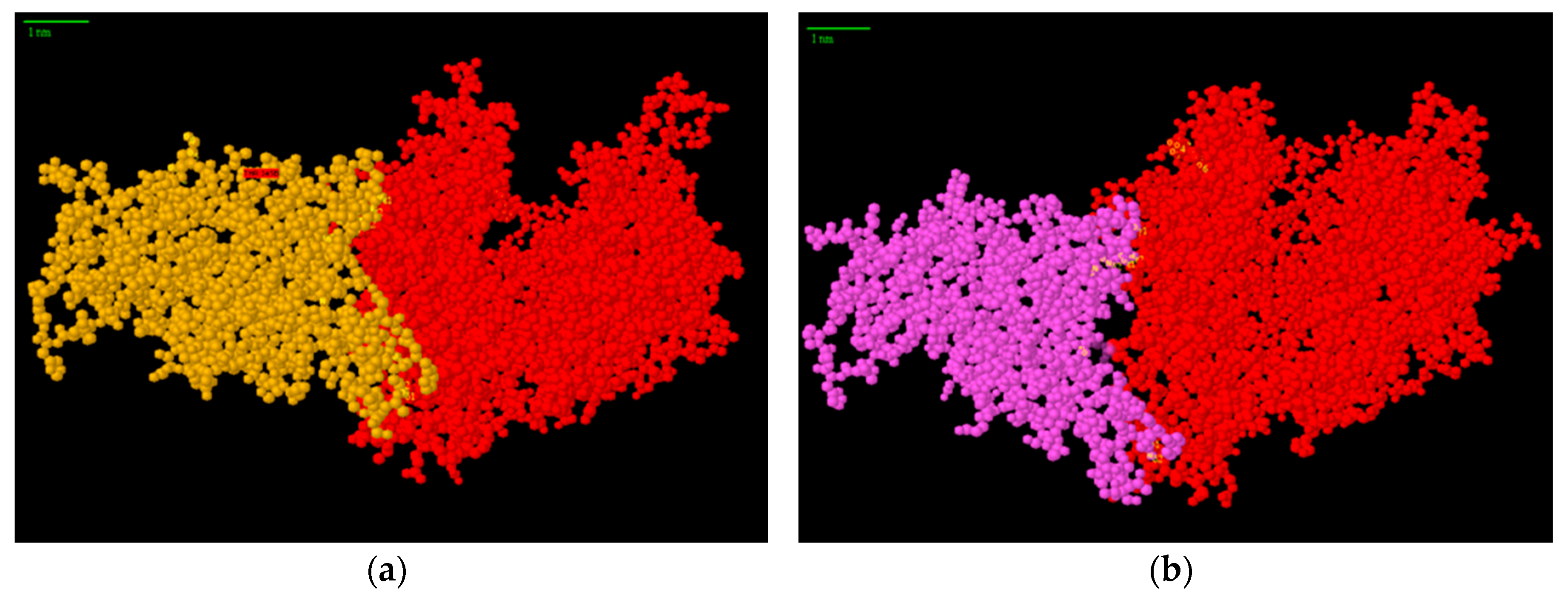
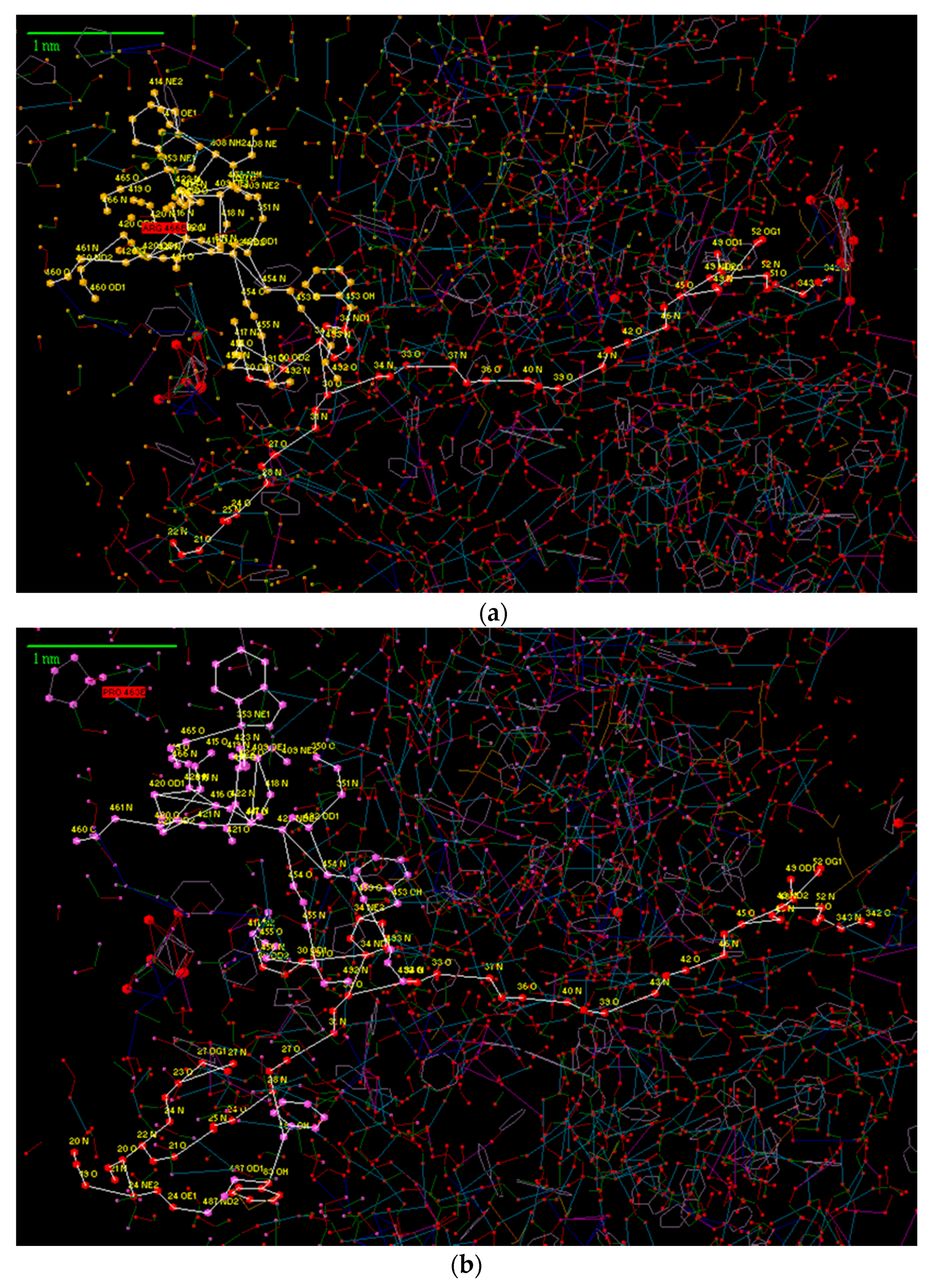
First, let us consider the general view of the complex (Figure 2a,b), from which it can be seen that both structures are practically identical. Their peculiarity lies in the fact that there are two clearly marked areas of contact between the spike and the cellular receptor—the upper and lower. Through examining these areas via their SSIVS (Figure 3), one can see that the first and second structures are practically the same.
There are several areas of contact between the SARS-CoV-2 coronavirus spike and the ACE-2 protein observed in these figures, which represented in Figure 3a,b.
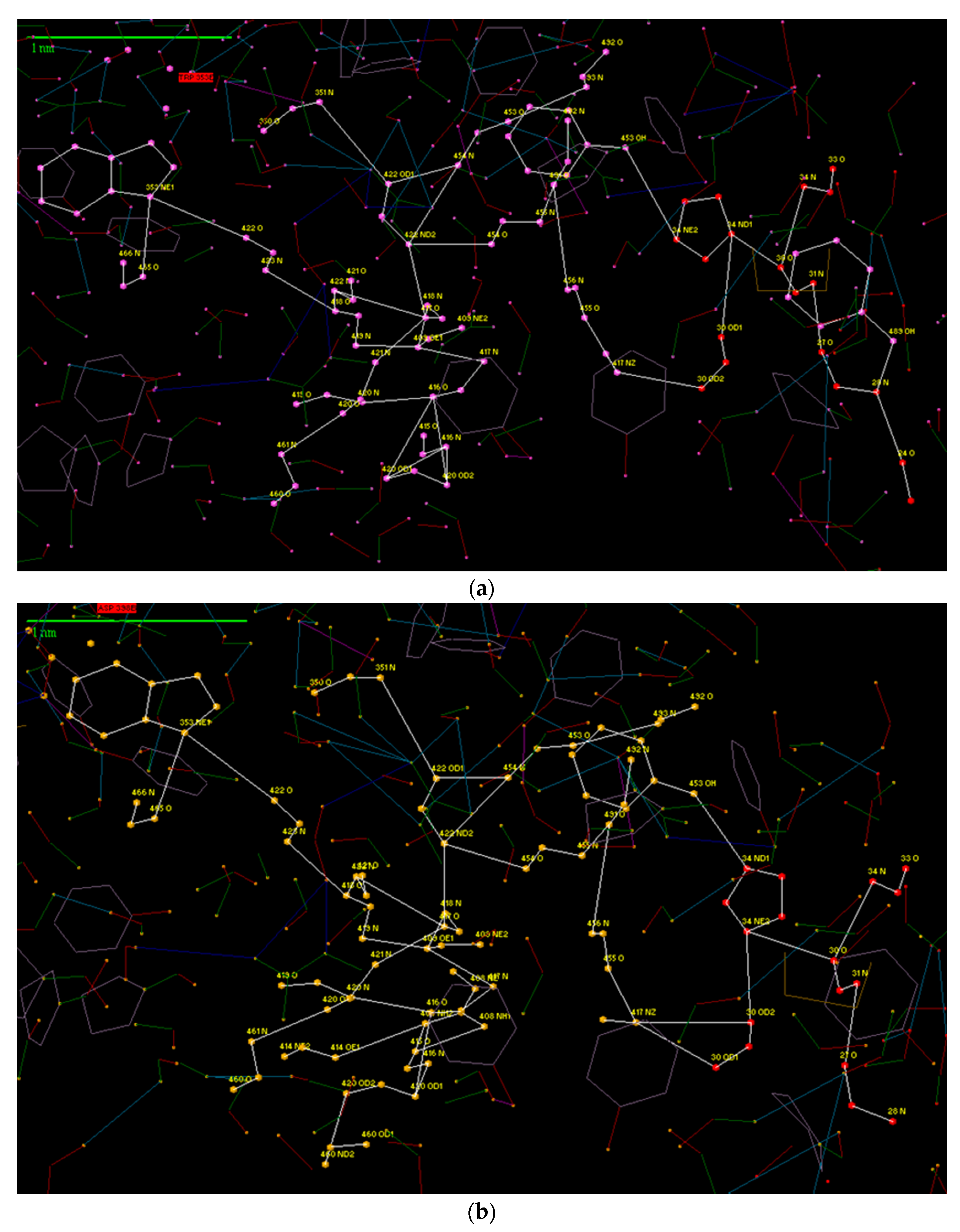
Most interestingly, in our opinion, is the interaction between His 34 protein ACE-2 and Tyr 453 KB. This fragment for both files (6LZG-1.pdb and 6m0j-1.pdb) is shown in Figure 4a,b.
Two sequences could be proposed as complementary structures to bound SARS-CoV-2. Sequence No. 1 contains only eight amino acids and is a helical fragment. It contains water-soluble side chains (THR, ASP, LYS, ASN, HIS). The peptide can be used both in the form of a solution and in the form of an anchor group (on a stem) in a diagnostic biochip.
As a second option, it is possible to propose increasing the length of the fragment to Tyr 41. This will provide even greater strength of binding of the anchor sequence to the spine of the virus (sequence No. 2).
Both structures of the SSVS are almost identical, which increases the reliability of the data presented. It can be assumed that the complex formed at this site is strong enough to ensure its attachment to the coronavirus spike and can compete for binding with the ACE-2 receptor.
The peptides can be used for virus inactivation, as shown in Figure 5.
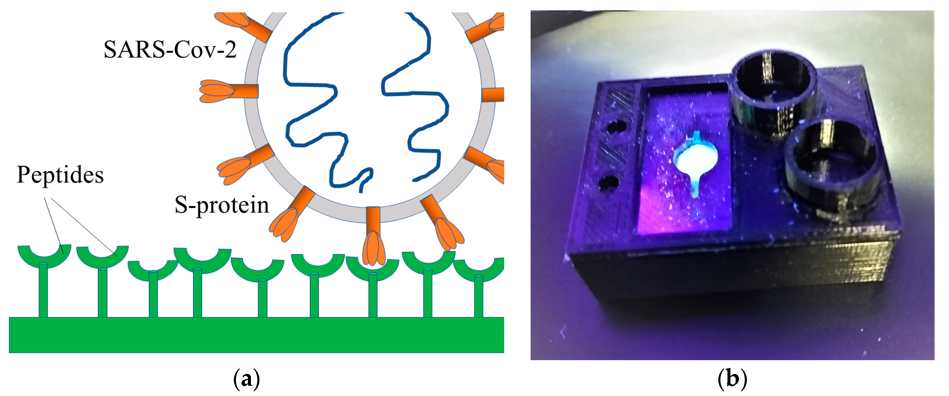
Peptides complementary to SARS-CoV2 spikes in biosensor were covalently bound to active sites on the glass surface via short linkers within the flow-through microfluidic system (Figure 6a). This configuration enables the sample preparation stage to be reduced considerably. The biochip laboratory sample is presented in Figure 6b.
Preliminary testing of biosensor was carried out at the Pasteur Institute and demonstrated promising results for peptide No. 2. Wide scale testing is currently in progress.

References

  1. WHO Coronavirus Disease (COVID-19) Dashboard. Available online: https://covid19.who.int (accessed on 25 October 2020).
  2. Wrapp, D.; Wang, N.; Corbett, K.S.; Goldsmith, J.A.; Hsieh, C.L.; Abiona, O.; Graham, B.S.; McLellan, J.S. Cryo-EM structure of the 2019-nCoV spike in the prefusion conformation. Science 2020, 367, 1260–1263. [Google Scholar] [CrossRef]
  3. Available online: http://protein-3d.ru (accessed on 16 April 2020).
  4. Karasev, V.A.; Luchinin, V.V. Introduction into Design of Bionic Nanosystems; Physmatlit: Moscow, Russia, 2011; 464p. [Google Scholar]
  5. Protein Data Bank. Available online: https://www.rcsb.org/ (accessed on 15 April 2020).
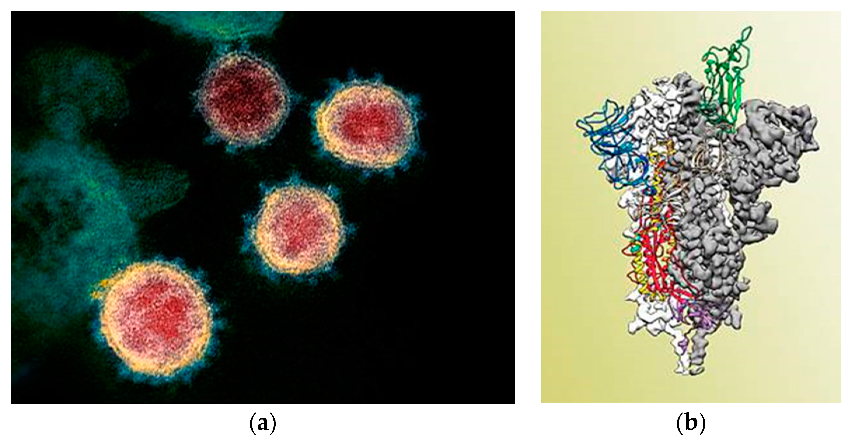
Figure 1. Image of severe acute respiratory syndrome (SARS)-CoV-2 coronaviruses obtained from a laboratory culture of infected cells (a); The structure of the protein spike of the SARS-CoV-2 coronavirus. The domain joining the host cell is colored green (b) [2] (UT Austin, McLellan Lab).
Proceedings 60 00052 g001
Figure 2. General view of the complex of a fragment of the thorn of the coronavirus SARS-CoV-2 and protein ACE-2: 6LZG.pdb (a); 6m0j.pdb (b).
Proceedings 60 00052 g002
Figure 3. General view of the complex of the SARS-2 coronavirus spike and the ACE-2 protein in the rendering of the Systems of Conjugated Ionic-Hydrogen Bonds (SSIVS). (a) 6LZG.pdb (yellow balls—a fragment of the coronavirus thorn, red balls—ACE-2 protein), (b) 6m0j.pdb (purple balls—a fragment of a coronavirus thorn, red balls—ACE-2 protein).
Proceedings 60 00052 g003
Figure 4. Fragment of the thorn of the coronavirus SARS-CoV-2 and fragment 27–34 of the ACE-2 protein in the rendering of the SSIVS: (a) 6m0j.pdb, (b) 6LZG.pdb.
Proceedings 60 00052 g004
Figure 5. Mechanism of virus inactivation. (a) Mechanism of coronavirus cell entry mediated by the viral S-protein; (b) inhibition of fusion S-protein with 3D complementary peptide.
Proceedings 60 00052 g005
Figure 6. Schematic representation of biosensor principle for SARS-CoV-2 determination in swab samples (a); laboratory sample of the biosensor (b).
Proceedings 60 00052 g006
Publisher’s Note: MDPI stays neutral with regard to jurisdictional claims in published maps and institutional affiliations.