Proceeding Paper

Optical Properties on Bone Analysis: An Approach to Biomaterials †

1
Federal University of Uberlandia, Uberlândia 38400-902, Brazil
2
Applied Research Institute of the Polytechnic of Coimbra, Coimbra 3030-199, Portugal
*
Author to whom correspondence should be addressed.
Presented at the 15th International Workshop on Advanced Infrared Technology and Applications (AITA 2019), Florence, Italy, 17–19 September 2019.
Proceedings 2019, 27(1), 36; https://doi.org/10.3390/proceedings2019027036
Published: 27 September 2019

Abstract

:
The objective of the present study was to investigate the influence of demineralization solution on the optical properties of chicken femoral samples. Biomaterials based on bone have gained importance in clinical applications due to their properties as better osseointegration and biocompatibility. Biomateriais (bone substitute) are essentials to auxiliary in treatment of diseases related to bones such as bone density disorder, low bone mineral mass and the deterioration of bone tissue. Our data shows that integrating sphere technique permits to determinate significant difference in optical properties between healthy and demineralized samples. In this work, the optical properties of bone samples from chicken femur have been measured over the wavelength range 700–1000 nm.

1. Introduction

In recent years, tissue characterization with optical techniques has singular relevance in the field of diagnosis [1]. The main physical parameter is a refractive indices and its spatial variation along the tissue. Optical techniques can provide important information and insight for early detection and diagnostic of diseases. Bone is a natural material constituting mineral, organic and water [2]. Bone tissue engineering material with significant properties as three-dimensional porous structure, large surface area, optical properties, non-hazardous, eco-friendly, and biocompatible are known as main factors for cell adhesion, proliferation, differentiation, and new tissue formation [3]. For many reasons, animal bone has been considered as the best choice among distinct synthetic biomaterials for acting as substitute. On the other hands, it is known that a challenge aspect to check the quality of the bone is the imbalance of mineral matter [4]. In this work, we report the optical properties by using and integrating sphere [5], in which such as absorption and extinction coefficient of samples of bone from chicken femur have been measured over the wavelength range 700–1000 nm. Comparative results between optical properties of healthy chicken bone and demineralized samples were investigated.

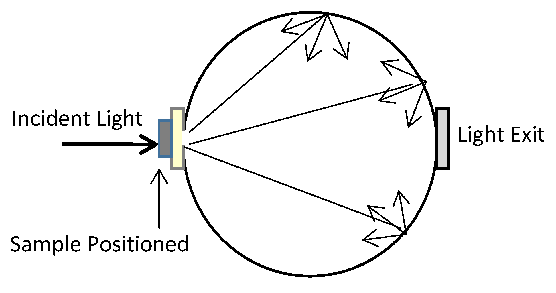
2. Materials and Methods

We examined chicken femoral bone samples on compact and trabecular regions in according Figure 1. We choose this biological material for practical opportunity to discriminate optical scattering centers inside each specific bone region showing main differences right after two distinct demineralization processes. The demineralization solution (20% w/v1) was prepared by adding citric acid to distilled water. The inner surface and all other internal parts of the integrating sphere are coated. Tissues were placed in a straight line at the aperture (light incident) in according to schematic view showed in Figure 2. Illumination of the tissue was provided by optical fiber positioned inside the integrating sphere. We have used a 2”, 4 ports, integrating sphere from Thorlabs Inc. We have used an integration sphere system for determination of optical parameters such as absorption (µa) and scattering (µs) coefficients.

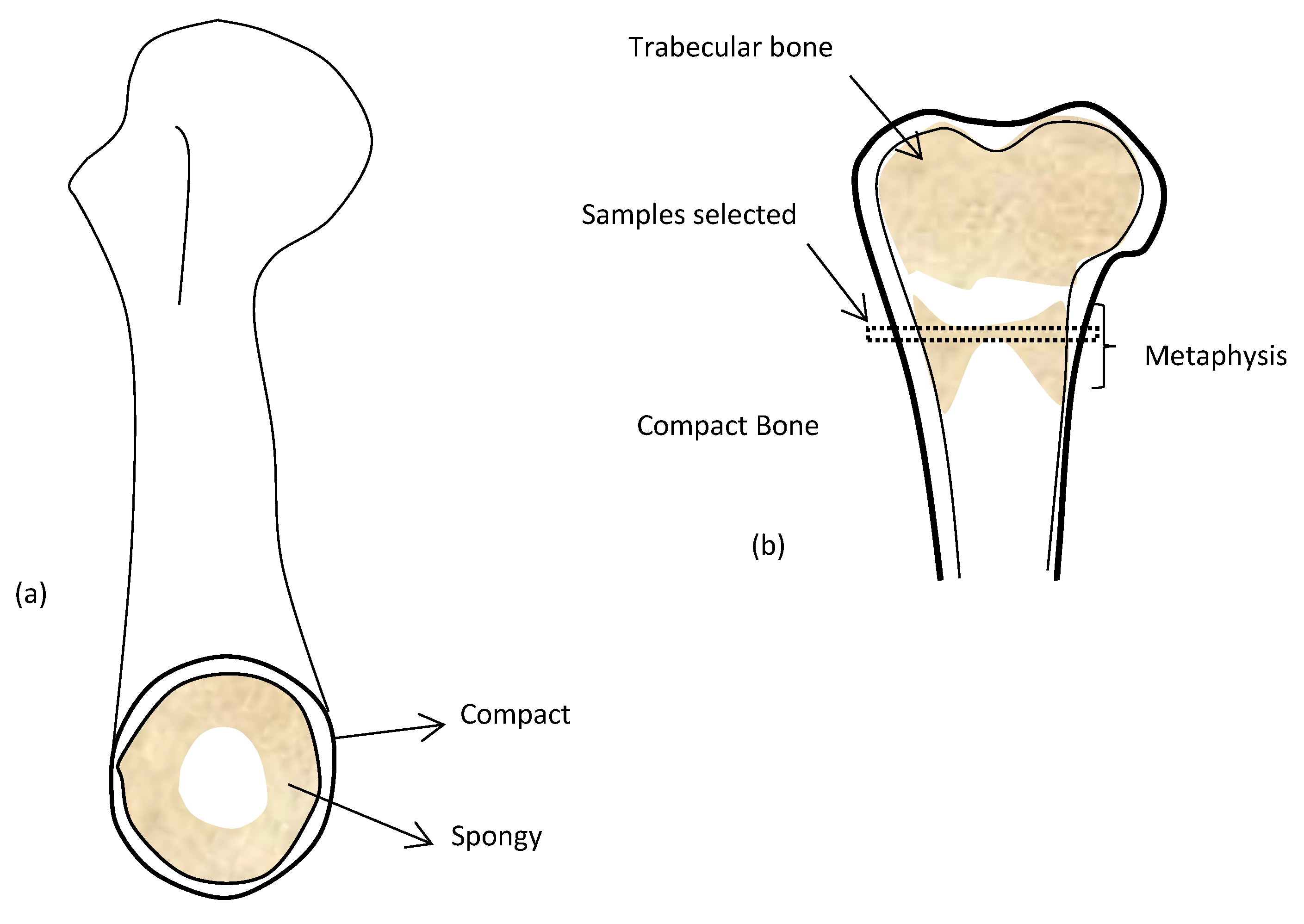
2.1. Bone Preparation

Freshly removed chicken bone were obtained from commercial butcher. After removing the soft tissue, the bones were ground into 2-sided blocks with a flat surface (5 × 5 mm) and an average thickness of approximately 3 mm. The measurements were carried out in native as well as in demineralized tissues. In advance of each measurement, the system was calibrated without sample. Quantitative characterization of bone in terms of optical properties was achieved with an integrating sphere. In Figure 2, a representative scheme of transmittance experiment is shown. Following acquisition of total reflection and transmission the Inverse adding doubling (IAD) technique is applied to obtain the scattering and absorption coefficients [5]. In this technique, measurements of total diffuse reflectance and transmittance of a sample are employed together with a mathematical model for optical parameters estimation.

3. Results and Discussion

The results obtained from chicken bone samples have shown that this is possible to discriminate the demineralization processes on each bone surface. Based on the distinct optical absorption and extinction of bone in infrared wavelength range may distinguish changes on scattering and absorption coefficients in according with preparation methods to demineralization processes. Figure 3 shows the absorption and scattering coefficients of healthy (non-treated) versus demineralized (treated) bone.
The hydroxyapatite is main chromophore in bone. The scattering and absorption pattern were qualitatively distinguishable in the samples due to coefficient shifts. Scattering coefficient alters in according refraction index and scatterer’s density in the tissue at specific wavelength.

Acknowledgments

This work was supported by the Brazilian Agencies, CAPES, CNPq and FAPEMIG.

Conflicts of Interest

The authors declare no conflict of interest.

References

  1. Saager, R.B.; Quach, A.; Rowland, R.A.; Baldado, M.L.; Durkin, A.J. Low-cost tissue simulating phantoms with adjustable wavelength-dependent scattering properties in the visible and infrared ranges. J. Biomed. Opt. 2016, 21, 67001. [Google Scholar] [CrossRef] [PubMed]
  2. Landis, W. The strength of a calcified tissue depends in part on the molecular structure and organization of its constituent mineral crystals in their organic matrix. Bone 1995, 16, 533–544. [Google Scholar] [CrossRef] [PubMed]
  3. Kim, D.; Thangavelu, M.; Cheolui, S.; Kim, H.S.; Choi, M.J.; Song, J.E.; Khang, G. Effect of different concentration of demineralized bone powder with gellan gum porous scaffold for the application of bone tissue regeneration. Int. J. Boil. Macromol. 2019, 134, 749–758. [Google Scholar] [CrossRef] [PubMed]
  4. Paschalis, E.P.; Mendelsohn, R.; Boskey, A.L. Infrared Assessment of Bone Quality: A Review. Clin. Orthop. Relat. Res. 2011, 469, 2170–2178. [Google Scholar] [CrossRef] [PubMed]
  5. Yamakoshi, T.; Lee, J.; Matsumura, K.; Yamakoshi, Y.; Rolfe, P.; Kiyohara, D.; Yamakoshi, K.-I. Integrating Sphere Finger-Photoplethysmography: Preliminary Investigation towards Practical Non-Invasive Measurement of Blood Constituents. PLOS ONE 2015, 10, 0143506. [Google Scholar] [CrossRef] [PubMed]
Figure 1. (a) Chicken bone transversal cut; (b) Sample selected region.
Proceedings 27 00036 g001
Figure 2. Representative scheme of transmittance analysis.
Proceedings 27 00036 g002
Figure 3. Average optical properties (scattering and absorption coefficients) measured on samples analyzed.
Proceedings 27 00036 g003
1
% w/v = g of solute/100 mL of solution.

