Proceeding Paper

The Effects of Oleuropein on Different Clinically Types of Human Neuroblastoma Cells †

1
Department of Basic Oncology, Dokuz Eylul University Institute of Oncology, Izmir 35340, Turkey
2
Department of Pediatric Oncology, Dokuz Eylul University Institute of Oncology, Izmir 35340, Turkey
*
Author to whom correspondence should be addressed.
Presented at the 2nd International Cell Death Research Congress, Izmir, Turkey, 1–4 November 2018.
Proceedings 2018, 2(25), 1591; https://doi.org/10.3390/proceedings2251591
Published: 12 December 2018
(This article belongs to the Proceedings of The 2nd International Cell Death Research Congress)

Abstract

:
Neuroblastoma is an embryonic tumor originating from the neural crest. It accounts for 8–10% of all childhood cancers. Although Cisplatin is used in neuroblastoma treatment, it has many side effects, such as ototoxicity, nephrotoxicity, and neurotoxicity. One herbal agent that has attracted attention in recent years is oleuropein (OLE), the active component of olive leaf. This component belongs to the polyphenol group and it has antioxidant, anti-microbial, anti-inflammatory, anti-hypertensive and anti-carcinogenic effects. It has beneficial effects against neurodegeneration in both culture cells and model organisms. Oleuropein has been shown to be increased apoptosis in SH-SY5Y neuroblastoma cell line in one study. Cisplatin (cis-diaminedichloroplatinum II (CDDP) is a widely used agent for the treatment of many different human cancers in childhood and adults with antimitotic and antineoplastic properties. CDDP is the most effective chemotherapeutic agent in specially treatment of neuroblastoma. Purpose of this study was to determine whether oleuropein and CDDP have possible anti-proliferative activity in different types of human neuroblastoma cells as representing different clinical features (bone marrow metastatic LAN-5 cells and treated with chemotherapy and beam therapy CHP-134 cells representing late relapse) investigated. Human bone marrow metastatic LAN-5 and treated with chemotherapy and beam therapy CHP-134 neuroblastoma cells representing late relapse were used in this study. The effects of OLE and CDDP on LAN-5 and CHP-134 neuroblastoma cell proliferation and apoptotic cell death was investigated using WST-1 cell proliferation and Annexin-V/PI flow cytometric assays. Oleuropein and CDDP have been shown to inhibit proliferation of LAN-5 and CHP-134 neuroblastoma cells. In further studies, it is planned to investigate different cell death mechanisms by using combination of oleuropein and cisplatin in different kind of human neuroblastoma cells.

1. Introduction

Neuroblastoma is a common malignancy in childhood and originates from the primitive neural crest cells in the sympathetic nervous system [1]. Although CDDP is used in neuroblastoma treatment, it has many side effects, such as ototoxicity, nephrotoxicity, and neurotoxicity [2]. Because of the limitations of treatment, new treatment protocols are still under investigation [1]. The only publication ever made about Oleuropein and Cisplatin belongs to Patocnjak et al. [3]. In this study, the renal protective effects of Oleuropein against Cisplatin-induced renal damage were investigated. This study showed that all these effects were reduced with oleuropein in a dose-dependent manner [3].
Purpose of this study was to determine whether oleuropein and CDDP have possible anti-proliferative activity in different types of human neuroblastoma cells as representing different clinical features (bone marrow metastatic LAN-5 cells and treated with chemotherapy and beam therapy CHP-134 cells representing late relapse) investigated. It is anticipated that in the case of detection of possible anti-tumoral effects in cells that reflect different clinical situations of oleuropein in neuroblastoma, new studies may be evaluated to assess clinical benefit after animal studies in-vivo.

2. Materials and Methods

2.1. Cell Culture

In this study, LAN-5 (bone marrow metastasis), CHP-134 (chemotherapy and irradiated, late relapse) human neuroblastoma cell lines were used. LAN-5 and CHP-134 were maintained in RPMI-1640 (RPMI-1640 Cell Culture Medium, Gibco™ Life Technologies, Waltham, MA, USA) containing 20% FBS (Fetal Bovine Serum, Gibco™ Life Technologies, Waltham, MA, USA), 1% penicillin/streptomycin (Gibco™ Life Technologies, Waltham, MA, USA), and 1% l-glutamine (Gibco™ Life Technologies, California, USA). The cells were incubated at 37 °C and 5% CO2 conditions. The medium of the cells was freshened 2 or 3 times weekly [4]. When cultures reach 80% congestion, they will be removed from culture dishes with trypsin/EDTA (Capricorn Scientific GmbH, TRY-1R10, Ebsdorfergrund, Germany) solution in 1:2 ratios. All reagents were freshly prepared with mediums before all experiments. The cell viability was determined using an automatic cell counter with a trypan blue (Eve Cell Counting, NanoEnTek Inc., EVS-050, Waltham, MA, USA) exclusion test.

2.2. Detection of Cell Proliferation

The cells were then treated with different concentrations of OLE (Oleuropein, Sigma-Aldrich Chemie GmbH, Taufkirchen, Germany) and CDDP (Cisplatin, Koçak Farma, Istanbul, Turkey) (50–1000 uM) for 24, 48, and 72 h [4,5]. Following the incubation periods, the WST-1 assay was performed by adding 10 μL of WST reagents (Cell Proliferation Reagent, Sigma-Aldrich Chemie GmbH, 5015944001, Taufkirchen, Germany) to each well, and all neuroblastoma cells were further incubated at 37 °C for 2 h. After incubation periods, the absorbances of the cells was measured test wavelength at 450 nm and the reference wavelength at 630 nm was measured at 630 nm on an ELISA reader. The half-maximal inhibitory or lethal concentrations (IC50 or LD50) of the agents were calculated according to control cells viability.

2.3. Detection of Apoptotic Cells

Apoptotic cell death was determined using annexin-V-fluorescein isothiocyanate (FITC)/Propidium Iodide (FITC Annexin V Apoptosis Detection Kit, BD Pharmingen, Franklin Lakes, NJ, U.S.A.) in all cells. The principle of the method is based on of the extent apoptosis determination using flow cytometry of cells stained with annexin-V-FITC and PI, a non-vital dye. Neuroblastoma cell lines were incubated for 24 h in the presence of OLE or CDDP. For all neuroblastoma cells, OLE was used at 500 μM and CDDP was used at 50 μM concentrations determined from cell proliferation assays. Analyses were carried out using a flow cytometer (Accuri, Franklin Lakes, NJ, U.S.A) at 488 nm excitation and 530 nm emission wavelengths for annexin-V and 488 nm excitation and 647 nm emission wavelengths for PI and evaluated using the BD Accuri C6 Software (BD Biosciences).

3. Results

3.1. Detection of Cell Proliferation

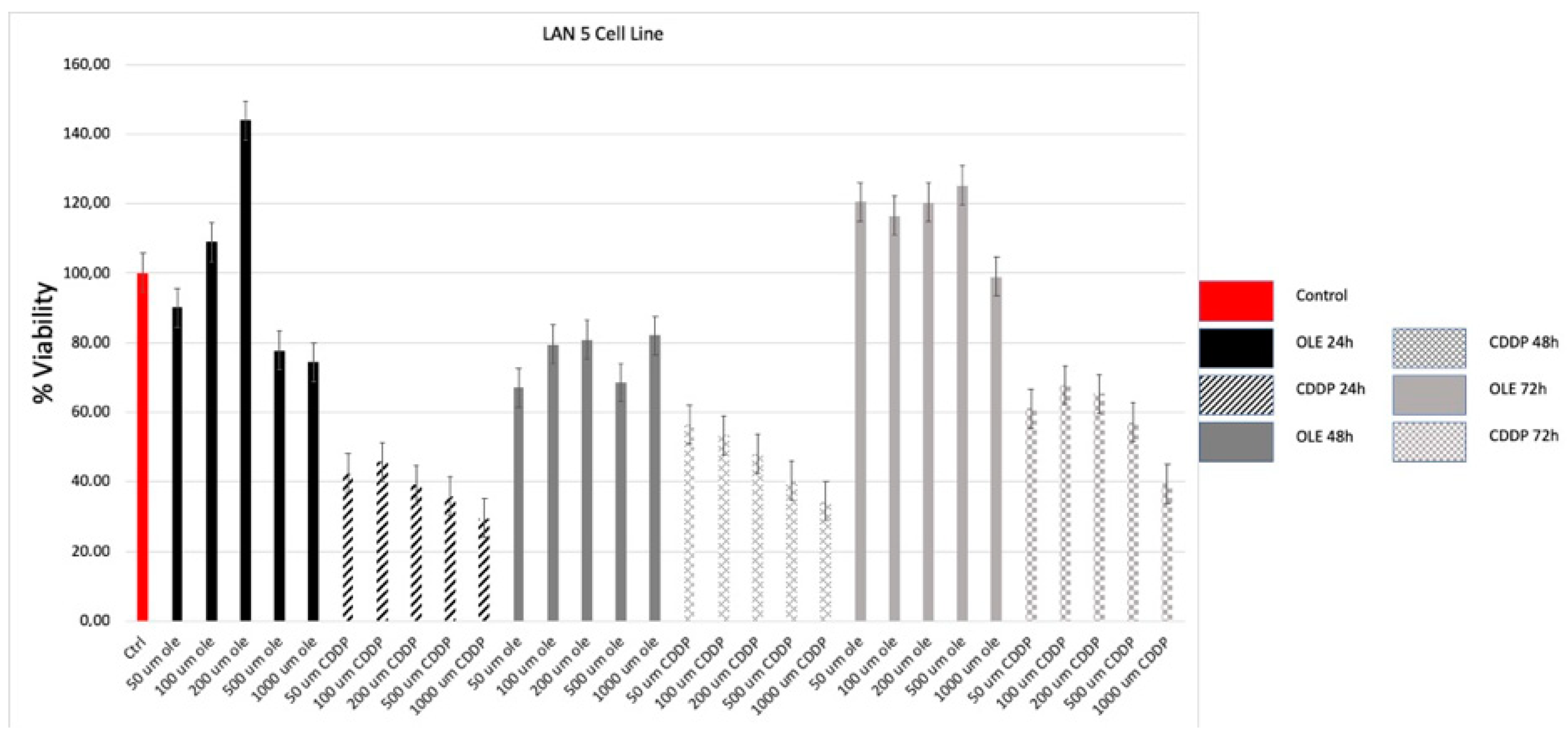
The cell viability of CHP-134 (chemotherapy and irradiated, late relapse) and LAN-5 (bone marrow metastasis) neuroblastoma cells treated with different doses of OLE (50–1000 μM) and CDDP (50–1000 μM) was determined using the WST-1 assay. OLE and CDDP inhibited neuroblastoma cell proliferations in a dose dependent manner.
In CHP-134 human neuroblastoma cells, the cell viability decreased at a rate of 56% and 35% with the treatment of 500 μM OLE and 50 μM CDDP, respectively, after 24 h of incubation (Figure 1).
In LAN-5 human neuroblastoma cells, the cell viability decreased at a rate of 77% and 42% with the treatment of 500 μM OLE and 50 μM CDDP, respectively, after 24 h of incubation (Figure 2).
500 μM OLE and 50 μM CDDP concentration applied for 24 h was designated as the IC50.
Moreover, we tested the apoptotic effects of the agents in these cells using the annexin-V analysis.

3.2. Apoptotic Cell Death Results of the Cells

We also compared apoptosis level at the lC50 concentration of OLE and CDDP on CHP-134 and LAN-5 cells. To compare the apoptosis level of OLE and CDDP, flow cytometry annexin-V/PI assays were used. While the IC50 dose of OLE in the CHP-134 cell line resulted in 12% apoptotic cell death and 45% necrotic cell death, the IC50 dose of CDDP resulted in 29% apoptotic cell death and 33% necrotic cell death (Figure 3). In the LAN5 cell line, the IC50 dose of OLE resulted in 10% apoptotic cell death and 3% necrotic cell death. However, the IC50 dose of CDDP resulted in 31% apoptotic cell death and 30% necrotic cell death (Figure 3).

4. Discussion

There are some studies showing that OLE also reduces the toxicity caused by CDDP. In a study by Patoncnjak et al. showed that oral usage of OLE protected against CDDP-induced renal damage [3].In addition to the protective effect of OLE, there are studies on the anticancer effect. The anticancer effects of OLE were investigated in a study by Seçme et al. [5]. In this study, IC50 was determined by XTT assay and it was found to be 350 uM at 48th hour.
Elamin et al. examined the dose-dependent cytotoxic effect of OLE on MDAMB-231 and MCF-7 breast cancer cell lines [6]. Like as our study, they showed similar results in breast cancer cells. Seçme et al. also similarly our results has been showed that OLE decreases cell proliferation and induces apoptotic cell death in neuroblastoma cells [5].

5. Conclusions

In this study, OLE was shown to inhibit cell proliferation and induce apoptosis in human neuroblastoma cells as dose-dependent manner. In further studies, it is planned to investigate different cell death mechanisms by using combination of oleuropein and cisplatin in different kind of human neuroblastoma cells.

References

  1. Olgun, N.; Kansoy, S.; Aksoylar, S.; Cetingul, N.; Vergin, C.; Oniz, H.; Sarialioglu, F.; Kantar, M.; Uysal, K.; Tuncyurek, M.; et al. Experience of the Izmir Pediatric Oncology Group on Neuroblastoma: IPOG-NBL-92 Protocol. Pediatr. Hematol. Oncol. 2003, 20, 211–218. [Google Scholar] [CrossRef] [PubMed]
  2. Altun, Z.; Olgun, Y.; Ercetin, P.; Aktas, S.; Kirkim, G.; Serbetcioglu, B.; Olgun, N.; Guneri, E.A. Protective effect of acetyl-l-carnitine against cisplatin ototoxicity: Role of apoptosis-related genes and pro-inflammatory cytokines. Cell Prolif. 2014, 47, 72–80. [Google Scholar] [CrossRef] [PubMed]
  3. Potočnjak, I.; Škoda, M.; Pernjak-Pugel, E.; Peršić, M.P.; Domitrović, R. Oral administration of oleuropein attenuates cisplatin-induced acute renal injury in mice through inhibition of ERK signaling. Mol. Nutr. Food Res. 2016, 60, 530–541. [Google Scholar] [CrossRef] [PubMed]
  4. Cecen, E.; Altun, Z.; Ercetin, P.; Aktas, S.; Olgun, N. Promoting effects of sanguinarine on apoptotic gene expression in human neuroblastoma cells. Asian Pac. J. Cancer Prev. 2014, 15, 9445–9451. [Google Scholar] [CrossRef] [PubMed]
  5. Seçme, M.; Eroğlu, C.; Dodurga, Y.; Bağcı, G. Investigation of anticancer mechanism of oleuropein via cell cycle and apoptotic pathways in SH-SY5Y neuroblastoma cells. Gene 2016, 585, 93–99. [Google Scholar] [CrossRef] [PubMed]
  6. Elamin, M.H.; Daghestani, M.H.; Omer, S.A.; Elobeid, M.A.; Virk, P.; Al-Olayan, E.M.; Hassan, Z.K.; Mohammed, O.B.; Abdelilah, A. Olive oil oleuropein has anti-Brest cancer properties with higher efficiency on Er-negative cells. Food Chem. Toxicol. 2013, 53, 310–316. [Google Scholar] [CrossRef] [PubMed]
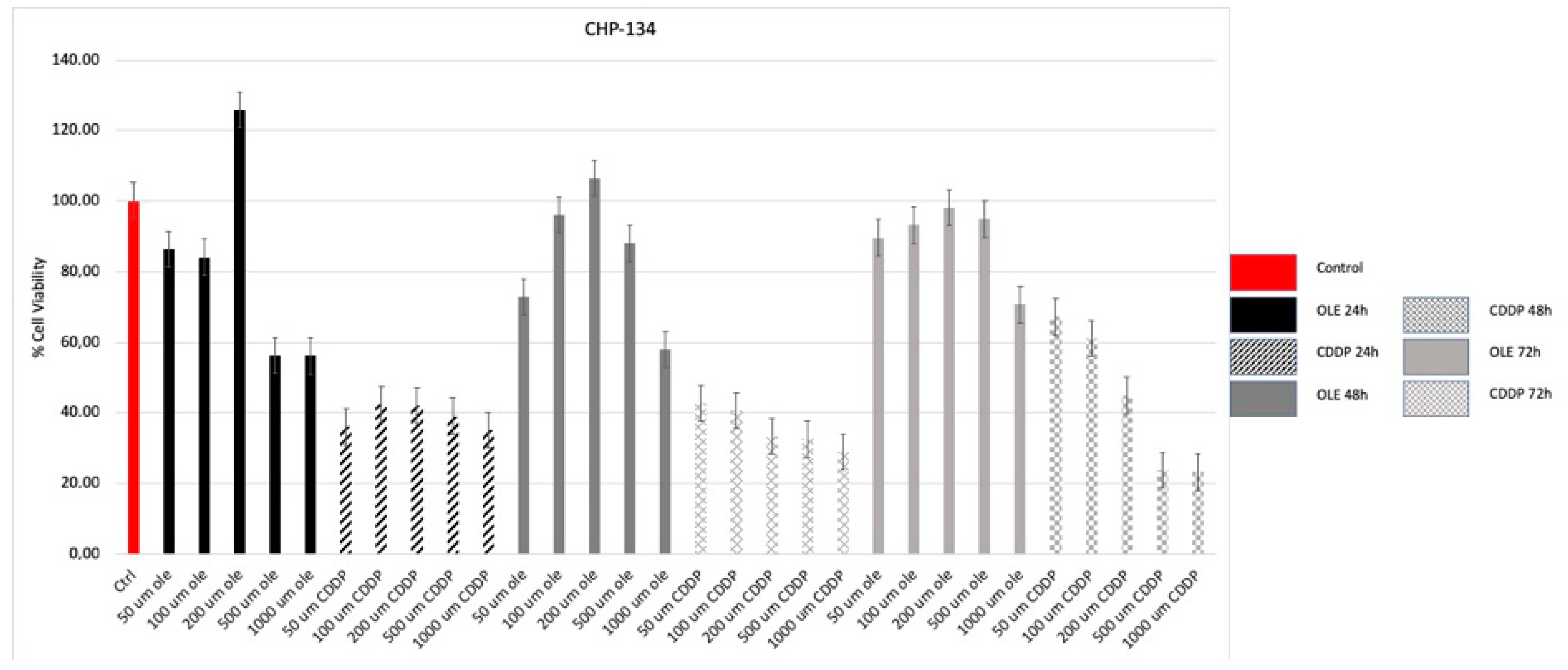
Figure 1. Relative cell proliferation assay results of OLE and CDDP in CHP-134 (chemotherapy and irradiated, late relapse) human neuroblastoma cells for 24, 48, and 72 h incubations.
Proceedings 02 01591 g001
Figure 2. Relative cell proliferation assay results of OLE and CDDP in LAN5 (bone marrow metastasis) human neuroblastoma cells for 24, 48, and 72 h incubations.
Proceedings 02 01591 g002
Figure 3. Apoptotic percentages of OLE (500 μM) and CDDP (50 μM) treated CHP-134 and LAN5 cells determined using flow cytometry analysis. OLE: oleuropein; CDDP: cisplatin.
Proceedings 02 01591 g003
Publisher’s Note: MDPI stays neutral with regard to jurisdictional claims in published maps and institutional affiliations.