Proceeding Paper

Combination of First Generation Proteasome Inhibitor Bortezomib with Temozolomide and Radiotherapy in Glioblastoma 2D and 3D Cell Cultures †

1
School of Medicine/Genetic and Metabolic Diseases Research and Investigation Center, Marmara University, 34854 İstanbul, Turkey
2
Advanced Nanomaterials Research Laboratory, Marmara University, 34854 İstanbul, Turkey
3
Department of Radiation Oncology, School of Medicine, Marmara University, 34854 İstanbul, Turkey
*
Author to whom correspondence should be addressed.
Presented at the 2nd International Cell Death Research Congress, Izmir, Turkey, 1–4 November 2018.
Proceedings 2018, 2(25), 1579; https://doi.org/10.3390/proceedings2251579
Published: 7 December 2018
(This article belongs to the Proceedings of The 2nd International Cell Death Research Congress)

Abstract

:
Glioblastoma is the most common high-grade brain tumor in adults, which has a rapidly developing radiotherapy/chemotherapy resistance and high recurrence rate. The aim of our study is to determine the effects of bortezomib that is a quite important proteasome inhibitor on glioblastoma tumors in the 3D environment. In this study, we have performed bortezomib and temozolomide drug treatments and radiation applications to U251 cell line in 2D and 3D cultures with PCL/gelatin/graphene nanofibrous scaffolds. The best result was reached in the combination of drugs with radiation therapy that has the most decreased viability in the experiment group.

1. Introduction

Proteasomal degradation is an important mechanism to prevent the accumulation of cellular damage. While removal of damage is a necessary process for maintaining integrity in healthy cells, may lead to treatment resistance in cancer cells. When cancer cells and healthy cells are compared, it is observed that in the case of anti-cancer stress, the defense mechanisms of cancer cells are much more active than healthy cells. Bortezomib, the first generation proteasome inhibitor, has been used for the treatment of different types of cancers in the clinic for about 15 years. Bortezomib is used extensively in hematological cancer types and has significantly increased patient survival.
Glioblastoma (GBM) is the most common and most aggressive grade IV astrocytoma. The median survival after diagnosis is usually less than one year. The standard treatment for GBM is usually the application of adjuvant radiotherapy (RT) and temozolomide, the alkylating agent, after surgical resection. The mechanism that causes the recurrence of the disease is the presence of stem cells that are resistant to radiotherapy and chemotherapy. In a recent study [1], GBM stem cells have been shown to have a high proteasome activity. Another mechanism causing treatment resistance is the repair of alkylating damage of temozolomide by cancer cells via O-methylguanine-DNA-methyltransferase. The proteasome inhibitor bortezomib has been shown to inhibit this methyltransferase enzyme and cause the resistance to be overcome [2]. These studies and many others confirm the need for the use of proteasome inhibitors in the treatment of GBM. Experimental models for cancer drugs are evolving to 3D scaffolds since 2D cell cultures and animal models have some limitations. Graphene oxide (GO) is a derivative of graphene exhibits many advantages such as superior biocompatibility, high dispersibility in water and organic solvents, great mechanical properties, considerable electrical and thermal conductivity [3,4]. PCL is aliphatic polyester, which has a great deal of interest in recent decades as its biodegradability, biocompatibility, and suitability for biomedical studies. Gelatin (GE) is a natural biopolymer derived from collagens exhibits excellent biocompatibility and biodegradability properties and has been widely used and studied in regard to many biomedical applications. The addition of graphene into PCL/gelatin blend results in great improvements in the mechanical and hydrophilicity properties of the nanofibrous scaffold, which makes it an excellent candidate for tissue engineering applications [5].
In this study, temozolomide and bortezomib (proteasome inhibitor) were used in combination with radiation therapy. In the experimental design, GBM microenvironment was formed in three-dimensional culture media with PCL/gelatin/graphene oxide nanofibrous scaffolds.

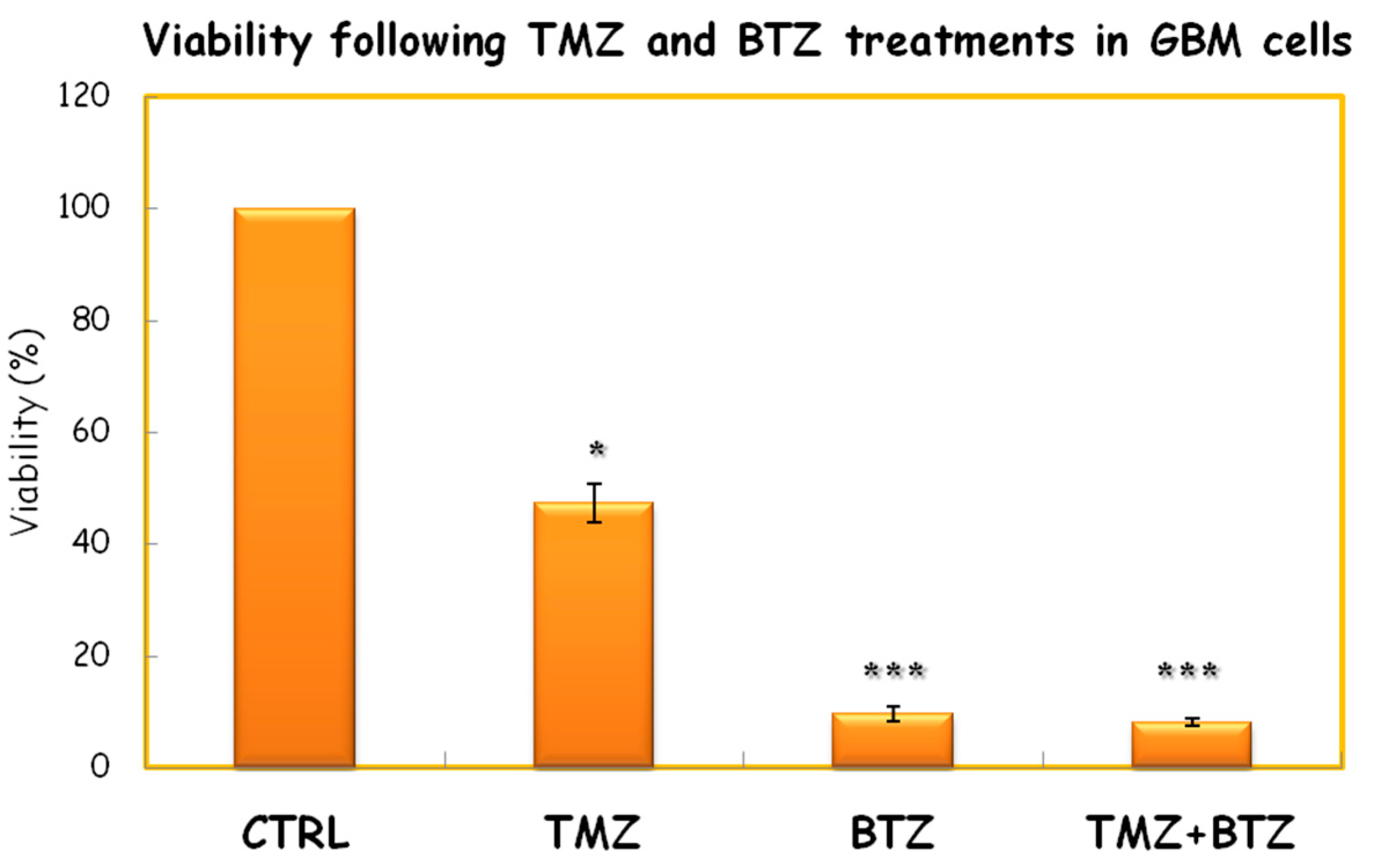
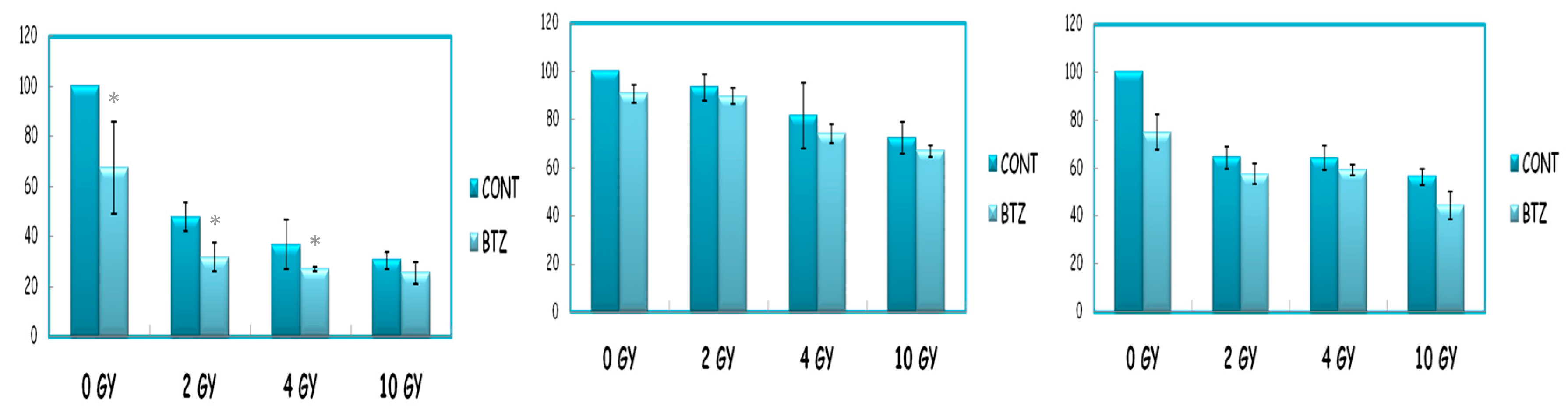
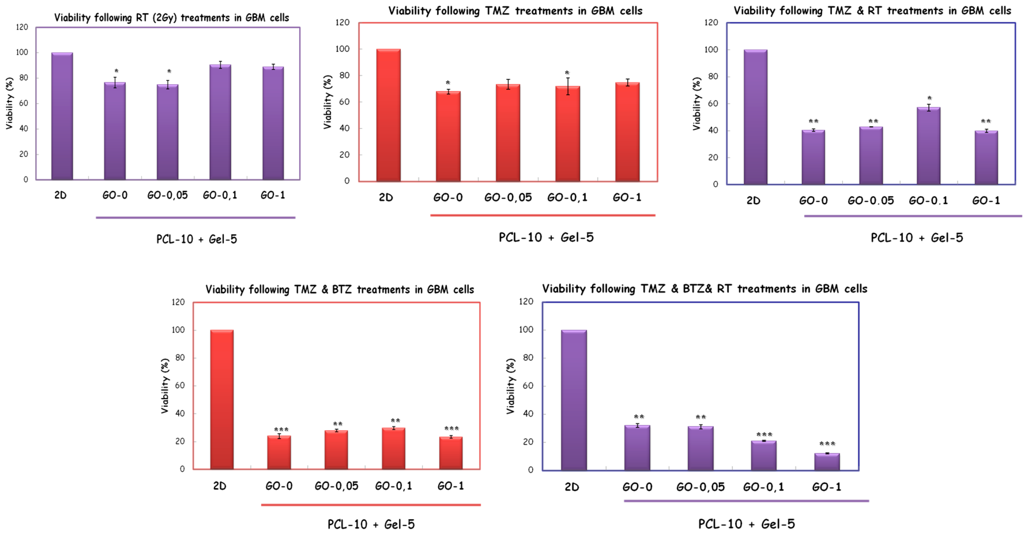
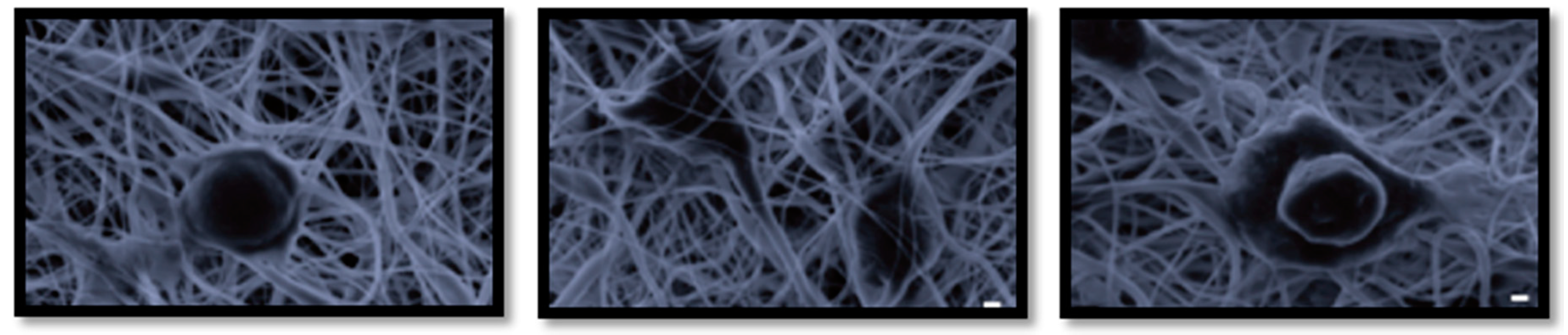
2. Materials and Methods

Human glioblastoma cancer cell line was purchased from the American Type Culture Collection (ATCC). For material independent samples, cells were cultured in DMEM containing 10% fetal bovine serum (FBS), 1% Penicillin/Streptomycin in the 24 well/plate with 100,000 cells per well and incubated at 37 °C and 5% CO2. For TMZ (50 mM), BTZ (330 nM) and radiation (2 Gy) exposures, the MTT protocol was applied at the end of 72 h of incubation. Cell viability was measured using MTT reagent (Sigma, St. Louis, MO, USA) dissolved in PBS (5 mg/mL). After 4 h incubation, the absorbance of formazan crystals dissolved in the DMSO solution were measured in 96 wells/plate at 570 nm.
Adhesion of cells on the materials was confirmed by scanning electron microscopy (SEM) following fixation with 2.5% glutaraldehyde for 1 h. The cells on the material were then washed with 25%, 50%, 75% and 100% ethanol for 10 min. Finally; the samples were dried overnight in a laminar flow flask and monitored under SEM.

3. Discussion

In this study, we investigated the effects of drug and radiotherapy in different concentrations on the viability of 2D and 3D cultures of the GBM cells. The median survival for glioblastoma after diagnosis is usually less than one year, and even under the best conditions, the vast majority of patients are lost within two years. Standard treatment for GBM is usually adjuvant radiotherapy (RT) and chemotherapy (CT) after surgical resection. In our study, we aimed to investigate the combinations of bortezomib, which is a proteasome inhibitor, in addition to these treatments. Bortezomib is used extensively in the clinics, especially in hematologic cancer types, and significantly increases patient survival [6]. First, we tried different treatment procedures (radiotherapy, TMZ, and TMZ + BTZ) in 2D culture and compared the results. The combination of BTZ with TMZ and radiotherapy significantly reduced the viability (Figure 1). Drug combinations with radiotherapy were performed with 2 Gy dose since there was no significant difference in cell viability following 2, 4 and 10 Gy radiotherapies (Figure 2). The poly ε-caprolactone (PCL) nanofibers were produced by electrospinning method to mimic the GBM tumor environment in our study. These nanofibers offer a unique advantage as cell culture models and they are well suited for neural cellular systems due to their topographical similarity to white matter in the brain. In addition, PCL, which is an electrospun nanofiber, produces the morphological and molecular markers of glioma migration ex vivo [7,8]. In this direction, we have repeated the entire experiment in 3D cell culture that is containing the gelatin and graphene oxide electrospun combination at different concentrations (PCL10-Gel5-GO0, GO0.05, GO0.1, GO1) by the electrospinning method. Among others, PCL10-Gel5-GO1 was less toxic to the cells and it was found to be well consistent with the drug treatments and radiotherapy (Figure 3 and 4). We can conclude from our 2D and 3D GBM data that, BTZ combination with TMZ and radiotherapy may increase the survival rates.

Acknowledgments

This work was supported by The Scientific and Technological Research Council of Turkey (TÜBİTAK, Project no: 118S522).

References

  1. André-Grégoire, G.; Bidère, N.; Gavard, J. Temozolomide affects Extracellular Vesicles Released by Glioblastoma Cells. Biochimie 2018, 9084. [Google Scholar] [CrossRef] [PubMed]
  2. Vlachostergios, P.J.; Hatzidaki, E.; Befani, C.D.; Liakos, P.; Papandreou, C.N. Bortezomib overcomes MGMT-related resistance of glioblastoma cell lines to temozolomide in a schedule-dependent manner. Investig. New Drugs 2013, 31, 1169–1181. [Google Scholar] [CrossRef] [PubMed]
  3. Mohammadi, S.; Shafiei, S.S.; Asadi-Eydivand, M.; Ardeshir, M.; Solati-Hashjin, M. Graphene oxide- enriched poly (ε-caprolactone) electrospun nanocomposite scaffold for bone tissue engineering applications. J. Bioact. Compat. Polym. 2017, 32, 325–342. [Google Scholar] [CrossRef]
  4. Wang, C.; Li, Y.; Ding, G.; Xie, X.; Jiang, M. Preparation and characterization of graphene oxide/poly (vinyl alcohol) composite nanofibers via electrospinning. J. Appl. Polym. Sci. 2013, 127, 3026–3032. [Google Scholar] [CrossRef]
  5. Jalaja, K.; Sreehari, V.S.; Kumar, P.A.; Nirmala, R.J. Graphene oxide decorated electrospun gelatin nanofibers: Fabrication, properties and applications. Mater. Sci. Eng. C 2016, 64, 11–19. [Google Scholar] [CrossRef] [PubMed]
  6. Jackson, G.; Einsele, H.; Moreau, P.; San Miguel, J. Bortezomib, a novel proteasome inhibitor, in the treatment of hematologic malignancies. Cancer Treat. Rev. 2005, 31, 591–602. [Google Scholar] [CrossRef] [PubMed]
  7. Johnson, J.; Nowicki, M.O.; Lee, C.H.; Chiocca, E.A.; Viapiano, M.S.; Lawler, S.E.; Lannutti, J.J. Quantitative analysis of complex glioma cell migration on electrospun polycaprolactone using time-lapse microscopy. Tissue Eng. Part C Methods 2009, 15, 531–540. [Google Scholar] [CrossRef] [PubMed]
  8. Rao, S.S.; Nelson, M.T.; Xue, R.; DeJesus, J.K.; Viapiano, M.S.; Lannutti, J.J.; Sarkar, A.; Winter, J.O. Mimicking white matter tract topography using core–shell electrospun nanofibers to examine migration of malignant brain tumors. Biomaterials 2013, 34, 5181–5190. [Google Scholar] [CrossRef] [PubMed]
Figure 1. Percentage of 2D culture GBM cell viability treated with different doses of radiotherapy (a) 24 h, (b) 48 h and (c) 72 h. * p < 0.05 versus controls.
Proceedings 02 01579 g001
Figure 2. Percentage of 2D culture GBM cell viability treated with temozolomide and bortezomib. * p < 0.05 versus control, *** p < 0.001 versus control .
Proceedings 02 01579 g002
Figure 3. Percentage of 3D culture GBM cell viability treated with Radiotherapy (2 Gy), TMZ, TMZ + Radiotherapy, TMZ + BTZ and TMZ + BTZ + Radiotherapy. * p < 0.05 versus control, ** p < 0.01 versus control , *** p < 0.001 versus control.
Proceedings 02 01579 g003
Figure 4. SEM images of GBM cells on the PCL/Gel/GO scaffold.
Proceedings 02 01579 g004
Publisher’s Note: MDPI stays neutral with regard to jurisdictional claims in published maps and institutional affiliations.

