Proceeding Paper

Calmodulin Antagonists as Potential Therapeutic Agents for Cancer Treatment “Breast Cancer” †

1
Department of Biomedical and Therapeutic, Faculty of Medicine, University Malaysia Sabah, Kota Kinabalu 88400, Sabah, Malaysia
2
Biotechnology Research Institute, University Malaysia, Kota kinabalu 88400, Sabah, Malaysia
*
Author to whom correspondence should be addressed.
Presented at the 2nd International Cell Death Research Congress, Izmir, Turkey, 1–4 November 2018.
Proceedings 2018, 2(25), 1550; https://doi.org/10.3390/proceedings2251550
Published: 6 December 2018
(This article belongs to the Proceedings of The 2nd International Cell Death Research Congress)

Abstract

:
Although cancer research undergone a rapid expansion, there is no potential cure and the disease remains one of the leading causes of mortality worldwide. Breast cancer continues as the female malignancy and a major cause of death in middle-aged women. Calmodulin can interact with large number of different target proteins and modulate their activity in different ways. Calmodulin antagonists have been reported to induce apoptosis and inhibit tumor cell growth. However, the potential effects of these target agents on various cancer within the molecular levels are poorly understood. The main objective of this study is to evaluate the potency of a number of calmodulin antagonists on the growth of human breast cancer cell lines. In addition, preliminary trials were carried out to find the role of these agents within the molecular levels. The finding indicates that Calmodulin antagonists might be the key for future breast cancer treatment strategy.

1. Introduction

Globally, breast cancer is the most common female malignancy and a major cause of death in middle-aged women [1,2]. Calmodulin (CaM) was first described in eukaryotic by Cheung [3] and in prokaryotic by Mohamad Salih [4]. Many studies had reported the possible use of calmodulin antagonist as human antitumor agents. The mechanism of CaM antagonist agents interfere with the activity of CaM as well the function of Estrogen/Receptor (E/R) in inhibiting breast cell growth. CaM also plays a key role in the regulation of cell proliferation in which their growth is highly sensitive to CaM antagonists which might become a new therapeutic approach in the treatment of breast cancer [5,6]. Recently few attempts carried out to explain the genetic role correlation of anti-breast cancer agents. In breast cancer research field, the 7.8 kb mRNA splicing profile of BRCA 1 has become increasingly important research topic as it includes 4.8 kb form of exon 11 sites which highly expressed in breast cancer cell [7]. In this research project we evaluate the potency number of selected Calmodulin antagonists on the growth of human breast cancer cell lines as well as its mechanism within the molecular levels. Furthermore, this is the first study indicating the significance of BRCA1 exon 11 gene expression in breast cancer treated with calmodulin antagonists.

2. Materials and Methods

Human breast cancer cell lines; ER-positive; MCF-7, T47D, and ER-negative MDAMB-231 were grown with calmodulin antagonists (Tamoxifen, Trifluoperazine and Phenothiazine); in DMEM containing 10% heat inactivated FBS, 100 U/mL penicillin, 100 µg/mL streptomycin and 2 mM L-glutamine and incubated 37 °C in humidified 5% CO2 incubator at different time interval continuously for 24, 48 and 72 h. The cytotoxicity ability was analyzed via WST-8 assay at density cells 3 × 10³ cells/well in 96 well microtiter plates. The IC50 was estimated using sigmoidal dose curve-fitting models via Excel and normalized using GraphPad Prism 7. Moreover, Gene expression of BRCA1 exon 11 gene in cell lines were analyzed and normalized with respect to GAPDH gene and compared to control via quantitative real time PCR.

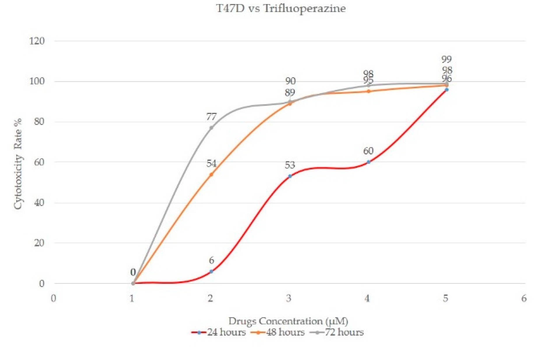
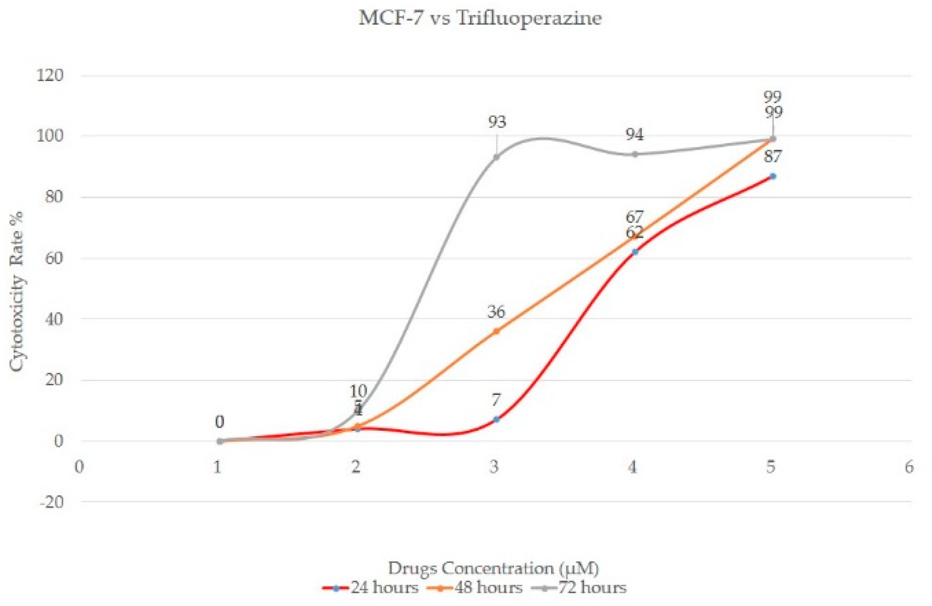
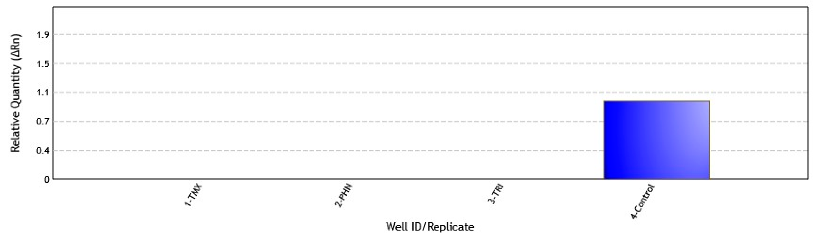
3. Results and Discussion

Calmodulin antagonists effectively reduced cell growth of both ER-positive and ER-negative human breast cancer cell lines. The IC50 values of CaM antagonists were between the ranges of 9 to 25µM as listed in Table 1, in which all calmodulin antagonists shows significant dose dependents decrease in survival fractions against breast cancer cell lines. The highest cytotoxicity rate (%) and low survival rate (%) were achieved after 72 h of calmodulin antagonist exposure. Among all three antagonists, Trifluoperazine shows the lowest dose dependent concentration against both positive and negative estrogenic breast cancer cell lines.
In Figure 1 and Figure 2, at 50 µM concentration the maximum cytotoxicity for 72 h incubation are 99% for Trifluoperazine against T47D and MCF-7, whereas the IC50 value were obtained at approximately 10 µM and 9 µM respectively. While as for MDAMB-231 cells, maximum cytotoxicity for 72 h incubation are 91% (Figure 3), whereas the IC50 value was obtained at approximately 19 µM. Preliminary findings indicates that, trifluoperazine shows more effective inhibition of cell growth in ER-negative cells as compared to ER-positive cells. As shown in Figure 3, cell cytotoxicity of MDAMB-231 cells starts from 10 µM with minimum of 28% of cell death as compared to ER-positive cells of MCF-7 and T47D cells which only shows minimum of cell death mechanisms starting from 20 µM (Figure 2 and Figure 3).
The molecular mechanism of Trifluoperazine in ER-negative cells was further determine via qPCR to determine calmodulin antagonist’s correlation towards gene expression correspond to highly mutated BRCA1 exon 11 gene. No expressions were observed in treated MDAMB-231 cells with calmodulin antagonists against control cells (untreated cells) (Figure 4). Results indicate that calmodulin antagonists able to suppress the expression of BRCA1 gene from undergo mutation specifically at exon 11 region which associated with the development and growth of breast cancer.

4. Conclusions

The findings suggested that calmodulin strongly involved in tumor cell growth and regulation in breast cancer. Preliminary finding shows the capabilities of Trifluoperazine in inhibiting the growth of breast cancer cells line as well suppressing the expression of BRCA1 exon 11 regions might be one of the promising therapeutic agents for the treatment of breast cancer other than depending towards endocrine therapy of Tamoxifen only. This is the first finding shows the correlation between BRCA1 gene expression towards cell cytotoxicity of Calmodulin antagonists in breast cancer. Further experimental are still carried out to fully determine the splicing expression of BRCA1 gene exons and its molecular pathway mechanism.

Acknowledgments

This work was supported by The Scientific and Technological Ministry of Malaysia (Project No. R/FRGS 2/2014). We also would like to thank the faculty of Medicine and Health sciences for their s logistic support.

References

  1. Siegel, R.L.; Miller, K.D.; Jemal, A.I. Cancer Statistics, 2017. CA Cancer J. Clin. 2017, 67, 7–30. [Google Scholar] [CrossRef] [PubMed]
  2. Torre, L.A.; Bray, F.; Siegel, R.L.; Ferlay, J.; Lortet-Tieulent, J.; Jemal, A. Global cancer statistics, 2012. CA Cancer J. Clin. 2015, 65, 87–108. [Google Scholar] [CrossRef] [PubMed]
  3. Cheung, WY. Cyclic 3',5’-nucleotide phosphodiesterase: pronounced stimulation by snake venom. Biochem. Biophys. Res. Commun. 1967, 29, 478–482. [Google Scholar] [CrossRef]
  4. Salih, F.A.M.; Bhatnagar, R.; Bhatnagar, N.; Singh, Y.; Murthy, P.S.; Venkitasubramanian, T.A. On the presence of Calmodulin-like protein in Mycobacteria. FEMS Microbiol. Lett. 1988, 56, 89–94. [Google Scholar]
  5. Salih, F.A.M.; Alabadi, R. Possible role of Calmodulin in gene activation and regulation. Short Communication (one-page preliminary result), Int. Med. J. Malaysia 2004, 3, 3. [Google Scholar]
  6. Audran, E.; Dagher, R.; Gioria, S.; Tsvetkov, P.O.; Kulikova, A.A.; Didier, B.; Villa, P.; Makarov, A.A.; Kilhoffer, M.C.; Haiech, J.A. General Framework to characterize inhibitors of calmodulin: use of calmodulin Inhibitors to study the interaction between calmodulin and its calmodulin binding domains. Biochim. Biophys Acta. 2013, 1833, 1720–1731. [Google Scholar] [CrossRef] [PubMed]
  7. Favy, D.A.; Lafarge, S.; Rio, P.; Vissac, C.; Bignon, Y.-J.; Bernard-Gallon, D.; Real-Time, P.C.R. Quantification of Full-Length and Exon 11 Spliced BRCA 1 Transcripts in Human Breast Cancer Cell Lines. Biochem. Biochem. Res. Commun. 2000, 274, 73–78. [Google Scholar] [CrossRef] [PubMed]
Figure 1. Cytotoxicity Rate in T47D cells after 24, 48 and 72 h exposure with Trifluoperazine. Cells are treated with various concentration starting with minimum dose of 10 µM to maximum dose of 50 µM respectively (1 = 10, 2 = 20, 3 = 30, 4 = 40 and 5 = 50 µM).
Proceedings 02 01550 g001
Figure 2. Cytotoxicity Rate in MCF-7 cells after 24, 48 and 72 h exposure with Trifluoperazine. Cells are treated with various concentration starting with minimum dose of 10 µM to maximum dose of 50 µM respectively (1 = 10, 2 = 20, 3 = 30, 4 = 40 and 5 = 50 µM).
Proceedings 02 01550 g002
Figure 3. Cytotoxicity Rate in MDAMB-231 cells after 24, 48 and 72 h exposure with Trifluoperazine. Cells are treated with various concentration starting with minimum dose of 10 µM to maximum dose of 50 µM respectively (1 = 10, 2 = 20, 3 = 30, 4 = 40 and 5 = 50 µM).
Proceedings 02 01550 g003
Figure 4. Relative quantity of BRCA1 exon 11 gene expression of MDAMB-231 cells against Calmodulin antagonists. 1-TMX (treated cells with Tamoxifen), 2-PHN (treated cells with Phenothiazine), 3-TRI (treated cells with Trifluoperazine) and 4-Control (untreated cells without antagonists).
Proceedings 02 01550 g004
Table 1. IC50 values of CaM antagonists (after 72 h exposure).
Calmodulin AntagonistsMCF-7T47DMDAMB-231
Tamoxifen10 µM13 µM25 µM
Phenothiazine11 µM14 µM20 µM
Trifluoperazine9 µM10 µM19 µM
Publisher’s Note: MDPI stays neutral with regard to jurisdictional claims in published maps and institutional affiliations.

