Proceeding Paper

Helicobacter Pylori Causes Oxidative Stress and Apoptosis in DNA Double Strand Break Repair Inhibited Human Gastric Adenocarcinoma Cells †

1
Department of Toxicology, Faculty of Pharmacy, Hacettepe University, 06100 Sıhhiye Ankara, Turkey
2
Department of Toxicology, Faculty of Pharmacy, Erzincan Binali Yildirim University, 24100 Erzincan, Turkey
3
Department of Medical Microbiology, Faculty of Medicine, Hacettepe University, 06100 Sıhhiye Ankara, Turkey
4
Department of Toxicology, Faculty of Pharmacy, Lokman Hekim University, Çankaya 06510 Ankara, Turkey
*
Author to whom correspondence should be addressed.
Presented at the 2nd International Cell Death Research Congress, Izmir, Turkey, 1–4 November 2018.
Proceedings 2018, 2(25), 1544; https://doi.org/10.3390/proceedings2251544
Published: 6 December 2018
(This article belongs to the Proceedings of The 2nd International Cell Death Research Congress)

Abstract

:
Helicobacter pylori, a helix-shaped gram-negative microaerophilic bacteria has been classified as a human carcinogen (Group I carcinogen) by the International Agency for Research on Cancer (IARC) on the basis of numerous animal and epidemiological studies. Chronic Helicobacter pylori infection can cause lead to high levels of intracellular reactive oxygen species (ROS) and genomic instability both directly by genetic and/or epigenetic pathways. It is known that high levels of intracellular ROS can trigger apoptosis and induce DSBs. In this study, we aimed to investigate intracellular ROS levels and apoptotic effects of Helicobacter pylori infection on AGS cells in the presence of a DSB repair inhibitor [8-(4-dibenzothienyl)-2-(4-morpholinyl)-4H-1-benzopyran-4-one, Nu7441]. After incubation of AGS cells with different multiplicities of infection (MOIs; 25, 50, 75, 100, 200 and 400), we observed that Helicobacter pylori causes MOI-dependent cytotoxicity and intracellular ROS levels significantly increased in all study groups vs. control. Both caspase 3 and caspase 8 levels were higher all of the study group when compared to control group. In HP+Nu7441 group, caspase 8 levels 3.5 times higher vs. control; indicating unrepaired DSBs in Helicobacter pylori infection can highly be induce apoptosis in AGS cells. Other cell death mechanisms, including autophagy, should be studies in order to fully understand the cytotoxicity mechanisms of the bacterium. Moreover, we can state that in organisms with unrepaired DSBs or with high DSB levels, Helicobacter pylori can cause more oppressive adverse effects, the importance of which should be elucidated with mechanistic studies.

1. Introduction

It is stated that over 15%–20% of malignancies worldwide can be related to infections caused by viruses, bacteria and schistosomes [1]. Helicobacter pylori is helix-shaped gram-negative microaerophilic bacteria [2]. It usually colonizes in upper gastrointestinal tract and the bacterium is strongly associated with development of peptic ulceration, gastric carcinoma and gastric lymphoma [3]. Helicobacter pylori has been classified as a human carcinogen (Group I carcinogen) by the International Agency for Research on Cancer (IARC) on the basis of numerous animal and epidemiological studies [3]. This bacterium has four major virulence factors; urease, the cytotoxin-associated gene product A (CagA), the vacuolating toxin A (VagA), the duodenal ulcer-promoting toxin A (DupA) and adhesion protein A (BabA). It has been demonstrated that, these toxins have major role in the Helicobacter pylori pathogenesis [4]. Apoptosis is a genetically controlled process in development and against chronic inflammation [5]. Besides, apoptosis occurs in response to several environmental chemicals. A variety of chronic infectious diseases can generate steady-state levels of intracellular reactive oxygen species (ROS). ROS within the infected cells may possibly lead to different types of DNA lesions. Accumulation of DNA lesions may finally lead to mutations that may activate oncogenes or inactivate tumor suppressor genes [6]. Helicobacter pylori has been shown to generate ROS/RNS, induce DNA damage and lead to chronic inflammation in gastric epithelial cells. Limited number of studies address the effects of Helicobacter pylori on DNA damage, particularly its impact on single strand and double strand DNA breaks (DSBs) [7]. The most dangerous DNA damage is DSB, which can be induced by several physical, chemical and biological agents. Chronic Helicobacter pylori infection can cause lead to high levels of intracellular ROS and genomic instability both directly by genetic and/or epigenetic pathways. It is known that high levels of intracellular ROS can trigger apoptosis and induce DSBs [8]. Several independent studies have suggested that Helicobacter pylori may lead to cytotoxicity and bacterial lipopolisaccharides have important roles on the apoptotic activity [3,4].
In this study, we aimed to investigate intracellular ROS levels and apoptotic effects of Helicobacter pylori infection on AGS cells in the presence of a DSB repair inhibitor [8-(4-dibenzothienyl)-2-(4-morpholinyl)-4H-1-benzopyran-4-one, Nu7441].

2. Materials and Methods
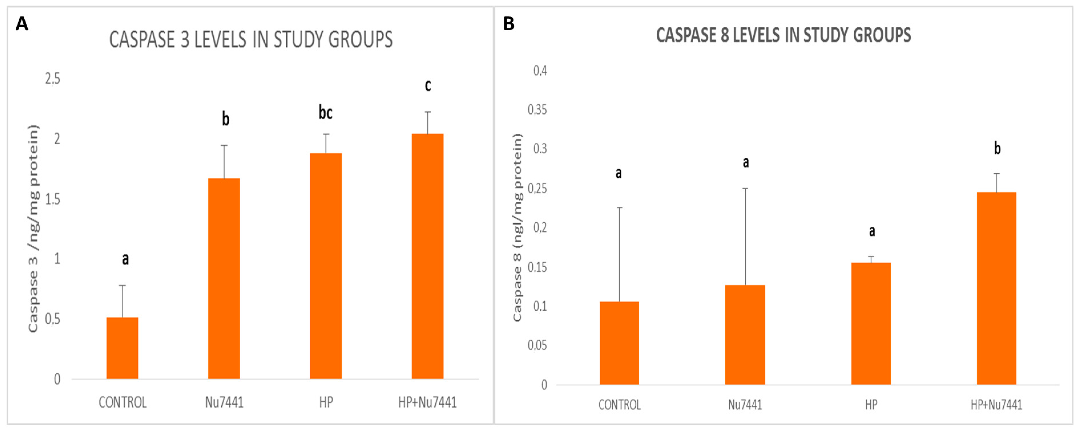
Helicobacter pylori (NCTC 11637) was grown under micro-aerobic conditions. After seeding the AGS cells (ATCC 1739, 10.000 cells in 90 µL DMEM) in a 96-well plate, cells were exposed to Helicobacter pylori strains with different multiplicities of infection (MOI: 25, 50, 75, 100, 150 and 200 bacterium per cell) for 24 h. Cell viability was assessed with 3-(4,5-dimethylthiazol-2-yl)-2,5-diphenyltetrazolium bromide (MTT) assay. The median inhibitory MOI (MOI50) was found to be 218 bacterium/live cell and MOI70 was 138 bacterium/live cell. Nu7441 was applied at a concentration of 10 µM in order to prevent DSB repair. The study groups were as follows: Control AGS cells (Control) Nu7441 treated AGS cells (Nu7441 group) AGS cells infected with Helicobacter pylori at MOI70 (HP group) AGS cells treated with Nu7441 and infected with Helicobacter pylori at MOI70 (HP+Nu7441 group)
ROS detection: The intracellular ROS detection kit is based on the conversion of the non-fluorescent probe 5-(and 6-)chloromethyl-2′,7′-dichlorodihydrofluorescein diacetate (CM-H2DCFA) to green-fluorescent chloromethyl-2′,7′-dichlorofluorescin (CM-DCF) by living cells. ROS reacts with a fluorogenic sensor contained in the cytoplasm to form a fluorometric product proportional to the amount of present ROS. Briefly, AGS cells (1 × 10³) were washed with 90 μL phosphate buffered saline (PBS). ROS Detection Reagent Stock Solution was prepared by mixing 40 μL DMSO with the ROS Detection Reagent. For control wells (untreated cells), only 10 μL PBS was added. MOI70 was used in all Helicobacter pylori-infected AGS cells. 96-well culture plate was incubated in a 5% CO2, 37 °C incubator for 30 min. Master reaction mixture (20 μL stock solution, 10 μL assay buffer) was prepared and 100 μL of this mixture was immediately added to each well. 96-well culture plate was incubated for 30 min. Fluorescence intensity was measured (λexitation = 640 nm, λemission = 675 nm) by using a spectrofluorometer (Spectramax M2, Molecular Devices, Sunnyvale, CA, USA). Control cells were accepted to have 100% ROS production. Results were expressed as the mean percentage of ROS production vs. control group.
Caspase 3 and Caspase 8 Levels: Caspase levels were evaluated by using commercial spectrophotometric kits. Results are expressed as ng/mL. The kits are based on the quantitative sandwich enzyme immunoassay technique. Basically, 100 μL AGS cell lysate or standard was added in pre-coated wells with human caspase-3 or caspase-8. After 90 min incubation at 37 °C, wells were washed out 2 times with washing buffer. Biotinylated human caspase-3 or caspase-8 antibody was added to each well and incubated 60 min at 37 °C. Plate was washed out with washing buffer and enzyme-conjugate liquid was added to each well. After 30 min incubation at 37 °C, plate was washed and color reagent and incubated at 37 °C until color gradient appear. The chromogenic reaction was controlled within 30 min and the reaction was stopped by adding stop solution to each well. The optic density of the color was measured at 450 nm.

3. Results

Helicobacter pylori causes MOI-dependent cytotoxicity. We found that ROS levels significantly increased in all study groups vs. control. Both caspase 3 and caspase 8 levels were higher in all of the study groups when compared to control group. In HP+Nu7441 group, caspase 8 levels 3.5 times higher vs. control. Besides, caspase 8 levels in HP+Nu7441 group were markedly higher than all other study groups (Figure 1). Our results are indicate that unrepaired DSBs in Helicobacter pylori infection can highly induce apoptosis in AGS cells.

4. Discussion

Different microorganisms can lead to intracellular toxicity after exposure to certain MOIs [8]. Helicobacter pylori is known to be carcinogenic to humans [2]. However, the intracellular toxicity mechanisms of this bacterium are not yet fully understood. The bacterium does not directly enter the cells; however its toxins can pass through cellular membranes leading to cytotoxicity and perhaps changes in cellular oxidant/antioxidant balance [3,4]. In this study, we have shown that Helicobacter pylori can lead to a MOI-dependent cytotoxicity in AGS cells. Moreover, we have observed that this bacterium can induce ROS, extrinsic caspase pathway and trigger apoptosis. Other cell death mechanisms, including autophagy, should be studied in order to fully understand the cytotoxicity mechanisms of the bacterium. Moreover, we can state that in organisms with unrepaired DSBs or with high DSB levels, Helicobacter pylori can cause more oppressive adverse effects and perhaps high incidence of carcinogenicity, the importance of which should be elucidated with mechanistic studies. Acknowledgments: This study is not yet supported by any organization and did not receive any funding.

Author Contributions

P.E. conceived and designed the experiments; D.O., Ü.S., G.Ö. and Y.A. performed the experiments; P.E. and B.G. analyzed the data, P.E. and D.O. wrote the paper.

Conflicts of Interest

The authors declare no conflict of interest.

References

  1. De Martel, C.; Ferlay, J.; Franceschi, S.; Vignat, J.; Bray, F.; Forman, D.; Plummer, M. Global burden of cancers attributable to infections in 2008: A review and synthetic analysis. Lancet Oncol. 2012, 13, 607–615. [Google Scholar] [CrossRef]
  2. Neelapu, N.R.R.; Nammi, D.; Pasupuleti, A.C.M.; Surekha, C. Helicobacter pylori ınduced gastric ınflammation, ulcer, and cancer: A pathogenesis perspective. Int. J. Inflamm. Cancer Integ. Ther. 2014, 1, 1000113. [Google Scholar]
  3. Toller, I.M.; Neelsen, K.J.; Steger, M.; Hartung, M.L.; Hottiger, M.O.; Stucki, M.; Kalali, B.; Gerhard, M.; Sartori, A.A.; Lopes, M.; et al. Carcinogenic bacterial pathogen Helicobacter pylori triggers DNA double-strand breaks and a DNA damage response in its host cells. Proc. Natl. Acad. Sci. USA 2011, 108, 14944–14949. [Google Scholar] [CrossRef] [PubMed]
  4. Paniagua, G.L.; Monroy, E.; Rodríguez, R.; Arroniz, S.; Rodríguez, C.; Cortés, J.L.; Camacho, A.; Negrete, E.; Vaca, S. Frequency of vacA, cagA and babA2 virulence markers in Helicobacter pylori strains isolated from Mexican patients with chronic gastritis. Ann. Clin. Microbiol. Antimicrob. 2009, 8, 14. [Google Scholar] [CrossRef] [PubMed]
  5. Elmore, S. Apoptosis: A review of programmed cell death. Toxicol. Pathol. 2007, 35, 495–516. [Google Scholar] [CrossRef] [PubMed]
  6. Jena, N.R. DNA damage by reactive species: Mechanisms, mutation and repair. J. Biosci. 2012, 37, 503–517. [Google Scholar] [CrossRef] [PubMed]
  7. Erkekoglu, P.; Oral, D.; Kocer-Gumusel, B.; Chao, M.W. DNA Double-Strand Breaks Caused by Different Microorganisms: A Special Focus on Helicobacter pylori. J. Environ. Pathol. Toxicol. Oncol. 2017, 36, 131–150. [Google Scholar] [CrossRef] [PubMed]
  8. Jackson, S.P.; Bartek, J. The DNA-damage response in human biology and disease. Nature 2009, 461, 1071–1078. [Google Scholar] [CrossRef] [PubMed]
Figure 1. (A) Caspase-3 levels in the study groups. (B) Caspase-8 levels in the study groups. a,b,c Bars that do not have the same superscripts are significantly different than each other. t-test was used to compare the study groups. p < 0.05 is considered to be statistically significant.
Proceedings 02 01544 g001
Publisher’s Note: MDPI stays neutral with regard to jurisdictional claims in published maps and institutional affiliations.

