Proceeding Paper

A Reliable and Sensitive Method Using Cyclic Voltammetry for the Detection of Airborne Fungi  †

1
Department for Integrated Sensor Systems, Center for Water and Environmental Sensors, Danube University Krems, 3500 Krems, Austria
2
Division Microbiological Diagnostics for Water and Health, Physiology und Microbiological Diagnostics, Karl Landsteiner University of Health Sciences, 3500 Krems, Austria
3
IMC FH Krems, University of Applied Sciences, 3500 Krems, Austria
*
Author to whom correspondence should be addressed.
Presented at the Eurosensors 2018 Conference, Graz, Austria, 9–12 September 2018.
Proceedings 2018, 2(13), 959; https://doi.org/10.3390/proceedings2130959
Published: 29 November 2018
(This article belongs to the Proceedings of EUROSENSORS 2018)

Abstract

:
Fungi are widespread throughout the environment and can cause health as well as indoor problems. One of the most common fungi involved in building damage are Aspergillus sp. This study describes a rapid method for the detection of fungal spores. It is based on the ability of fungi to produce the enzyme cellulase, which can cleave the substrate Aminophenyl-β-d-cellobioside (APC) resulting into 4-Aminophenyl (AP). This cleavage product can then be oxidized leading to an increased output current signal. In the experimental setup several growth media and different parameters were tested over time, for instance growth conditions like pH or incubation temperature. Furthermore, various spore concentrations, which are related to the cellulase activities, were examined. This method presents a technique that makes it possible to specifically detect low concentrations of fungi spores in short time.

1. Introduction

Fungi are responsible for the biological decay of building materials and works of art. Furthermore, microbes, especially fungi and their metabolic products can have serious implications on human health [1]. Nowadays, fungal contaminations are examined using airborne microorganism samplers collecting fungal spores or by taking directly samples from the contaminated materials. The following cultivation takes several days to finally make quantitative statements.
Aspergillus niger is known for its biodeteriorative effects on cultural artefacts, which can lead to serious threats for objects. Therefore, the aim of this study was the development of a method detecting airborne cellulolytic fungi using cyclic voltammetry as a measurement method. The optimized methodology should further be integrated into a biosensor for a fast and sensitive detection of indoor fungal contaminations.
The electrochemical detection of cellulolytic fungi is based on the ability of the fungi to produce a specific enzyme, called cellulase. Furthermore, the reaction of this enzyme with the substrate 4-Aminophenyl-β-d-cellobioside (APC) leads to the hydrolyzed and electroactive product 4-Aminophenyl (AP). AP is then oxidized on an electrode using a potentiostat resulting in an increase of current at a specific potential range [2,3].

2. Materials and Methods

At first, the general principle of the method was examined. Therefore, pure fungal cellulase-solutions were tested at various concentrations with different types of substrates, to find out whether the cellulase is capable of utilizing one of the substrates. The most promising, APC, was used for further studies. A cellulase solution (100 units) was incubated with the APC substrate solution, leading to an increase of current signal at a potential range between 0 and 250 mV (see Figure 1). For the electrochemical analysis, screen-printed electrodes (Gwent Electronic Materials Ltd., Pontypool, UK) were used. Cyclic voltammetry (CV) was used for all experiments using an applied potential ranging from 0 to 1500 mV, a step size of 1 mV and a scan speed of 50 mV s−1. All measurements were performed at room temperature with a working volume of 100 µL that ensured complete coverage of all three electrode areas. CV measurements were performed using the Gamry Reference 600+ (Gamry Instruments, USA) as well as an instrument developed at our working group [4].
After verification of the general principle of the assay, first optimization experiments were performed. Therefore, different parameters of the setup, like pH and growth media composition were evaluated before a range of incubation temperatures (22 to 37 °C) was tested (see Figure 2) [5]. Afterwards, several fungal concentrations of Aspergillus niger (3000, 300 and 30 colony forming units, CFU Ml1) as well as the influence of possible contaminating bacteria, e.g. Pseudomonas stutzeri, on the assay were tested (see Figure 3).

3. Results and Discussion

3.1. Proof of Principle

The data presented in Figure 1 indicate that the enzyme is capable of cleaving the substrate APC leading to the electroactive product AP. The oxidation process of the cleavage product on the screen-printed electrode leads to a distinct increase of current between 0–250 mV.

3.2. Temperature Optimization

The results in Figure 2 show that the temperature plays an important role in the enzyme production and its activity, since the samples incubated at 37 °C already gave a signal after 24 h. Furthermore, considerably higher current responses could be measured for the cellulose solution as well as the fungi incubated at 37 °C compared to samples analyzed at room temperature.

3.3. Specificity and Cross Reactivity

The data in Figure 3 reflect that it was possible to specifically detect approximately 300 CFU of spores of A. niger after 26 h of incubation. This signal continuously increased over the next hours of incubation from 700 nA to 3.45 µA after 32 h. For the sample containing 30 CFU, a longer incubation period was necessary to observe a low current signal. After 32 h the fungal sample delivered a current signal of 400 nA. The tested bacterium Pseudomonas stutzeri did not produce an increase in output current. Therefore, it can be concluded that bacteria, which do not bear the ability of cellulase production and cannot metabolize APC, will not display a current output signal. Furthermore, the correlation between fungal growth, which is linked to time of incubation and intensity of the signal, could be clearly observed.
In conclusion, the technique described, represents a reliable method for an easy and fast detection of cellulolytic airborne fungi. Future experiments aim to evaluate the general specificity within the Kingdom Fungi, cross reactivity and detection limits with other cellulolytic fungi under laboratory as well as real life conditions. Finally, our goal is to integrate the designed methodology into a miniaturized and automated biosensor system as an early warning system for detection of airborne fungi and fungal spores.

Acknowledgments

The authors thank the Danube University Krems for their financial support.

Conflicts of Interest

The authors declare no conflict of interest. The founding sponsors had no role in the design of the study; in the collection, analyses, or interpretation of data; in the writing of the manuscript, and in the decision to publish the results.

References

  1. Commission of the European Communities. Biological Particles in Indoor Environments. In European Collaborative Action—Indoor Air Quality & its Impact on Man; Report No. 12; Commission of the European Communities: Brussels, Belgium, 1993. [Google Scholar]
  2. Cruys-Bagger, N.; Tatsumi, H.; Borch, K.; Westh, P. A graphene screen-printed carbon electrode for real-time measurements of unoccupied active sites in a cellulase. Anal. Biochem. 2014, 447, 162–168. [Google Scholar] [CrossRef] [PubMed]
  3. Kubicek, C.P.; Mikus, M.; Schuster, A.; Schmoll, M.; Seiboth, B. Metabolic engineering strategies for the improvement of cellulase production by Hypocrea jecorina. Biotechnol. Biofuels 2009, 2, 19. [Google Scholar] [CrossRef] [PubMed]
  4. Kellner, K.H.; Posnicek, T.; Ettenauer, J.; Zuser, K.; Brandl, M. A new, low-cost potentiostat for environmental measurements with an easy-to-use PC interface. Procedia Eng. 2015, 120, 956–960. [Google Scholar] [CrossRef]
  5. Sohail, M.; Siddiqi, R.; Ahmad, A.; Khan, S.A. Cellulase production from Aspergillus niger MS82: Effect of temperature and pH. New Biotechnol. 2009, 25, 437–441. [Google Scholar] [CrossRef] [PubMed]
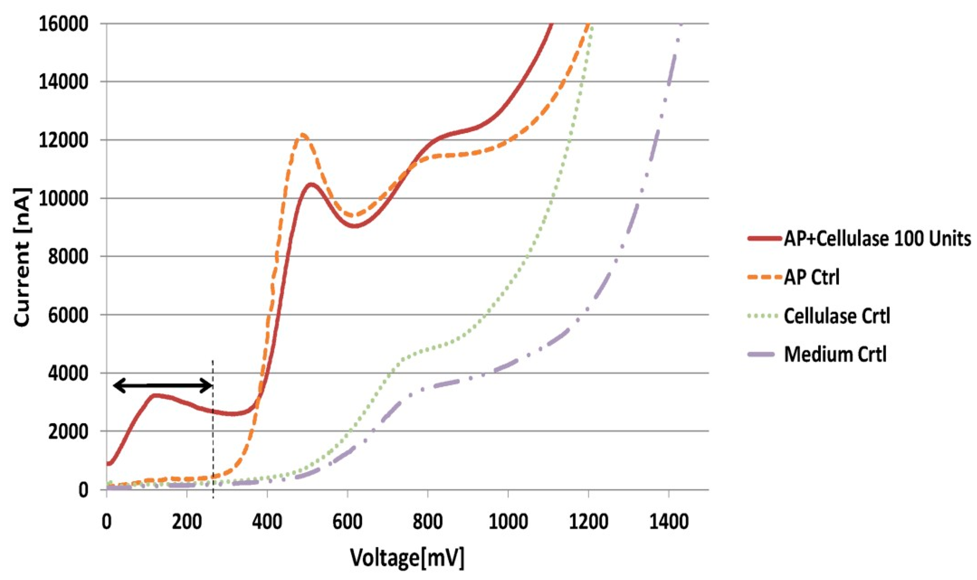
Figure 1. Evaluation of the capability of the cellulase (100 Units) to cleave the electroactive substrate APC (1 mM) after 300 min incubation (including different controls—Crtl) at a specific current rage, indicated with arrows in the figure.
Proceedings 02 00959 g001
Figure 2. Investigation of the influence of temperature on the production of cellulase through the fungi (106 spores/mL) and on the activity of the enzyme (25 Units). The mean values of the current signal [nA] between 0–250 mV are shown for all samples over the total measurement time range (24–32 h).
Proceedings 02 00959 g002
Figure 3. Mean output signals (between 0–250 mV) of different concentrations of Aspergillus niger spores (3000 CFU, 300 CFU and 30 CFU) and the bacterium Pseudomonas stutzeri (1.9 × 10⁸ CFU) including 1 mM APC.
Proceedings 02 00959 g003
Publisher’s Note: MDPI stays neutral with regard to jurisdictional claims in published maps and institutional affiliations.