1. Introduction
Recent advancements in dental adhesive technology have demonstrated the significant benefits of incorporating magnetic nanoparticles (MNPs) into resin adhesives. A pioneering study revealed that MNP-doped adhesives, when activated by an external magnetic field, enhanced dentin bond strength by 59% compared to conventional systems. This improvement was attributed to increased resin infiltration, evidenced by longer and denser resin tags [1]. The MNPs, due to their nanoscale size, effectively penetrated dentinal tubules and interfibrillar spaces, promoting hybrid layer integrity and minimizing microleakage [2]. Additionally, magnetic field application reduced adhesive layer thickness by up to 86.5%, depending on exposure duration. These findings support the potential of magnetically guided nanotechnology to improve the performance and longevity of dental restorations [3].
Iron oxide MNPs have shown potential to inhibit biofilm formation, particularly when used in conjunction with antimicrobial agents. These findings pave the way for the development of bioactive restorative materials with intrinsic antibacterial properties, aimed at preventing restoration failure due to secondary caries and enhancing the long-term clinical success of dental treatments [4].
2. Materials and Methods
2.1. MNP Synthesis
The functionalized magnetic nanoparticles (MNPs) were synthesized via a multi-step procedure comprising three environmentally friendly and cost-effective stages.
Fe3O4 MNPs were synthesized by coprecipitating Fe(II) and Fe(III) salts in basic medium. Fe3+:Fe2+ = 2:1 molar ratio, were dissolved in 200 mL of distilled water, and 200 mL of NH3 solution (25%) was added under vigorous stirring at 70 °C under continuous flow of argon. After 2h at 70 °C at the pH of 12, the system was cooled at room temperature, the precipitate was magnetically separated and washed with distilled water to a neutral pH.
Silica-coated nanoparticles (MNPs-SiO2) were synthesized by the hydrolysis of the tetraethyl orthosilicate (TEOS) in basic medium on the surface of the MNPs; 320 mL ethanol and 80 mL water were mixed with 0.4g MNPs, and the solution was sonicated for 10 min. Then it was mixed with 8 mL aqueous ammonia solution and 6 mL TEOS, added dropwise under stirring. The reaction was allowed to continue at room temperature for 1 h, under magnetic stirring; then, the product was separated using an external magnet and washed several times with distilled water. The final product was collected and dried at 60 °C for 12 h.
Dual-coated nanoparticles (MNPs-SiO2-Ca(OH)) were obtained by covering MNPs-SiO2 with calcium hydroxide. First, 94.5 g of calcium nitrate tetrahydrate, Ca(NO3)2 × 4 H2O was dissolved in distilled water (8.5 wt.%) and 1 g of MNPs-SiO2 were added. Then, a solution of NaOH (16 g, 1.6%) was added dropwise, with an addition rate of 1 mL/min, under magnetic stirring at 1200 rpm at room temperature. After 3 h the solution was filtered, and the resulting precipitate was washed 3 times successively with 100 mL of water then dried in the oven for 1 h at 60 °C [5]. The double-layered surface functionalization of the MNPs with SiO2 and Ca(OH) provides chemical inertness needed to prevent interference with the adhesive’s photopolymerization.
2.2. Photocolorimetry
Four groups of artificial teeth were prepared (n = 5 each). Group 0 (G0) served as the control and was restored using a conventional dental adhesive, Single Bond 2 (3M ESPE, Two Harbors, MN, USA), sourced in Romania. G1 used the same adhesive supplemented with MNPs-SiO2. G2 involved the adhesive loaded with MNPs-SiO2-Ca(OH). G3 was treated with the adhesive mixed with uncoated MNPs. The teeth were prepared for similar class I cavities of 4 mm depth. The magnetic dental adhesive was obtained by mixing one curette of MNP powder and one drop of adhesive for 15 s to obtain a homogenous mixture.
Group 0 samples were prepared following the standard protocol for direct restorations. An etching gel was applied for 30 s, and the surface was thoroughly rinsed and dried. Single Bond 2 adhesive was then applied and gently brushed onto the cavity surface for 15 s. To eliminate the solvent, air was blown over the surface for 5 s. The adhesive was light-cured for 10 s and composite resin was applied in increments; each layer was light-cured for 20 s until the cavity was filled.
For Groups 1, 2, and 3, the cavity surface was etched with gel for 30 s, rinsed, and dried; the adhesive containing magnetic nanoparticles (MNPs) was applied and brushed onto the surface, and air blown for 5 s; then, a permanent magnet was positioned as close as possible to the sample and held in place for 2 min. to attract and align the MNPs; the adhesive was light-cured for 10 s; and composite layers were added and light-cured for 20 s each until the cavity was fully restored. Finally, we removed 2 mm from the occlusal surface to prepare a flat surface for colorimetric testing.
To analyze color differences between the adhesive layer and the bulk of the restoration, samples from each group were immobilized in dental impression material (Zetaplus, Zhermack, Badia Polesine, Italy) and photographed, on a gray background, in polarized light, using a DSLR D5300 camera (Nikon, Tokyo, Japan) with an MK-14EXT flash (Meike, Chengdu, China) [6]. Both the flash and the camera’s objective were covered by polarizing filters with mutually perpendicular axes of transmission. Each digital picture was processed in IrfanView version 4.56 (https://www.irfanview.com/64bit.htm, accessed on 2 July 2025) using Jazzman’s White Balance I filter version 0.1(http://www.westbrookjazz.de/filter/f0.html, accessed on 2 July 2025) to render the background neutral gray. Then, the image was displayed in IrfanView and the corresponding CIELAB coordinates were assessed, as averages of 3 × 3 pixel domains, using the Digital Colorimeter software (https://apps.microsoft.com/detail/digital-colorimeter-precise-pixel-color-picker/9MWT4TWC53MH?hl=en-US&gl=US, accessed on 2 July 2025). For each sample, 12 locations were measured on the image of the adhesive layer as well as on the image of the restoration, halfway between its center and periphery (Figure 1A). A typical measurement is shown in Figure 1B.
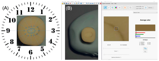
Figure 1.
Digital photocolorimetry. (A) The set of 12 points assessed on the image of the adhesive layer (circles) and on that of the composite filling (squares); (B) snapshot of a representative measurement in a square region of 3 × 3 pixels centered on the adhesive layer’s image.
3. Results and Discussion
We evaluated whether the adhesive layer differs in color from the bulk of the restoration. The Bland–Altman plots from Figure 2 indicate that individual chromatic differences between the adhesive layer and the bulk are larger for MPN-laden adhesives (G1–G3) than for the commercial adhesive (G0).
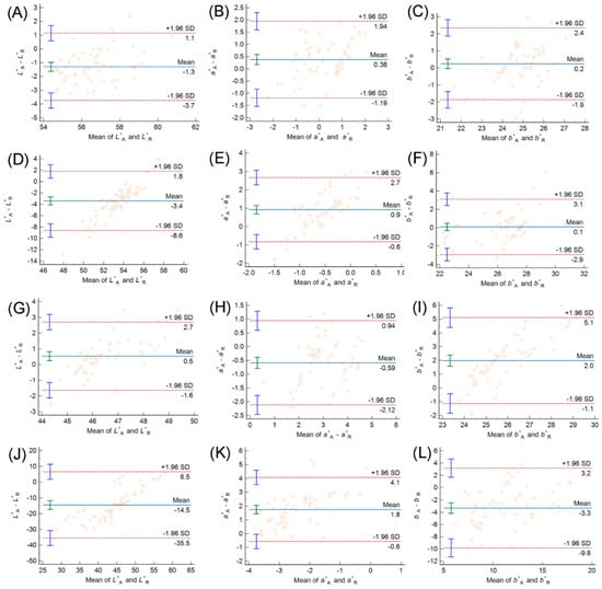
Figure 2.
Bland–Altman analysis of differences vs. means of CIELAB color space coordinates of the adhesive layer (A) and the bulk of the restoration (R) evaluated by digital photocolorimetry: (A–C) Group 0; (D–F) Group 1; (G–I) Group 2; (J–L) Group 3.
Figure 2G,H indicate that G2 is closest in color to the bulk of the restoration. This result agrees with the spectrophotometric assessment of MNP powders with SiO2 and dual coatings [5].
The Bland–Altman analysis of Figure 2 also reveals differences between individual regions of the same material. In each plot, the interval of agreement, delimited by the red dashed lines, includes about 95% of the differences between the color coordinates of pairs of evaluated points. The wide intervals of agreement observed for G1 and G3 suggest that the adhesive loaded with MNPs-SiO2 or with uncoated MNPs is more heterogeneous chromatically than the commercial adhesive (G0) or that augmented with MNPs-SiO2-Ca(OH).
In conclusion, MNP coating is an effective method for masking the dark color of iron oxide nanoparticles. Future research should explore the mechanical properties of the augmented dental adhesives and the long-term stability of the coating layers in the oral environment.
Author Contributions
Conceptualization, C.S. and V.-F.D.; methodology, R.T., I.C. and V.M.S., software, R.-A.T.; validation, L.-M.N.; formal analysis, R.T., I.C. and V.M.S.; investigation, C.-S.N.; resources, R.-A.T.; data curation, C.-S.N. and C.S.; writing—original draft preparation, C.-S.N., L.-M.N. and C.S.; writing—review and editing, V.-F.D.; visualization, L.-M.N.; supervision, C.S. and V.-F.D. All authors have read and agreed to the published version of the manuscript.
Funding
This research was funded by the “Victor Babes” University of Medicine and Pharmacy of Timisoara.
Institutional Review Board Statement
Not applicable.
Informed Consent Statement
Not applicable.
Data Availability Statement
The data supporting the findings of this study are available from the corresponding author upon reasonable request.
Conflicts of Interest
The authors declare no conflict of interest.
References
- Ji, Y.; Choi, S.K.; Sultan, A.S.; Chuncai, K.; Lin, X.; Dashtimoghadam, E.; Melo, M.A.; Weir, M.; Xu, H.; Tayebi, L.; et al. Nanomagnetic-mediated drug delivery for the treatment of dental disease. Nanomed. Nanotechnol. Biol. Med. 2018, 14, 919–927. [Google Scholar] [CrossRef] [PubMed]
- Langer, A.; Ilie, N. Dentin infiltration ability of different classes of adhesive systems. Clin. Oral Investig. 2012, 17, 205–216. [Google Scholar] [CrossRef] [PubMed]
- Zaharia, C.; Sinescu, C.; Gabor, A.-G.; Socoliuc, V.; Talpos, S.; Hajaj, T.; Sfirloaga, P.; Oancea, R.; Miclau, M.; Negrutiu, M.-L. Influences of Polymeric Magnetic Encapsulated Nanoparticles on the Adhesive Layer for Composite Materials Used for Class I Dental Fillings. Mater. Plast. 2019, 56, 399–404. [Google Scholar] [CrossRef]
- Wysokińska-Miszczuk, J.; Piotrowska, K.; Paulo, M.; Madej, M. Composite Materials Used for Dental Fillings. Materials 2024, 17, 4936. [Google Scholar] [CrossRef] [PubMed]
- Crăciunescu, I.; Ispas, G.M.; Ciorîta, A.; Turcu, R.P. Functionalized Magnetic Nanomaterial Based on SiO2/Ca(OH)2-Coated Clusters Decorated with Silver Nanoparticles for Dental Applications. Crystals 2025, 15, 615. [Google Scholar] [CrossRef]
- Bezerra, A.; Oshima, S.; Feldmann, A.; Tango, R.N.; Duque, T.; Philippi, A.; Gonçalves, T. Digital Photocolorimetric Analysis of In Vitro Tooth Color Changes. Oper. Dent. 2024, 49, 336–344. [Google Scholar] [CrossRef] [PubMed]
Disclaimer/Publisher’s Note: The statements, opinions and data contained in all publications are solely those of the individual author(s) and contributor(s) and not of MDPI and/or the editor(s). MDPI and/or the editor(s) disclaim responsibility for any injury to people or property resulting from any ideas, methods, instructions or products referred to in the content. |
© 2025 by the authors. Licensee MDPI, Basel, Switzerland. This article is an open access article distributed under the terms and conditions of the Creative Commons Attribution (CC BY) license (https://creativecommons.org/licenses/by/4.0/).