Article

Bovine Leptospirosis in Caatinga Biome, Brazil: New Insights into Diagnosis and Epidemiology

by
Nathanael Natércio da Costa Barnabé
1,
Rafael Rodrigues Soares
1,
Deivyson Kelvis Silva Barros
1,
Denise Batista Nogueira
2,
Flávia Teresa Ribeiro da Costa
1,
João Pessoa Araújo Júnior
3,
Camila Dantas Malossi
3,
Leila Sabrina Ullmann
3,
Diego Figueiredo da Costa
4,
Maria Luana Cristiny Rodrigues Silva
1,
Severino Silvano dos Santos Higino
1,
Carolina de Sousa Américo Batista Santos
1,
Sérgio Santos de Azevedo
1,* and
Clebert José Alves
1
1
Academic Unit of Veterinary Medicine (UAMV), Federal University of Campina Grande (UFCG), Patos 58708-110, Brazil
2
Faculty of Veterinary Medicine and Zootechny (FMVZ), University of São Paulo (USP), São Paulo 05508-220, Brazil
3
Institute of Biosciences, Department of Microbiology and Immunology, University of the São Paulo State (UNESP), Botucatu 18618-687, Brazil
4
Department of Veterinary Science, Federal University of Paraíba (UFPB), Areia 58397-000, Brazil
*
Author to whom correspondence should be addressed.
Trop. Med. Infect. Dis. 2023, 8(3), 177; https://doi.org/10.3390/tropicalmed8030177
Submission received: 3 January 2023 / Revised: 10 March 2023 / Accepted: 10 March 2023 / Published: 17 March 2023

Abstract

:
Bovine leptospirosis causes economic losses and raises public health concerns. It is possible that there are peculiarities in the epidemiology of leptospirosis in regions with a semiarid climate, such as the Caatinga biome in Brazil, where the climate is hot and dry, and the etiological agent require alternative routes of transmission. This study aimed to close knowledge gaps to the diagnosis and epidemiology of Leptospira spp. infection in cows from the Caatinga biome, Brazil. Samples of the blood, urinary tract (urine, bladder and kidney) and reproductive tract (vaginal fluid, uterus, uterine tube, ovary and placenta) were collected from 42 slaughtered cows. Diagnostic tests included were the microscopic agglutination test (MAT), polymerase chain reaction (PCR) and bacterial isolation. Anti-Leptospira spp. antibodies were found in 27 (64.3%) of the animals analyzed using MAT at a 1:50 dilution (cut-off 50), while 31 (73.8%) animals had at least one organ/fluid where the presence of Leptospira spp. DNA was identified, and 29 animals (69%) were positive at bacteriological culture. The highest sensitivity values for MAT were obtained at the cut-off point of 50. In conclusion, even under hot and dry climate conditions, it is possible that Leptospira spp. can spread through alternative routes such as venereal transmission; moreover, a cut-off of 50 is recommended for the serological diagnosis of cattle from the Caatinga biome.

1. Introduction

Leptospirosis is widespread globally and is among the diseases that impair livestock production rates. It raises public health concerns because of its zoonotic impact and that, even after more than a century of research, the vaccines available do not confer lasting immunity or cross-protection between serovars [1]. The agent, pathogenic Leptospira species, has several hosts, and exposure ensues through direct contact with infected animals or indirectly via water and soil contaminated with urine [2,3]. Transmission can also occur through contact with the vaginal fluid and placental remains, copulation and also vertically [4]. The losses in livestock production result from abortions, stillbirth, weak offspring, diminished growth rate, diminished milk production or agalactia and death [5]. In cattle, this disease is more associated with subfertility and early embryonic death when the serovars is host-adapted, such as serovar Hardjo [6]. Studies conducted in the semiarid region of Brazil have suggested that both domestic [7,8,9,10,11,12] and wild animals [13,14] present high frequencies of Leptospira spp. This occurs despite the hot and dry climate conditions of this region, which are adverse to the survival of this bacterium in the environment.
Among the methods for diagnosing leptospirosis, there are indirect methods that investigate the immune response of the host and direct methods that target the detection of leptospires or their DNA. The microscopic agglutination test (MAT) has been recommended as a serological test by the World Organization for Animal Health (OIE) because of its capacity to demonstrate the most likely infecting serogroup [15]. Moreover, polymerase chain reaction (PCR) is a fast technique with high sensitivity and specificity and is, therefore, very reliable [16,17], although microbiological isolation is considered to be the gold standard [18,19]. The paired use of these diagnostic methods increases the capacity for detecting a positive animal in order to cover all situations where leptospires are not shed in urine, or chronic phase where antibody titers can be low; in addition, microbiological culture and isolation is still the only way to confirm the autochthonous Leptospira spp. serovars.
Given that there are differences in incidence related to climatic conditions [20] and to individual adaptation to the agent, such that they remain serologically unidentified, the test protocol that fits best needs to be established so as to ensure greater diagnostic accuracy. Sheep maintained in semiarid conditions have shown resistance to Leptospira spp. as well as short seroconversion periods [21,22]. Furthermore, surveys have found that the MAT had better performance with a cut-off point of 50, considering PCR and bacteriological culturing as gold standards [10,12]. Thus, the selection of an appropriate cut-off point for serology is relevant to increasing sensitivity and avoiding false-negative results. However, surveys focusing on improving the serology cut-off points applied to the diagnosis of cattle leptospirosis are lacking concerning Brazilian semiarid regions.
Low rainfall and high temperatures characterize the semiarid region of Brazil, and when associated with the peculiarities of the existing vegetation, the Caatinga, a biome exclusive to Northeastern Brazil with abundant wild fauna—such as tamanduá-mirim (Tamandua tetradactyla), preá (Cavia aperea), mocó (Kerodon rupestris) and cachorro-do-mato (Cerdocyon thous), offers unique epidemiological conditions that can influence the occurrence of infectious diseases such as leptospirosis. Therefore, this study aimed to evaluate the serological, molecular and microbiological methods applied to the diagnosis of leptospirosis in cows from the Caatinga biome in Northeastern Brazil.

2. Materials and Methods

2.1. Study Area

Biological samples were collected from the public slaughterhouse of Patos county (latitude: 7°00′19″ south; longitude: 37°16′48″ west) in the state of Paraíba, Northeastern Brazil. The animals originated from areas located in the Caatinga biome and were slaughtered within a maximum period of two days. This biome is an exclusively Brazilian biome and has a semiarid climate characterized by long periods of water scarcity and stunted vegetation. The climate is hot and dry, with a rainy season in summer/autumn and rainfall concentrated between March and April. However, precipitation may occur at any time between January and May. Droughts can last for more than a year, resulting in a negative water balance plus high solar radiation [23,24]. The period during which the present study was conducted corresponded to the dry season, with average rainfall and temperature of 0.47 mm and 29.28 °C, respectively [25].

2.2. Sampling

The minimum sample size was determined using the following formula for proportion analysis [26]:
n = p 0 × q 0 × Z 1 β + Z α 2 × p 1 × q 1 p 0 × q 0 2 p 1 p 0
where
n = minimum sample size
Zα/2 = 1.96 (Z value for 95% confidence level)
Z1−β = 1.64 (Z value for power of 95%)
P0 = 33% (reference proportion for PCR positivity) [27]
P1 = 61.40% (estimate for the experimental proportion of positivity in PCR) [28]
q0 = 1−p0
q1 = 1−p1
According to these parameters, a minimum of 37 animals were needed to assess true prevalence within a confidence interval of 95%; however, 42 cattle were used. They were all healthy female, aged greater than or equal to 24 months, cross-bred and had no history of vaccination against leptospirosis. According to the data taken from animal movement forms held by the State Veterinary Service of Paraíba, these female cattle came from rural farms located in the semiarid region, from municipalities belonging to two federative units: Paraíba (Cacimba de Areia, Condado, Olho D’Água, Patos, Pombal, Santa Terezinha, São José de Espinharas, São José do Bonfim and São Mamede) and Pernambuco (Buíque, Capoeiras and Exu).

2.3. Biological Sample Collection

The blood samples were collected from jugular vein using 8 mL labeled sterile tubes containing a coagulation activator just prior to slaughter of the animals. After collection, the tubes were sent to the laboratory, where they were centrifuged at 1512 g for 10 min, and the serum samples were stored in microtubes at −20 °C.
The bladder, kidney, ovary, uterine tube, uterus and placenta (placentome region) were collected for the direct detection of Leptospira spp. The tissues were immediately fragmented by using autoclaved surgical materials and sterile slides for each tissue. After that, the fragments were immediately transferred to a specific room in the slaughterhouse, where there was a Bunsen burner, and then they were deposited onto autoclaved Petri dishes while avoiding contact between the fragments. The material was fragmented into smaller portions of ≈ 2 gm (in duplicate), placed into DNA and RNA-free microtubes and stored at −20 °C for molecular detection and bacteriological isolation. Sterile swabs were used to collect vaginal fluid directly from the cervical–vaginal region, and urine was taken by cystocentesis during evisceration, using 5 mL sterile syringes. Both materials were also stored in duplicates in DNA- and RNA-free microtubes with 0.5 mL of phosphate-buffered saline [10].

2.4. Microscopic Agglutination Test (MAT)

The microscopic agglutination test (MAT) was used to detect anti-Leptospira spp. antibodies using a collection of 24 serovars belonging to 17 different pathogenic serogroups of five species provided by the Laboratory of Veterinary Bacteriology of the Fluminense Federal University, Niterói, Rio de Janeiro, Brazil, originating from the Pasteur Institute, France. The Leptospira species and serovars were L. interrogans serovars Copenhageni, Canicola, Autumnalis, Wolffi, Hardjoprajitno, Icterohaemorrhagiae, Pomona, Kennewicki, Hebdomadis, Pyrogenes, Bratislava and Australis; L. santarosai seorovars Guaricura, Shermani and Canalzoni; L. borgpetersenii serovars Javanica, Tarassovi, Ballum, Mini and Castellonis; L. kirschneri serovars Grippotyphosa and Cynopteri; L. noguchi serovars Panama and Lousiana [15]. The sera were screened at a 1:50 ratio, and the positive ones were two-fold diluted, in which the final result was the respective highest titer achieved, making use of the ranking technique [16].

2.5. Microbiological Culture

Immediately after the collection, three drops of urine, the swab with vaginal fluid and approximately 2 gm of each tissue were inoculated into tubes containing 5 mL of liquid EMJH medium (Difco, BD Franklin Lakes, NJ, USA) with amphotericin B (0.05 mg/mL), 5-fluorouracil (0.1 mg/mL), fosfomycin (0.4 mg/mL), trimethoprim (0.2 mg/mL) and sulfamethoxazole (0.4 mg/mL) [17]. After 24 h, 1 mL of the primary culture was inoculated into a semi-solid EMJH medium without antibiotics, and then incubated in a biological oxygen demand incubator (BOD) at 30 °C. Regardless of the presence of Dinger ring zone, the tubes were examined weekly for 12 weeks using dark-field microscopy.

2.6. Leptospira spp. DNA Detection and Sequencing

The Dneasy Blood and Tissue Kit (Qiagen, Hilden, Germany) was used to extract the DNA from the urine, vaginal fluid and tissues, as well as vaginal fluid and urine EMJH cultures that exhibited the growth of leptospires under dark-field microscopy examination, according to the manufacturer’s recommendations. The gene LipL32, specific to pathogenic leptospires and, therefore, of public health importance, was amplified with LipL32-45F (5′-AAG CAT TAC CGC TTG TGG TG-3′) and LipL32-286R (5′-GAA CTC CCA TTT CAG CGA TT-3′) primers [29] following the procedures for polymerase chain reaction (PCR) previously described [18]. Primers were used in a concentration of 0.6 μM, 1.0 U Taq polymerase, 2.4 μM MgCl2, 0.3 mM dNTP in a final volume of 25 μL. One cycle of initial denaturation at 94 °C for two minutes, followed by 35 cycles of denaturation at 94 °C for 30 s, annealing the primers to 53 °C for 30 s and one minute extension with 72 °C and final extension cycle at 72 °C for five minutes were used. PCR products were developed by 2% ultrapure agarose gel electrophoresis stained with Evans Blue (Thermo Fisher Scientific, Waltham, MA, USA) and 100 bp ladder, and DNA bands (≅260 bp) were visualized under ultraviolet light. Strain Leptospira interrogans serovar Copenhageni, Fiocruz L1-130 (ATCC BAA-1198) was used as positive control, and ultrapure water was used as a negative control.
LipL32-45F and LipL32-286R primers [29] were used in the sequencing reactions with the Sequencing Kit Big Dye Terminator v3.1 (Applied Biosystems, Foster City, CA, USA). 3130xl Genetic Analyzer and POP-7 polymer [30] were used for capillary electrophoresis, sequence alignment was conducted by using BioEdit [31], and the dataset strings were obtained from GenBank (National Biotechnology Information Center, Bethesda, MD, USA) (http://www.ncbi.nlm.nih.gov, accessed on 19 November 2022) using the BLAST tool (http://www.ncbi.nlm.nih.gov/BLAST/, accessed on 19 November 2022). SeaView4 software [32] was applied during the phylogenetic analysis, and the neighbor’s association model was used to build a phylogenetic tree with a bootstrap value of 1000 repetitions (http://tree.bio.ed.ac.uk/software/figtree/, accessed on 26 November 2022), as viewed through the FigTree v1.4.3 (http://tree.bio.ed.ac.uk/, accessed on 26 November 2022). The phylogenetic reconstruction included Leptospira sequences for comparison.

2.7. Statistical Analysis

The proportions of positive animals and samples were compared by using the chi-squared test with Yates’ continuity correction or Fisher’s exact test using the BioEstat 5.3 software [33] with a 5% significance level (p ≤ 0.05). The sensitivity and specificity of MAT at the cut-off points of 50, 100, 200 and 400 were calculated using DAG_Stat software [34], deeming the PCR and microbiological culture singly results as gold standards.

3. Results

3.1. Leptospira spp. Antibody Detection

Twenty-seven out of 42 animals (64.3%; 95% CI = 49.2–77%) presented with anti-Leptospira spp. antibodies at the cut-off point of 50, and Sejroe, Tarassovi, Australis, Ballum, Djasiman and Hebdomadis were the reactive serogroups. At the cut-off of 100, 16 animals (38.1%; 95% CI = 25–53.2%) were seroreactive for Sejroe, Tarassovi and Hebdomadis serogroups, and at titer of 200, 10 animals (23.8%; 95% CI = 10.9–36.7%) reacted for the serogroups of Sejroe, Tarassovi and Hebdomadis, and eight animals (19%; 95% CI = 10–33.3%) were seroreactive for Sejroe, Tarassovi and Hebdomadis at the titer of 400. Overall, the most frequent serogroups were Sejroe (55.6%) and Tarassovi (22.2%), with titers ranging from 50 to 1600 (Table 1), considering only the seroreactive animal (n = 27).

3.2. Leptospira spp. Molecular Results

Leptospiral DNA was found in at least one sample in 31 animals (73.8%; 95% CI = 58.9–84.7%). Among the 309 samples, PCR detected leptospires DNA in 90 (29.1%). The most frequent PCR positive samples were the placenta (13 samples: 86.7%), uterus (17 samples: 40.5%) and kidneys (14 samples: 33.3%). There were statistically significant differences (p < 0.05) between urine and kidney, urine and uterus, uterus and uterine tube, and the placenta differed from all of the biological materials (Table 2).

3.3. Microbiological Culture

In microbiological culture, the pathogen was visualized in at least one sample in 29 animals (69%; 95% CI = 54–80.9%), and 73 cultures (23.6%) out of 309 were found to be positive, highlighting the placenta (10 samples: 66.7%), uterus (13 samples: 31%), ovary (11 samples: 26.2%) and kidneys (11 samples: 26.2%). Urine differed statistically (p < 0.05) from the kidney, uterus and ovary, and the placenta differed from all of the biological materials (Table 2). Leptospira spp. DNA was identified in at least one microbiological culture in 19 animals (45.3%; 95% CI = 30.2–60.3%), and the biological materials with the highest frequencies were uterus (eight samples: 19.1%) and placenta (two samples: 13.3%).

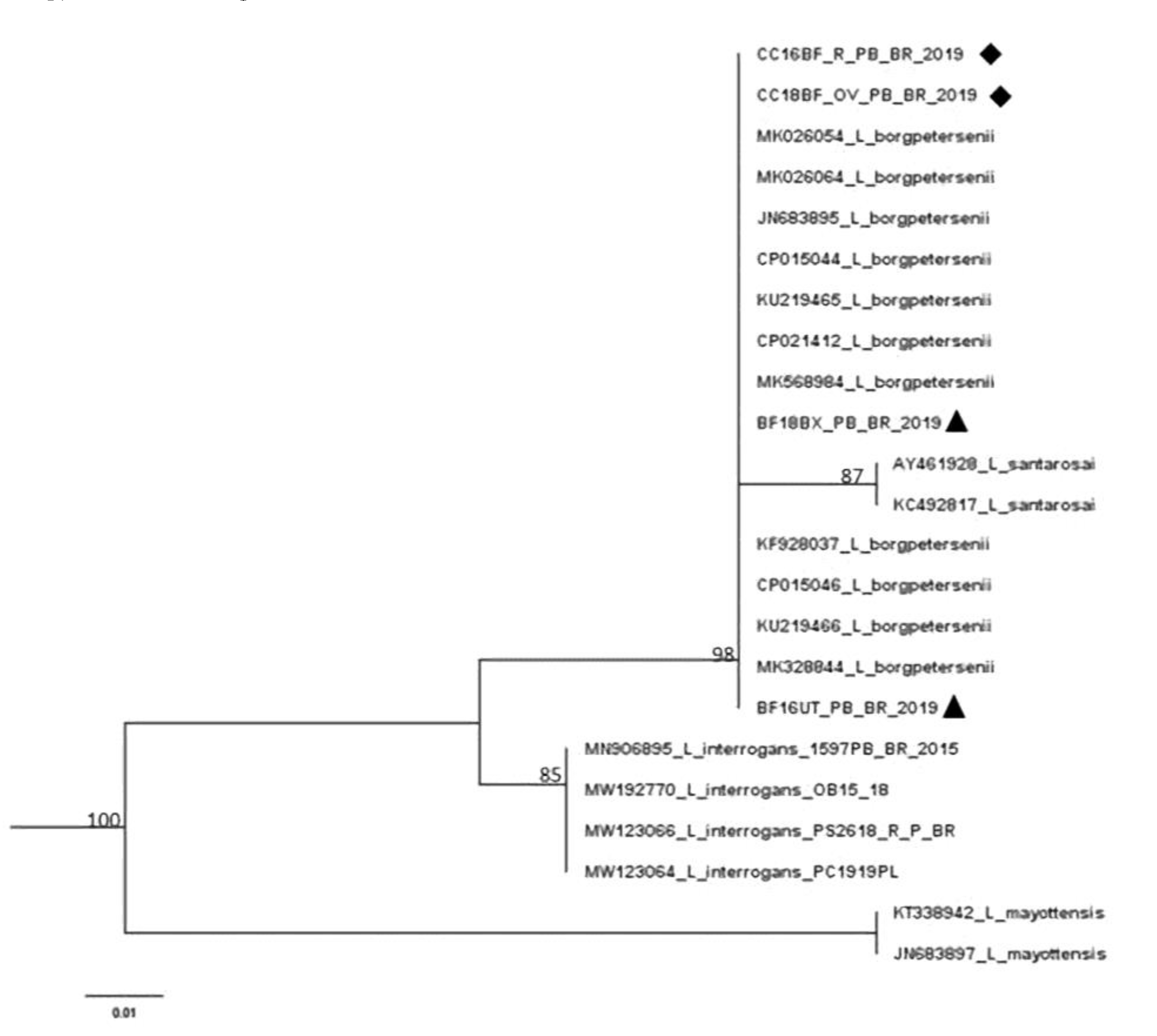
3.4. Leptospira spp. DNA Sequencing

Due to budget issues, DNA sequencing from the PCR products was possible in two samples taken from tissues (bladder and uterus) and two from cultures (kidney and ovary) from different animals. These samples showed a 99% similarity with Leptospira borgpetersenii (Figure 1).

3.5. Performance of Diagnostic Tests

Overall, the molecular test showed good performance in diagnosing Leptospira spp. infection, with high sensitivity (100.00%) and specificity (84.6%) deeming microbiological culture results as gold standards, especially for detecting leptospiral DNA from the uterine tube, with 100% sensitivity and specificity (Table 3). Table 4 and Table 5 show the sensitivity and specificity of the MAT for the different cut-off points (50, 100, 200 and 400) based on using the PCR or microbiological culture results as gold standards. It was verified that, regardless of the biological material used and the gold standard, the highest sensitivity values in the MAT were obtained for the titer of 50.

4. Discussion

The high frequency of seroreactivity found (64.3%) indicates that, even under adverse climatic conditions, Leptospira spp. can be present in herds in the Caatinga biome of Brazil. There was variation in the serogroups found (Sejroe, Tarassovi, Australis, Ballum, Djasiman and Hebdomadis), suggesting that there are different sources of infection, although Sejroe (55.6%) and Tarassovi (22.2%) were more prevalent. In other investigations conducted in semiarid regions, Sejroe (58.17%) was the most prevalent serogroup, followed by Icterohaemorrhagiae (17.32%) and Australis (4.58%) [7], as well as Sejroe (36.8%), Hebdomadis (26.3%), Australis (10.5%), Djasiman (10.5%), Ballum (5.3%) and Pomona (5.3%) [35]. Surveys of different regions of Brazil have demonstrated that regardless of the biome involved, Sejroe was most prevalent: 50.68% average prevalence in northeastern region [7,35,36,37]; 72.65% in northern region [38,39]; 30.3% in central-western region [40]; 47.01% in southeastern region [41,42,43,44]; and 48.56% in southern region [45,46]. The plurality of types of Leptospira suggests that contact with other animals exists, but there is the possibility that cattle without signs and symptoms of Leptospira spp. infection may carry and spread other strains within the species (adaptive process) [47].
The contrast between unfavorable climatic conditions (average rainfall and temperature of 0.47 mm and 29.28 °C, respectively, in the dry season) and a significant proportion of seroreactive animals, especially regarding the Sejroe serogroup, provides evidence for intraspecies transmission, which is less dependent on environmental factors because cattle are maintenance hosts [5,48]. Tarassovi is one of the main serogroups found in cattle [49]; however, there have been few reports of it in Brazil [44]. The Australis serogroup is associated with pigs and causes reproductive failure, abortion, stillbirth, fetal mummification and weak piglets [50]; it has also been described in mammals maintained in zoos [51]. The reservoirs of the serogroup Ballum are rodents, especially mice (Mus musculus). Asymptomatic mice harbor bacterium in their kidneys, which makes them an important source of infection for humans or animals [50]. The confirmation of the presence of this serogroup in cattle suggests that rodents have had access to cattle food, which usually consists of protein concentrate stored to ensure supplementation during periods in which there is low production of phytomass, or the rodents have had access to the facilities and feeders/water troughs.
PCR data can generate difficulties in interpreting the interrelation of results, as some factors, such as the amount of bacterial DNA in the sample and its quality, may not be favorable [38,52]. Leptospiral DNA was detected in 73.8% of the animals, but the true frequency may be higher, as negativity can also be correlated with DNA concentrations below the detectable threshold [53]. When comparing the positivity rates between different biological materials, there were statistically significant differences (p < 0.05) that demonstrated that leptospires accumulate preferentially in the placenta, uterus and kidneys when compared to the urine. This can be explained by the fact that the size, morphology (spirochete/helicoidal) and translational motility of the agent facilitate access to these organs, allowing it to escape the immune system due to the physical barrier that prevents contact between antibody and antigen molecules. The broad presence of leptospires in the reproductive tract reinforces that this is a site of extra renal bacterial colonization in cattle, which can act as adapted hosts, as has already been elucidated in other studies [28,54].
When analyzed from an epidemiological perspective, it is clear that, especially in the Caatinga biome, where climatic conditions are adverse for bacterial survival in the environment, extra renal colonization by leptospires is a strong indication that, in this region, venereal transmission and vertical transmission of leptospires are alternative routes in contrast to transmission through the urine, in which the external environment is part of the epidemiological cycle. The PCR results confirmed that, for animals maintained under field conditions, as well as urine, vaginal fluid could be valuable for identifying carriers both in slaughterhouses and farms.
Leptospira spp. was isolated from 29 animals (69%). By comparing the positivity rates between the different materials, there were statistically significant differences (p < 0.05) that demonstrated the tropism of the microorganism for the placenta, uterus, kidney and ovary, while the frequency of occurrence in the urine was lower, as explained earlier. The results from the cultures revealed that working on microbiological isolation, using tissue from the placenta, uterus, kidney and ovary can increase the chances of success. However, considering that access to tissue from the uterus, kidney and ovary is difficult, the collection of such material is appropriate in postmortem cases. Leptospires-specific DNA was identified in 19 cultures, among which there was one kidney sample and one ovary sample from different animals. The isolation and characterization of autochthonous strains are important with regard to understanding epidemiology and refining diagnostic tools. Moreover, this introduces the possibility of discovering new species and/or variants. If these circulating strains can be incorporated into vaccines, greater protection for animals in the region can be provided [55].
The sequenced DNA from four samples demonstrated 99% similarity with L. borgpetersenii, which belongs to the pathogenic clade and, according to virulence, to subgroup 2, along with the species L. santarosai, L. mayottensis, L. weilii and L. alexanderi [1]. L. borgpetersenii causes early embryonic loss and estrus repetition, resulting from uterine inflammation and damage caused by embryo invasion [6]. The identification of this species through genetic sequencing provided yet another indication of venereal transmission, and perhaps this pathway was responsible for influencing the high frequency of positive findings during the dry period because any other route that depends on external variables is less likely to succeed. In addition, this species has been reported to infect humans in other countries [56,57,58,59,60,61,62].
Out of the 31 positive animals on PCR, 15 showed positivity in both the reproductive and urinary tracts, 13 were positive only in the reproductive tract and three only in the urinary tract. Animals with urogenital tract involvement potentiate the diffusion of the agent, especially in the rainy season of a semiarid region, due to the possibility of simultaneous transmission via the urinary, venereal and congenital/transplacental routes. The occurrence of a bovine genital leptospirosis syndrome has been proposed [6], and it is an infection dissociated from renal/systemic leptospire colonization, which is very important in the dry period when the bacterium only has a short survival window in the environment. Therefore, in drought seasons, the urinary tract may be less relevant to the dissemination of Leptospira spp., except in microclimates, which would have been unlikely in the present study, considering that the animals were living on several different farms.
Under the conditions of the Caatinga biome, molecular detection proved to be reliable for diagnosing pathogenic Leptospira spp. infection and showed high sensitivity (100%) and specificity (84.6%). This reinforces the importance of molecular methods, and many factors contribute to this accuracy, e.g., the extraction kit, thermostable DNA polymerase, laboratory equipment and operating procedure. Reports of leptospiral DNA detection in animals negative upon MAT are common, and if the animal presents with a high antibody titer, the outcome of PCR tends to be negative. The high sensitivity of PCR often eliminates the need for isolation as a confirmatory result, and it is suitable for emergency situations, given that it is fast and enables early diagnosis based on blood analysis during leptospiremia [29].
The findings of this research reinforce the hypothesis that, although very useful for herd diagnosis, serology may be an insufficient tool to identify carriers individually, thus making it necessary to detect the presence of the agent to identify and safely treat the carriers [10]. Therefore, PCR proved to be a great diagnostic alternative because it is fast and has high sensitivity and specificity. The higher proportion of animals positive for PCR compared to MAT is due to the fact that, in some cases, depending on the serovar–host interaction and individual host responses, leptospires have low antigenicity and, in addition, the infection may be in the chronic phase [48]. Thus, a seronegative animal is not always free from infection. The analysis of the cut-off points for serology showed that, regardless of the biological material investigated, the highest sensitivity values of MAT were obtained at a titer of 50, deeming both PCR and microbiological culture as gold standards. In cattle from Colombia [63], a titer of 50 presented 95% and 89% sensitivity and specificity, respectively, in comparison to microbiological culture; however, the authors only used urine samples for the direct diagnosis of Leptospira spp. infection. Similar results were also found by our research group in sheep from Brazilian semiarid regions [10,12].
It is worth mentioning that the diagnostic tests for Leptospira spp. infection are imperfect, and the appropriate combination of methods and specimens for each stage of infection improves diagnostic accuracy. The principle behind MAT is simple, but it requires maintaining a panel of live leptospires, representing a biological risk and restricting its practice to specialized laboratories [64]. In PCR, some biological materials require proper treatment to avoid inhibitory substances [65]; in addition, it is limited by its inability to identify specific strains [54]. Microbiological confirmation is laborious and time-consuming [43]; in addition, contamination by secondary microorganisms’ compromises culture performance [44].
Our data show that a cut-off point of 50 may be more suitable for cattle from the Caatinga biome, and, in this scenario, high sensitivity values are essential to reduce the number of false-negative results upon MAT. In addition, given the results of this survey, cut-off points below 50 should also be considered as they are relevant in other species, such as sheep and swine. This is very important from epidemiological and infection control points of view as it avoids the maintenance of infected animals in the herd since subclinical infections are very common [19].
It is important to emphasize that although the recommendation by the World Organization for Animal Health (OIE) of the use of cut-off point of 100 for the serological diagnosis of leptospirosis in cattle [15], this statement has limitations regarding the detection of renal and genital Leptospira spp. carriers, which generally have low antibody titers [48]. In addition, the use of a lower cut-off point than the conventionally used was based on the question whether this methodology demonstrates favorable results only for small ruminants [10,22] or also for cattle, being both in semiarid conditions. The central idea is to assess whether increasing the sensitivity of the MAT increases the correlation between serological results and Leptospira spp. carrier condition.

5. Conclusions

This study provides important information relating to the diagnosis and epidemiology of Leptospira spp. infection in cattle from the Caatinga biome of Brazil. The results indicate that, even under the adverse environmental conditions of the Brazilian Caatinga biome, leptospires may survive and propagate through alternative routes of transmission, such as sexual pathways, and the high proportion of PCR-positive cows in the genital tract highlights the possible role of females in venereal transmission. Moreover, a cut-off of 50 should be considered for the serological diagnosis of cattle from the Caatinga biome. The study also shows that cows are commonly exposed to leptospires in the Caatinga biome, and this constitutes a One Health-based concern, demonstrating the importance of broad studies where large numbers of humans and animals coexist when investigating zoonotic infections and when planning and implementing control measures for cattle-associated leptospirosis.

Author Contributions

All authors have made substantial contributions to the following: Conceptualization and methodology, S.S.d.A. and C.J.A.; performed the experiments, N.N.d.C.B., R.R.S., D.K.S.B., D.B.N., F.T.R.d.C., J.P.A.J., C.D.M., L.S.U. and M.L.C.R.S.; writing—original draft preparation, N.N.d.C.B.; writing—review and editing, D.F.d.C., S.S.d.S.H., S.S.d.A., C.d.S.A.B.S. and C.J.A.; supervision, S.S.d.A. and C.J.A.; project administration, C.J.A.; funding acquisition, S.S.d.A. All authors have read and agreed to the published version of the manuscript.

Funding

This research was funded by National Council for Scientific and Technological Development (CNPq), grant numbers 302222/2016-2 and 423836/2018-8, and Research Support Foundation of the State of Paraíba (FAPESQ), grant number 46360.673.28686.05082021.

Institutional Review Board Statement

All experimental protocols were approved by the Federal University of Campina Grande Animal Care and Use Committee (CEUA-UFCG) ID# 069-2018. All procedures were undertaken in accordance with the relevant guidelines and regulations.

Informed Consent Statement

Not applicable.

Data Availability Statement

The datasets generated during and/or analyzed during the current study are available from the corresponding author upon request.

Acknowledgments

We thank Walter Lilenbaum (Laboratory of Veterinary Bacteriology of the Fluminense Federal University, Niterói, Rio de Janeiro, Brazil, for providing the serovars used in serology.

Conflicts of Interest

The authors declare no conflict of interest.

References

  1. Picardeau, M. Virulence of the zoonotic agent of leptospirosis: Still terra incognita? Nat. Rev. Microbiol. 2017, 15, 297–307. [Google Scholar] [CrossRef] [PubMed]
  2. Costa, F.; Hagan, J.E.; Calcagno, J.; Kane, M.; Torgerson, P.; Martinez-Silveira, M.S.; Stein, C.; Abela-Ridder, B.; Ko, A.I. Global morbidity and mortality of leptospirosis: A systematic review. PLoS Negl. Trop. Dis. 2015, 17, e0003898. [Google Scholar] [CrossRef] [PubMed] [Green Version]
  3. DE Oliveira, D.; Figueira, C.P.; Zhan, L.; Pertile, A.C.; Pedra, G.G.; Gusmão, I.M.; Wunder, E.A.; Rodrigues, G.; Ramos, E.A.G.; Ko, A.I.; et al. Leptospira in breast tissue and milk of urban Norway rats (Rattus norvegicus). Epidemiol. Infect. 2016, 144, 2420–2429. [Google Scholar] [CrossRef] [PubMed] [Green Version]
  4. Lilenbaum, W.; Varges, R.; Brandão, F.; Cortez, A.; de Souza, S.; Brandão, P.; Richtzenhain, L.; Vasconcellos, S. Detection of Leptospira spp. in semen and vaginal fluids of goats and sheep by polymerase chain reaction. Theriogenology 2008, 69, 837–842. [Google Scholar] [CrossRef] [PubMed]
  5. ughini-Gras, L.; Bonfanti, L.; Natale, A.; Comin, A.; Ferronato, A.; LA Greca, E.; Patregnani, T.; Lucchese, L.; Marangon, S. Application of an integrated outbreak management plan for the control of leptospirosis in dairy cattle herds. Epidemiol. Infect. 2014, 142, 1172–1181. [Google Scholar] [CrossRef]
  6. Loureiro, A.P.; Lilenbaum, W. Genital bovine leptospirosis: A new look for an old disease. Theriogenology 2020, 141, 41–47. [Google Scholar] [CrossRef]
  7. Pimenta, C.L.R.M.; Castro, V.; Clementino, I.J.; Alves, C.J.; Fernandes, L.G.; Brasil, A.W.L.; Santos, C.S.A.B.; Azevedo, S.S. Bovine leptospirosis in Paraíba State: Prevalence and risk factors associated with the occurrence of positive herds. Pesq. Vet. Bras. 2014, 34, 332–336. [Google Scholar] [CrossRef] [Green Version]
  8. De Andrade Morais, D.; da Costa, D.F.; Nunes, B.C.; de Sousa Américo Batista Santos, C.; Alves, C.J.; de Azevedo, S.S. Seroepidemiological survey for leptospirosis in equines from semiarid region of Paraíba state, Northeastern Brazil. Semin. Ciências Agrárias 2019, 40, 2079–2086. [Google Scholar] [CrossRef] [Green Version]
  9. Silva, A.F.; Farias, P.J.A.; Silva, M.L.C.R.; Júnior, J.P.A.; Malossi, C.D.; Ullmann, L.S.; Costa, D.F.; Higino, S.S.S.; Azevedo, S.S.; Alves, C.J. High frequency of genital carriers of Leptospira sp. in sheep slaughtered in the semi-arid region of northeastern Brazil. Trop. Anim. Health Prod. 2019, 51, 43–47. [Google Scholar] [CrossRef] [Green Version]
  10. Nogueira, D.B.; da Costa, F.T.R.; Bezerra, C.D.S.; Silva, M.L.C.R.; da Costa, D.F.; Viana, M.P.; da Silva, J.D.; Júnior, J.P.A.; Malossi, C.D.; Ullmann, L.S.; et al. Use of serological and molecular techniques for detection of Leptospira sp. carrier sheep under semiarid conditions and the importance of genital transmission route. Acta Trop. 2020, 207, 105497. [Google Scholar] [CrossRef]
  11. Nogueira, D.B.; da Costa, F.T.R.; Bezerra, C.D.S.; Soares, R.R.; Barnabé, N.N.D.C.; Falcão, B.M.R.; Silva, M.L.C.R.; da Costa, D.F.; Araújo, J.P.; Malossi, C.D.; et al. Leptospira sp. vertical transmission in ewes maintained in semiarid conditions. Anim. Reprod. Sci. 2020, 219, 106530. [Google Scholar] [CrossRef]
  12. Soares, R.R.; Barnabé, N.N.D.C.; Nogueira, D.B.; da Silva, L.S.C.; Júnior, J.P.A.; Malossi, C.D.; Ullmann, L.S.; da Costa, D.F.; Silva, M.L.C.R.; Higino, S.S.D.S.; et al. Serological, molecular and bacteriological approaches for detecting Leptospira sp. carrier rams maintained in semiarid conditions. Acta Trop. 2021, 213, 105759. [Google Scholar] [CrossRef]
  13. Fernandes, J.J.; Peixoto, A.D.L.; de Farias, A.S.S.; Pinheiro, T.J.; da Costa, D.F.; Silva, M.L.C.R.; Júnior, J.P.A.; Malossi, C.D.; Ullmann, L.S.; de Azevedo, S.S.; et al. Didelphis albiventris as a carrier of Leptospira sp. in the central nervous tissue in the semiarid region of Northeast, Brazil. Comp. Immunol. Microbiol. Infect. Dis. 2020, 73, 101560. [Google Scholar] [CrossRef]
  14. Fernandes, J.J.; Pinheiro, T.J.; Costa, D.F.; Júnior, J.P.A.; Malossi, C.D.; Ullmann, L.S.; Silva, M.L.C.R.; Azevedo, S.S.; Alves, C.J.; Higino, S.S.D.S. Leptospira interrogans infection in tegu lizard (Tupinambis merianae), Brazil. Cienc. Rural 2020, 50, e20200424. [Google Scholar] [CrossRef]
  15. OIE—World Organization for Animal Health. Reference Laboratory Reports Activities; World Organization for Animal Health: Ulster, Northern Ireland, 2014. [Google Scholar]
  16. Guedes, I.B.; de Souza, G.O.; Castro, J.F.D.P.; Cavalini, M.B.; Filho, A.F.D.S.; Heinemann, M.B. Usefulness of the Ranking Technique in the Microscopic Agglutination Test (MAT) to Predict the Most Likely Infecting Serogroup of Leptospira. Front. Vet. Sci. 2021, 8, 654034. [Google Scholar] [CrossRef]
  17. Chakraborty, A.; Miyahara, S.; Villanueva, S.Y.A.M.; Saito, M.; Gloriani, N.G.; Yoshida, S.-I. A novel combination of selective agents for isolation of Leptospira species. Microbiol. Immunol. 2011, 55, 494–501. [Google Scholar] [CrossRef]
  18. Hamond, C.; Martins, G.; Loureiro, A.P.; Pestana, C.; Lawson-Ferreira, R.; Medeiros, M.A.; Lilenbaum, W. Urinary PCR as an increasingly useful tool for an accurate diagnosis of leptospirosis in livestock. Vet. Res. Commun. 2014, 38, 81–85. [Google Scholar] [CrossRef]
  19. Martins, G.; Lilenbaum, W. Comments of Environmental Conditions for the Maintenance of Leptospira in Tropical Scenarios. Curr. Microbiol. 2015, 71, 624–625. [Google Scholar] [CrossRef]
  20. Picardeau, M. Diagnosis and epidemiology of leptospirosis. Med. Mal. Infect. 2013, 43, 1–9. [Google Scholar] [CrossRef]
  21. da Costa, D.F.; da Silva, A.F.; Brasil, A.W.D.L.; Loureiro, A.P.P.; dos Santos, F.A.; de Azevedo, S.S.; Lilenbaum, W.; Alves, C.J. Leptospirosis in native mixed-breed sheep slaughtered in a semiarid region of Brazil. Cienc. Rural 2017, 47, e20160563. [Google Scholar] [CrossRef] [Green Version]
  22. da Costa, D.F.; Silva, M.L.C.R.; Martins, G.; Dantas, A.F.M.; de Melo, M.A.; de Azevedo, S.S.; Lilenbaum, W.; Alves, C.J. Susceptibility among breeds of sheep experimentally infected with Leptospira interrogans Pomona serogroup. Microb. Pathog. 2018, 122, 79–83. [Google Scholar] [CrossRef] [PubMed]
  23. Batista, J.S.; Riet-Correa, F.; Teixeira, M.M.G.; Madruga, C.R.; Simões, S.D.V.; Maia, T.F. Trypanosomiasis by Trypanosoma vivax in cattle in the Brazilian semiarid: Description of an outbreak and lesions in the nervous system. Vet. Parasitol. 2007, 143, 174–181. [Google Scholar] [CrossRef] [PubMed]
  24. Alvares, C.A.; Stape, J.L.; Sentelhas, P.C.; de Moraes Gonçalves, J.L.; Sparovek, G. Köppen’s climate classification map for Brazil. Meteorol. Z. 2014, 22, 711–728. [Google Scholar] [CrossRef] [PubMed]
  25. INMET—Instituto Nacional de Meteorologia. Estação Meteorológica de Observação de Superfície Automática. Available online: https://www.gov.br/agricultura/pt-br/assuntos/inmet?r=estacoes/estacoesAutomaticas (accessed on 10 May 2021).
  26. Arango, H.G. Bioestatística Teórica e Computacional, 3rd ed.; Guanabara Koogan: Rio de Janeiro, Brasil, 2009; p. 438. [Google Scholar]
  27. Pimenta, C.L.R.M.; da Costa, D.F.; Silva, M.L.C.R.; Pereira, H.D.; Araújo Júnior, J.P.; Malossi, C.D.; Ullmann, L.S.; Alves, C.J.; de Azevedo, S.S. Strategies of the control of an outbreak of leptospiral infection in dairy cattle in Northeastern Brazil. Trop. Anim. Health Prod. 2019, 51, 237–241. [Google Scholar] [CrossRef] [Green Version]
  28. Loureiro, A.P.; Pestana, C.; Medeiros, M.A.; Lilenbaum, W. High frequency of leptospiral vaginal carriers among slaughtered cows. Anim. Reprod. Sci. 2017, 178, 50–54. [Google Scholar] [CrossRef]
  29. Stoddard, R.A.; Gee, J.E.; Wilkins, P.P.; Mccaustland, K.; Hoffmaster, A.R. Detection of pathogenic Leptospira spp. through TaqMan polymerase chain reaction targeting the LipL32 gene. Diagn. Microbiol. Infect. Dis. 2009, 64, 247–255. [Google Scholar] [CrossRef]
  30. Platt, A.R.; Woodhall, R.W.; George, A.L., Jr. Improved DNA sequencing quality and efficiency using an optimized fast cycle sequencing protocol. BioTechniques 2007, 43, 58–62. [Google Scholar] [CrossRef] [Green Version]
  31. Gouy, M.; Guindon, S.; Gascuel, O. SeaView version 4: A multiplatform graphical user interface for sequence alignment and phylogenetic tree building. Mol. Biol. Evol. 2010, 27, 221–224. [Google Scholar] [CrossRef] [Green Version]
  32. Hall, T.A. BioEdit: A user-friendly biological sequence alignment editor and analysis program for Windows 95/98/NT. Nucleic Acids Symp. Ser. 1999, 41, 95–98. [Google Scholar]
  33. Ayres, M.; Ayres Junior, M.; Ayres, D.L.; Santos, A.S. Bioestat 5.0 Aplicações Estatísticas nas Áreas das Ciências Biomédicas; ONG Mamiraua: Belém, Brasil, 2007; p. 364. [Google Scholar]
  34. Mackinnon, A. A spreadsheet for the calculation of comprehensive statistics for the assessment of diagnostic tests and inter-rater agreement. Comput. Biol. Med. 2000, 30, 127–134. [Google Scholar] [CrossRef]
  35. Pimenta, C.L.R.M.; Nogueira, D.B.; Bezerra, C.D.S.; Morais, D.A.; Silva, M.L.C.R.; Costa, D.F.; Higino, S.S.D.S.; Santos, C.S.A.B.; Alves, C.J.; Azevedo, S.S. High proportion of cattle and sheep seropositive and renal carriers of Leptospira sp. under semiarid conditions. Rev. Bras. Ciênc. Vet. 2020, 27, 22–28. [Google Scholar] [CrossRef]
  36. Mineiro, A.L.B.B.; Vieira, R.J.; Costa, E.A.; Santos, R.L.; Gonçalves, L.M.F.; Carvalho, S.M.; Bomfim, M.R.Q.; Costa, F.A.L. Serology, polymerase chain reaction and histopathology for leptospirosis in samples collected at slaughter from dairy cows of Parnaiba region, state of Piauí, Brazil. Pesq. Vet. Bras. 2011, 31, 859–866. [Google Scholar] [CrossRef] [Green Version]
  37. Silva, F.J.; Conceição, W.L.F.; Fagliari, J.J.; Girio, R.J.S.; Dias, R.A.; Borba, M.R.; Mathias, L.A. Prevalence and risk factors of bovine leptospirosis in the State of Maranhão, Brazil. Pesq. Vet. Bras. 2012, 32, 303–312. [Google Scholar] [CrossRef] [Green Version]
  38. Guedes, I.B.; Araújo, S.A.D.A.; de Souza, G.O.; Silva, S.O.D.S.; Taniwaki, S.A.; Cortez, A.; Brandão, P.E.; Heinemann, M.B. Circulating Leptospira species identified in cattle of the Brazilian Amazon. Acta Trop. 2019, 191, 212–216. [Google Scholar] [CrossRef]
  39. Guedes, I.B.; de Souza, G.O.; Castro, J.F.D.P.; Filho, A.F.D.S.; Rocha, K.D.S.; Gomes, M.E.T.; de Moraes, C.C.G.; Heinemann, M.B. Development of a pooled antigen for use in the macroscopic slide agglutination test (MSAT) to detect Sejroe serogroup exposure in cattle. J. Microbiol. Methods 2019, 166, 105737. [Google Scholar] [CrossRef]
  40. Miashiro, A.F.; Vasconcellos, S.A.; de Morais, Z.M.; de Souza, G.O.; Filho, J.M.L.; Figueiredo, A.D.O.; Pellegrin, A.O. Prevalência de leptospirose em rebanhos bovinos no Pantanal de Mato Grosso do Sul. Pesq. Vet. Bras. 2018, 38, 41–47. [Google Scholar] [CrossRef] [Green Version]
  41. Favero, M.; Pinheiro, S.R.; Vasconcellos, S.A.; Morais, Z.M.; Ferreira, F.; Ferreira Neto, J.S. Bovine leptospirosis—Most frequent serovars in blood collections performed between 1984 to 1997 from herds of 21 brazilian states. Arq. Inst. Biol. 2001, 68, 29–35. [Google Scholar]
  42. Araújo, V.E.M.; Moreira, E.C.; Naveda, L.A.B.; Silva, J.A.; Contreras, R.L. Frequency of anti-Leptospira interrogans agglutinins in bovine serum samples in Minas Gerais, Brazil, 1980 to 2002. Arq. Bras. Med. Vet. Zootec. 2005, 57, 430–435. [Google Scholar] [CrossRef] [Green Version]
  43. Pinto, P.S.; Loureiro, A.P.; Penna, B.; Lilenbaum, W. Usage of Leptospira spp. local strains as antigens increases the sensitivity of the serodiagnosis of bovine leptospirosis. Acta Trop. 2015, 149, 163–167. [Google Scholar] [CrossRef]
  44. Pinna, M.H.; Martins, G.; Loureiro, A.P.; Lilenbaum, W. Detection of bovine carriers of Leptospira by serological, bacteriological, and molecular tools. Trop. Anim. Health Prod. 2018, 50, 883–888. [Google Scholar] [CrossRef]
  45. Herrmann, G.P.; Rodrigues, R.O.; Machado, G.; Moreira, E.C.; Lage, A.; Leite, R.C. Seroprevalence of leptospirosis in cattle in the southeast and southwest regions of the state of Rio Grande do Sul, Brazil. Ciênc. Anim. Bras. 2012, 13, 131–138. [Google Scholar] [CrossRef]
  46. Hashimoto, V.Y.; Chideroli, R.T.; Ribeiro, J.; Alfieri, A.A.; Da Costa, G.M.; Pereira, U.D.P.; De Freitas, J.C. Serological and molecular findings in diagnosis of leptospirosis serovar hardjo in a dairy bovine herd. Semin. Cienc. Agrar. 2017, 38, 3155–3164. [Google Scholar] [CrossRef] [Green Version]
  47. Pinto, P.S.; Pestana, C.; Medeiros, M.A.; Lilenbaum, W. Plurality of Leptospira strains on slaughtered animals suggest a broader concept of adaptability of leptospires to cattle. Acta Trop. 2017, 172, 156–159. [Google Scholar] [CrossRef]
  48. Ellis, W.A. Animal leptospirosis. Curr. Top. Microbiol. Immunol. 2015, 387, 99–137. [Google Scholar] [CrossRef] [PubMed]
  49. Soo, Z.M.P.; Khan, N.A.; Siddiqui, R. Leptospirosis: Increasing importance in developing countries. Acta Trop. 2020, 201, 105183. [Google Scholar] [CrossRef]
  50. Adler, B.; de la Pena Moctezuma, A. Leptospira and leptospirosis. Vet. Microbiol. 2010, 140, 287–296. [Google Scholar] [CrossRef]
  51. Lenharo, D.K.; Santiago, M.E.B.; Lucheis, S.B. Avaliação sorológica para leptospirose em mamíferos silvestres procedentes do parque zoológico municipal de Bauru, SP. Arq. Inst. Biol. 2012, 79, 333–341. [Google Scholar] [CrossRef] [Green Version]
  52. Hamond, C.; Pestana, C.P.; Medeiros, M.A.; Lilenbaum, W. Genotyping of Leptospira directly in urine samples of cattle demonstrates a diversity of species and strains in Brazil. Epidemiol. Infect. 2016, 144, 72–75. [Google Scholar] [CrossRef] [Green Version]
  53. Latosinski, G.S.; Fornazari, F.; Babboni, S.D.; Caffaro, K.; Paes, A.C.; Langoni, H. Serological and molecular detection of Leptospira spp. in dogs. Rev. Soc. Bras. Med. Trop. 2018, 51, 364–367. [Google Scholar] [CrossRef]
  54. Pires, B.C.; Grapiglia, J.B.; Moreira, L.; Jaeger, L.H.; Carvalho-Costa, F.A.; Lilenbaum, W. Occurrence of uterine carriers for Leptospira interrogans on slaughtered cows. Microb. Pathog. 2018, 114, 163–165. [Google Scholar] [CrossRef]
  55. Zarantonelli, L.; Suanes, A.; Meny, P.; Buroni, F.; Nieves, C.; Salaberry, X.; Briano, C.; Ashfield, N.; Silveira, C.D.S.; Dutra, F.; et al. Isolation of pathogenic Leptospira strains from naturally infected cattle in Uruguay reveals high serovar diversity, and uncovers a relevant risk for human leptospirosis. PLoS Negl. Trop. Dis. 2018, 12, e0006694. [Google Scholar] [CrossRef] [Green Version]
  56. Merien, F.; Portnoi, D.; Bourhy, P.; Charavay, F.; Berlioz-Arthaud, A.; Baranton, G. A rapid and quantitative method for the detection of Leptospira species in human leptospirosis. FEMS Microbiol. Lett. 2005, 249, 139–147. [Google Scholar] [CrossRef] [Green Version]
  57. Wynwood, S.J.; Craig, S.B.; Graham, G.C.; Blair, B.R.; Burns, M.A.; Weier, S.L.; Collet, T.A.; McKay, D.B. The emergence of Leptospira borgpetersenii serovar Arborea as the dominant infecting serovar following the summer of natural disasters in Queensland, Australia 2011. Trop. Biomed. 2014, 31, 281–285. [Google Scholar]
  58. Barragan, V.; Chiriboga, J.; Miller, E.; Olivas, S.; Birdsell, D.; Hepp, C.; Hornstra, H.; Schupp, J.M.; Morales, M.; Gonzalez, M.; et al. High Leptospira Diversity in Animals and Humans Complicates the Search for Common Reservoirs of Human Disease in Rural Ecuador. PLoS Negl. Trop. Dis. 2016, 10, e0004990. [Google Scholar] [CrossRef] [Green Version]
  59. Grillová, L.; Angermeier, H.; Levy, M.; Giard, M.; Lastère, S.; Picardeau, M. Circulating genotypes of Leptospira in French Polynesia: An 9-year molecular epidemiology surveillance follow-up study. PLoS Negl. Trop. Dis. 2020, 14, e0008662. [Google Scholar] [CrossRef]
  60. Allan, K.J.; Maze, M.J.; Galloway, R.L.; Rubach, M.P.; Biggs, H.M.; Halliday, J.E.B.; Cleaveland, S.; Saganda, W.; Lwezaula, B.F.; Kazwala, R.R.; et al. Molecular Detection and Typing of Pathogenic Leptospira in Febrile Patients and Phylogenetic Comparison with Leptospira Detected among Animals in Tanzania. Am. J. Trop. Med. Hyg. 2020, 103, 1427–1434. [Google Scholar] [CrossRef]
  61. Bandara, A.G.N.M.K.; Kalaivarny, G.; Perera, N.; Indrakumar, J. Aseptic meningitis as the initial presentation of Leptospira borgpetersenii serovar Tarassovi: Two case reports and a literature review. BMC Infect. Dis. 2021, 21, 488. [Google Scholar] [CrossRef]
  62. Grillová, L.; Robinson, M.T.; Chanthongthip, A.; Vincent, A.T.; Nieves, C.; Oppelt, J.; Mariet, J.-F.; Lorioux, C.; Vongsouvath, M.; Mayxay, M.; et al. Genetic diversity of Leptospira isolates in Lao PDR and genome analysis of an outbreak strain. PLoS Negl. Trop. Dis. 2021, 15, e0010076. [Google Scholar] [CrossRef]
  63. Hernández-Rodríguez, P.; Díaz, C.A.; Dalmau, E.A.; Quintero, G.M. A comparison between polymerase chain reaction (PCR) and traditional techniques for the diagnosis of leptospirosis in bovines. J. Microbiol. Methods 2011, 84, 1–7. [Google Scholar] [CrossRef]
  64. Koizumi, N.; Micardeau, M. Leptospira spp. Methods and Protocols; Humana: New York, NY, USA, 2020; pp. 277–286. [Google Scholar]
  65. Ahmed, A.; Engelberts, M.F.M.; Boer, K.R.; Ahmed, N.; Hartskeerl, R.A. Development and validation of a real-time PCR for detection of pathogenic Leptospira species in clinical materials. PLoS ONE 2009, 4, e7093. [Google Scholar] [CrossRef]
Figure 1. A phylogenetic tree based on the nucleotide sequence alignment of the LipL32 gene of Leptospira spp. constructed using the neighbor-joining method with 1000 replicates. ▲ ♦ Samples sequenced from tissues. Samples sequenced from cultures.
Tropicalmed 08 00177 g001
Table 1. Predominant serogroups and respective antibody titers at MAT in cattle from the Caatinga biome, Brazil.
SerogroupsTiters *Total (%)
501002004008001600
Sejroe35122215 (55.6)
Tarassovi311--16 (22.2)
Australis2-----2 (7.4)
Ballum2-----2 (7.4)
Djasiman1-----1 (3.7)
Hebdomadis---1--1 (3.7)
Total (%)11 (40.7)6 (22.2)2 (7.4)3 (11.1)2 (7.4)3 (11.1)27 (100)
* Only the highest antibody titer was counted.
Table 2. Leptospira spp. molecular and microbiological diagnoses according to different types of biological material from cattle from the Caatinga biome, Brazil.
SampleTotalPCRCulturePCR of Culture
31 */42 (73.8%)29 */42 (69%)13 */42 (31%)
+ (%)Sequencing+ (%)+ (%)Sequencing
Urine426 (14.3) a-4 (9.5) a1 (2.4)-
Bladder4210 (23.8) ab18 (19.1) ab1 (2.4)-
Kidney4214 (33.3) b-11 (26.2) b2 (4.8)1
Vaginal fluid4210 (23.8) ab-9 (21.4) ab1 (2.4)-
Uterus4217 (40.5) b113 (31) b8 (19.1)-
Uterine tube427 (16.7) a-7 (16.7) ab2 (4.8)-
Ovary4213 (31) ab-11 (26.2) b2 (4.8)1
Placenta1513 (86.7) c-10 (66.7) c2 (13.3)-
Total30990 (29.1)273 (23.6)19 (6.2)2
* = number of positive animals; + = positive samples. Different lowercase letters in the same column indicate significantly different proportions (p ≤ 0.05).
Table 3. Performance of molecular tests for diagnosing Leptospira spp. infection in cattle from the Caatinga biome, Brazil, based on the microbiological result as the gold standard.
Biological MaterialMicrobiological CulturePCR
29 */42 (69.05%)31 */42 (73.81%)
+-+-SENSPE
Urine42562510094.7
Bladder821102110094.1
Kidney1118141710090.3
Vaginal fluid920102110097
Uterus1316171410086.2
Uterine tube722724100100
Ovary1118131810093.6
Placenta10513210040
Total2913311110084.6
* = number of positive animals; + = positive samples; - = negative samples; SEN = sensitivity (%); SPE = specificity (%).
Table 4. Performance of different MAT cut-offs using molecular analysis (PCR) as gold standard per biological material.
Biological MaterialAntibody Titers (Cut-Off)
50100200400
27 */42 (64.3%)16 */42 (38.1%)10 */42 (23.8%)8 */42 (19%)
PCRMATPCRMATPCRMATPCRMAT
+-SENSPE+-SENSPE+-SENSPE+-SENSPE
Urine42366.736.13135063.92833.377.82633.383.3
Bladder7207037.55115065.6464081.3353084.4
Kidney91864.335.761042.964.35535.782.14428.685.7
Vaginal fluid6216034.45115065.6373078.1353084.4
Uterus81947.12461035.3604623.5764423.584
Uterine tube42357.134.311514.357.11914.374.31714.380
Ovary81961.534.531323.155.23723.175.93523.182.8
Placenta7253.905138.5503123.1503123.150
* = number of MAT positive animals at MAT; + = positive samples; - = negative samples; SEN = sensitivity (%); SPE = specificity (%).
Table 5. Performance of different MAT cut-offs using microbiological culture as gold standard per biological material.
Biological MaterialAntibody Titers (Cut-Off)
50100200400
27 */42 (64.3%)16 */42 (38.1%)10 */42 (23.8%)8 */42 (19%)
MCMATMCMATMCMATMCMAT
+-SENSPE+-SENSPE+-SENSPE+-SENSPE
Urine42310039.53137565.8285079265084.2
Bladder6217538.24125064.73737.579.43537.585.3
Kidney8197238.751145.564.55545.583.94436.487.1
Vaginal fluid62166.736.451155.666.73733.378.83533.384.9
Uterus72053.93151138.562.13723.175.93523.182.8
Uterine tube42357.134.311514.357.11914.374.31714.380
Ovary72063.635.531327.358.13727.377.43527.383.9
Placenta636040424060222060222060
* = number of positive animals at MAT; + = positive samples; - = negative samples; SEN = sensitivity; SPE = specificity; MC = microbiological culture.
Disclaimer/Publisher’s Note: The statements, opinions and data contained in all publications are solely those of the individual author(s) and contributor(s) and not of MDPI and/or the editor(s). MDPI and/or the editor(s) disclaim responsibility for any injury to people or property resulting from any ideas, methods, instructions or products referred to in the content.

