Review

Comparison of the PF07598-Encoded Virulence-Modifying Proteins of L. interrogans and L. borgpetersenii

Section of Infectious Diseases, Department of Internal Medicine, Yale University School of Medicine, New Haven, CT 06510, USA
*
Author to whom correspondence should be addressed.
Trop. Med. Infect. Dis. 2023, 8(1), 14; https://doi.org/10.3390/tropicalmed8010014
Submission received: 16 November 2022 / Revised: 22 December 2022 / Accepted: 23 December 2022 / Published: 26 December 2022
(This article belongs to the Special Issue New Insights in Leptospirosis)

Abstract

:
Leptospirosis is an emerging infectious disease, with increasing frequency and severity of outbreaks, a changing epidemiology of populations at risk, and the emergence of new strains, serovars, serogroups, and species. Virulence-modifying (VM) proteins encoded by the PF07598 gene family are hypothesized to be Leptospira-secreted exotoxins that mediate the molecular and cellular pathogenesis of severe and fatal leptospirosis. If confirmed experimentally, this concept could revolutionize the treatment, diagnosis, prognosis, and vaccine-mediated prevention of leptospirosis by enabling a novel array of targeted interventions. VM proteins, as with other bacterial-secreted protein exotoxins, mediate their virulence effects by attaching to eukaryotic cells, competing with other microorganisms for limited resources in environmental niches, directly intoxicating target cells, and disrupting their function in the mammalian host. In contrast with the most pathogenic group of Lept ospira, particularly L. interrogans, whose genomes contain 12–15 PF07598 paralogs, strains of the livestock and human pathogen L. borgpetersenii have two PF07598 paralogs. Given the possible non-environmentally mediated transmission of some L. borgpetersenii strains and the much smaller number of VM proteins in this species, their role in infection and disease may well differ from other leptospiral species. Comparison of VM proteins from different clades of pathogenic Leptospira may deepen our understanding of leptospirosis’s pathogenesis, leading to novel approaches to ameliorating Leptospira infection in humans and animals.

1. Introduction

Human deaths due to infectious diseases remain the leading scourge of humanity [1]. Many of these emerging infectious diseases are zoonotic, originating from both domestic and wild animals [2]. Leptospirosis, a leading yet neglected zoonotic bacterial disease, is responsible for widespread morbidity and disproportionately impacts people in regions with warm, humid climates in which the bacteria can easily proliferate [3]. Difficulties in diagnosing human and animal leptospirosis remain an important problem, and despite the availability of molecular diagnostics, diagnosis still largely depends on archaic serology [4]. The disease is caused by Gram-negative spirochetes of the genus Leptospira, which has 64 genomic species and 300 serovars identified to date [5]. A wide variety of animals—rodents, livestock, and wild animals—are sources of leptospiral infection by excreting Leptospira in their urine. Humans are accidental hosts and acquire the infection from either direct or indirect sources such as contaminated water, like rivers, puddles, and rice paddies [6,7,8,9,10,11]. Animal leptospirosis is of public health and economic importance as it negatively impairs livestock production and represents an infectious threat to humans; dogs, which both serve as companion animals and roam ferally, are susceptible to leptospiral infection in diverse settings. Cattle leptospirosis can reflect in abortion, stillbirth, premature birth, general reproductive failure, and milk production drop syndrome [12]. Bovines are susceptible to the infection from multiple serovars and Leptospira species, including L. borgpetersenii serovar Hardjo and L. interrogans serovar Pomona [13,14]. Infected animals may be seronegative and still excrete Leptospira [13]. The serious health effect on livestock is an important motivation to develop a pan-leptospiral vaccine that would be long-lasting and provide sterile immunity and cross-protection among diverse species and serovars [15]. To achieve a successful vaccination process, it is very important to understand the risk and assessments of the disease in a population.
Fundamental biological insights are essential for understanding how to prevent infectious disease transmission and intervene against disease in both humans and animals. The recently discovered virulence-modifying (VM) proteins, encoded by the PF07598 gene family, are found only in group 1 pathogenic Leptospira [16,17,18]. The use of in silico and in vitro approaches have begun to show how these proteins can be an asset in combatting and preventing leptospirosis in humans and animals. This review aims to further understand the potential roles of the PF07598 gene family in the biology of Leptospira and the pathobiology of leptospirosis by a comparative analysis of this gene family in L. interrogans and L. borgpetersenii.

2. Leptospirosis and Vaccination

A human being who comes into contact with urine-infected animals or contaminated environments, whether directly or indirectly, is considered at risk for leptospiral infection [19]. Finding a way to prevent and treat this disease is always one of the goals of researchers working with these bacteria. To date, naturally acquired anti-leptospiral immunity has been considered primarily to be antibody-mediated, with immune responses directed against leptospiral lipopolysaccharide (LPS) [20,21,22]. However, it also appears that cell-mediated responses contribute to protection against some serovars such as Hardjo in cattle. In animals, such as dogs, there is currently no gold standard methodology for in vitro prediction of experimental in vivo evaluation of vaccine efficacy [23]. In the case of recombinant vaccines, several vaccine approaches have been tested, including bacterial DNA, viral delivery vaccines, live attenuated bacteria, and subunit vaccines [24]. In the complex extensive reverse vaccinology study to date, a total of 238 where proteins identified and evaluated as potential vaccine candidates [25]. A hamster colonization model was used to evaluate pool of recombinant proteins (5 proteins/pools) and >70% were immunogenic [25]. However, none of the recombinant protein pools conferred protection against kidney colonization [25]. In metanalysis research involving vaccines and vaccination for dogs, researchers estimated a general 84% protection against carrier status [23]. The American Veterinary Medical Association explains that currently available vaccines effectively prevent leptospirosis and protect dogs for at least 12 months [26,27,28]. Annual vaccination is recommended for at-risk dogs and required for bovines. Leptospirosis is a nearly silent infection, rarely inducing outward clinical signs in cattle, as was reflected in peripheral blood mononuclear cell profiles [29].
With few outwardly observable clinical signs, cattle are a chronic host of serologically identical but genetically distinct members of serovar Hardjo, namely L. borgpetersenii serovar Hardjo (type Hardjo-bovis) and L. interrogans serovar Hardjo (type Hardjo-prajitno) [12]. Cattle are the major reservoir of these agents, which can infect humans and other animals and cause acute disease [30,31]. In North America, L. borgpetersenii serovar Hardjo type Hardjo-bovis is most often isolated from cattle [13,31]. Vaccination with bacterins does reduce livestock-human transmission, and it can also reduce the impact of leptospirosis on cattle, which improves animal production [29]. Information is a key feature in the prevention of diseases like leptospirosis. Recently, in research with VM proteins, Chaurasia et al. [32] found that vaccination of C3H/HeJ mice with L. interrogans serovar Lai VM proteins protected mice from any clinical manifestations of the disease. The authors observed that this led to ~3–4 log10 reduction in bacterial load in the liver and kidney, two key organs in the pathogenesis of leptospirosis and transmission of Leptospira, respectively. Hence, recombinant protein vaccines may be key to global immunization against pathogenic leptospirosis, eliminating kidney colonization and spread in the environment [32]. However, so far, we believe this is a very promising hyphothesis.

3. Virulence Factors in Leptospira and PF07598 Paralogous Gene Family

Virulence in Leptospira still has several unknown aspects. Fouts et al. [16] described virulence in terms of survival mechanisms of Leptospira bacteria such as adhesion to the extracellular matrix (ECM), complement evasion and ECM degradation via metalloproteases, motility, chemotaxis, resistance to oxidative stress. As well evading/motility mechanism like the immunodominant proteins of Leptospira and the exotoxin PF07598 paralogous gene family. Members of the PF07598 gene family are expressed and upregulated to various extents, as shown in vivo in a hamster model of acute leptospirosis. It has been established that group 1 (pathogenic Leptospira) includes L. interrogans serovars Icterohaemorrhagiae and Copenhageni, which are the strains most often associated with severe and fatal outcomes [5]. The members of this group uniquely encode in their genomes virulence-related protein families, such as the metalloproteases-associated protein family and the VM protein family [16], which suggests the potential importance of these protein families in the pathogenesis of leptospirosis [5]. An ultimate objective for clinical leptospirosis research is to combat the lack of wide cross-protection provided by current serogroup/serovar-specific bacterin vaccines by developing a pan-leptospirosis vaccine [33,34,35]. The premise of much ongoing research in the field is that identifying pathogenic virulence factors conserved across species/serovar/strain will enable the use of such proteins for vaccine development to target host immune responses that protect against numerous serovars of Leptospira [33].
Virulence factors are often the focus of characterization since a bacterin vaccine that exposes an immunized host to more known virulence factors are likely to produce improved protection [33,34,35]. To date, only a limited number of known virulence factors have been identified in pathogenic Leptospira, and they have been more intensely studied in L. interrogans than in L. borgpetersenii [16,33]. The outer membrane, surface-exposed virulence factors are of particular focus as they interact directly with the host during infection and are thus available to antibodies [33].
The large novel gene family PF07598, previously of unknown function, encodes the VM proteins found uniquely in group 1 pathogenic Leptospira [17,18,36]. The identification of these new virulence-associated genes should spur additional experimental inquiry into their potential roles in mediating Leptospira pathogenesis and, perhaps, in enabling Leptospira to persist in the hostile and competitive environmental microbial world [17]. Here we take diverse in vitro, in vivo, and in silico approaches to analyze features of the PF07598 gene family at several levels in Leptospira.

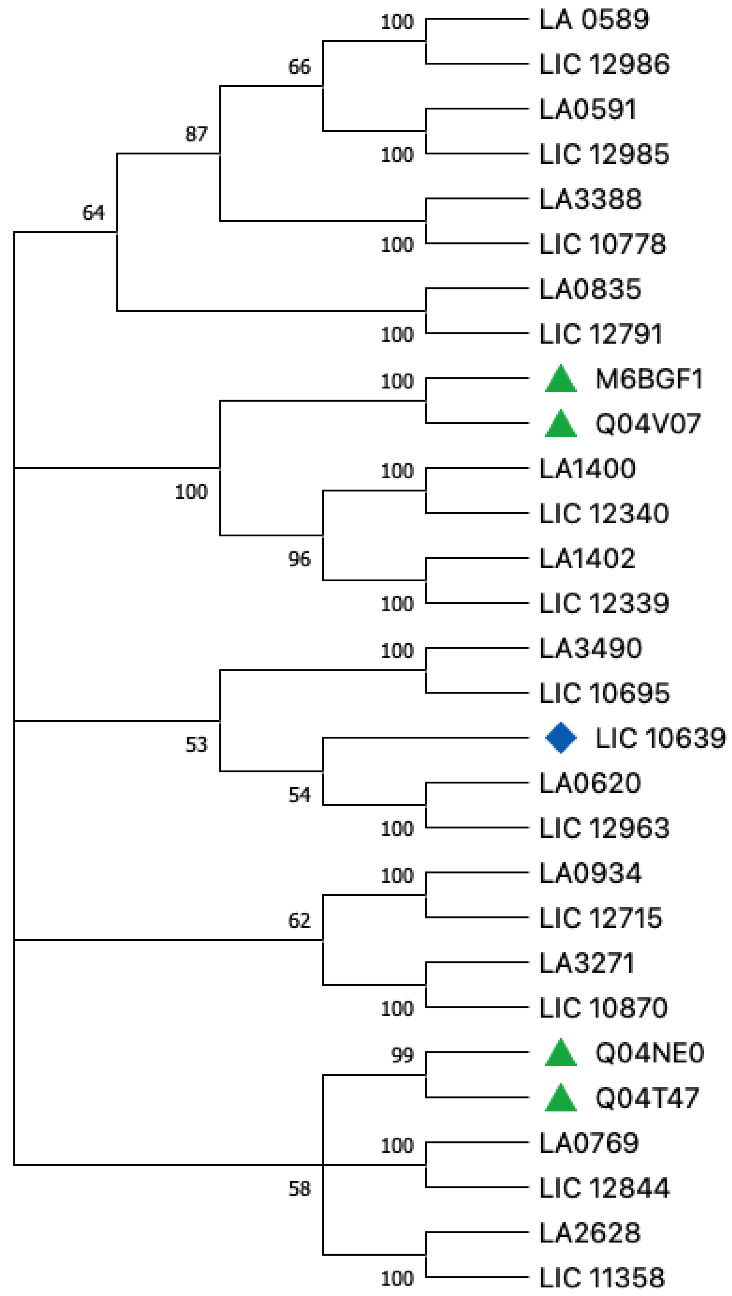
4. Proteins of PF07598 Paralogous Gene Family: General Comparison between Leptospirosis Agents

Host Tropism

Two of the largest phylogenetically distinct pathogenic species are Leptospira borgpetersenii and Leptospira interrogans, which, combined, cause most cases of leptospirosis [37]. Although the clinical symptoms of infection due to these two species are similar, they are transmitted differently; epidemiological data support a host-to-host transmission cycle for L. borgpetersenii [38]. After the ongoing ecological niche switch from free-living to a symbiotic lifestyle (concomitant with gene expansion), this group of bacteria stabilizes and restricts their lifestyle in specific niches [39]. Overall, so far, is not clear if is the reduction in the number of virulence-modifying proteins in L. borgpetersenii associated with a switch to a symbiotic lifestyle that causes protein production changes, although they remain as virulent as L. interrogans, which has more VM proteins [12,13,14,15].
In a study comparing L. borgpetersenii and L. interrogans survival in water at 20 °C, the former lost >90% viability within 48 h whereas L. Interrogans retained 100% viability over the same period [38]. L. interrogans retained 30% viability over a 3-week incubation, by which time no viable L. borgpetersenii were detected (R.L.Z. unpublished data) [38]. The authors concluded that L. borgpetersenii does not tolerate nutrient deprivation and does not survive passage through water [38]. L. borgpetersenii has a limited capacity to acquire nutrients and survive in environments external to a mammalian host [38]. Although the reason for this is not yet completely clear, it is possible that the large range of potential hosts that can be infected by Leptospira species requires some specificity and that horizontal gene transfers may be one of the methods allowing fast adaptation to these hosts [39]. Adherence is only one of the possible mechanisms underlying the host specificity [40]. In some instances, the ability of a pathogen to infect a host has been correlated with its ability to adhere to cells from that species [40]. This leads to more evolutionary questions about the Leptospira species and host dynamics.

5. General Hypothesis

Our body of work has shown that the PF07598 gene family is restricted to group 1 pathogenic Leptospira and is not found in either intermediate or saprophytic Leptospira [16,17,18,36,41]. VM proteins encode the paralogs, which vary in their numbers in L. interrogans serovar Lai (12), L. interrogans serovar Copenhageni (13), L. borgpetersenii serovar Hardjo (4), and several others using system biology and “pathogenomic” approaches [16]. This raises several questions. For instance, why does L. borgpetersenii have four VM proteins? There may be one protein and three isomers, or there may be four different proteins. Perhaps there have also been mildly deleterious mutations or, similarly, new insertions of intrinsic elements that might facilitate intragenomic recombination.
A tenet of evolution is that alleles that are under negative selection are often deleterious and confer no evolutionary advantage; these are removed from the gene and are eventually extinguished from the population [42]. Conversely, alleles under positive selection do confer an evolutionary advantage and lead to an increase in the overall fitness of the organism; thus, they increase in frequency until they eventually become fixed in the population [42]. Comparing the complete genome of two strains of L. borgpetersenii serovar Hardjo, one hypothesis is that the bacteria are undergoing a process of insertion sequence (IS)-mediated genome reduction [38], leading simultaneously to the perception of genome reduction in L. borgpetersenii and genome expansion in L. interrogans, which reflects differences in the environments during transmission between hosts. That is one theory that could support the evolution of L. borgpetersenii. Based on several comparisons [16,17,18,43], the interpretation is that Lai has orthologs in L. interrogans Copenhageni (LIC) apart from LIC_10639, and each protein from L. borgpetersenii has copies inside the species. Answering the general question about why this happens is a work in progress.

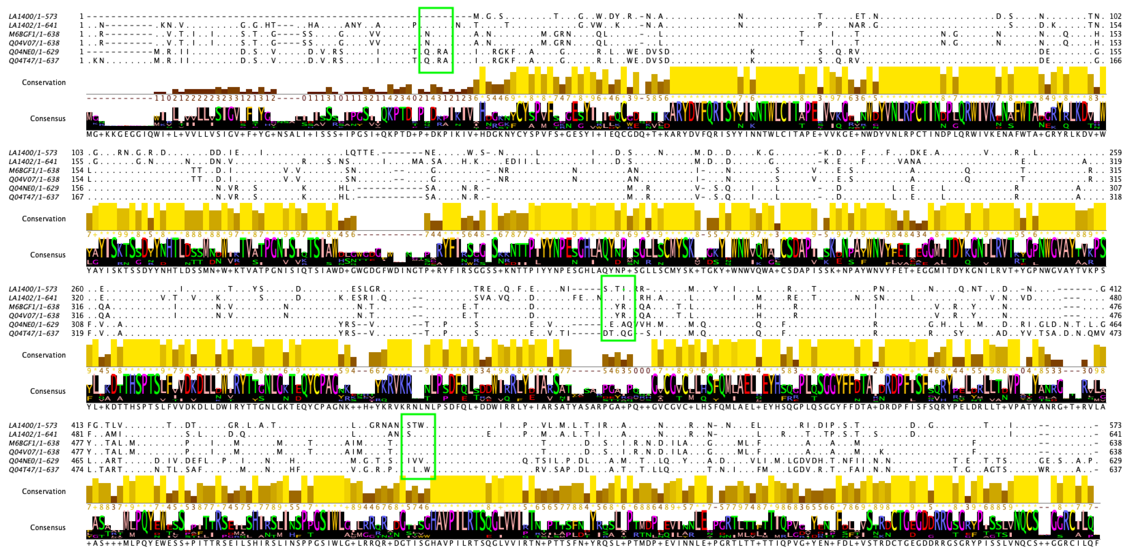
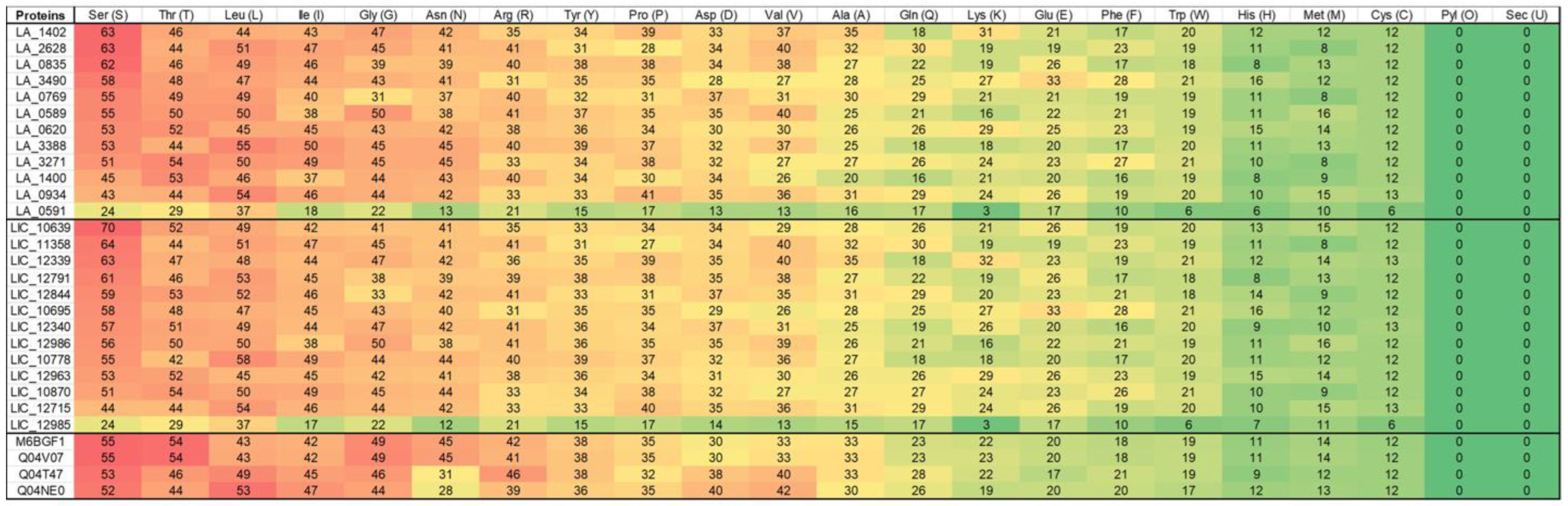
6. Amino Acids and Basic Evolutionary Differences

Since LIC and L. interrogans Lai cause severe leptospirosis in humans and L. borgpetersenii Hardjo-bovis causes fetal loss in cattle, we focused on the phylogenetic comparison between the VM proteins from these species and serovars (Table 1). In the phylogenetic tree, the evolutionary history was inferred using the neighbor-joining method and MEGA 11 [44,45]. The selection from the L. borgpeterseni protein sequences was based on a previous result from Putz et al. [43], where the authors demonstrated that the genes Q04NE0, Q04T47, and Q04V07 are present in a clinical isolate from cattle.
Leptospira borgpetersenii serovar Hardjo strains HB203 and JB197 have a high level of genetic homology but cause different clinical presentations in the hamster model of infection [43]; HB203 colonizes the kidney and presents with chronic shedding while JB197 causes severe organ failure and mortality [43].
Comparative analysis of protein content suggests that different protein profiles are expressed by Leptospira strain JB197 compared to strain HB203 (both cultivated at 29 and 37 °C) [43]. For the genome of JB197, the proteins Q04T47 and Q04V07 were highly expressed (proteomics analysis) compared with Q04NE0, but with a low expression still present in the genome. In UniProt Reference Clusters using sequence identity thresholds (100%, 90%, and 50%), we determined that M6BFG1 from L. borgpetersenii serovar Hardjo-bovis strain Sponselee showed 90% identity with Q04V07. Based on this finding, L. borgpetersenii serovar Hardjo-bovis presents four VM proteins, and further experimental validations could be used to develop a potential vaccine candidate for cattle and other animals worldwide.
As expected, in the phylogenetic tree, L. borgpetersenii serovar Hardjo-bovis M6BFG1 was clustered with Q04V07 with a bootstrap value of 100%, and with Q04NE0 and Q04T47 with a bootstrap value of 99% (Figure 1). Sequence data of protein from multiple species help in understanding evolutionary patterns. Multiple Sequence Alignment (MSA) of protein sequences obtained from across species or a cohort is usually the starting point for such analyses [46].
Comparative multiple sequence analysis of VM proteins from L. interrogans Lai (ancient VM protein sequences LA1400 and LA1402, vaccine candidates) and L. borgpeterseni serovar Hardjo-bovis (Figure 2) showed non-conserved amino acids specific to Lai and Hardjo-bovis. This clearly differentiates the significance of species-specific amino acids’ variation-conservation and their functional role in the pathogenesis of human and animal leptospirosis. Conservation of amino acids at the N-terminal and variation at the C-terminal revealed that these VM proteins can present different degrees of host tropism. However, more research must be performed to further investigate that aspect.
When evaluating the level of conservation of individual amino acids [46], a highly conserved amino acid is likely to have an important role in either structure or function. This is especially true for perfectly conserved amino acids, which are mostly identified in the functional sites of proteins [46]. Focusing on the differences between proteins in the same gene family can lead to future studies about evolutionary aspects beyond cross-reaction in vaccine research. This suggests that the amino acid specific to Hardjo-bovis and non-conserved on Lai could be a potential target for animal vaccines and therapeutics.
According to Anfinsen’s dogma of molecular biology, the native structure and function of proteins are uniquely determined by their amino acid sequence [48]. To identify the amino acid composition, the Expasy ProtParam [49] tool was used to create figures that would highlight the differences between L. borgpeterseni and L. interrogans (Figure 3), thus capturing the importance of understanding a phylogenetic analysis.
Knowledge of the complete amino acid sequence of each protein (Figure 3) is crucial not only to the basic understanding of its molecular structure, but also for defining the correlation of the protein structure with the biological and immunologic properties of, say, bacteria [50]. Understanding VM proteins helps develop one strong final product since amino acid deletion can strongly affect protein function. Based on the similarity and homology of sequences curated from different species, protein sequences are classified into families that are likely to share structural and functional similarities [46]. Most sites tolerate a degree of change, which reflects either a polymorphism or a mutation under a selection pressure [46]. However, co-evolution, where another amino acid simultaneously undergoes a mutation along with it, has recently gained considerable interest [46]. The correlation between the phylogenetic tree (Figure 1) and the content of the amino acid (Figure 3) represents a range of data that can give a background for more in vitro and in vivo analysis of these proteins. Besides that, it is worthwhile to consider which protein characteristics are important for which processes or functions.

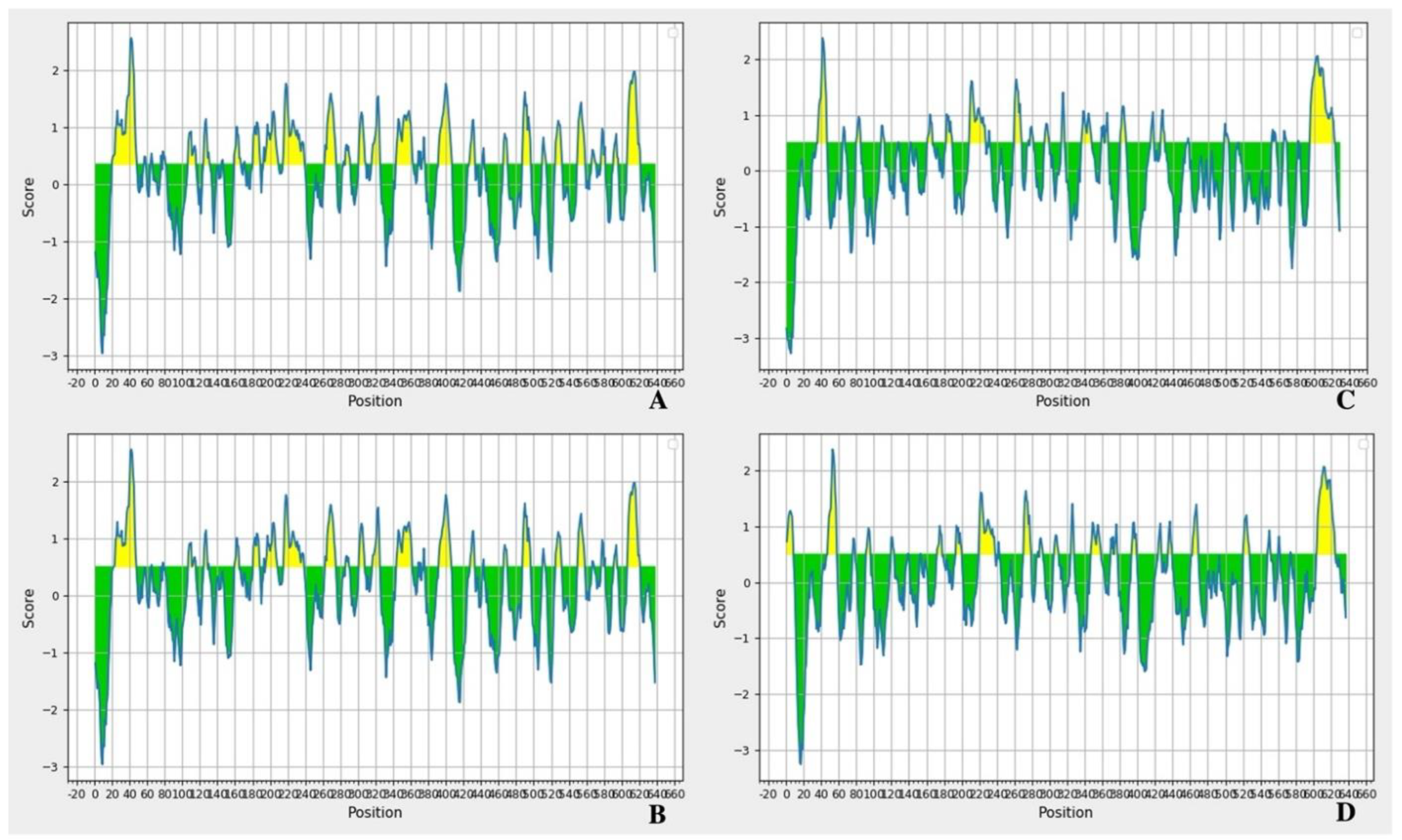
7. Antigenicity and Epitopes

Vaccination, which involves several natural and artificial proteins, is a key strategy for controlling various infectious diseases. This gives another layer to understanding antigenicity and epitopes, as well as their importance in the protein structure. Computational vaccine design, also known as computational vaccinology, encompasses epitope mapping (Figure 4), antigenicity (Table 2), and immunogen design tools. In silico prediction of immune response to emerging infectious diseases can accelerate the next generation of vaccines. The capacity of an antigen to bind with the receptors is called the antigenicity [47]. Characterizing the antigenicity of a protein can be important for predicting vaccine efficacy.
However, such experiments are labor-intensive, time-consuming, and not suitable for the early stage detection [47]. Computational prediction of antigenic dissimilarity using amino acid sequences enables large-scale antigenic characterization [51]. This approach for example can be applied as well, to reverse vaccinology (RV), which is a technique that has been widely used for screening surface-exposed proteins (PSEs) of important pathogens, including outer membrane proteins (OMPs), and extracellular proteins (ECPs) as potential vaccine candidates [52]. One of the goals of using this method is the initial screening is achieved by using bioinformatics to identify all surface-exposed proteins (potential vaccine candidates) and this typically reduces the number of targets 10-fold, from thousands to hundreds of proteins [51,53]. Surface-exposed proteins generally comprise a wide array of virulence factors involved in pathogen-host interactions and are responsible for causing disease [52]. Overall, Screening using in vitro assays further reduces the number of vaccine candidates and hence the number of laboratory animals required for efficacy testing.
Importantly, sequence-based computational methods make it possible to characterize antigenicity without requiring high biosafety levels [47]. The use of programs such as VaxiJen showcases the idea that antigens possess common, underlying physicochemical features that are independent of species and conventional global sequence similarity [54]. The VM proteins presented here, based on our results, can be recognized by the binding sites of the antibodies.
With the main objective of producing vaccines for humans and animals, after understanding that the protein can be recognized by antibodies, the next step is to understand the localization of epitopes inside the amino acid sequences. A B-cell epitope is the antigen portion binding to the immunoglobulin or antibody [55]. These epitopes recognized by B-cells may constitute any solvent-exposed region in the antigen and can be of different chemical natures and can be subjects for epitope prediction methods [55]. B-cell epitope prediction aims to identify B-cell epitopes with the practical purpose of replacing the antigen for antibody production or for carrying structure-function studies [55]. Any solvent-exposed region in the antigen can be subject to recognition by antibodies [55].
These data support the next steps for protein in vivo research (Figure 4 and Table 2). VM proteins are members of the PF07598 family, which is gaining attention as studies lead to potential human and animal vaccination. Based on what was previously explained about Leptospira vaccines, cross-reaction between vaccines from a protein produced by different species is very relevant to the field. This leads to challenges that need to be overcome.

8. Clinical Relevance and Challenges

The highly innovative aspect of the present body of work is the discovery of VM proteins and the hypothesis that these molecules are possible exotoxins that mediate severe and fatal leptospirosis at the cellular level. Despite years of study and the availability of commercial vaccines, detailed analysis of the animal immune response to vaccination and the Leptospira challenge is lacking [29]. This remains a problem even in the face of technology related to vaccine production. Vaccines for Leptospira with cross-reaction potential and strong protective aspects are still not available for humans and animals. Not just that, but there are several serovars and strains related to L. borgpetersenii (Table 3). The use of VM proteins could help close this gap or at least be a starting point.
In addition, overall surveillance should prioritize species where economic losses have been identified, species with a known history of maintaining the occurrence of leptospirosis, and novel species identified in epidemiological studies of the human disease [12]. This will establish the relative importance of serovars and host species, leading to possibilities for reducing/managing risk factors, vaccination programs, rodent control programs, and animal health programs [12].
Therefore, vaccine development would be a critical, and possibly the first, demonstration of the importance of conserved proteins for the vaccine at the species level, which would represent a major advancement in human and animal leptospirosis vaccine and adjunct biologics development.

9. Concluding Remarks

The unique “lifestyle” of Leptospira species has given a different direction to their genome features. Virulence-modifying (VM) proteins are still a novel approach to discovering more about leptospirosis pathogenesis, presenting an opportunity for new treatments and prevention protocols. The use of in silico and in vitro approaches have started to show how these proteins can be an asset in combatting and preventing leptospirosis in humans and, in the future, animals. While previous mouse immunization experiments suggest that VM proteins are important virulence factors in vivo, the proposed mechanism of protective immunity in the animal system remains unknown. Demonstrating that immunization with VM proteins from one Leptospira species and serogroup cross-protects against challenge infection containing a different serogroup would be an important advancement in the field. Such research would form the basis for leptospirosis vaccine development for several species.

Supplementary Materials

The following supporting information can be downloaded at: https://www.mdpi.com/article/10.3390/tropicalmed8010014/s1, , Figure S1: Epitope analysis based on iedb.org from the amino acid sequence of Leptospira interrogans Lai, LA1400 which has the ortholog LIC_12340; Figure S2: Epitope analysis based on iedb.org from the amino acid sequence of Leptospira interrogans Lai, LA1402 which has the ortholog ortholog LIC_12339; Figure S3: Epitope analysis based on iedb.org from the amino acid sequence of Leptospira borgpetersenii, M6BGF1 which has the ortholog Q04V07; Figure S4: Epitope analysis based on iedb.org from the amino acid sequence of Leptospira borgpetersenii, Q04V07 which has the ortholog M6BGF1; Figure S5: Epitope analysis based on iedb.org from the amino acid sequence of Leptospira borgpetersenii, Q04NE0 which has the ortholog Q04T47; Figure S6: Epitope analysis based on iedb.org from the amino acid sequence of Leptospira borgpetersenii, Q04T47 which has the ortholog Q04NE0; Table S1: Predicted peptides analysis of the amino acids sequence from Leptospira interrogans Lai, protein LA1400, based on iedb.org server; Table S2: Predicted peptides analysis of the amino acids sequence from Leptospira interrogans Lai, protein LA1402, based on iedb.org server; Table S3: Predicted peptides analysis of the amino acids sequence from Leptospira borgpetersenii, protein M6BGF1, based on iedb.org server; Table S4: Predicted peptides analysis of the amino acids sequence from Leptospira borgpetersenii, protein Q04V07, based on iedb.org server; Table S5: Predicted peptides analysis of the amino acids sequence from Leptospira borgpetersenii, protein Q04NE0, based on iedb.org server; Table S6: Predicted peptides analysis of the amino acids sequence from Leptospira borgpetersenii, protein Q04T47, based on iedb.org server; Table S7: Online tools used for analysis of L. interrogans Lai, Copenhageni and L. borgpetersenii.

Author Contributions

Conceptualization: J.M.V.; Investigation, Formal analysis, Writing—Original Draft: D.S.V.; Formal analysis, Writing—Review and Editing: R.C.; Resources, Supervision: J.M.V. All authors have read and agreed to the published version of the manuscript.

Funding

This work was supported by the Americas Foundation, Inc., and the United States Public Health Service through National Institutes of Health, NIAID grants R01AI108276 and U19AI115658 (JMV).

Institutional Review Board Statement

Not applicable.

Informed Consent Statement

Not applicable.

Data Availability Statement

The datasets presented in this study can be obtained from the authors when resquested.

Acknowledgments

The authors would like to thank the late Gustavo da Silva Vieira (lovely and intelligent brother) wistfully for the success of D.S.V. in the manuscript preparation.

Conflicts of Interest

The work reported here has been filed in patent applications from Yale University. JV and spouse have an equity interest in LeptoX BioPharma, Inc. which may have a future interest in licensing this work.

References

  1. Lopez, A.D.; Mathers, C.D. Measuring the global burden of disease and epidemiological transitions: 2002–2030. Ann. Trop. Med. Parasitol. 2006, 100, 481–499. [Google Scholar] [CrossRef] [PubMed]
  2. Garba, B.; Bahaman, A.R.; Bejo, S.K.; Zakaria, Z.; Mutalib, A.R.; Bande, F. Major epidemiological factors associated with leptospirosis in Malaysia. Acta Trop. 2018, 178, 242–247. [Google Scholar] [CrossRef] [PubMed]
  3. Costa, F.; Hagan, J.E.; Calcagno, J.; Kane, M.; Torgerson, P.; Martinez-Silveira, M.S.; Stein, C.; Abela-Ridder, B.; Ko, A.I. Global Morbidity and Mortality of Leptospirosis: A Systematic Review. PLoS Negl. Trop. Dis. 2015, 9, e0003898. [Google Scholar] [CrossRef] [PubMed] [Green Version]
  4. Martins, G.; Lilenbaum, W. Control of bovine leptospirosis: Aspects for consideration in a tropical environment. Res. Vet. Sci. 2017, 112, 156–160. [Google Scholar] [CrossRef]
  5. Picardeau, M. Virulence of the zoonotic agent of leptospirosis: Still terra incognita? Nat. Rev. Microbiol. 2017, 15, 297–307. [Google Scholar] [CrossRef]
  6. Agampodi, S.B.; Karunarathna, D.; Jayathilala, N.; Rathnayaka, H.; Agampodi, T.C.; Karunanayaka, L. Outbreak of leptospirosis after white-water rafting: Sign of a shift from rural to recreational leptospirosis in Sri Lanka? Epidemiol. Infect. 2014, 142, 843–846. [Google Scholar] [CrossRef]
  7. Agampodi, S.; Peacock, S.J.; Thevanesam, V. The potential emergence of leptospirosis in Sri Lanka. Lancet Infect. Dis. 2009, 9, 524–526. [Google Scholar] [CrossRef]
  8. Agampodi, S.B. Spatial epidemiology of leptospirosis in Sri Lanka. Epidemiol. Infect. 2012, 140, 1530–1531. [Google Scholar] [CrossRef] [Green Version]
  9. Agampodi, S.B.; Dahanayaka, N.J.; Bandaranayaka, A.K.; Perera, M.; Priyankara, S.; Weerawansa, P.; Matthias, M.A.; Vinetz, J.M. Regional differences of leptospirosis in Sri Lanka: Observations from a flood-associated outbreak in 2011. PLoS Negl. Trop. Dis. 2014, 8, e2626. [Google Scholar] [CrossRef] [Green Version]
  10. Agampodi, S.B.; Nugegoda, D.B.; Thevanesam, V. Determinants of leptospirosis in Sri Lanka: Study protocol. BMC Infect. Dis. 2010, 10, 332. [Google Scholar] [CrossRef]
  11. Agampodi, S.B.; Peacock, S.J.; Thevanesam, V.; Nugegoda, D.B.; Smythe, L.; Thaipadungpanit, J.; Craig, S.B.; Burns, M.A.; Dohnt, M.; Boonsilp, S.; et al. Leptospirosis outbreak in Sri Lanka in 2008: Lessons for assessing the global burden of disease. Am. J. Trop. Med. Hyg. 2011, 85, 471–478. [Google Scholar] [CrossRef] [PubMed]
  12. Ellis, W.A. Animal leptospirosis. Curr. Top. Microbiol. Immunol. 2015, 387, 99–137. [Google Scholar] [CrossRef] [PubMed]
  13. Nally, J.E.; Hornsby, R.L.; Alt, D.P.; Bayles, D.; Wilson-Welder, J.H.; Palmquist, D.E.; Bauer, N.E. Isolation and characterization of pathogenic leptospires associated with cattle. Vet. Microbiol. 2018, 218, 25–30. [Google Scholar] [CrossRef]
  14. Stringfellow, D.A.; Brown, R.R.; Hanson, L.E.; Schnurrenberger, P.R.; Johnson, J. Can antibody responses in cattle vaccinated with a multivalent leptospiral bacterin interfere with serologic diagnosis of disease? J. Am. Vet. Med. Assoc. 1983, 182, 165–167. [Google Scholar] [PubMed]
  15. Draghi, M.G.; Brihuega, B.; Benitez, D.; Sala, J.M.; Biotti, G.M.; Pereyra, M.; Homse, A.; Guariniello, L. Leptospirosis outbreak in calves from Corrientes Province, Argentina. Rev. Argent. Microbiol. 2011, 43, 42–44. [Google Scholar] [CrossRef]
  16. Fouts, D.E.; Matthias, M.A.; Adhikarla, H.; Adler, B.; Amorim-Santos, L.; Berg, D.E.; Bulach, D.; Buschiazzo, A.; Chang, Y.F.; Galloway, R.L.; et al. What Makes a Bacterial Species Pathogenic?:Comparative Genomic Analysis of the Genus Leptospira. PLoS Negl. Trop. Dis. 2016, 10, e0004403. [Google Scholar] [CrossRef] [Green Version]
  17. Lehmann, J.S.; Matthias, M.A.; Vinetz, J.M.; Fouts, D.E. Leptospiral pathogenomics. Pathogens 2014, 3, 280–308. [Google Scholar] [CrossRef] [Green Version]
  18. Lehmann, J.S.; Fouts, D.E.; Haft, D.H.; Cannella, A.P.; Ricaldi, J.N.; Brinkac, L.; Harkins, D.; Durkin, S.; Sanka, R.; Sutton, G.; et al. Pathogenomic inference of virulence-associated genes in Leptospira interrogans. PLoS Negl. Trop. Dis. 2013, 7, e2468. [Google Scholar] [CrossRef]
  19. Grassmann, A.A.; Souza, J.D.; McBride, A.J. A Universal Vaccine against Leptospirosis: Are We Going in the Right Direction? Front. Immunol. 2017, 8, 256. [Google Scholar] [CrossRef] [Green Version]
  20. Chen, X.; Xie, X.; Wu, D.; Zhang, S.; Zhang, W.; Cao, Y. The pre-activated immune response induced by LPS protects host from leptospirosis. PLoS ONE 2020, 15, e0242742. [Google Scholar] [CrossRef]
  21. Bonhomme, D.; Santecchia, I.; Vernel-Pauillac, F.; Caroff, M.; Germon, P.; Murray, G.; Adler, B.; Boneca, I.G.; Werts, C. Correction: Leptospiral LPS escapes mouse TLR4 internalization and TRIF-associated antimicrobial responses through O antigen and associated lipoproteins. PLoS Pathog. 2020, 16, e1009173. [Google Scholar] [CrossRef] [PubMed]
  22. Werts, C.; Tapping, R.I.; Mathison, J.C.; Chuang, T.H.; Kravchenko, V.; Saint Girons, I.; Haake, D.A.; Godowski, P.J.; Hayashi, F.; Ozinsky, A.; et al. Leptospiral lipopolysaccharide activates cells through a TLR2-dependent mechanism. Nat. Immunol. 2001, 2, 346–352. [Google Scholar] [CrossRef] [PubMed]
  23. Bergmann Esteves, S.; Moreira Santos, C.; Ferreira Salgado, F.; Paldes Goncales, A.; Gil Alves Guilloux, A.; Marinelli Martins, C.; Kuribaiashi Hagiwara, M.; Alonso Miotto, B. Efficacy of commercially available vaccines against canine leptospirosis: A systematic review and meta-analysis. Vaccine 2022, 40, 1722–1740. [Google Scholar] [CrossRef] [PubMed]
  24. Dellagostin, O.A.; Grassmann, A.A.; Hartwig, D.D.; Felix, S.R.; da Silva, E.F.; McBride, A.J. Recombinant vaccines against leptospirosis. Hum. Vaccin. 2011, 7, 1215–1224. [Google Scholar] [CrossRef] [PubMed]
  25. Murray, G.L.; Lo, M.; Bulach, D.M.; Srikram, A.; Seemann, T.; Quinsey, N.S.; Sermswan, R.W.; Allen, A.; Adler, B. Evaluation of 238 antigens of Leptospira borgpetersenii serovar Hardjo for protection against kidney colonisation. Vaccine 2013, 31, 495–499. [Google Scholar] [CrossRef]
  26. Sant’Anna da Costa, R.; Di Azevedo, M.I.N.; Dos Santos Baptista Borges, A.L.; Aymee, L.; Martins, G.; Lilenbaum, W. Effect of Vaccination against Leptospira on Shelter Asymptomatic Dogs Following a Long-Term Study. Animals 2022, 12, 1788. [Google Scholar] [CrossRef]
  27. Smith, A.M.; Stull, J.W.; Moore, G.E. Potential Drivers for the Re-Emergence of Canine Leptospirosis in the United States and Canada. Trop. Med. Infect. Dis. 2022, 7, 377. [Google Scholar] [CrossRef]
  28. Eric Klaasen, H.L.; Adler, B. Recent advances in canine leptospirosis: Focus on vaccine development. Vet. Med. 2015, 6, 245–260. [Google Scholar] [CrossRef] [Green Version]
  29. Wilson-Welder, J.H.; Alt, D.P.; Nally, J.E.; Olsen, S.C. Bovine Immune Response to Vaccination and Infection with Leptospira borgpetersenii Serovar Hardjo. mSphere 2021, 6, e00988-20. [Google Scholar] [CrossRef]
  30. Dreyfus, A.; Wilson, P.; Collins-Emerson, J.; Benschop, J.; Moore, S.; Heuer, C. Risk factors for new infection with Leptospira in meat workers in New Zealand. Occup. Environ. Med. 2015, 72, 219–225. [Google Scholar] [CrossRef]
  31. Bolin, C.A.; Cassells, J.A.; Zuerner, R.L.; Trueba, G. Effect of vaccination with a monovalent Leptospira interrogans serovar hardjo type hardjo-bovis vaccine on type hardjo-bovis infection of cattle. Am. J. Vet. Res. 1991, 52, 1639–1643. [Google Scholar] [PubMed]
  32. Chaurasia, R.; Salovey, A.; Guo, X.; Desir, G.; Vinetz, J.M. Vaccination with Leptospira interrogans PF07598 gene family-encoded Virulence Modifying Proteins protects mice from severe leptospirosis and reduces bacterial load in liver and kidney. Front. Cell. Infect. Microbiol. 2022, 28, 822. [Google Scholar] [CrossRef] [PubMed]
  33. Putz, E.J.; Fernandes, L.G.V.; Sivasankaran, S.K.; Bayles, D.O.; Alt, D.P.; Lippolis, J.D.; Nally, J.E. Some like it hot, some like it cold; proteome comparison of Leptospira borgpetersenii serovar Hardjo strains propagated at different temperatures. J. Proteom. 2022, 262, 104602. [Google Scholar] [CrossRef] [PubMed]
  34. Maia, M.A.C.; Bettin, E.B.; Barbosa, L.N.; de Oliveira, N.R.; Bunde, T.T.; Pedra, A.C.K.; Rosa, G.A.; da Rosa, E.E.B.; Seixas Neto, A.C.P.; Grassmann, A.A.; et al. Challenges for the development of a universal vaccine against leptospirosis revealed by the evaluation of 22 vaccine candidates. Front. Cell. Infect. Microbiol. 2022, 12, 940966. [Google Scholar] [CrossRef] [PubMed]
  35. Wunder, E.A.; Adhikarla, H.; Hamond, C.; Owers Bonner, K.A.; Liang, L.; Rodrigues, C.B.; Bisht, V.; Nally, J.E.; Alt, D.P.; Reis, M.G.; et al. A live attenuated-vaccine model confers cross-protective immunity against different species of the Leptospira genus. eLife 2021, 10, e64166. [Google Scholar] [CrossRef]
  36. Chaurasia, R.; Marroquin, A.S.; Vinetz, J.M.; Matthias, M.A. Pathogenic Leptospira Evolved a Unique Gene Family Comprised of Ricin B-Like Lectin Domain-Containing Cytotoxins. Front. Microbiol. 2022, 13, 859680. [Google Scholar] [CrossRef]
  37. Brenner, D.J.; Kaufmann, A.F.; Sulzer, K.R.; Steigerwalt, A.G.; Rogers, F.C.; Weyant, R.S. Further determination of DNA relatedness between serogroups and serovars in the family Leptospiraceae with a proposal for Leptospira alexanderi sp. nov. and four new Leptospira genomospecies. Int. J. Syst. Bacteriol. 1999, 49 Pt 2, 839–858. [Google Scholar] [CrossRef]
  38. Bulach, D.M.; Zuerner, R.L.; Wilson, P.; Seemann, T.; McGrath, A.; Cullen, P.A.; Davis, J.; Johnson, M.; Kuczek, E.; Alt, D.P.; et al. Genome reduction in Leptospira borgpetersenii reflects limited transmission potential. Proc. Natl. Acad. Sci. USA 2006, 103, 14560–14565. [Google Scholar] [CrossRef] [Green Version]
  39. Vincent, A.T.; Schiettekatte, O.; Goarant, C.; Neela, V.K.; Bernet, E.; Thibeaux, R.; Ismail, N.; Mohd Khalid, M.K.N.; Amran, F.; Masuzawa, T.; et al. Revisiting the taxonomy and evolution of pathogenicity of the genus Leptospira through the prism of genomics. PLoS Negl. Trop. Dis. 2019, 13, e0007270. [Google Scholar] [CrossRef] [Green Version]
  40. Baumler, A.; Fang, F.C. Host specificity of bacterial pathogens. Cold Spring Harb. Perspect. Med. 2013, 3, a010041. [Google Scholar] [CrossRef]
  41. Ricaldi, J.N.; Fouts, D.E.; Selengut, J.D.; Harkins, D.M.; Patra, K.P.; Moreno, A.; Lehmann, J.S.; Purushe, J.; Sanka, R.; Torres, M.; et al. Whole Genome Analysis of Leptospira licerasiae Provides Insight into Leptospiral Evolution and Pathogenicity. PLoS Negl. Trop. Dis. 2012, 6, e1853. [Google Scholar] [CrossRef] [PubMed] [Green Version]
  42. Gunnell, M.K.; Robison, R.A.; Adams, B.J. Natural Selection in Virulence Genes of Francisella tularensis. J. Mol. Evol. 2016, 82, 264–278. [Google Scholar] [CrossRef] [PubMed]
  43. Putz, E.J.; Sivasankaran, S.K.; Fernandes, L.G.V.; Brunelle, B.; Lippolis, J.D.; Alt, D.P.; Bayles, D.O.; Hornsby, R.L.; Nally, J.E. Distinct transcriptional profiles of Leptospira borgpetersenii serovar Hardjo strains JB197 and HB203 cultured at different temperatures. PLoS Negl. Trop. Dis. 2021, 15, e0009320. [Google Scholar] [CrossRef]
  44. Saitou, N.; Nei, M. The neighbor-joining method: A new method for reconstructing phylogenetic trees. Mol. Biol. Evol. 1987, 4, 406–425. [Google Scholar] [CrossRef]
  45. Tamura, K.; Stecher, G.; Kumar, S. MEGA11: Molecular Evolutionary Genetics Analysis Version 11. Mol. Biol. Evol. 2021, 38, 3022–3027. [Google Scholar] [CrossRef] [PubMed]
  46. Sruthi, C.K.; Prakash, M. Amino acid impact factor. PLoS ONE 2018, 13, e0198645. [Google Scholar] [CrossRef] [PubMed] [Green Version]
  47. Zhou, X.; Yin, R.; Kwoh, C.K.; Zheng, J. A context-free encoding scheme of protein sequences for predicting antigenicity of diverse influenza A viruses. BMC Genom. 2018, 19, 936. [Google Scholar] [CrossRef] [Green Version]
  48. Anfinsen, C.B. Principles that govern the folding of protein chains. Science 1973, 181, 223–230. [Google Scholar] [CrossRef] [Green Version]
  49. Wilkins, M.R.; Gasteiger, E.; Bairoch, A.; Sanchez, J.C.; Williams, K.L.; Appel, R.D.; Hochstrasser, D.F. Protein identification and analysis tools in the ExPASy server. In 2-D Proteome Analysis Protocols; Link, A.J., Ed.; Methods in Molecular Biology; Humana Press: Clifton, NJ, USA, 1999; Volume 112, pp. 531–552. [Google Scholar] [CrossRef]
  50. Waterhouse, A.M.; Procter, J.B.; Martin, D.M.A.; Clamp, M.; Barton, G.J. Jalview Version 2—A multiple sequence alignment editor and analysis workbench. Bioinformatics 2009, 25, 1189–1191. [Google Scholar] [CrossRef]
  51. Grassmann, A.A.; Kremer, F.S.; Dos Santos, J.C.; Souza, J.D.; Pinto, L.D.S.; McBride, A.J.A. Discovery of Novel Leptospirosis Vaccine Candidates Using Reverse and Structural Vaccinology. Front. Immunol. 2017, 8, 463. [Google Scholar] [CrossRef] [PubMed] [Green Version]
  52. Zeng, L.; Wang, D.; Hu, N.; Zhu, Q.; Chen, K.; Dong, K.; Zhang, Y.; Yao, Y.; Guo, X.; Chang, Y.F.; et al. A Novel Pan-Genome Reverse Vaccinology Approach Employing a Negative-Selection Strategy for Screening Surface-Exposed Antigens against leptospirosis. Front. Microbiol. 2017, 8, 396. [Google Scholar] [CrossRef] [PubMed] [Green Version]
  53. Dellagostin, O.A.; Grassmann, A.A.; Rizzi, C.; Schuch, R.A.; Jorge, S.; Oliveira, T.L.; McBride, A.J.; Hartwig, D.D. Reverse Vaccinology: An Approach for Identifying Leptospiral Vaccine Candidates. Int. J. Mol. Sci. 2017, 18, 158. [Google Scholar] [CrossRef] [PubMed] [Green Version]
  54. Doytchinova, I.A.; Flower, D.R. Identifying candidate subunit vaccines using an alignment-independent method based on principal amino acid properties. Vaccine 2007, 25, 856–866. [Google Scholar] [CrossRef] [PubMed]
  55. Sanchez-Trincado, J.L.; Gomez-Perosanz, M.; Reche, P.A. Fundamentals and Methods for T- and B-Cell Epitope Prediction. J. Immunol. Res. 2017, 2017, 2680160. [Google Scholar] [CrossRef] [PubMed]
Figure 1. Preliminary phylogenetic tree using amino acid sequences from VM proteins from L. interrogans Lai (LA), L. interrogans Copenhageni (LIC), and L. borgpeterseni, based on Fouts et al. [17] and Putz et al. [37], data. Legend: LA is related with L. interrogans Lai; Green (triangle)—L. borgpeteersenii VM proteins; Blue (rhombus)—LIC is the VM protein that does not have an ortholog as a match and is related with L. interrogans Copenhageni.
Tropicalmed 08 00014 g001
Figure 2. Multiple alignments for consensus, conservation, and direct differences were performed with Jalview [47] using amino acid sequences of L. interrogans Lai and L. borgpeterseni VM proteins. Legend: The green box shows highlights os similarities and differences in the amino acids of different strains of Leptospira.
Tropicalmed 08 00014 g002
Figure 3. Comparative amino acids conservation from L. interrogans Lai, L. interrogans Copenhageni (LIC), and L. borgpeterseni. The prediction of amino acid conservation across the spp. among the paralogs was performed using Expasy ProtParam tool [49]. Legend: Redder, the bigger the amount of that amino acid at the VM protein sequence. Greener, less the amount of that particular amino acid at the VM protein sequence.
Tropicalmed 08 00014 g003
Figure 4. Epitope modeling using amino acids sequences of L. borgpeterseni VM proteins applied at the Bepipred Linear Epitope Prediction tool [51]. Legend: (A) (M6BGF1), (B) (Q04V07), (C) (Q04NE0) and (D) (Q04T47). Better visualization of the figures can be found at the Supplementary File as well as the figures related to L. interrogans LAI epitope modeling.
Tropicalmed 08 00014 g004
Table 1. General List of virulence-modifying proteins from L. interrogans Lai, L. interrogans Copenhageni and L. borgpeterseni, gene code, amino acids amount, and signal peptide prediction using Signal P 6.0 server, all based on Fouts et al. [16] and Putz et al. [43] data.
Lepstospira StrainGeneProtein IDAmino AcidsCleavage Site
L. interrogans LaiLA_3490Q8F0K363918aa–19aa
LA_0620Q8F8D763729aa–30aa
LA_1402Q8F6A764127aa–28aa
LA_1400Q8F6A9573NA
LA_0591Q8F8G631322aa–23aa
LA_3388Q8F0V363122aa–23aa
LA_0835Q8F7V763122aa–23aa
LA_0589Q8F8G863222aa–23aa
LA_3271Q8F16663631aa–32aa
LA_0934Q8F7L063831aa–32aa,
LA_0769Q8F820602NA
LA_2628Q8F2Y363822aa–23aa
L. borgpeterseniLBJ_1339Q04T4763730aa–31aa
LBJ_0577Q04V0763822aa–23aa
LBJ_4195Q04NE062919aa–20aa
LEP1GSC016_2732M6BGF163822aa–23aa
L. interrogans CopenhageniLIC_12986Q72N5263222aa–23aa
LIC_12339Q72PX8663NA
LIC_12340Q72PX762730aa–31aa
LIC_10695Q72UG263930aa–31aa
LIC_10639Q72UL864033aa–34aa
LIC_12963Q72N7463731aa–32aa
LIC_12844Q72NJ063924aa–25aa
LIC_10870Q72TZ463631aa–32aa
LIC_12715Q72NW363831aa–32aa
LIC_1135872SM163822aa–23aa
LIC_12791Q72NP163122aa–23aa
LIC_10778Q72U8363122aa–23aa
LIC_12985Q72U8363122aa–23aa
Table 2. Antigenicity prediction using amino acids sequences from L. interrogans Lai and L. borgpeterseni VM proteins, using VaxiJen [29] server.
ProteinAntigenicityPrediction for the Protective Antigen
LA1400+0.4561
LA1402+0.3814
M6BGE1+0.3925
Q04V07+0.3998
Q04NE0+0.3928
Q04T47+0.4243
Table 3. List of taxon ID (NCBI) of L. borgpetersenii, with different serovars.
Taxon IdScientific Name
1303729 *Leptospira borgpetersenii serovar Hardjo-bovis str. Sponselee
280504Leptospira borgpetersenii serovar Javanica
280505Leptospira borgpetersenii serovar Ballum
280506Leptospira borgpetersenii serovar Tarassovi
280507Leptospira borgpetersenii serovar Mini
328971Leptospira borgpetersenii serovar Hardjo
338217Leptospira borgpetersenii serovar Hardjo-bovis
338220Leptospira borgpetersenii serovar Sejroe
355277 *Leptospira borgpetersenii serovar Hardjo-bovis str. JB197
376921Leptospira borgpetersenii serovar Castellonis
400680Leptospira borgpetersenii serovar Saxkoebing
400681Leptospira borgpetersenii serovar Wolfii
508535Leptospira borgpetersenii serovar Balcanica
508536Leptospira borgpetersenii serovar Ceylonica
508537Leptospira borgpetersenii serovar Dikkeni
508538Leptospira borgpetersenii serovar Jules
508539Leptospira borgpetersenii serovar Kisuba
508540Leptospira borgpetersenii serovar Kwale
508541Leptospira borgpetersenii serovar Moldaviae
508542Leptospira borgpetersenii serovar Nero
508543Leptospira borgpetersenii serovar Nyanza
508544Leptospira borgpetersenii serovar Pina
508545Leptospira borgpetersenii serovar Poi
508546Leptospira borgpetersenii serovar Polonica
508547Leptospira borgpetersenii serovar Sorexjalna
508548Leptospira borgpetersenii serovar Tunis
508549Leptospira borgpetersenii serovar Worsfoldi
508550Leptospira borgpetersenii serovar Srebarna
561995Leptospira borgpetersenii serovar Whitticombi
577404Leptospira borgpetersenii serovar Istrica
652580Leptospira borgpetersenii serovar Arborea
1049771Leptospira borgpetersenii serovar Kenya
1049780Leptospira borgpetersenii str. Brem 328
1049781Leptospira borgpetersenii str. Noumea 25
1049782Leptospira borgpetersenii str. UI 09149
1049786Leptospira borgpetersenii str. Brem 307
1141102Leptospira borgpetersenii str. 4E
1192865Leptospira borgpetersenii serovar Pomona
1193007Leptospira borgpetersenii str. 200701203
1193008Leptospira borgpetersenii str. 200801773
1193009Leptospira borgpetersenii str. 200801926
2034701Leptospira borgpetersenii serovar Piyasena
2814707Leptospira borgpetersenii serovar Guangdong
2814708Leptospira borgpetersenii serovar Hamptoni
2814709Leptospira borgpetersenii serovar Nigeria
Observation: The list from UNIPROT (https://www.uniprot.org/taxonomy?query=parent:174; accessed on 8 November 2022 ) contains 43 “children” from L. borgpetersenii, two strains that have been used in applied research were added to the list and are marked (*). In the same link is possible to find taxon ID that presents proteomics data.
Disclaimer/Publisher’s Note: The statements, opinions and data contained in all publications are solely those of the individual author(s) and contributor(s) and not of MDPI and/or the editor(s). MDPI and/or the editor(s) disclaim responsibility for any injury to people or property resulting from any ideas, methods, instructions or products referred to in the content.

