Article

Immunization with EmCRT-Induced Protective Immunity against Echinococcus multilocularis Infection in BALB/c Mice

1
Laboratory of Pathogenic Biology, School of Basic Medical Sciences and Forensic Medicine, Baotou Medical College, Baotou 014040, China
2
Parasitology Research Laboratory, School of Life Sciences, Xiamen University, Xiamen 361102, China
3
Department of Pediatrics, National School of Tropical Medicine, Baylor College of Medicine, Houston, TX 77030, USA
*
Authors to whom correspondence should be addressed.
These authors contributed equally to this work and share first authorship.
Trop. Med. Infect. Dis. 2022, 7(10), 279; https://doi.org/10.3390/tropicalmed7100279
Submission received: 22 August 2022 / Revised: 27 September 2022 / Accepted: 27 September 2022 / Published: 1 October 2022
(This article belongs to the Special Issue The Immunology of Zoonotic Infection)

Abstract

:
Alveolar echinococcosis (AE) is a severe parasitic zoonosis caused by the larval stage of Echinococcus multilocularis. The identification of the antigens eliciting acquired immunity during infection is important for vaccine development against Echinococcus infection. Here, we identified that E. multilocularis calreticulin (EmCRT), a ubiquitous protein with a Ca2+-binding ability, could be recognized by the sera of mice infected with E. multilocularis. The native EmCRT was expressed on the surface of E. multilocularis larvae as well as in the secreted products of metacestode vesicles and protoscoleces (PSCs). The coding DNA for EmCRT was cloned from the mRNA of the E. multilocularis metacestode vesicles and a recombinant EmCRT protein (rEmCRT) was expressed in E. coli. Mice immunized with soluble rEmCRT formulated with Freund’s adjuvant (FA) produced a 43.16% larval vesicle weight reduction against the challenge of E. multilocularis PSCs compared to those that received the PBS control associated with a high titer of IgG, IgG1 and IgG2a antibody responses as well as high levels of Th1 cytokines (IFN-γ and IL-2) and Th2 cytokines (IL-4, IL-5 and IL-10), produced by splenocytes. Our results suggest that EmCRT is an immunodominant protein secreted by E. multilocularis larvae and a vaccine candidate that induces partial protective immunity in vaccinated mice against Echinococcus infection.

1. Introduction

Alveolar echinococcosis (AE) is a severe parasitic zoonosis caused by the larval stage of Echinococcus multilocularis, which is rarer but far more severe than cystic echinococcosis (CE) resulting from an infection of E. granulosus [1,2]. Humans get infected by the ingestion of infective eggs and the larval stage of the cestode is developed in the liver as cyst-like metacestode vesicles [3]. The vesicles aggressively grow in the liver tissue. Similar to cancer, the vesicles spread within the liver tissue or even metastasize to distant organs, such as the spleen and brain [4]. It is estimated that more than 1 million people are affected with echinococcosis worldwide. Most infected people experience severe clinical syndromes which are life-threatening. The 10-year mortality of untreated AE patients is greater than 90% after diagnosis [5]. Currently, the treatment of AE is mainly through surgery accompanied with anthelminthic chemotherapy [5,6]. However, surgery cannot completely remove the parasite tissue in most cases due to the diffusive and infiltrative feature of the metacestode tissue. Treatment with drugs mainly relies on benzimidazole drugs that usually restrain the growth and dispersion of metacestodes only and cannot eliminate the parasite from an infected liver [6]. These anthelminthic drugs show a limited efficiency against AE, with certain side effects. Therefore, it is necessary to develop a vaccine as an alternative approach to control the infection of E. multilocularis which cause AE in endemic areas.
Calreticulin (CRT) is a well-conserved and ubiquitous protein with a Ca2+-binding ability among helminths [7]. It mainly exists in the endoplasmic reticulum of all kinds of cells [8], with multifunctions to maintain their parasitism in the hosts [9,10,11]. CRT secreted by Trichinella spiralis (TsCRT) binds to the human complement C1q to inhibit the C1q-dependent complement activation and attack the invaded parasite as a survival strategy [12]. A further study identified that the C1q-binding domain was located in the S-domain of the protein and the binding of TsCRT to C1q also inhibited neutrophils to release oxygen species and the formation of neutrophil extracellular traps, so as to inhibit neutrophil-related inflammation [13]; therefore, CRT has been targeted as a vaccine candidate. Immunization with recombinant CRT has induced partial protection against Schistosoma mansoni [14], hookworm [15] and Taenia solium [16,17]. In this study, we cloned and characterized EmCRT, a CRT homologue in E. multilocularis, and identified that immunization with bacterial-expressed recombinant EmCRT elicited partial protection against E. multilocularis infection with Th1/Th2 mixed immune responses in mice.

2. Materials and Methods

2.1. Parasite and Animals

Mice used in this study include 6–8-week-old female BALB/c or Kunming mice with weight of 18–21 g or 30–35 g, respectively, purchased from Xiamen University Laboratory Animals Center (XMULAC). All mice were raised at Xiamen University Animal Facility with free access to food and water. The protocols used in this study were prepared in strict accord with good animal practice and approved by Institutional Animal Care and Use Committee of Xiamen University with approval Number: 2013-0053.
E. multilocularis used in this study was originally obtained from an infected fox in Hulunbeier Pasture of Inner Mongolia, China [18], and maintained in Kunming mice as metacestode vesicles as described [19,20,21]. Briefly, the metacestode vesicles were collected from an infected Kunming mouse, then cultivated in DMEM medium supplemented with 10% FBS in a culture flask covered with host feeder Hela cells. The cultivated hydatid tissue was collected and the protoscoleces (PSCs) were isolated by filtering the homogenized hydatid tissue through a 70 μm cell strainer, then washed three times in phosphate-buffered saline (PBS). The collected PSCs were used to infect Kunming or BALB/c mice by intraperitoneal injection of 2000 PSCs per mouse.

2.2. Cloning of Emcrt

The total RNA of E. multilocularis was extracted from cultivated metacestode vesicle tissue using RNeasy Mini Kit (Qiagen, Hilden, Germany). The total cDNAs were reverse transcribed from the total RNA using the Evo M-MLV Reverse Transcription Kit with gDNA Clean (ACCURATE BIOLOGY, Changsha, China). The DNA encoding the full-length EmCRT without signal peptide was amplified by PCR from the total cDNA using specific primers designed based on the nucleotide sequences of EmCRT (UniProtKB accession No. A0A068YA41): the forward primer 5′-ATGGGTCGCGGATCCATGGAAGTTTACTTC-3′ and the reverse primer 5′-GTGGTGGTGCTCGAGCTACAATTCATCCTT-3′. The amplified Emcrt cDNA was cloned into the bacterial expression vector pET-28a (Novagen, Darmstadt, Germany) using BamH I and Xho I sites. The recombinant EmCRT protein (rEmCRT) was expressed in E. coli BL21 (DE3) under induction of 0.4 mM IPTG at 25 °C for 4 h. The soluble rEmCRT with His-tags at N-terminus was purified by Ni-affinity chromatography (Beyotime Biotechnology, Shanghai, China), according to the manufacturer’s instructions. Endotoxin (LPS) was removed by ToxinEraserTM Endotoxin Removal Kit (GenScript Biotech Corporation, Nanjing, China) and measured by the ToxinsensorTM Chromogrnic LAL Endotoxin Assay Kit (GenScript Biotech Corporation, Nanjing, China), according to the manufacturer’s protocol [12]. The purity of rEmCRT was analyzed by SDS-PAGE and the antigenicity confirmed by Western blot with anti-EmCRT mouse sera and E. multilocularis infected mouse sera. The protein concentration was determined using BCA Protein Assay Kit (Beyotime Biotechnology, Shanghai, China).

2.3. Calcium-Binding Staining

The calcium-binding property of rEmCRT was assessed by staining with Stains-all (Sigma, St. Louis, MO, USA), a cationic carbo-cyanine dye that stains Ca2+-binding proteins as blue and non-calcium-binding proteins as red [22,23]. In the assay, bovine serum albumin (BSA) and the recombinant protein of EmBmi-1 (EmuJ_001132200) were used as non-calcium-binding protein controls.

2.4. Expression and Localization of Native EmCRT in E. multilocularis Metacestode Larvae

To determine the mRNA transcription level of Emcrt in larval stage of E. multilocularis, total RNA was extracted from E. multilocularis metacestode vesicles or from isolated PSCs and reversely transcribed into total cDNA as described above, qPCR was performed with HieffTM qPCR SYBR® Green Master Mix (Yeasen Biotechnology (Shanghai) Co., Ltd, China) using Emcrt specific primers (forward 5′-TACACGCTCATCATCCGACC-3′ and reverse 5′-TTCCGGTTGTTTGGCATTCG-3′). The housekeeper gene Emelp (E. multilocularis ERM-like protein, GenBank accession No. AJ012663) was used as an internal control with forward primer 5′-CAGGATCTCTTCGATCAAGTG-3′ and reverse primer 5′-GACCATACTTGGCAACACAG-3′. The results of the threshold cycle (Ct) were calculated using 2^−ΔCt method after being normalized by Emelp, and the fold-change of the Emcrt gene transcriptional level in E. multilocularis PSCs was calculated relative to that in metacestode vesicles.
To determine EmCRT protein expression in the different parts of parasite, anti-EmCRT serum was generated by immunizing mice with rEmCRT as described [12]. PSCs and metacestode vesicles were isolated from infected Kunming mice and cultivated in vitro as described above. Crude somatic extracts of PSCs and metacestode vesicles were obtained by homogenizing the tissue and centrifugation. The vesicle fluid was drawn from inside of the metacestode vesicles and centrifuged at 1000 rpm for 10 min. The fluid supernatant was used as vesicle fluid protein. In addition, the excretory and secretory products (ES) of metacestode vesicles and PSCs were obtained by cultivating PSCs and metacestode vesicles in an RPMI 1640 (Shanghai Basalmedia Technologies Co., LTD, Shanghai, China) without FCS for 48 h at 37 °C with 5% CO2. The culture supernatants containing metacestode vesicles and PSCs ES products were concentrated by centrifugation and buffer exchanged into PBS [12]. Afterward, the same amount of all the different tissue samples was separated in 10% polyacrylamide gel, then transferred to PVDF membranes. The EmCRT protein was recognized by the anti-rEmCRT mouse sera followed by a secondary anti-mouse IgG antibody conjugated with HRP (1:5000 dilution; BOSTER Biological Technology Co. LTD, Chengmai, China). The density of antibody-recognized bands was scanned and semi-quantitatively assessed using the analytic software Image J.
To determine the localization of EmCRT in the metacestode of E. multilocularis, immunofluorescence assay (IFA) was performed using anti-rEmCRT sera as described previously [3]. Briefly, collected PSCs and metacestode vesicles were probed with anti-rEmCRT mouse sera (1:1000 dilutions) at 4 °C overnight and then with the secondary antibody Alexa 488-conjugated anti-mouse IgG (Life Technologies, Carlsbad, CA, USA) for 1 h at 37 °C. DNA was counterstained with 4′, 6-diamidino-2-phenylindole (DAPI). Fluorescence images were taken using a Nikon 80i fluorescence microscope (Tokyo, Japan). Contrast and light adjusting and merge of pictures were performed by using Nikon image software supplied by the microscope manufacturer or Adobe Photoshop 8.0 software (San Jose, CA, USA) [24,25,26].

2.5. Immunization and Challenge Infection

A total of 39 BALB/c mice were randomly divided into three groups with 13 each. The mice in the first group were each vaccinated subcutaneously with 25 μg of rEmCRT emulsified with complete Freund’s adjuvant (FA) [27,28]. The mice were boosted twice with the same amount of protein emulsified with incomplete FA with two weeks interval. Other two groups of mice were injected with PBS + FA or PBS alone as controls. Two weeks after the final boost, 5 mice from each group were euthanized, the sera and spleens were collected for measuring humoral and cellular immune responses. The remaining 8 mice in each group were challenged with 2000 PSCs by intraperitoneal injection in total volume of 100 µL. All challenged mice were sacrificed 14 weeks after infection and the total metacestode vesicles developed in the abdominal cavity were collected to measure the parasite weight [29].

2.6. ELISA Measurement of the Antibody Response

Serum was collected from each mouse one week post each immunization. The EmCRT-specific IgG and subtype IgG1 and IgG2a were measured in these sera using an indirect enzyme-linked immunosorbent assay (ELISA). Briefly, flat-bottom 96-well plates (Thermo Fisher, Waltham, MA, USA) were coated with 100 μL/well of rEmCRT at a concentration of 1.0 μg/mL in bicarbonate buffer (pH 9.6) overnight at 4 °C. After three washes with PBS + 0.05% Tween-20 (PBST), the plates were blocked with blocking buffer (5% BSA in PBS) for 1 h at 37 °C, then probed with serial dilutions of mouse sera for 1 h at 37 °C. After being washed 3 times with PBST, the plates were incubated with HRP-conjugated goat anti-mouse IgG or IgG1 or IgG2a (Invitrogen, Carlsbad, CA, USA) for 1 h at 37 °C. After the final wash, the substrate 3, 3’, 5, 5’-tetramethylbenzidine (TMB, Beyotime Biotechnology, Shanghai,, China) was added to each well, and the reaction was stopped with stop solution (Beyotime Biotechnology, Shanghai,, China). Quantification of the reaction was determined by measuring the absorbance at 450 nm in an ELISA reader [30,31].

2.7. Cytokine Analysis

Two weeks after the last immunization, 5 mice from each group were sacrificed and their spleens were collected. The real time qPCR was performed to assess the expression of IL-2 and IFN-γ (Th1), and IL-4, IL-10, IL-5 (Th2) mRNAs in splenocytes collected from different groups of mice using the same method described above by using specific primers designed from their sequences. GAPDH was used as an internal control. The results of the threshold cycle (Ct) were calculated using 2^−ΔCt method after being normalized by GAPDH, the fold-change of the group of rEmCRT immunization was calculated relative to that of PBS + adjuvant control or PBS control.

2.8. Statistical Analysis

The data were expressed as the means ± standard error. p < 0.05 was regarded as statistically significant. Statistical analysis was performed using the Prism 7.0 software (GraphPad Prism software, San Diego, CA, USA). Analysis was carried out using the two-tailed Mann–Whitney test.

3. Results

3.1. Cloning and Expression of EmCRT

Based on the sequence analysis, calreticulin is genetically conserved among helminths. The sequence alignment of EmCRT with other helminth CRTs reveals that EmCRT shares a 98% sequence identity with calreticulin from E. granulosus (EgCRT) and an 89% identity with that from cestode Taenia solium (TsCRT) (Figure 1A). The phylogenic tree reflects the evolutionary relationships of the organisms involved based on the CRT sequence difference (Figure 1B). We generated a model for the EmCRT tertiary structure by SWISS MODEL, which contains three major domains, including the N-domain, internal proline-rich P-domain and C-terminal (Figure 1C).
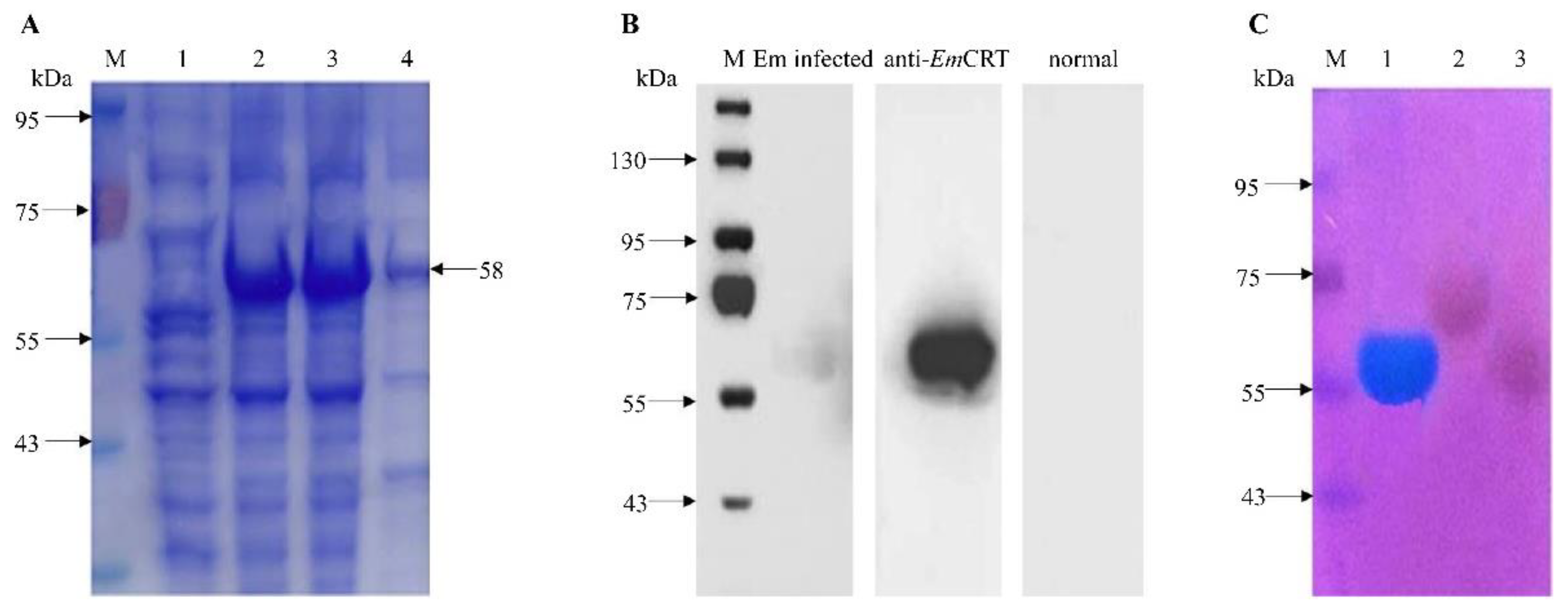
The recombinant EmCRT protein (rEmCRT) was expressed in BL21(DE3) under the induction of 0.4 mM IPTG, and the rEmCRT with the 6×His-tag expressed at the N-terminus was purified with immobilized metal affinity chromatography (IMAC). The SDS-PAGE analysis showed that the rEmCRT was expressed as a soluble protein with an apparent molecular weight of approximately 58 kDa, which is higher than the predicted size (47 kDa) based on the sequence, possibly due to the high negative charge of EmCRT (pI = 4.7) or other structural features [32,33,34,35] (Figure 2A). The purified rEmCRT was strongly recognized by the anti-EmCRT-antisera raised in the immunized mice as well as by the E. multilocularis-infected mouse sera (Figure 2B). The results indicate that the native EmCRT can be exposed to the host immune system and induce an antibody response during natural infection. Further staining with the cationic carbocyanine dye “Stains-all” showed rEmCRT was stained as dark blue (Figure 2C), indicating it is a Ca2+-binding protein [22].

3.2. Expression of Native EmCRT in E. multilocularis Metacestode Larvae

The RT-qPCR was performed to determine the transcriptional level of the Emcrt mRNA in the E. multilocularis PSCs and the total metacestode vesicles. As shown in Figure 3A, we found that the transcriptional level of Emcrt in the PSCs was significantly higher (4.1-fold) than that in the total metacestode vesicles (* p < 0.05).
The protein expression level of the native EmCRT was determined by Western blot, using anti-rEmCRT mouse sera. As shown in Figure 3B, one single 58 kDa protein band was clearly identified in the extracts from both the PSCs and metacestode vesicles with a much higher level in the PSCs, which is consistent with the results of the RT-qPCR, indicating EmCRT is mostly expressed in PSCs. The EmCRT protein was also detected in the metacestode vesicle fluid and the culture supernatants of both the PSCs and metacestode vesicles, indicating EmCRT is secreted mostly by PSCs and metacestode vesicles that contain PSCs.
The IFA with anti-EmCRT antisera showed that EmCRT was expressed in the germinal layer of the metacestode vesicle (Figure 4A), with a higher level on the surface of the PSCs mainly around the suckers (Figure 4B). In contrast, nothing was shown in both the PSCs and metacestode vesicle when probed with normal mouse sera. Taken together, these results demonstrate that EmCRT is constitutively present throughout the E. multilocularis larval stages and is especially abundant on the surface of PSCs.

3.3. Immune Responses to the Immunization with rEmCRT

The serum samples were gathered from mice one week after each immunization, and the antigen-specific IgG titers against rEmCRT were measured using ELISA. The immunization of the mice with rEmCRT formulated with FA induced high titers of rEmCRT-specific IgG, with the highest titer of 1:512,000 after the third immunization (Figure 5A). The IgG subclass of anti-EmCRT antibodies was measured. The results showed that the levels of IgG1 and IgG2a were significantly elevated after the last immunizations, with IgG1 predominant (Figure 5B).
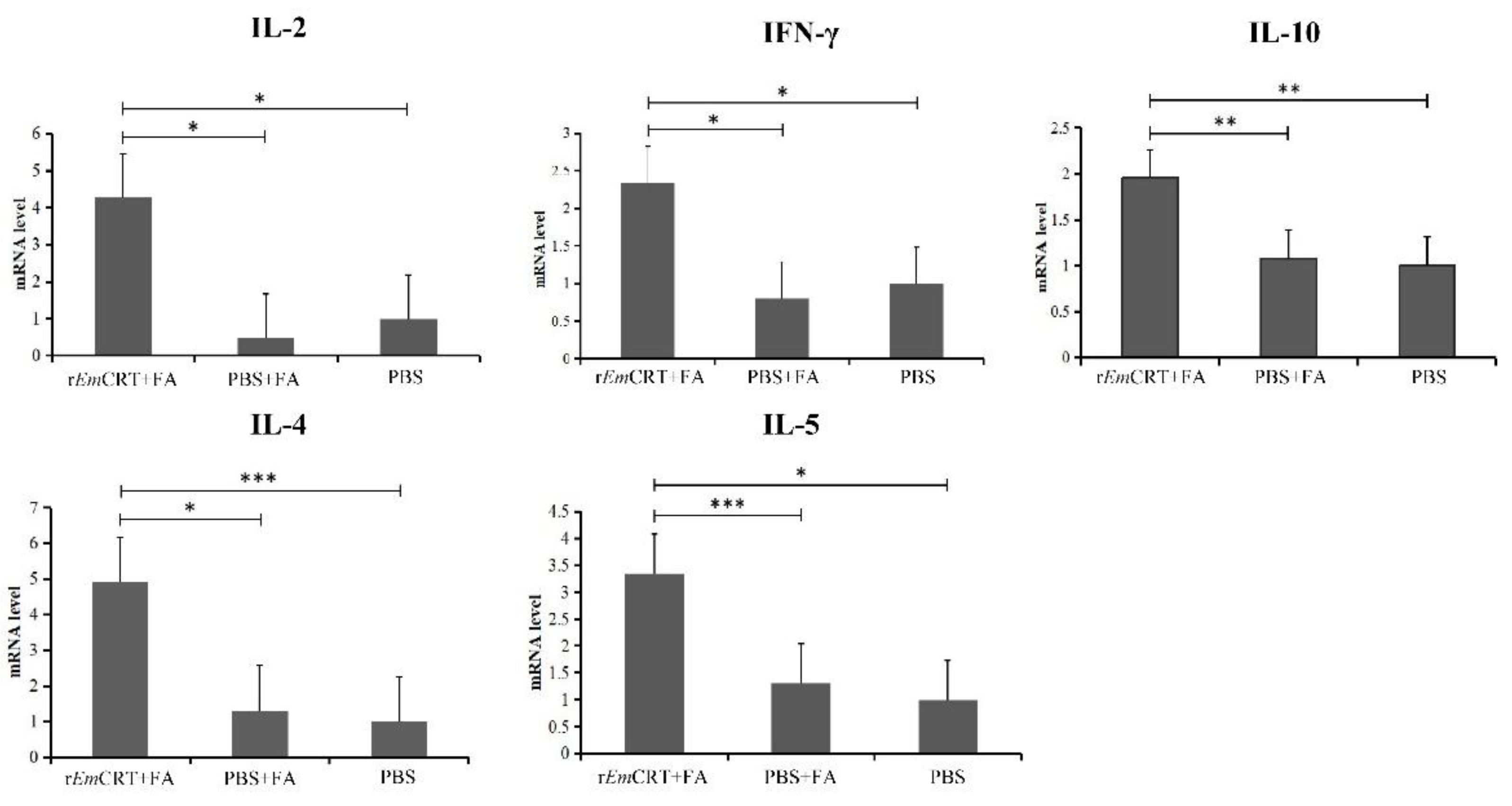
To determine the cellular immune responses upon immunizations with rEmCRT, the cytokine responses upon the immunization with rEmCRT were measured at the transcription level in splenocytes by a qPCR. Our results showed that the levels of the typical Th1 cytokines (IFN-γ and IL-2) and the Th2 cytokines (IL-4, IL-5 and IL-10) mRNA levels were all increased significantly in the mice vaccinated with rEmCRT compared with the mice that received the adjuvant or the PBS control groups, indicating that the rEmCRT vaccination induced mixed Th1 and Th2 responses. In addition, the Th2 cytokines IL-4 increased at the highest level, suggesting that rEmCRT may favor inducing a Th2-biased immune response (Figure 6), consistent with the result of the IgG subclass antibody levels (with IgG1 dominant over IgG2a).

3.4. Partially Protective Immunity Elicited by the Immunization with rEmCRT

The vaccine efficacy of the EmCRT immunization was observed by the reduced metacestode vesicle weight in the immunized mice. The result showed that immunization with EmCRT exhibited a 43.16% mean reduction in the weight of the total metacestode vesicles developed in the abdominal cavity 14 weeks after the challenge compared to the PBS control group (p < 0.05). The adjuvant control group also showed a 21.98% parasite weight reduction compared to the PBS group without significance. These results indicate that immunization with rEmCRT induced partial protective immunity against E. multilocularis infection in mice (Table 1).

4. Discussion

The metacestode stage of E. multilocularis is characterized by its tumor-like proliferation and metastasis, causing the life-threatening AE in humans [1,2,4]. The development of effective vaccines for AE has become urgent due to the lack of effective treatment and control means. The identification of the vaccine antigens that induce infective protective immunity and can be manufactured in a large scale is crucial for the vaccine development against AE [36]. In the effort to identify antigens that induce protective immunity against E. multilocularis infection, we cloned EmCRT from E. multilocularis metacestode cDNA and expressed it as a soluble recombinant protein in bacteria. Similar to other CRT, EmCRT has a typical structure of three domains, including a globular N-domain, an extended proline-rich P-domain and an acidic C-domain, and also has ability to bind to Ca2+ based on the staining with Stains-all [13]. The sequence alignment shows that EmCRT shares up to a 50–90% amino acid sequence identity with CRTs from other helminths. Importantly, EmCRT shares up to a 98% sequence identity with its counterpart in the closely related E. granulosus and an 89% identity with cestode T. solium, indicating immunization with EmCRT may produce cross-protective immunity to other cestode infections if EmCRT is a good vaccine candidate against E. multilocularis infection. The Western blot with anti-EmCRT mouse sera identified EmCRT was dominantly expressed in the PSCs of E. multilocularis and less in the metacestode vesicles, possibly because the vesicles contain some PSCs inside as well. Significantly, EmCRT was observed in the vesicle fluid and ES products of cultured PSCs and vesicles. IFA staining showed its expression on the germinal layer of the vesicles and more around the sucker of the scolex, indicating native EmCRT is secreted in the host environment and possibly involved in the protection of the invading parasite as a survival strategy. TsCRT from Trichinella spiralis has been identified as binding to the human complement component C1q and inhibiting the C1q-involved complement attack and neutrophil reaction to the infection of the nematode. However, the function of EmCRT is not clear yet in the role of E. multilocularis parasitism in the host.
Due to its secretory property and the potential functions involved in the survival of the parasitic helminth within the infected host, the recombinant EmCRT expressed in E. coli was applied to immunize mice for testing its immunogenicity and vaccine efficacy against the challenge of E. multilocularis PSCs. The results of the vaccine trial with EmCRT demonstrated that mice immunized with EmCRT formulated with Freund’s adjuvant produced a 43.16% reduction in the metacestode vesicles weight with statistical significance compared to mice that received the PBS only. It also showed a 21.98% reduction in the metacestode vesicle weight in the group that received the adjuvant only compared to the PBS control even though it is not statistically significant; possibly, the adjuvant itself boosts the mouse immune response that induces a certain resistance. The metacestode vesicle reduction in EmCRT-immunized mice is associated with significantly high titers of the IgG response. An IgG subtype analysis revealed IgG1 was dominant over IgG2a, indicating a Th2-biased immune response. The cytokine profile based on the mRNA expression showed that immunization with EmCRT induced splenocytes to produce both Th1 (IFN-γ and IL-2) and Th2 cytokines (IL-4, IL-5 and IL-10). However, the IL-4 was mostly induced to the highest level, combining with the dominant IgG1 response over IgG2a, and it indicates that the immunization of EmCRT formulated with FA induced both Th1 and Th2 responses, with Th2 dominant [37,38,39]. In general, our data indicated that EmCRT is a highly immunogenic antigen and induces a strong antibody response and Th1/2 cytokine response in immunized mice that may contribute to the protective immunity against the E. multilocularis infection in this study. The protective immunity was also observed in the immunization of CRTs from other helminths. More than one-third of hamsters infected with T. solium elicited anti-TsCRT IgG antibodies and IL-10 production [17]. Hamsters immunized with TsCRT produced 40–100% protection against T. solium oral infection, depending on the type of infected cysticerci [16]. Mice immunized with hookworm calreticulin intraperitoneally without adjuvant produced 43–49% fewer worms in their lungs following a hookworm larva challenge associated with low levels of serum IgE and moderate lung eosinophilia [15]. The spleen T cells from mice immunized with irradiated S. mansoni cercariae and acquired protection against S. mansoni infection strongly reacted to SmCRT with IL-4 production [14,39], indicating that CRT is able to induce a Th2 response and protective immune effect during helminth infections [40]. This evidence, combined with our observation in this study, further supports that EmCRT is a good vaccine candidate against E. multilocularis infection. However, the EmCRT immunization-induced metacestode vesicle weight reduction in this study was not high enough (43.16% compared to the PBS control). Further research is needed to optimize the protective immunity, such as the immunization route, different adjuvants or dosage, in order to achieve higher protection against E. multiocularis infection. Due to the complex life cycle and diversity of antigens in different stages, it may be necessary to use the combination of multiple antigens from different developmental stages or a multiepitope vaccine combined with T- and B-cell epitopes from different protective antigens [41] to increase protection against parasitic helminth infections. Some T-cell and B-cell epitopes have been identified from E. multiocularis based on the bioinformatic analysis [42], which is under investigation for a vaccine trial against AE.
In conclusion, this work demonstrates that EmCRT plays an important role in inducing partially protective immunity in the immunized mice and therefore could be considered a potential candidate for vaccine development against AE. It is necessary to optimize the protective immunity by changing the immunization route or regime or combining multiple effective vaccines to improve the vaccine efficacy against alveolar echinococcosis.

Author Contributions

L.Z. and L.C. conceived and designed the study. L.C., Z.X. and Z.C. performed the experiments. L.C., Z.X., S.X., Z.X., Y.Y. and J.C. analyzed the data. L.C. and Z.C. wrote the paper. L.Z., Y.W., Z.C., L.C. and B.Z. revised the manuscript. All authors read and approved the final manuscript.

Funding

This study was financially supported by grants from the National Natural Science Foundation of China (81860362) and the Natural Science Foundation of Inner Mongolia (2018BS08013).

Institutional Review Board Statement

The study was conducted in accordance with the Declaration of Helsinki and approved by the Institutional Animal Care and Use Committee of Xiamen University, with approval Number: 2013-0053.

Informed Consent Statement

Not applicable.

Data Availability Statement

Data included in this article are available from the corresponding author upon request and are also available at preprint Research Squire with DOI: https://doi.org/10.21203/rs.3.rs-1740725/v1 accessed on 26 September 2022.

Acknowledgments

We would like to thank Tang Chongti for kindly providing the parasite material and Yaying Wu, Zheni Xu and C.C. Xie for the mass spectrometry experiments and data analysis.

Conflicts of Interest

The authors declare that there is no conflict of interest.

References

  1. Deplazes, P.; Rinaldi, L.; Alvarez, R.C.; Torgerson, P.R.; Harandi, M.F.; Romig, T.; Antolova, D.; Schurer, J.M.; Lahmar, S.; Cringoli, G.; et al. Global Distribution of Alveolar and Cystic Echinococcosis. Adv. Parasitol. 2017, 95, 315–493. [Google Scholar]
  2. Yangdan, C.; Wang, C.; Zhang, L.; Ren, B.; Fan, H.; Lu, M. Recent advances in ultrasound in the diagnosis and evaluation of the activity of hepatic alveolar echinococcosis. Parasitol. Res. 2021, 120, 3077–3082. [Google Scholar] [CrossRef] [PubMed]
  3. Cheng, Z.; Liu, F.; Zhu, S.; Tian, H.; Wang, L.; Wang, Y. A rapid and convenient method for fluorescence analysis of in vitro cultivated metacestode vesicles from Echinococcus multilocularis. PLoS ONE 2015, 10, e118215. [Google Scholar] [CrossRef] [PubMed] [Green Version]
  4. Buttenschoen, K.; Kern, P.; Reuter, S.; Barth, T.F. Hepatic infestation of Echinococcus multilocularis with extension to regional lymph nodes. Langenbecks Arch. Surg. 2009, 394, 699–704. [Google Scholar] [CrossRef]
  5. Kern, P.; Menezes da Silva, A.; Akhan, O.; Müllhaupt, B.; Vizcaychipi, K.A.; Budke, C.; Vuitton, D.A. The Echinococcoses: Diagnosis, Clinical Management and Burden of Disease. Adv. Parasitol. 2017, 96, 259–369. [Google Scholar] [PubMed]
  6. Hemphill, A.; Stadelmann, B.; Rufener, R.; Spiliotis, M.; Boubaker, G.; Müller, J.; Müller, N.; Gorgas, D.; Gottstein, B. Treatment of echinococcosis: Albendazole and mebendazole—What else? Parasite 2014, 21, 70. [Google Scholar] [CrossRef]
  7. Ferreira, V.; Molina, M.C.; Valck, C.; Rojas, A.; Aguilar, L.; Ramírez, G.; Schwaeble, W.; Ferreira, A. Role of calreticulin from parasites in its interaction with vertebrate hosts. Mol. Immunol. 2004, 40, 1279–1291. [Google Scholar] [CrossRef]
  8. Schcolnik-Cabrera, A.; Oldak, B.; Juárez, M.; Cruz-Rivera, M.; Flisser, A.; Mendlovic, F. Calreticulin in phagocytosis and cancer: Opposite roles in immune response outcomes. Apoptosis 2019, 24, 245–255. [Google Scholar] [CrossRef]
  9. Wijeyesakere, S.J.; Bedi, S.K.; Huynh, D.; Raghavan, M. The C-Terminal Acidic Region of Calreticulin Mediates Phosphatidylserine Binding and Apoptotic Cell Phagocytosis. J. Immunol. 2016, 196, 3896–3909. [Google Scholar] [CrossRef] [Green Version]
  10. Ramírez-Toloza, G.; Aguilar-Guzmán, L.; Valck, C.; Ferreira, V.P.; Ferreira, A. The Interactions of Parasite Calreticulin With Initial Complement Components: Consequences in Immunity and Virulence. Front. Immunol. 2020, 11, 1561. [Google Scholar] [CrossRef]
  11. Fucikova, J.; Spisek, R.; Kroemer, G.; Galluzzi, L. Calreticulin and Cancer. Cell Res. 2021, 31, 5–16. [Google Scholar] [CrossRef] [PubMed]
  12. Zhao, L.; Shao, S.; Chen, Y.; Sun, X.; Sun, R.; Huang, J.; Zhan, B.; Zhu, X. Trichinella spiralis Calreticulin Binds Human Complement C1q As an Immune Evasion Strategy. Front. Immunol. 2017, 8, 636. [Google Scholar] [CrossRef] [PubMed] [Green Version]
  13. Shao, S.; Hao, C.; Zhan, B.; Zhuang, Q.; Zhao, L.; Chen, Y.; Huang, J.; Zhu, X. Trichinella spiralis Calreticulin S-Domain Binds to Human Complement C1q to Interfere With C1q-Mediated Immune Functions. Front. Immunol. 2020, 11, 572326. [Google Scholar] [CrossRef] [PubMed]
  14. Gengehi, N.E.; Ridi, R.E.; Tawab, N.A.; Demellawy, M.E.; Mangold, B.L. A Schistosoma mansoni 62-kDa band is identified as an irradiated vaccine T-cell antigen and characterized as calreticulin. J. Parasitol. 2000, 86, 993–1000. [Google Scholar] [CrossRef]
  15. Winter, J.A.; Davies, O.R.; Brown, A.P.; Garnett, M.C.; Stolnik, S.; Pritchard, D.I. The assessment of hookworm calreticulin as a potential vaccine for necatoriasis. Parasite Immunol. 2005, 27, 139–146. [Google Scholar] [CrossRef]
  16. León-Cabrera, S.; Cruz-Rivera, M.; Mendlovic, F.; Avila-Ramírez, G.; Carrero, J.C.; Laclette, J.P.; Flisser, A. Standardization of an experimental model of human taeniosis for oral vaccination. Methods 2009, 49, 346–350. [Google Scholar] [CrossRef]
  17. Mendlovic, F.; Cruz-Rivera, M.; Ávila, G.; Vaughan, G.; Flisser, A. Cytokine, antibody and proliferative cellular responses elicited by Taenia solium calreticulin upon experimental infection in hamsters. PLoS ONE 2015, 10, e121321. [Google Scholar]
  18. Tang, C.T.; Quian, Y.C.; Kang, Y.M.; Cui, G.W.; Lu, H.C.; Shu, L.M.; Wang, Y.H.; Tang, L. Study on the ecological distribution of alveolar Echinococcus in Hulunbeier Pasture of Inner Mongolia, China. Parasitology 2004, 128, 187–194. [Google Scholar] [CrossRef]
  19. Brehm, K.; Spiliotis, M. Recent advances in the in vitro cultivation and genetic manipulation of Echinococcus multilocularis metacestodes and germinal cells. Exp. Parasitol. 2008, 119, 506–515. [Google Scholar] [CrossRef]
  20. Spiliotis, M.; Mizukami, C.; Oku, Y.; Kiss, F.; Brehm, K.; Gottstein, B. Echinococcus multilocularis primary cells: Improved isolation, small-scale cultivation and RNA interference. Mol. Biochem. Parasitol. 2010, 174, 83–87. [Google Scholar] [CrossRef]
  21. Wang, H.; Li, J.; Guo, B.; Zhao, L.; Zhang, Z.; McManus, D.P.; Wen, H.; Zhang, W. In vitro culture of Echinococcus multilocularis producing protoscoleces and mouse infection with the cultured vesicles. Parasites Vectors 2016, 9, 411. [Google Scholar] [CrossRef] [Green Version]
  22. Campbell, K.P.; MacLennan, D.H.; Jorgensen, A.O. Staining of the Ca2+-binding proteins, calsequestrin, calmodulin, troponin C, and S-100, with the cationic carbocyanine dye “Stains-all”. J. Biol. Chem. 1983, 258, 11267–11273. [Google Scholar] [CrossRef]
  23. Sharma, N.; Sharma, R.; Rajput, Y.S.; Mann, B.; Gandhi, K. Distinction between glycomacropeptide and β-lactoglobulin with ‘stains all’ dye on tricine SDS-PAGE gels. Food Chem. 2021, 340, 127923. [Google Scholar] [CrossRef]
  24. Cabezón, C.; Cabrera, G.; Paredes, R.; Ferreira, A.; Galanti, N. Echinococcus granulosus calreticulin: Molecular characterization and hydatid cyst localization. Mol. Immunol. 2008, 45, 1431–1438. [Google Scholar] [CrossRef]
  25. Li, Y.; Xu, H.; Chen, J.; Gan, W.; Wu, W.; Wu, W.; Hu, X. Gene cloning, expression, and localization of antigen 5 in the life cycle of Echinococcus granulosus. Parasitol. Res. 2012, 110, 2315–2323. [Google Scholar] [CrossRef]
  26. Cao, S.; Gong, W.; Zhang, X.; Xu, M.; Wang, Y.; Xu, Y.; Cao, J.; Shen, Y.; Chen, J. Arginase promotes immune evasion of Echinococcus granulosus in mice. Parasites Vectors 2020, 13, 49. [Google Scholar] [CrossRef] [PubMed]
  27. Habjanec, L.; Halassy, B.; Tomasić, J. Immunomodulatory activity of novel adjuvant formulations based on Montanide ISA oil-based adjuvants and peptidoglycan monomer. Int. Immunopharmacol. 2008, 8, 717–724. [Google Scholar] [CrossRef] [PubMed]
  28. Lal, R.; Dhaliwal, J.; Dhaliwal, N.; Dharavath, R.N.; Chopra, K. Activation of the Nrf2/HO-1 signaling pathway by dimethyl fumarate ameliorates complete Freund’s adjuvant-induced arthritis in rats. Eur. J. Pharmacol. 2021, 899, 174044. [Google Scholar]
  29. Liu, C.; Han, X.; Lei, W.; Yin, J.; Wu, S.; Zhang, H. The efficacy of an alternative mebendazole formulation in mice infected with Echinococcus multilocularis. Acta Trop. 2019, 196, 72–75. [Google Scholar] [CrossRef]
  30. Bi, K.; Yang, J.; Wang, L.; Gu, Y.; Zhan, B.; Zhu, X. Partially Protective Immunity Induced by a 20 kDa Protein Secreted by Trichinella spiralis Stichocytes. PLoS ONE 2015, 10, e136189. [Google Scholar] [CrossRef] [PubMed]
  31. Li, R.; Yang, Q.; Guo, L.; Feng, L.; Wang, W.; Liu, K.; Tang, F.G. Immunological features and efficacy of the recombinant subunit vaccine LTB-EMY162 against Echinococcus multilocularis metacestode. Appl. Microbiol. Biotechnol. 2018, 102, 2143–2154. [Google Scholar] [CrossRef] [PubMed]
  32. Ostwald, T.J.; MacLennan, D.H. Isolation of a high affinity calcium-binding protein from sarcoplasmic reticulum. J. Biol. Chem. 1974, 249, 974–979. [Google Scholar] [CrossRef]
  33. Nagano, I.; Wu, Z.; Takahashi, Y. Functional genes and proteins of Trichinella spp. Parasitol. Res. 2009, 104, 197–207. [Google Scholar] [CrossRef]
  34. Rzepecka, J.; Rausch, S.; Klotz, C.; Schnöller, C.; Kornprobst, T.; Hagen, J.; Ignatius, R.; Lucius, R.; Hartmann, S. Calreticulin from the intestinal nematode Heligmosomoides polygyrus is a Th2-skewing protein and interacts with murine scavenger receptor-A. Mol. Immunol. 2009, 46, 1109–1119. [Google Scholar] [CrossRef] [PubMed]
  35. Tiwari, P.; Kaila, P.; Guptasarma, P. Understanding anomalous mobility of proteins on SDS-PAGE with special reference to the highly acidic extracellular domains of human E- and N-Cadherins. Electrophoresis. 2019, 40, 1273–1281. [Google Scholar] [CrossRef] [PubMed]
  36. Hein, W.R.; Harrison, G.B. Vaccines against veterinary helminths. Vet. Parasitol. 2005, 132, 217–222. [Google Scholar] [CrossRef]
  37. Hussaarts, L.; Yazdanbakhsh, M.; Guigas, B. Priming dendritic cells for th2 polarization: Lessons learned from helminths and implications for metabolic disorders. Front. Immunol. 2014, 5, 499. [Google Scholar] [CrossRef] [Green Version]
  38. Wang, L.; Wei, W.; Zhou, P.; Liu, H.; Yang, B.; Feng, L.; Ge, R.; Li, R.; Tang, F. Enzymatic characteristics and preventive effect of leucine aminopeptidase against Echinococcus multilocularis. Acta Trop. 2021, 222, 106066. [Google Scholar] [CrossRef]
  39. Lothstein, K.E.; Gause, W.C. Mining Helminths for Novel Therapeutics. Trends Mol. Med. 2021, 27, 345–364. [Google Scholar] [CrossRef]
  40. Jin, Q.; Zhang, N.; Li, W.; Qin, H.; Liu, Y.; Ohiolei, J.A.; Niu, D.; Yan, H.; Li, L.; Jia, W.Z.; et al. Trichinella spiralis Thioredoxin Peroxidase 2 Regulates Protective Th2 Immune Response in Mice by Directly Inducing Alternatively Activated Macrophages. Front. Immunol. 2020, 11, 2015. [Google Scholar] [CrossRef]
  41. Ortega-Pierres, G.; Vaquero-Vera, A.; Fonseca-Liñán, R.; Bermúdez-Cruz, R.M.; Argüello-García, R. Induction of protection in murine experimental models against Trichinella spiralis: An up-to-date review. J. Helminthol. 2015, 89, 526–539. [Google Scholar] [CrossRef] [PubMed]
  42. Chen, L.; Cheng, Z.; Wang, Y.; Zhao, L. Eukaryotic expression of calreticulin of Echinococcus multilocularis (EmCRT) and prediction of T-and B-cell epitopes of EmCRT. J. Pathog. Biol. 2020, 15, 1397–1403. [Google Scholar]
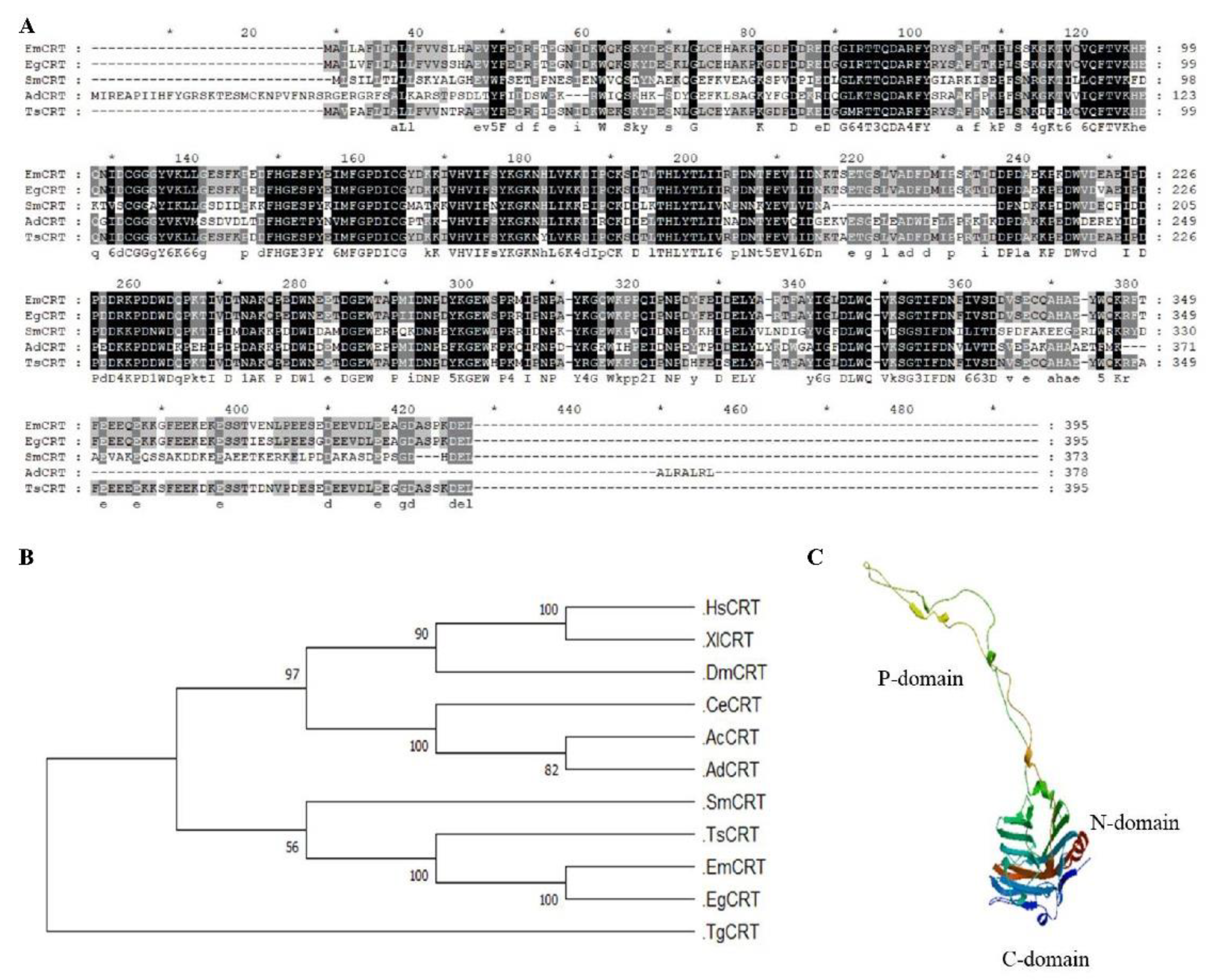
Figure 1. Amino acid sequence analysis and predicted three-dimensional structure of EmCRT. (A) Amino acid sequence alignment of EmCRT with its homologs from Echinococcus granulosus (EgCRT), Schistosoma mansoni (SmCRT), Taenia solium (TsCRT) and Ancylostoma duodenale (AdCRT) using the GeneDoc software. Numbers on the right refer to the last amino acid in each corresponding organism. Residues with dark shading indicate the same amino acids. Grey shading represents 80–90% similarity and light grey represents 60–70% similarity. (B) Phylogenetic analysis of the EmCRT and its homologues. Phylogenetic tree was generated with MEGA 6.0 by the neighbor-joining method (bootstrap = 1000). The numbers on the branches represent bootstrap values. Homo sapiens (HsCRT), Xenopus laevis (XlCRT), Drosophila melanogaster (DmCRT), Caenorhabditis elegans (CeCRT), Angiostrongylus cantonensis (AcCRT) and Trypanosoma grayi (TgCRT). (C) Modeled 3D structure of EmCRT.
Tropicalmed 07 00279 g001
Figure 2. Expression and characterization of rEmCRT. (A) Expression of rEmCRT in E. coli BL21 as soluble protein. After being induced with 0.4 mM IPTG for 4 h, the soluble fraction of induced (lane 3) or the whole induced lysate (lane 2) compared with lysate without induction (lane 1). Lane 4, purified rEmCRT (2 ug). (B) Recognition of rEmCRT by specific antibodies. Western blot analysis of rEmCRT (500 ng) with E. multilocularis-infected mouse sera, anti-rEmCRT mouse sera and with normal mouse sera. (C) Purified rEmCRT and other control proteins (100 ug each) stained with Stains-all. Lane 1, rEmCRT stained in blue; Lane 2, rEmBmi-1-GST as a E. multilocularis irrelevant protein control; and Lane 3, BSA control stained in red. M, molecular weight marker.
Tropicalmed 07 00279 g002
Figure 3. Expression of EmCRT in E. multilocularis metacestode larvae. (A) RT-qPCR analysis of transcription level of Emcrt mRNA in PSCs and metacestode vesicle tissue. After being normalized with Emelp, the fold-change of the Emcrt mRNA transcription in PSCs was calculated relative to that in metacestode vesicles. * p < 0.05. (B) Analysis of EmCRT protein expression in the lysates of metacestode vesicles (Lane 1), metacestode vesicle fluid (Lane 2), PSCs (Lane 3) and the excretory–secretory products released by cultured metacestode vesicles (Lane 4) and by PSCs (Lane 5) by Western blot with anti-rEmCRT mouse sera. Equal amount of total protein (100 ug), determined by Bradford assay, was loaded for each sample.
Tropicalmed 07 00279 g003
Figure 4. Immunolocalization of EmCRT in E. multilocularis metacestode larvae. Metacestode vesicles (A) and PSCs (B) were incubated with anti-EmCRT mouse serum and normal mouse serum, respectively, then probed with Alexa 488-conjugated anti-mouse IgG. The nuclei were stained with 4’, 6-diamidino-2-phenylindole (DAPI, blue). Scale bar = 100 μm. The arrows indicate the area with highly expressed EmCRT.
Tropicalmed 07 00279 g004
Figure 5. Antibody responses to the immunization with rEmCRT. (A) Total anti-EmCRT IgG titers in sera of all groups mice after last immunization. (B) OD450 value of anti-EmCRT IgG1 and IgG2a in sera of immunized mice with dilution 1:200. The values are presented as the arithmetic mean of five mice in the rEmCRT group ± standard error.
Tropicalmed 07 00279 g005
Figure 6. Splenocyte cytokine transcriptional profile of mice immunized with rEmCRT. Total cDNA was prepared from total RNA obtained from spleens of mice immunized with rEmCRT three times. The mRNA levels of IL-2, IFN-γ, IL-4, IL-5 and IL-10 were determined by RT-qPCR. Data are expressed as the mean of the ratio of each cytokine relative to GAPDH (housekeeping gene). * p < 0.05, ** p < 0.01, *** p < 0.001.
Tropicalmed 07 00279 g006
Table 1. Alveolar echinococcus weight in mice.
GroupsMetacestode Vesicle WeightMean Reduction in Parasite Weight (Compared with PBS)
rEmCRT + FA0.8539125 ± 0.33720192143.16% *
PBS + FA1.1720375 ± 0.49476575421.98%
PBS1.50224 ± 0.69753474
Metacestode vesicle weight in each group of mice after challenge with 2000 protoscoleces. Metacestode vesicle weight is presented as the mean ± S.D. The asterisks indicate statistically significant differences in metacestode vesicle weight compared to the PBS group (* p < 0.05).
Publisher’s Note: MDPI stays neutral with regard to jurisdictional claims in published maps and institutional affiliations.