Article

18-Fluorine-Fluorodeoxyglucose Positron Emission Computer Tomography Imaging in Melioidosis: Valuable but Not Essential

1
Infectious Diseases Department, Royal Darwin Hospital, Darwin, NT 0810, Australia
2
Infectious Diseases Department, Monash Health, Melbourne, VIC 3168, Australia
3
Division of Nuclear Medicine, Royal Darwin Hospital, Darwin, NT 0810, Australia
4
College of Medicine and Public Health, Flinders University, Adelaide, SA 5042, Australia
5
Global and Tropical Health Division, Menzies School of Health Research, Charles Darwin University, Darwin, NT 0811, Australia
*
Author to whom correspondence should be addressed.
Trop. Med. Infect. Dis. 2025, 10(3), 69; https://doi.org/10.3390/tropicalmed10030069
Submission received: 10 February 2025 / Revised: 3 March 2025 / Accepted: 5 March 2025 / Published: 6 March 2025

Abstract

:
Melioidosis is an endemic tropical disease caused by Burkholderia pseudomallei. It typically causes pulmonary disease and bacteraemia but can disseminate to cause multi-organ disease. 18-F FDG PET/CT has an evolving role in diagnosing other infectious diseases, especially where the pathogen or extent of infection is challenging to elucidate clinically and with conventional imaging (CT, US and MRI). We present a case series of patients diagnosed with melioidosis who also underwent 18-F FDG PET/CT from December 18th 2018 to September 30th 2022. Indications for imaging were categorised and analysed as to whether 18-F FDG PET/CT changed management over conventional imaging. Twenty-one 18-F FDG PET/CT scans were performed for sixteen patients. Two scans (9.5%) performed for pyrexia of unknown origin changed management in both cases. Twelve scans (57.1%) performed to ascertain the extent of dissemination of melioidosis changed management in only three (25%) cases. Five scans (23.8%) performed to monitor the response to treatment of known foci changed management in all five cases. Five scans (23.8%) performed for suspected or known malignancy changed management in three (60%) cases. 18-F FDG PET/CT is an emerging tool which improves diagnosis and changes the management of melioidosis when applied judiciously and for well-selected indications.

1. Introduction

Melioidosis is an infectious disease caused by Burkholderia pseudomallei, a Gram-negative bacterium which can infect humans and a wide range of animals. B. pseudomallei is commonly found in soil and surface groundwater and is endemic to tropical areas, especially in northern Australia and Southeast Asia [1]. Infection is acquired via exposure through damaged skin, inhalation or ingestion and usually results in subclinical disease that is cleared by immunocompetent hosts. B. pseudomallei infection that causes symptoms is termed melioidosis, with the majority of patients (88%) presenting with an acute infection, and less commonly chronic infection (9%) and reactivation of latent infection (3%) [2,3]. Bacteraemia occurs in over half of all patients with melioidosis, and the most common clinical presentations are pneumonia, skin infection and genitourinary infection, with less common presentations including soft tissue abscesses, septic arthritis, osteomyelitis and neurological infection [4].
18-Fluorine-Fluorodeoxyglucose Positron Emission Computer Tomography (18-F FDG PET/CT) has an increasing role in the investigation and monitoring of infectious diseases, with applications in pyrexia and bacteraemia of unknown origin, Staphylococcus aureus bacteraemia, as well as prosthetic valve infective endocarditis and vascular graft infections [5,6,7,8,9,10]. In this context, 18-F FDG PET/CT has had utility in identifying and monitoring foci of disseminated infection, suggesting that it may have a role in melioidosis as well, where the location and extent of foci are often not readily apparent and where the monitoring of these foci over a prolonged treatment course is essential.
The role of 18-F FDG PET/CT in the investigation of melioidosis is not defined in the literature but has been described in case reports and small case series involving patients presenting with pyrexia of unknown origin to identify foci of disseminated melioidosis [11]. Other case series have found utility in identifying both symptomatic and asymptomatic sites of infection in patients with known melioidosis [12,13]. It has also been used serially to assess treatment efficacy where other imaging modalities are confounded by residual post-infective changes [11]. 18-F FDG PET/CT has also been used in the workup of lesions suspicious for malignancy which, on biopsy, ultimately cultured B. pseudomallei [14,15].
The Darwin Prospective Melioidosis Study began in October 1989, and 18-F FDG PET/CT first became available at Royal Darwin Hospital on 5 December 2018. We investigated all 18-F FDG PET/CT scans performed from this date to the last case of melioidosis in the 2021-22 wet season, which occurred on 30 September 2022, with the aim to determine the diagnostic and management impacts of 18-F FDG PET/CT in patients with melioidosis.

2. Materials and Methods

We included all patients with melioidosis confirmed on blood, sputum, urine, fluid or tissue culture who also had 18-F FDG PET/CT performed between 5 December 2018 and 30 September 2022. The indications for 18-F FDG PET/CT were determined, and timing and comparisons with other radiology and patient clinical course were analysed. Therapy for cases and duration of intravenous and oral eradication phases were as in the 2020 Darwin melioidosis treatment protocol [16].
This study was approved by the ethics committee of the Northern Territory Department of Health and Menzies School of Health Research (approval number 02/38). A waiver of consent from individual participants was granted. Data were accessed for research purposes only and included only deidentified patient information. All methods were performed in accordance with the relevant guidelines and regulations. This study did not receive funding.

3. Results

There were 192 melioidosis cases between 5 December 2018 and 30 September 2022, 10 (5.2%) of which died due to melioidosis and 25 (13.0%) of which had one or more 18-F FDG PET/CT procedures performed for any reason. Upon review, nine of the 18-F FDG PET/CT scans performed were entirely unrelated to the diagnosis or management of melioidosis and were excluded from our study. Notably, we included patients who had 18-F FDG PET/CT performed for the diagnosis or management of malignancy but where the PET diagnosed or changed the management of their melioidosis. We therefore analysed the 21 18-F FDG PET/CT scans performed for the remaining 16 patients, with some 18-F FDG PET/CT scans having multiple indications.
The indications for 18-F FDG PET/CT were placed into four categories:
1.
Two 18-F FDG PET/CT scans performed for pyrexia of unknown origin (9.5%);
2.
Twelve 18-F FDG PET/CT scans for ascertaining the extent of dissemination of melioidosis (57.1%);
3.
Five 18-F FDG PET/CT scans for monitoring the response to treatment of known foci (23.8%);
4.
Five 18-F FDG PET/CT scans for suspected or known malignancy (23.8%).
Demographics, comorbidities, clinical scenarios, indications for 18-F FDG PET/CT, the results of 18-F FDG PET/CT and the changes in management (if any) for the 16 cases are summarised in Table A1.
For each 18-F FDG PET/CT indication, it was determined whether the 18-F FDG PET/CT changed management based on the following:
1.
Pyrexia of unknown origin 2/2 (100%);
2.
The extent of dissemination of melioidosis 3/12 (25%);
3.
The monitoring response to the treatment of known foci 5/5 (100%);
4.
Suspected or known malignancy 3/5 (60%).
When 18-F FDG PET/CT was performed for pyrexia of unknown origin, it changed management by prompting biopsy in both cases. In case 6 (Table A1), it prompted biopsy of an 18-F FDG PET/CT avid lesion, which cultured B. pseudomallei 26 days after presenting to the hospital and subsequent to three CT scans and one MRI over the 26 days. In case 8 (Table A1, Figure 1), CT identified multiple splenic lesions; however, 18-F FDG PET/CT identified an 18-F FDG PET/CT avid lymph node not identified on CT, prompting a lymph node biopsy, which was culture-positive for B. pseudomallei, enabling the diagnosis of melioidosis.
18-F FDG PET/CT performed for determining the extent of dissemination was helpful in a minority of cases, changing management in only 3 of 12 18-F FDG PET/CT scans. Case 15 (Table A1) provides an example of circumstances in which 18-F FDG PET/CT identified metabolic activity in prosthetic material at the thoracic endovascular aortic repair (TEVAR) site not identified on CT angiography after a positive blood culture for B. pseudomallei, clarifying the need for lifelong suppressive antibiotics. It also identified a splenic infarct suggesting septic emboli and suggesting to clinicians that the original penetrating ulcer requiring TEVAR was very likely a mycotic pseudoaneurysm. In case 10, a man with prostate and cutaneous melioidosis with poor treatment response prompted clinicians to look for further disseminated foci of melioidosis. 18-F FDG PET/CT identified possible ischial osteomyelitis underlying known cutaneous melioidosis. This extended the treatment duration to a minimum of 6 weeks [16]; however, this duration was subsequently over-ridden by ongoing urine culture positivity at 6 weeks due to an insufficiently drained prostatic abscess also seen on 18-F FDG PET/CT.
When 18-F FDG PET/CT was used to monitor treatment response, it was most helpful when treatment had already extended beyond the minimum recommended by the Darwin treatment protocol due to poor response to therapy or when the patient had persistent symptoms at the time of planned completion of intravenous treatment [16]. This was exemplified in case 2, when a 10-week induction phase of intravenous therapy (rather than 6 weeks) was chosen for osteomyelitis disseminated throughout the skeleton due to clinical and radiological progression on MRI. In addition, the oral eradication phase was also extended from 6 to 9 months due to persistent symptoms. The 18-F FDG PET/CT results showing complete resolution allowed both the clinician and the patient to feel comfortable with stopping treatment despite persistent arthralgia. Similarly in case 8, the 18-F FDG PET/CT showed increasing avidity of splenic foci at 3.5 weeks of a planned 4-week treatment period despite no volumetric change seen on CT, prompting extension of induction intravenous therapy (Figure 1). 18-F FDG PET/CT also gave clinicians confidence to transition from intensive intravenous phase to oral eradication phase in case 11, where the patient recently had a relapse of melioidosis despite having undergone treatment. Furthermore, in case 15, complete resolution of 18-F FDG PET/CT avidity at the TEVAR graft site while taking suppressive cotrimoxazole once daily suggested that this was sufficient for the long-term management of the infected graft. Notably, every patient with melioidosis who had 18-F FDG PET/CT performed to monitor treatment response already had a preceding 18-F FDG PET/CT result for comparison.
In the five cases in which 18-F FDG PET/CT was performed for the workup of suspected or known malignancy, 18-F FDG PET/CT excluded malignancy in two cases (cases 8 and 9) by prompting biopsy, which subsequently cultured B. pseudomallei (Figure 1). In case 1, 18-F FDG PET/CT staged the patient’s breast cancer which was previously diagnosed when the patient presented with B. pseudomallei breast abscess and therefore guided the oncological treatment approach. In case 4, 18-F FDG PET/CT appearance was not consistent with malignancy, and it was planned for the patient to undergo serial imaging, presenting in the interim with B. pseudomallei sepsis, with the 18-F FDG PET/CT not significantly changing management.

4. Discussion

CT and ultrasound imaging have been available for all three decades of the Darwin Prospective Melioidosis Study. MRI has only been available since 1993, and 18-F FDG PET/CT has only been available since 2018. Mortality from melioidosis in the Darwin study improved from 31% in the 1989–1994 period to 6% in the 2014–2019 period as a result of advances in the overall diagnostic and management approach of which developments in diagnostic imaging are a part [2]. In most locations where melioidosis is endemic, 18-F FDG PET/CT, MRI and, in many cases, even CT are unavailable. Our study indicates that there is likely a small but important role for 18-F FDG PET/CT when performed for specific indications to optimise the diagnosis and management of melioidosis. As suggested in the literature, 18-F FDG PET/CT has a role in pyrexia of unknown origin, and this holds true for melioidosis, guiding biopsy to isolate B. pseudomallei despite unhelpful CTs being performed prior. 18-F FDG PET/CT also changes management when it is performed to monitor sites of known infection, guiding clinicians when melioidosis does not clinically, biochemically or radiologically respond to treatment as expected. 18-F FDG PET/CT can help guide antibiotic duration for clinicians and can reassure patients with ongoing symptoms when treatment has extended beyond the guidelines outlined in the 2020 Darwin melioidosis treatment protocol [16]. 18-F FDG PET/CT was never performed at a time point earlier than the planned completion of the intensive or eradication phase with the intention of cutting short the duration of therapy, so from our data, we cannot draw any conclusions about using 18-F FDG PET/CT to shorten therapy. 18-F FDG PET/CT, however, adds little to conventional CT imaging in the majority of cases when used to determine the extent of infection. Our data suggest that 18-F FDG PET/CT could be reserved for specific indications such as suspected infected prostheses, where the sensitivity of CT may be limited, or for patients in which an occult uncontrolled source is clinically suspected but has not been identified using conventional imaging. It is important to note, however, that when 18-F FDG PET/CT was used to monitor infection, it was the most helpful if it was able to be compared to a prior 18-F FDG PET/CT scan performed for another indication, usually one performed to determine the extent of infection. 18-F FDG PET/CT performed for the workup of suspected or known malignancy has variable utility and is challenging to draw conclusions from; diagnosing melioidosis on biopsy was an unexpected finding. However, it highlights how melioidosis can frequently mimic malignancy radiographically and that biopsies in melioidosis endemic regions should be sent fresh for bacterial culture in addition to histology.
Our study has a number of limitations: First is its generalisability, as there are few other healthcare settings with such high incidence of melioidosis with the same availability and access to 18-F FDG PET/CT. Melioidosis is known to be endemic in northern Australia and Southeast Asia and has increasingly been identified across South Asia, Africa, the Americas and the Pacific, most frequently in low- and middle-income countries [17]. Of 192 patients diagnosed with melioidosis while 18-F FDG PET/CT was available in the Darwin study, only 16 (8.3%) underwent 18-F FDG PET/CT, and of these, only 9 (4.7% of total cases) underwent 18-F FDG PET/CT that changed management. This suggests that for the majority of cases, conventional imaging is sufficient in terms of diagnostics. The significant mortality reduction seen over the 30 years of the Darwin Prospective Melioidosis Study occurred prior to the availability of 18-F FDG PET/CT2.
Another limitation is the retrospective nature of this study, which requires judgements to be made regarding the expected clinical course if 18-F FDG PET/CT had not been performed. This is especially difficult to determine for 18-F FDG PET/CT scans performed at the end of a planned course of treatment for patients with ongoing symptoms. While we determined that these 18-F FDG PET/CT scans changed management in each case, this may overestimate 18-F FDG PET/CT’s utility here; a viable alternative of ceasing therapy and monitoring progress may have achieved similar results. However, this could have come at the cost of clinician and patient confidence that melioidosis had been adequately treated, with the results of 18-F FDG PET/CT providing reassurance, especially in patients who had previously had relapse of melioidosis or had experienced prolongation of treatment to over 1 year.

5. Conclusions

In conclusion, this study demonstrates the key indications for which 18-F FDG PET/CT changes the diagnosis and management of melioidosis compared to conventional imaging. This method most notably facilitates pathogen identification in pyrexia of unknown origin via biopsy and guides management when treatment has already extended beyond treatment guidelines due to poor response to planned therapy. It also supports the strength of conventional imaging, primarily with CT, in identifying the extent of infection to determine treatment duration, with 18-F FDG PET/CT being especially useful if there is strong clinical suspicion of an uncontrolled source despite an unrevealing CT.

Author Contributions

Conceptualization, B.J.C. and J.J.M.; methodology, J.B., B.J.C., J.J.M. and N.K.; formal analysis and data curation, J.B. and N.K.; writing—original draft preparation, J.B.; writing—review and editing, J.B., B.J.C. and J.J.M.; supervision and project administration, B.J.C. and J.J.M. All authors have read and agreed to the published version of the manuscript.

Funding

This research received no external funding.

Institutional Review Board Statement

This study was conducted in accordance with the Declaration of Helsinki. This study was approved by the ethics committee of the Northern Territory Department of Health and Menzies School of Health Research (approval number 02/38).

Informed Consent Statement

Patient consent was waived by the ethics committee as data were accessed retrospectively for research purposes only and included only deidentified patient information.

Data Availability Statement

All data relevant to the article are included in the article. Further inquiries can be directed to the corresponding authors upon reasonable request.

Acknowledgments

We acknowledge and are grateful for the expertise and support of our laboratory and clinical colleagues in the diagnosis and management of the patients in this study.

Conflicts of Interest

The authors declare no conflicts of interest.

Abbreviations

The following abbreviations are used in this manuscript:
PETPositron Emission Tomography
FDGFluorodeoxyglucose
CTComputed Tomography
USUltrasound
MRIMagnetic Resonance Imaging
TEVARThoracic Endovascular Aortic Repair

Appendix A

Table A1. Demographics, comorbidities, clinical scenarios, indications for 18-F FDG PET/CT, results of 18-F FDG PET/CT and changes in management (if any). If 18-F FDG PET/CT, results changed management this has been highlighted in green, and if it did not this is highlighted in red.
Demographics and ComorbiditiesClinical ScenarioIndication Category18-F FDG PET/CT ResultChange in Management
1. 69F
T2DM, CKD, IHD, asthma
Previous pulmonary melioidosis, represented with concurrent breast cancer and B. pseudomallei breast abscess1st PET: Extent of dissemination AND suspected/known malignancy1st PET: Study identified breast lesion with no disseminated or metastatic disease1st PET: Did not change management of melioidosis

1st PET: Guided oncological management approach
2. 56F
T2DM
Disseminated melioidosis with pulmonary, splenic, liver, peripancreatic and widespread upper and lower limb osteomyelitis with multiple septic joints1st PET: Extent of dissemination

2nd PET: Monitoring response to treatment
1st PET: Identified extent of infection but did not add to previous MRI results

2nd PET: Performed 9 months later; showed resolution of PET avidity
1st PET: Did not change management over clinical examination and MRI findings

2nd PET: Gave treating clinicians confidence to cease treatment despite ongoing pain thought to be due to mechanical arthritis
3. 81M
Myelodysplastic syndrome, CKD, IHD
Initially thought to be B. pseudomallei bacteraemia with no focus as it had unremarkable CT CAP results 1st PET: Extent of dissemination1st PET: Multiple FDG avid nodules in lung, mediastinum and duodenum1st PET: Identified pulmonary involvement not seen on CT 2 weeks earlier, altering duration of IV intensive phase of therapy
4. 73F
T2DM, asthma/COPD, cognitive impairment
Presented 1 month prior to diagnosis of melioidosis with pneumonia and lung lesion, improved and discharged home, PET ordered for malignancy investigation given persistent lung lesion; represented following PET with respiratory sepsis1st PET: Suspected/known malignancy1st PET: Partially cystic lung lesion thought unlikely to be malignant; no metastatic or disseminated disease1st PET: Did not change management compared to conventional imaging
5. 42M
Asthma
Fibula ORIF
Disseminated melioidosis with pulmonary splenic and prostatic and cutaneous abscesses Clinical concern for infected metalware at ORIF site1st PET: Extent of dissemination1st PET: PET performed 2 months after diagnosis; showed active prostate infection, no other foci of infection1st PET: Did not change management compared to conventional imaging and clinical impression.
6. 31F
T2DM, RHD
Disseminated melioidosis with preceding pyrexia of unknown origin; presented with back pain and rising melioidosis serology titre; subcarinal lymphadenopathy identified on CT1st PET: Pyrexia of unknown origin1st PET: Identified multiple nodal, splenic and small bowel foci1st PET: Prompted lymph node biopsy; identified small bowel and splenic FDG avid lesions that were not identified on CT or abdominal ultrasound; guided duration of IV intensive therapy
7. 29F
Hazardous alcohol use
Disseminated melioidosis with bacteraemia, scalp and extradural abscess1st PET: Extent of dissemination1st PET: Large left temporal melioidosis site and few subcutaneous and cervical nodularities; no distant foci1st PET: Did not change management of conventional imaging with CT and MRI
8. 50M
T2DM, hazardous alcohol use
Disseminated melioidosis with splenic hepatic and nodal foci; presented with pyrexia of unknown origin, arthralgia and abdominal pain; CT findings of disseminated lesions concerning for metastatic malignancy1st PET: Pyrexia of unknown origin AND suspected/confirmed malignancy

2nd PET: Monitoring response to treatment

3rd PET: Monitoring response to treatment
1st PET: FDG avid paratracheal lymphadenopathy and splenic foci

2nd PET: New axillary FDG avid axillary lymph nodes and increased avidity of known splenic foci

3rd PET: No positive foci of infection identified
1st PET: Identified new foci which guided biopsy, leading to diagnosis of melioidosis

2nd PET: Prompted extension of IV intensive phase from 4 weeks to total of 12 weeks due to increasing SUVmax of splenic foci at 3.5 weeks of IV therapy; CT performed prior to PET showed that these lesions were volumetrically unchanged

3rd PET: Gave clinicians confidence to cease antibiotics at planned duration despite ongoing non-specific symptoms
9. 68M
T2DM
Concurrent melioidosis and tuberculosis diagnosed after positive PET and lymph node biopsy performed during investigation for hoarse voice initially presumed to be due to cancer1st PET: Suspected/confirmed malignancy1st PET: Low-grade mediastinal and cervical lymphadenopathy1st PET: Identified foci were limited to lymphadenitis in mediastinum and cervical lymph nodes guiding duration of therapy
10. 49M
CKD3b, urethral stricture, COPD, latent TB, chronic hepatitis B, right AKA due to previous necrotising fasciitis, anal SCC in remission
Disseminated melioidosis with osteomyelitis, prostatic and cutaneous foci; relapsed 18 months later with prostatic, splenic and hepatic abscesses1st PET: Extent of dissemination1st PET: Identified prostatic, ischial, stomach and colonic foci1st PET: Identified bony FDG avidity suggestive of osteomyelitis not previously identified on other imaging, which guided planned duration, but duration extended beyond this due to persistently positive urine cultures
11. 65F
T1DM, HFrEF, pulmonary HTN
Pulmonary melioidosis with incomplete eradication phase; relapse representation 4 months later with 1st PET performed at that time and 2nd performed 6 weeks later1st PET: Extent of dissemination

2nd PET: Monitoring response to treatment
1st PET: Bilateral pulmonary and mediastinal nodal foci; identified nonspecific uptake in small bowel

2nd PET: Near resolution of pulmonary and mediastinal foci; resolution of small bowel focus
1st PET: Did not change management compared to chest CT

2nd PET: Gave clinicians confidence to transition to oral eradication phase therapy given previous relapse
12. 45F
T2DM, asthma/COPD
Pulmonary melioidosis with pleural effusion; delay in isolating B. pseudomallei with multiple courses of ineffective antibiotics1st PET: Extent of dissemination1st PET: No hypermetabolic findings to suggest active melioidosis1st PET: Did not change management over CT
13. 66M
T2DM, CKD3a, multiple myeloma
Pulmonary melioidosis concurrent with active treatment with lenalidomide for multiple myeloma1st PET: Extent of dissemination AND suspected/known malignancy1st PET: Multiple hypermetabolic lesions in mediastinal lymph nodes and skeleton1st PET: Did not change management compared to CT
14. 71M
Hazardous alcohol use, CP-A cirrhosis
Cutaneous melioidosis with dissemination and subsequent osteomyelitis 1st PET: Extent of dissemination1st PET: Active soft tissue and possible osteomyelitis 1st PET: Did not change management compared to CT; MRI was only modality that clearly showed evidence of osteomyelitis
15. 65M
No comorbidities
Underwent TEVAR for penetrating thoracic aortic ulcer 10 days prior to diagnosis of bacteraemic melioidosis after representing febrile1st PET: Extent of dissemination

2nd PET: Monitoring response to treatment
1st PET: Active infection with paraortic and retrocrural collections

2nd PET: Complete metabolic response to antibiotic therapy
1st PET: Confirmed clinical suspicion of graft infection not identified on CT

2nd PET: Complete resolution of metabolic changes while on lifelong suppressive cotrimoxazole, suggesting adequate suppression
16. 71F
Bronchiectasis
Pulmonary melioidosis after presenting with 6-month history of productive cough1st PET: Extent of dissemination1st PET: No hypermetabolic active foci of infection1st PET: Did not change management of melioidosis compared to CT

References

  1. Wiersinga, W.J.; Virk, H.S.; Torres, A.G.; Currie, B.J.; Peacock, S.J.; Dance, D.A.; Limmathurotsakul, D. Melioidosis. Nat. Rev. Dis. Primers 2018, 4, 17107. [Google Scholar] [CrossRef] [PubMed]
  2. Currie, B.J.; Mayo, M.; Ward, L.M.; Kaestli, M.; Meumann, E.M.; Webb, J.R.; Woerle, C.; Baird, R.W.; Price, R.N.; Marshall, C.S.; et al. The Darwin Prospective Melioidosis Study: A 30-year prospective, observational investigation. Lancet Infect. Dis. 2021, 21, 1737–1746. [Google Scholar] [CrossRef] [PubMed]
  3. Currie, B.J. Melioidosis: Evolving concepts in epidemiology, pathogenesis, and treatment. Semin. Respir. Crit. Care Med. 2015, 36, 111–125. [Google Scholar] [CrossRef] [PubMed]
  4. Birnie, E.; Virk, H.S.; Savelkoel, J.; Spijker, R.; Bertherat, E.; Dance, D.A.; Limmathurotsakul, D.; Devleesschauwer, B.; Haagsma, J.A.; Wiersinga, W.J. Global burden of melioidosis in 2015: A systematic review and data synthesis. Lancet Infect. Dis. 2019, 19, 892–902. [Google Scholar] [CrossRef] [PubMed]
  5. Habib, G.; Lancellotti, P.; Antunes, M.J.; Bongiorni, M.G.; Casalta, J.P.; Del Zotti, F.; Dulgheru, R.; El Khoury, G.; Erba, P.A.; Iung, B.; et al. 2015 ESC Guidelines for the management of infective endocarditis: The Task Force for the Management of Infective Endocarditis of the European Society of Cardiology (ESC)Endorsed by: European Association for Cardio-Thoracic Surgery (EACTS), the European Association of Nuclear Medicine (EANM). Eur. Heart J. 2015, 36, 3075–3128. [Google Scholar] [PubMed]
  6. Kung, B.T.; Seraj, S.M.; Zadeh, M.Z.; Rojulpote, C.; Kothekar, E.; Ayubcha, C.; Ng, K.S.; Ng, K.K.; Au-Yong, T.K.; Werner, T.J.; et al. An update on the role of 18F-FDG-PET/CT in major infectious and inflammatory diseases. Am. J. Nucl. Med. Mol. Imaging 2019, 9, 255–273. [Google Scholar] [PubMed]
  7. van der Vaart, T.W.; Fowler, V.G. Positron emission tomography-computed tomography in patients with Staphylococcus aureus bacteraemia: Worth the wait? Clin. Microbiol. Infect. 2023, 29, 1361–1363. [Google Scholar] [CrossRef] [PubMed]
  8. Ghanem-Zoubi, N.; Kagna, O.; Abu-Elhija, J.; Mustafa-Hellou, M.; Qasum, M.; Keidar, Z.; Paul, M. Integration of FDG-PET/CT in the Diagnostic Workup for Staphylococcus aureus Bacteremia: A Prospective Interventional Matched-cohort Study. Clin. Infect. Dis. 2021, 73, e3859–e3866. [Google Scholar] [CrossRef] [PubMed]
  9. Gomes, A.; Glaudemans, A.W.; Touw, D.J.; van Melle, J.P.; Willems, T.P.; Maass, A.H.; Natour, E.; Prakken, N.H.; Borra, R.J.; van Geel, P.P.; et al. Diagnostic value of imaging in infective endocarditis: A systematic review. Lancet Infect. Dis. 2017, 17, e1–e14. [Google Scholar] [CrossRef] [PubMed]
  10. Lauri, C.; Signore, A.; Glaudemans, A.W.; Treglia, G.; Gheysens, O.; Slart, R.H.; Iezzi, R.; Prakken, N.H.; Debus, E.S.; Honig, S.; et al. Evidence-based guideline of the European Association of Nuclear Medicine (EANM) on imaging infection in vascular grafts. Eur. J. Nucl. Med. Mol. Imaging 2022, 49, 3430–3451. [Google Scholar] [CrossRef] [PubMed]
  11. Subran, B.; Ackermann, F.; Watin-Augouard, L.; Rammaert, B.; Rivoisy, C.; Vilain, D.; Canzi, A.M.; Kahn, J.E. Melioidosis in a European traveler without comorbidities: A case report and literature review. Int. J. Infect. Dis. 2013, 17, e781–e783. [Google Scholar] [CrossRef] [PubMed]
  12. Kulkarni, P.; Shelley, S.; Elangoven, I.M.; Jaykanth, A.; Ejaz, A.P.; Rao, N.S. 18-Fluorine-Fluorodeoxyglucose Positron Emission Tomography-Computed Tomography in the Evaluation of the Great Masquerader Melioidosis: A Case Series. Indian J. Nucl. Med. 2020, 35, 222–225. [Google Scholar] [CrossRef] [PubMed]
  13. Ayub, I.I.; Thangaswamy, D.; Krishna, V.; Sridharan, K.S. Role for Positron Emission Tomography-Computed Tomography in Melioidosis. Indian J. Nucl. Med. 2021, 36, 354–356. [Google Scholar] [CrossRef] [PubMed]
  14. Zaw, K.K.; Wasgewatta, S.L.; Kwong, K.K.; Fielding, D.; Heraganahally, S.S.; Currie, B.J. Chronic Pulmonary Melioidosis Masquerading as lung malignancy diagnosed by EBUS guided sheath technique. Respir. Med. Case Rep. 2019, 28, 100894. [Google Scholar] [CrossRef] [PubMed]
  15. Zhao, J.; Yap, A.; Wu, E.; Yap, J. A mimic of bronchogenic carcinoma—Pulmonary melioidosis. Respir. Med. Case Rep. 2020, 29, 101006. [Google Scholar] [CrossRef] [PubMed]
  16. Sullivan, R.P.; Marshall, C.S.; Anstey, N.M.; Ward, L.; Currie, B.J. 2020 Review and revision of the 2015 Darwin melioidosis treatment guideline; paradigm drift not shift. PLoS Negl. Trop. Dis. 2020, 14, e0008659. [Google Scholar] [CrossRef] [PubMed]
  17. Currie, B.J.; Meumann, E.M.; Kaestli, M. The Expanding Global Footprint of Burkholderia pseudomallei and Melioidosis. Am. J. Trop. Med. Hyg. 2023, 108, 1081–1083. [Google Scholar] [CrossRef] [PubMed]
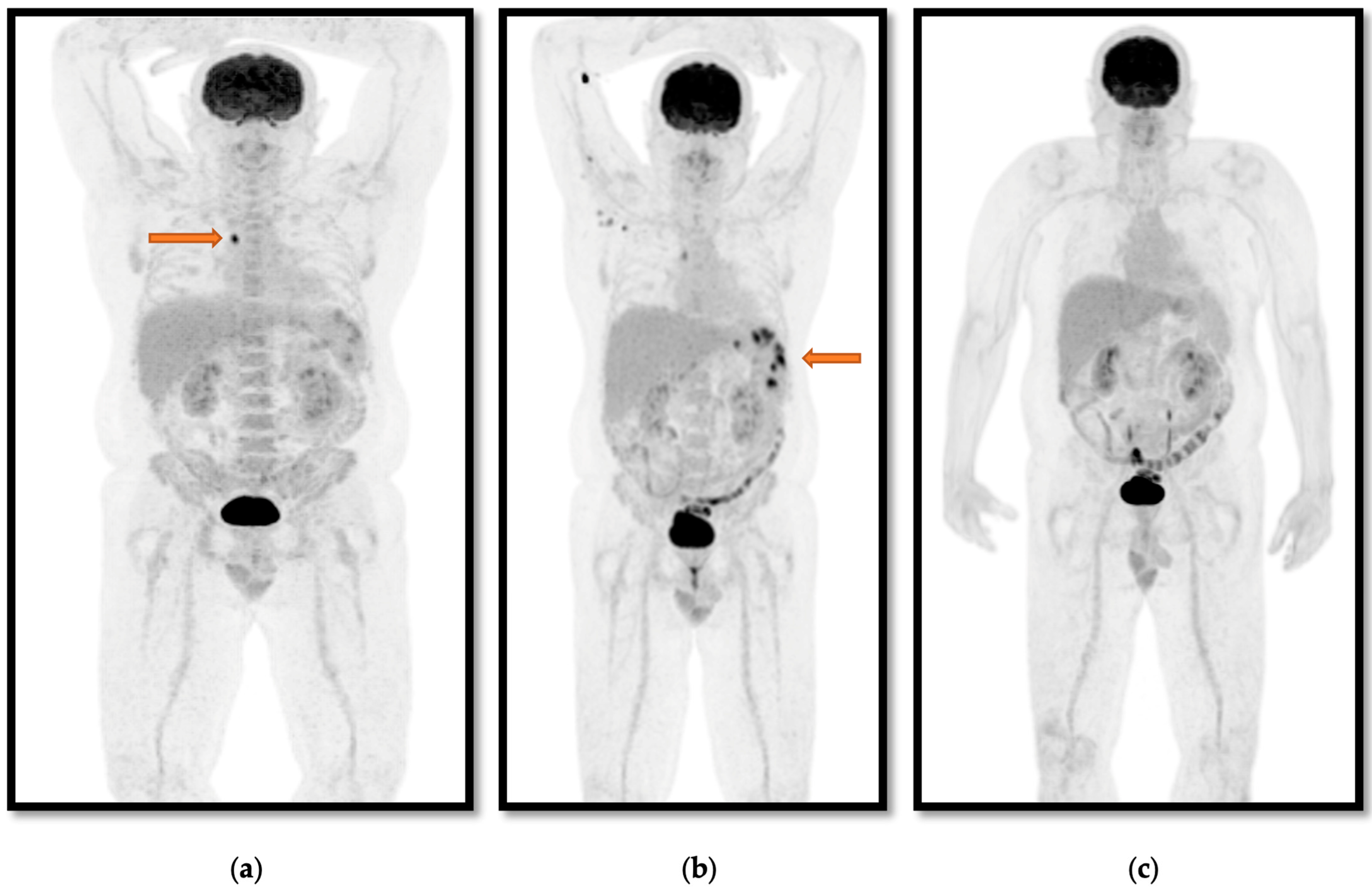
Figure 1. An illustrative case (case 8) of a patient who presented with pyrexia of unknown origin despite undergoing blood cultures and CT. (a) 18-F FDG PET/CT identifying an FDG avid lymph node (arrow), which was biopsied and cultured B. pseudomallei. (b) The result of 18-F FDG PET/CT performed after completing 4 weeks of intensive phase therapy as the patient remained febrile. This showed increased FDG avidity of splenic foci (arrow), extending the IV intensive therapy phase to 12 weeks. (c) The result of 18-F FDG PET/CT performed due to persistent symptoms at 30 weeks of treatment, showing complete resolution of foci, giving clinicians and the patient confidence to cease antibiotics.
Tropicalmed 10 00069 g001
Disclaimer/Publisher’s Note: The statements, opinions and data contained in all publications are solely those of the individual author(s) and contributor(s) and not of MDPI and/or the editor(s). MDPI and/or the editor(s) disclaim responsibility for any injury to people or property resulting from any ideas, methods, instructions or products referred to in the content.