Review

Current Progress on Passiflora caerulea L. In Vitro Culturing

by
Pervin Halkoglu-Hristova
1,
Alexandra Garmidolova
1,*,
Teodora Yaneva
2 and
Vasil Georgiev
1,*
1
Laboratory of Cell Biosystems, Institute of Microbiology, Bulgarian Academy of Sciences, 139 Ruski Blvd., 4000 Plovdiv, Bulgaria
2
Department of Food Technology, Institute of Food Preservation and Quality—Plovdiv, Agricultural Academy of Bulgaria, 154 Vasil Aprilov Blvd., 4000 Plovdiv, Bulgaria
*
Authors to whom correspondence should be addressed.
Submission received: 2 April 2025 / Revised: 4 June 2025 / Accepted: 25 June 2025 / Published: 1 July 2025
(This article belongs to the Section Biology Research and Life Sciences)

Abstract

Passiflora caerulea L., commonly known as the blue passionflower, is traditionally grown as an ornamental plant, but has a diverse chemical composition resulting in a wide range of biological activities that determine its pharmacological properties and use in medicine. Traditional propagation methods, including seed germination and vegetative cuttings, are often inefficient due to low germination rates, susceptibility to pathogens, and slow growth. In particular, P. caerulea presents significant challenges in germination due to its slow development. In this context, in vitro cultivation is used to enable rapid, large-scale plant production while maintaining genetic fidelity. The study of Passiflora tissue cultures began in 1966 and has since attracted increasing attention from researchers around the world. However, despite growing interest, studies specifically focused on the in vitro propagation of P. caerulea remain limited. This review aims to summarize existing knowledge on the main techniques used for in vitro culturing and propagation of P. caerulea, including organogenesis, somatic embryogenesis, and callogenesis. Particular attention is paid to the key factors that influence the initiation, growth, and regeneration of cultures, including the type of explant, the composition of the media, and the environmental conditions. Advances in the in vitro cultivation of P. caerulea have greatly improved the understanding and propagation of this species. Although in vitro cultivation offers several advantages, it is crucial to conduct thorough research on the selection of explants, their age, and the appropriate culture media to ensure optimal growth and development.

1. Introduction

For centuries, plants have been a vital source of medicine, yielding a wide range of chemical compounds and secondary metabolites with therapeutic properties. About 80% of all current medications are derived from plants, directly or indirectly. Currently, there is growing utilization of plant-derived metabolites across multiple sectors: pharmaceuticals, food additives, stimulants, hallucinogens, therapeutic agents, pesticides, perfumes, cosmetics, and more [1,2,3] Moreover, the development of innovative plant-based products continues to progress. However, the continuous availability of plant-derived medications has often been compromised. Thus, in vitro technologies play a vital role in supplying significant quantities of plant material, regardless of climate conditions and the risk of disease development. Plant tissue culturing has emerged as a remarkable method for the micropropagation of plants within a controlled environment using a nutrient medium [4]. In vitro technologies are gaining recognition for their numerous advantages, enabling rapid and efficient mass propagation, the induction of genetic diversity, the creation of new varieties, and fundamental biological research [4]. Additionally, in vitro conservation is a highly effective approach for safeguarding plant germplasm, especially for rare and endangered species, as well as for those with recalcitrant seeds and those that are propagated vegetatively [5]. This technology serves as a crucial instrument across various domains, and when integrated with biotechnological methods, it opens up means for sustainable agricultural advancement, addressing significant food security challenges. The exploration of novel species that may yield various secondary metabolites and demonstrate diverse biological activities is a significant field that contributes to advancements in drug discovery, biotechnology, and ecological studies.
Passiflora caerulea L., also known as passionflower, belongs to the family Passifloraceae and is cultivated for its ornamental value as a climbing plant for landscaping [6], but also has various medicinal properties [7]. The family consists of more than 550 species [8] of which 50–60 bear edible fruits [9]. The primary focus of research on P. caerulea has been its calming and anxiety-reducing effects, leading to the development of various dietary supplements and medications aimed at relaxing the nervous system [10,11]. The native range of the species is Argentina, Brazil, Bolivia, Chile, Paraguay, and Uruguay, but it is also cultivated as a garden plant in North America, Europe, and Asia Melissa officinalis and Passiflora caerulea infusion as physiological stress [7]. The ecological impact of P. caerulea includes reports of its uncontrolled spread and takeover of habitats, affecting native plant species. Comparable invasions have been documented in South Africa and Australia. Germinating P. caerulea presents a significant challenge [12,13]. It is certainly possible to germinate the plant from seed, though it may take as long as a year for the seeds to develop. Alternatively, the seeds may be sown in spring in well-drained soil, allowing them the opportunity to germinate during the summer, though they might stay dormant until the next spring. Another important consideration is that the seeds are collected from their natural environment, which may raise conservation issues. In this regard, micropropagation stands out as an important technique for enhancing their propagation, due to its many benefits. Tissue culture studies on Passiflora began in 1966, and since that time, there have been several publications on techniques related to tissue culture applied to this genus [14,15,16,17].
P. caerulea is a species that has not been investigated in depth. Few studies concerning in vitro micropropagation of the plant exist in the scientific literature, and this review aims to summarize the main techniques for in vitro culturing of P. caerulea to date and emphasize its significance.

Research Methodology

To prepare this review, an in-depth scientific investigation was conducted using multiple academic databases to gather and synthesize relevant information on P. caerulea L. in vitro cultivation techniques. The primary focus was on identifying studies related to callus induction, shoot and root regeneration, and the effects of various culture media and plant growth regulators on the species’ development. The earliest report found dates back to 1966. Our literature review aimed to cover a comprehensive range of protocols and findings that contribute to the micropropagation and phytochemical analysis of P. caerulea to date.
Key databases used included Scopus (https://www.scopus.com/, accessed on 9 January 2025), R Discovery (https://discovery.researcher.life/, accessed on 9 January 2025), and Semantic Scholar (https://www.semanticscholar.org/, accessed on 9 January 2025), with search terms such as “Passiflora caerulea”, “in vitro culturing”, “plant growth regulators”, “nutrient medium composition”, “organogenesis”, “callus”, “cell suspension”, “secondary metabolites”. Additionally, the tool Connected Papers (https://www.connectedpapers.com/, accessed on 9 January 2025) was used to visually explore related publications and expand the network of relevant studies. This program generates a citation-based graph that highlights semantically and topically linked research articles, aiming to identify influential relevant works.

2. Challenges Related to the Culturing of P. caerulea

Traditional propagation methods often do not satisfy the increasing need for mass multiplication, conservation, and research of this species due to several biological and environmental limitations, such as contamination, low germination rates, and seed dormancy. The three principal components influencing the cultivation of P. caerulea are as follows [18].

2.1. Seed Dormancy and Low Germination Rate

The issues of seed dormancy and germination in the Passifloraceae family are considerable. In order to maintain plant viability, a natural process known as seed dormancy stops germination in unfavorable circumstances. Overcoming this dormancy is essential for successful culture and proliferation, particularly for species like P. caerulea, which are important for both decoration and medicine [19]. Multiple factors influence seed germination: seed coat anatomy, seeds viability, suitable environmental conditions (moisture, temperature, and oxygen), appropriate storage, and the ability to overcome dormancy [20]. The seed coat often acts as a barrier to water and gas exchange, contributing to dormancy [21]. It has been investigated that the presence of inhibitory substances in the fruit pulp and juice of P. caerulea can also suppress seed germination. Usually, untreated seeds of various passion vines require two weeks to several months to germinate, depending on the species [19].
Studies have shown that under normal conditions (20–30 °C), seeds from P. caerulea usually begin to germinate after approximately 15 days, but the overall germination rate remains suboptimal [19]. Pretreatments for higher germination levels of the seeds include aril removal, desiccation, storage at appropriate temperature and light conditions, fermentation, the addition of gibberellin to the substrate, and soaking in water for a precise period. The aril might act like the mucilage in other seeds, which constitutes a barrier against oxygen. It is considered that diaspores of this species are orthodox, factors that are of great importance when related to germination. Mendiondo and Amela García obtained the highest germination percentages—and the lowest—when the seeds were devoid of their arils and soaked in water at room temperature for 24 h [19]. In their work, germination was enhanced when the aril was removed in seeds stored under fermentation for two months.

2.2. Contamination Issues

Contamination of P. caerulea is a significant challenge. This issue is not unique to P. caerulea but is a common problem in the Passifloraceae family. The plant secretes a sweet sap that is a nutritious environment for many microorganisms [22]. Plants of this genus are often infected with Xanthomonas plant tissue cultures, where bacterial and fungal contaminants can interfere with plant growth and development. Particularly in humid or poorly drained environments, Passiflora is susceptible to fungal diseases such as Fusarium wilt and root rot [23].
In this context, in vitro micropropagation suggests the cultivation of seeds in controlled in vitro environments with specific growth regulators that can promote germination and subsequent plant development. This approach enables the precise control of environmental conditions and nutrient availability.

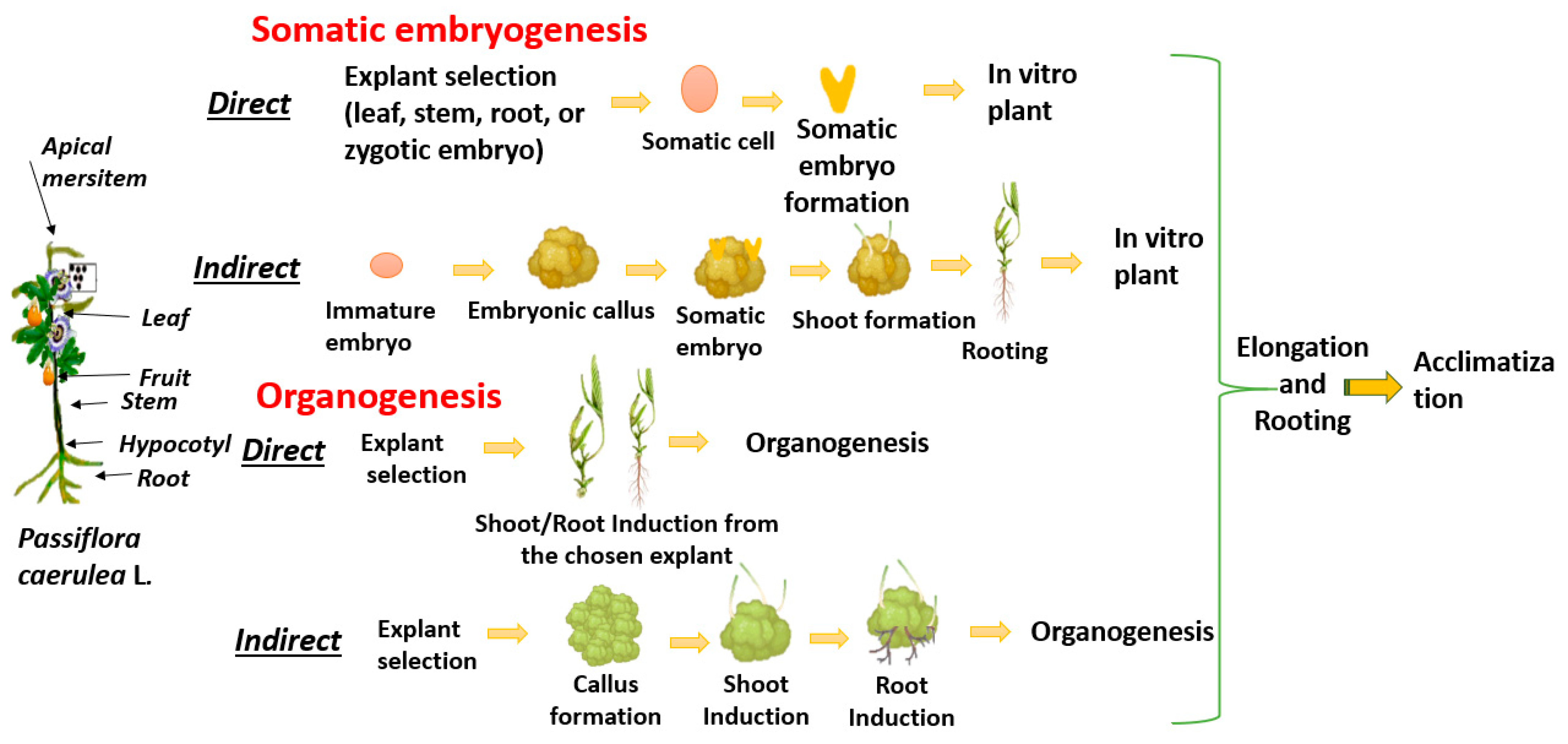
3. In Vitro Micropropagation of P. caerulea

In vitro micropropagation techniques and conventional breeding methods represent two different approaches to plant propagation, each with its advantages and limitations [22,23]. In vitro micropropagation offers several advantages over conventional propagation methods, particularly in terms of efficiency, plant quality, and adaptability: (1) it allows the sustainable production of biomass and active compounds throughout the year, independent of external climatic conditions; (2) it offers the possibility of producing plant resources without the need for extensive cultivation in natural environments, thus contributing to the conservation of rare and endangered species; (3) it supports the genetic manipulation and selection of plants with improved biological potential, which may lead to the development of new, more effective drugs and products; and (4) it enables the cultivation of plants in a more sustainable way. In vitro methods also facilitate the cultivation of plants and their tissues in controlled laboratory environments, allowing for the optimization of growth conditions and the production of secondary metabolites, which not only increases the yield of target compounds but also opens the horizon for the discovery of new biologically active substances that may not be present under natural conditions, etc. [24,25,26,27,28,29,30]. This biotechnological approach allows the rapid production of large quantities of genetically uniform and disease-free plants, which is essential to meet the demands of modern agriculture. Table 1 presents the main culturing methods used for in vitro micropropagation, while Table 2 summarizes the methods and analyses conducted to date for P. caerulea.
The micropropagation of P. caerulea typically begins with the selection and sterilization of suitable explants, such as nodal segments or shoot tips. One of the primary challenges in culturing Passiflora species is contamination, as the endogenous microbial load can be high, particularly in field-grown plants. Therefore, the first and essential step is sterilization. In the research by [15], successful sterilization was achieved by cutting leaf explants of P. caerulea, washing them with running water, and subsequently treating them with a 0.1% (w/v) HgCl2 solution for 3–5 min. Following that, they were washed with sterile distilled water. The authors of [31] demonstrated that leaf explants underwent sterilization through surface washing with a 5% antibacterial agent for 30 min, followed by rinsing with distilled water. Then, they were exposed for 5 min to a 5% sodium hypochlorite solution and subsequently rinsed three times with distilled water. Jafari et al. documented a successful sterilization procedure for seeds intended for in vitro introduction, achieved by washing them for 30 min with running water to eliminate surface haze. Subsequently, the sample was subjected to a 70% ethyl alcohol treatment for 40 s, followed by immersion in a 10% sodium hypochlorite solution for 10 min [32]. Once the treatment period concludes, the seeds undergo multiple washes with sterile distilled water.
According to a study by Fernando et al., the primary mechanism for the in vitro regeneration of Passiflora is direct organogenesis [29]. Organogenesis plays a crucial role in plant micropropagation, enabling the efficient regeneration of plant tissues and the production of genetically uniform plantlets. It can occur in two ways: direct, where shoots or roots develop directly from explant tissues without an intervening callus stage; and indirect, where shoots or roots arise from a callus (an undifferentiated mass of cells) [33]. This involves the development of shoots and roots from explants under controlled conditions, using specific PGRs and culture media to enhance organogenesis. Previous studies have shown that a suitable culture medium to induce organogenesis in vitro is MS [34] and ½ MS supplemented with varying concentrations of PGRs [35,36]. In research by Martinelli, a modified MS medium was created, according to the nutrient levels found in the leaves of P. edulis flavicarpa [37]. The new medium, without Cl or I, showed increased levels of P, Ca, Mg, S, Mn, Cu, Na, and FeEDTA while showing reduced levels of Zn. Direct organogenesis has been recorded in Passiflora species using BAP, combined with TDZ at different concentrations to stimulate axillary buds. Jafari et al. examined the in vitro micropropagation of P. caerulea, utilizing explants sourced from zygotic embryo-supers and cotyledonary nodules. These were cultured on a modified MS medium containing 1.5 mg/L BAP and 0.15 mg/L IBA, indicating that IBA was more effective in promoting root formation than NAA and IAA [32]. Rathod et al. reported direct regeneration from leaf explants of P. caerulea L. cultured on MS basal medium enriched with the growth regulators 2 mg/L BAP and 1 mg/L KIN, successfully progressing through all stages of multiplication and achieving functional plant regeneration within 54 days [31].
The shoot regeneration capacity of P. caerulea has drawn considerable attention from researchers seeking to optimize micropropagation protocols. Shoot induction is a critical step in the process of in vitro plant regeneration depending on several factors such as explant type, genotype, culture medium composition, and hormonal balance. Id and Daneshvar evaluated the potential of cotyledonary nodes and shoot tips for shoot multiplication. Sterilization was carried out with a % sodium hypochlorite solution for 10 min [38]. The study demonstrated shoot tips achieving up to 96.66% regeneration using Murashige and Skoog (MS) medium supplemented with 1.5 mg/L BAP and 0.15 mg/L IBA. The MS medium and the absence of phytohormones were found to be effective for the in vitro establishment of P. caerulea, thereby reducing production costs while maintaining plant quality [17]. The results of Jafari et al. show that direct shoot regeneration can be successfully achieved by using higher levels of BAP and kinetin. Results indicate that machine learning techniques, such as General Regression Neural Networks (GRNNs) and Random Forests (RFs), can effectively predict and optimize indirect shoot regeneration [39]. The models showed impressive predictive accuracy, highlighting the promise of machine learning in enhancing regeneration protocols.
Numerous studies have explored the enhancement of callogenesis in P. caerulea, employing various techniques and methodologies to boost both efficiency and effectiveness [15,31,38]. Id and Daneshvar used machine learning models, including multilayer perceptron, to predict callogenesis responses based on PGRs and explant types [38]. It was found that the optimal combination to achieve the highest callogenesis rate (100%) involved leaf explants cultured with specific concentrations of 2,4-D (2 mg/L) and BAP 0.2 (mg/L). Sensitivity analysis showed that the type of explant had a significant effect on the efficacy of plant growth regulators in callogenesis. The results confirmed the hypothesis that explant selection plays a key role in tissue culture protocols. P. caerulea was successfully cultured in vitro by using leaf explants inoculated on MS medium. This medium contained a combination of growth regulators: 0.5 mg/L NAA, 0.5 mg/L IAA, 0.5 mg/L 2,4-D, 0.5 mg/L KIN, and 1 mg/L BAP, which resulted in callus formation. Each explant responded differently depending on the hormonal balance and physical and chemical factors, highlighting their influence [31].
Several authors have reported that callus induction of Passiflora sp. can take place under photoperiodic conditions (16/8 h light/dark) [40,41]. The research carried out by [18] indicated that homogeneous callusing obtained from epicotyl and hypocotyl on MS medium supplemented with NAA (0.5 mg/L) and KIN (0.1 mg/L) obtained the highest value of growth index on a medium supplemented with 2,4-D (2.0 mg/L).
Rooting is an essential phase in the micropropagation of P. caerulea. It is a complex process influenced by several factors, including auxin types and concentrations, explant types, and environmental conditions. A recent study by Jafari et al. showed that a hybrid model integrating a generalized regression neural network with a genetic algorithm was effective in predicting and optimizing rooting responses, achieving high prediction accuracy [42]. Rooting experiments indicated that the use of 1 mg/L IBA in MS medium led to higher rooting percentages, and plantlet acclimatization in soil reached a survival rate exceeding 90% [32]. Additionally, the selection of rooting substrate, including vermiculite or peat mixes, can greatly influence root development [43]. Research has also explored the combination of biostimulants with rooting substrates to enhance the success rates of rooting. Figure 1 illustrates a schematic representation of the in vitro cultivation process of P. caerulea.

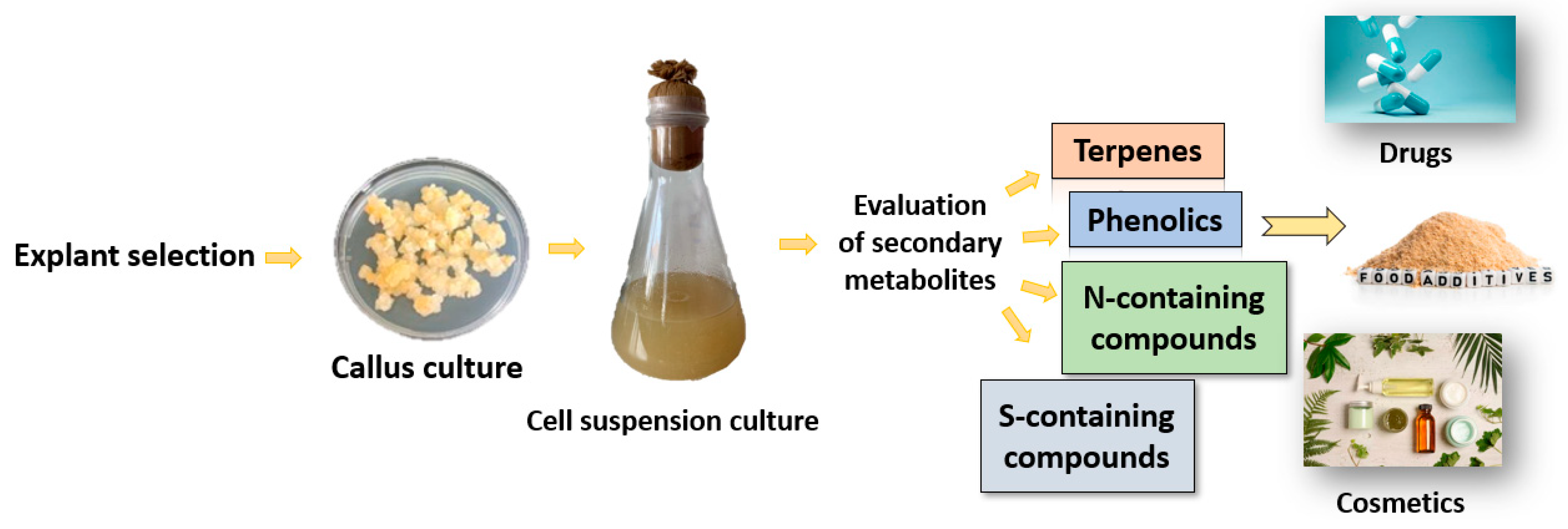
4. Regenerated Plantlets from P. caerulea and Phytochemical Analysis

P. caerulea is rich in various phytochemical compounds, including alkaloids, phenolics, flavonoids, and glycosides. Plant tissue culture techniques, used to obtain cultures rich in phytochemical compounds, have been used to determine the quantities of bioactive compounds present in this species under controlled in vitro conditions. Ozarowski et al.’s review focused on various micropropagation techniques for culturing P. caerulea. Table 3 summarises some of the studies on regenerated platelets from the plant, as well as the analysis of phytochemicals found in callus cultures [22].
As demonstrated by Busilacchi et al., who reported a 70% shoot regeneration rate from proliferating tissue masses, successful induction of direct organogenesis without preceding callus development is a consistent trend throughout the research. The lack of callus during this procedure promotes the genetic integrity of regenerated plantlets and suggests a lower probability of somaclonal variation. A crucial component of the multiplication of medicinal plants is biochemical consistency, which is further confirmed by the similarity in TLC chromatograms between mother plants and in vitro regenerants [44].
To elaborate, Ozarowski et al. showed 100% callus induction on MS media supplemented with combinations of BA, GA3, and 2,4-D, demonstrating both direct and indirect organogenesis. BA (4.4 μM) and GA3 (2.88 μM) produced the highest shoot regeneration rate (106.4%) and bud-forming index (3.1), highlighting the significance of particular cytokinin/gibberellin ratios in boosting morphogenetic responses [22]. The phytochemical profile showed comparatively low quantities of important chemicals such as rosmarinic acid, chlorogenic acid, isovitexin, and vitexin, despite effective organogenesis.
These patterns were supported by further research on P. caerulea by Ozarowski et al., which reported up to 16 shoots per explant when BA was present; however, callusing also had a noticeable impact [22]. Crucially, the use of a rotational liquid medium containing 2,4-D (18.1 μM) markedly enhanced root growth (100%), acclimation (100%), and regeneration efficiency (90%), underscoring the benefit of dynamic culture conditions in fostering tissue differentiation and plantlet survival.
The integration of phytochemical analyses by Ozarowski et al. facilitated a more comprehensive understanding of metabolic results [22]. They found that isovitexin was the most common compound in plantlets that had regenerated on BA-supplemented media, followed by rutin, chlorogenic acid, and other compounds using HPLC. While cytokinins, especially at higher concentrations, more efficiently support organogenesis and secondary metabolite biosynthesis, auxins may favor dedifferentiation over redifferentiation, as evidenced by the presence of morphogenic versus non-morphogenic callusing in response to BA and 2,4-D, respectively.
In combination, these findings underscore the significance of the PGR composition, explant source, and culture system in Passiflora’s morphogenetic responses and phytochemical outcomes. The most efficient method for creating genetically stable, biochemically consistent plantlets seems to be direct organogenesis, especially in liquid media systems. Indirect pathways, however, are nevertheless useful for metabolite identification and optimization, indicating a complementary function in phytopharmaceutical and propagation applications.
In general, there is a lack of scientific evidence regarding the phytochemical evaluation of in vitro clones for the detection of flavonoids, phenolic acids, and alkaloids in regenerated plantlets of P. caerulea. Additionally, the micropropagation processes based on organogenic callusing require further development; however, it appears that methods such as TCL and micropropagation in bioreactors may be employed to optimize the mass propagation of healthy regenerated plants [19].

Future Prospects and Conclusions

The developments that have been made in the in vitro culturing of P. caerulea have greatly enhanced the knowledge and propagation of this species. Various parts of the plant, leaves, stems, flowers, and fruits are known to produce a wide spectrum of bioactive compounds such as flavonoids, alkaloids, phenolic acids, cyanogenic glycosides, and others. In our previous review, we discussed the reported different biological activities of P. caerulea to date [45]. Despite the well-documented chemical richness of naturally grown P. caerulea, there is a significant gap in the literature regarding the phytochemical behavior of this species under in vitro culture conditions. In vitro culture techniques, including callogenesis, shoot induction, and somatic embryogenesis, are primarily used for mass propagation, germplasm conservation, and genetic transformation. However, in many medicinal plants, these systems also serve as platforms for secondary metabolite production, offering a controlled environment for studying and enhancing phytochemical synthesis. The natural production of these metabolites in wild plants can be affected by environmental factors, seasonal changes, and both biotic and abiotic stressors. In vitro cultures can produce bioactive compounds similar to or even exceeding those in naturally grown plants, particularly when optimized through elicitors, growth regulators, or precursor feeding. Plant tissue culture is an effective tool for their isolation and processing [46,47]. The process of extracting secondary metabolites from P. caerulea encompasses a sequence of biotechnological procedures designed to enhance the production of bioactive compounds such as flavonoids, alkaloids, and phenolic compounds. Figure 2 presents a technological scheme for producing secondary metabolites of P. caerulea.
The effective use of tissue culture methods, such as micropropagation and somatic embryogenesis, has shown the capability for extensive production of robust and genetically consistent plants. Although there are advantages to in vitro cultivation of P. caerulea, it is important to carry out extensive research into the selection of explants, age, and appropriate culture media to achieve optimal growth and development. These methods provide a sustainable way to conserve P. caerulea while also acting as a useful resource for examining its physiological and genetic traits.
The advancement of new technologies and the integration of machine learning and genetic algorithms enable the prediction and optimization of in vitro cultures, providing practical solutions to these challenges. In recent years, omics methods have proven to be useful and have been applied in various areas to identify different biological molecules. These methods not only enable the precise generation of bioactive compounds but also offer a solid basis for research in genetics, biochemistry, and pharmacology. Utilizing these approaches allows researchers to further study metabolite production, improve the investigation of metabolic pathways, and investigate novel uses in medicine, cosmetics, and biotechnology. With the ongoing progress in tissue culture and metabolic engineering, in vitro methods will be crucial for realizing the complete potential of P. caerulea and other plants of medicinal significance.

Author Contributions

Conceptualization, P.H.-H., A.G., T.Y. and V.G.; methodology, P.H.-H.; resources, V.G.; data curation, A.G.; writing—original draft preparation, A.G.; writing—review and editing, P.H.-H., A.G., T.Y. and V.G.; visualization, A.G.; supervision, V.G.; project administration, P.H.-H., A.G. and T.Y.; funding acquisition, P.H.-H., A.G. and T.Y. All authors have read and agreed to the published version of the manuscript.

Funding

This research was funded by the Bulgarian Scientific Fund, grant No. KP-06-M86/3; BG-175467353-2024-10-0027-C01.

Institutional Review Board Statement

Not applicable.

Informed Consent Statement

Not applicable.

Data Availability Statement

Not applicable.

Conflicts of Interest

The authors declare no conflicts of interest.

Abbreviations

The following abbreviations are used in this manuscript:
PGRsPlant growth regulators
MSMurashige and Skoog
WPMWoody plant medium
IAAIndole-3-acetic acid
IBAIndole-3-butyric acid
NAA1-naphthaleneacetic acid
2,4-D2,4-dichlorophenoxyacetic acid
iPIsopentenyl adenine
BAPBenzylaminopurine
KINKinetin
TDZThidiazuron
GRNNGeneral Regression Neural Networks
RFRandom Forests
BABenzyladenine or BAP—6-Benzylaminopurine
GA3Gibberellic acid
TLCThin-layer chromatography
HPLCHigh-performance liquid chromatography
HPTLCHigh-performance thin-layer chromatography

References

  1. Davis, C.C.; Choisy, P. Medicinal plants meet modern biodiversity science. Curr. Biol. 2024, 34, R158–R173. [Google Scholar] [CrossRef]
  2. Mishuk, A.U.; Hossain, A.; Karmakar, U.K. Medicinal potential of Passiflora foetida L. plant extracts: Biological and pharmacological activities. J. Integr. Med. 2014, 12, 121–126. [Google Scholar] [CrossRef]
  3. De Moura, T.; Araújo, F.; Cerqueira, S.; De, R.G. Current topics in medicinal chemistry. Curr. Top. Med. Chem. 2022, 22, 2315–2328. [Google Scholar] [CrossRef]
  4. Resources, P.G.; De Carlo, A. In vitro biotechnology for conservation and sustainable use of plant genetic resources. Plants 2024, 13, 1897. [Google Scholar] [CrossRef]
  5. Oros, P.B.; Cătană, C. In vitro plant tissue culture: Means for production of Passiflora species. Int. J. Innov. Approaches Agric. Res. 2020, 4, 505–523. [Google Scholar] [CrossRef]
  6. Berberich, S.; Snyder, J.; Geneve, R.; Williams, M.A. Growth and flowering response of container grown passion flower cultivars to fertilizer and paclobutrazol. J. Environ. Hortic. 1962, 24, 109–114. [Google Scholar] [CrossRef]
  7. Gerasimova, A.; Nikolova, K.; Petkova, N.; Ivanov, I.; Dincheva, I.; Tumbarski, Y.; Yanakieva, V.; Todorova, M.; Gentscheva, G.; Gavrilova, A.; et al. Metabolic Profile of Leaves and Pulp of Passiflora caerulea L. (Bulgaria) and Their Biological Activities. Plants 2024, 13, 1731. [Google Scholar] [CrossRef]
  8. Smruthi, R.; Divya, M.; Archana, K.; Ravi, M. The active compounds of Passiflora spp. and their potential medicinal uses from both in vitro and in vivo evidences. J. Adv. Biomed. Pharm. Sci. 2021, 4, 45–55. [Google Scholar] [CrossRef]
  9. Silva, C.G. Tissue Culture and Phytochemical Studies of Podophyllum, Diphylleia and Passiflora Species. Bachelor’s Thesis, University of Nottingham, Nottingham, UK, 2000. Available online: http://eprints.nottingham.ac.uk/28994/1/311836.pdf (accessed on 14 April 2025).
  10. Yi, S.-S. A Composition for Sleep Induction Comprising Passiflora incarnata Extract. KR 102240706B1, 2021. Available online: https://patents.google.com/patent/KR102240706B1/en (accessed on 19 February 2025).
  11. Feliú-Hemmelmann, K.; Monsalve, F.; Rivera, C. Melissa officinalis and Passiflora caerulea infusion as physiological stress decreaser. Int. J. Clin. Exp. Med. 2013, 6, 444–451. [Google Scholar]
  12. Nagabhushan Arun, M.; Mahender Kumar, R.; Sreedevi, B.; Padmavathi, G.; Revathi, P.; Pathak, N.; Venkatanna, B. The Rising Threat of Invasive Alien Plant Species in Agriculture. In The Rising Threat of Invasive Alien Plant Species in Agriculture; IntechOpen: London, UK, 2023. [Google Scholar] [CrossRef]
  13. Barbault, R.; Atramentowicz, M. Les Invasions Biologiques, une Question de Natures et de Sociétés; Cairn.info: Paris, France, 2010; p. 180. ISBN 978-2-7592-0373-4. [Google Scholar] [CrossRef]
  14. Silva, C.V.; Oliveira, L.S.; Loriato, V.A.P.; Silva, L.C.; de Campos, J.M.S.; Viccini, L.F.; de Oliveira, E.J.; Otoni, W.C. Organogenesis from Root Explants of Commercial Populations of Passiflora edulis Sims and a Wild Passionfruit Species, P. cincinnata Masters. Plant Cell Tissue Organ Cult. 2011, 107, 407–416. [Google Scholar] [CrossRef]
  15. Prithviraj, H.S.; Hemanth Kumar, N.K.; Prakasha; Shobha, J. An efficient in vitro regeneration of multiple shoots from leaf explant of Passiflora caerulea L., an important medicinal plant. Int. J. Recent Sci. Res. Res. 2015, 6, 7263–7265. Available online: https://recentscientific.com/sites/default/files/3767.pdf (accessed on 18 May 2025).
  16. Jafari, M.; Daneshvar, M.H.; Lotfi, A. Control of in vitro contamination of Passiflora caerulea using sodium hypochlorite. Indo-Am. J. Agric. Vet. Sci. 2016, 4, 7–16. [Google Scholar]
  17. Faria, G.A.; Oliveira, C.P.M.; Lopes, B.G.; Rocha, P.S.; Peron, G.M.; Souza, K.S.; Garcia, C.K.; Furlani Junior, E.; Cavichioli, J.C.; Felizardo, L.M. Establishment of a Protocol for In Vitro Propagation of Passiflora caerulea. Res. Soc. Dev. 2020, 9, e157997158. [Google Scholar] [CrossRef]
  18. Ożarowski, M. Influence of the Physico-Chemical Factors, Plant Growth Regulators, Elicitors and Type of Explants on Callus Cultures of Medicinal Climbers of Passiflora L. Herba Pol. 2011, 57, 58–75. [Google Scholar]
  19. Mendiondo, G.M.; Amela García, M.T. Germination of stored and scarified seeds of Passiflora caerulea L. (Passifloraceae). Plant Biosyst. 2009, 143, 369–376. [Google Scholar] [CrossRef]
  20. Phartyal, S.S.; Baskin, J.M.; Baskin, C.C.; Thapliyal, R.C. Physical dormancy in seeds of Dodonaea viscosa (Sapindaceae) from India. Seed Sci. Res. 2005, 15, 59–61. [Google Scholar] [CrossRef]
  21. Upretee, P.; Bandara, M.S.; Tanino, K.K. The role of seed characteristics on water uptake preceding germination. Seeds 2024, 3, 559–574. [Google Scholar] [CrossRef]
  22. Ożarowski, M.; Thiem, B. Progress in micropropagation of Passiflora spp. to produce medicinal plants: A mini-review. Rev. Bras. Farmacogn. 2013, 23, 937–947. [Google Scholar] [CrossRef]
  23. Rezazadeh, A.; Bailey, M.; Sarkhosh, A. Passion fruit problems in the home landscape. Univ. Fla. EDIS 2020, 2020, 6. [Google Scholar] [CrossRef]
  24. Espinosa, C.A.; César, L.; Garza, A.P.; García, S. In vitro plant tissue culture: Means for production of biological active compounds. Planta 2018, 248, 1–18. [Google Scholar] [CrossRef]
  25. Hasnain, A.; Atif, S.; Naqvi, H.; Ayesha, S.I.; Khalid, F.; Ellahi, M.; Abbas, A.; Adamski, R.; Markowska, D. Plants in vitro propagation with its applications in food, pharmaceuticals and cosmetic industries: Current scenario and future approaches. Front. Plant Sci. 2022, 13, 1009395. [Google Scholar] [CrossRef] [PubMed]
  26. Jain, S.M.; Nakhooda, M. Clonal and Micropropagation. In Encyclopedia of Applied Plant Sciences, 2nd ed.; Thomas, B., Murray, B.G., Murphy, D.J., Eds.; Academic Press: Oxford, UK, 2017; pp. 428–432. ISBN 978-0-12-394808-3. [Google Scholar] [CrossRef]
  27. Ahmad, N.; Strnad, M. (Eds.) Meta-Topolin: A Growth Regulator for Plant Biotechnology and Agriculture, 1st ed.; Springer: Singapore, 2021; Volume XIII, p. 339. [Google Scholar] [CrossRef]
  28. Micheli, M.; Prosperi, F.; Facchin, S.; Fernandes, D. Safeguard of plant germplasm through the in vitro culture. Hortic. Int. J. 2020, 4, 50–52. [Google Scholar] [CrossRef]
  29. Fernando, J.A.; Lu, M.; Appezzato-da-Glo, S.R.M. New insights into the in vitro organogenesis process: The case of Passiflora. Plant Cell Tissue Organ Cult. 2007, 91, 37–44. [Google Scholar] [CrossRef]
  30. Aparecida, C.; Brito, R.; Faria, D.; Pinheiro, P.; Carvalho, D.; Izabel, A.; Fealho, I.; Carvalho, D.; Monteiro, E.; Matos, D.; et al. In vitro regeneration of triploid plants from mature endosperm culture of commercial passionfruit (Passiflora edulis Sims). Sci. Hortic. 2018, 238, 408–415. [Google Scholar] [CrossRef]
  31. Rathod, H.P.; Pohare, M.; Bhor, S.; Jadhav, K.P.; Batule, B.S. In vitro micropropagation of blue passion flower (Passiflora caerulea L.). Trends Biosci. 2015, 7, 3079–3082. [Google Scholar]
  32. Jafari, M.; Daneshvar, M.H.; Lotfi, A. In vitro shoot proliferation of Passiflora caerulea L. via cotyledonary node and shoot tip explants. J. Biotechnol. Comput. Biol. Bionanotechnol. 2017, 98, 113–119. [Google Scholar] [CrossRef]
  33. Liu, Y.; Zhan, Y.; Fu, Q.; Li, S.; Sun, X.; Wang, Y.; Yu, M.; Qin, D.; Huo, J.; Zhu, C. Plant Regeneration via Somatic Embryogenesis and Indirect Organogenesis in Blue Honeysuckle (Lonicera caerulea L.). Horticulturae 2023, 9, 996. [Google Scholar] [CrossRef]
  34. Murashige, T.; Skoog, F. A revised medium for rapid growth and bio assays with tobacco tissue cultures. Physiol. Plant. 1962, 15, 473–497. [Google Scholar] [CrossRef]
  35. Nhut, D.T.; Khiet, B.L.T.; Thi, N.N.; Thuy, D.T.T.; Duy, N.; Hai, N.T.; Huyen, P.X. High Frequency Shoot Formation of Yellow Passion Fruit (Passiflora edulis F. flavicarpa) via Thin Cell Layer (TCL) Technology. In Protocols for Micropropagation of Woody Trees and Fruits; Jain, S.M., Häggman, H., Eds.; Springer: Dordrecht, The Netherlands, 2007; pp. 417–426. ISBN 978-1-4020-6352-7. [Google Scholar] [CrossRef]
  36. Vieira, L.M.; Rocha, D.I.; Taquetti, M.F. In vitro plant regeneration of Passiflora setacea D.C. (Passifloraceae): The influence of explant type, growth regulators, and incubation conditions. In Vitr. Cell. Dev. Biol. Plant 2014, 50, 738–745. [Google Scholar] [CrossRef]
  37. Monteiro, A.D.A.; Higashi, E.N.; Gonçalves, A.N.; Rodriguez, A.P.M. A Novel Approach for the Definition of the Inorganic Medium Components for Micropropagation of Yellow Passionfruit (Passiflora edulis Sims f. flavicarpa Deg.). In Vitr. Cell. Dev. Biol. Plant 2000, 36, 527–531. [Google Scholar] [CrossRef]
  38. Id, M.J.; Daneshvar, M.H. Machine learning-mediated Passiflora caerulea callogenesis optimization. PLoS ONE 2024, 19, e0292359. [Google Scholar] [CrossRef]
  39. Jafari, M.; Daneshvar, M.H. Prediction and optimization of indirect shoot regeneration of Passiflora caerulea using machine learning and optimization algorithms. BMC Biotechnol. 2023, 23, 27. [Google Scholar] [CrossRef]
  40. Antognoni, F.; Zheng, S.; Pagnucco, C.; Baraldi, R.; Poli, F.; Biondi, S. Induction of flavonoid production by UV-B radiation in Passiflora quadrangularis callus cultures. Fitoterapia 2007, 78, 345–352. [Google Scholar] [CrossRef]
  41. Rocha, D.I.; Vieira, L.M. Somatic embryogenesis of a wild passion fruit species Passiflora cincinnata Masters: Histocytological and histochemical evidences. Protoplasma 2012, 249, 747–758. [Google Scholar] [CrossRef] [PubMed]
  42. Jafari, M.; Daneshvar, M.H.; Jafari, S.; Hesami, M. Machine learning-assisted in vitro rooting optimization in Passiflora caerulea. Forests 2022, 13, 2020. [Google Scholar] [CrossRef]
  43. Oros, P.B.; Catana, C.; Gocan, T.; Moldovan, G.; Székely-Varga, Z. Influence of culture substrates and biostimulators on Passiflora rooting. Horticulture 2020, 77, 12. [Google Scholar] [CrossRef]
  44. Busilacchi, C.; Sapio, D. Field culture of micropropagated Passiflora caerulea L. histological and chemical studies. Bol. Latinoam. Caribe Plant. Med. Aromaticas. 2008, 7, 257–263. [Google Scholar] [CrossRef]
  45. Garmidolova, A.; Halkoglu-Hristova, P.; Georgiev, V. Exploring the multifunctionality of Passiflora caerulea L.: From traditional remedies to modern applications. Appl. Sci. 2025, 15, 3251. [Google Scholar] [CrossRef]
  46. Nawaz, M.S.; Ali, A.; Shahani, A.; Khalid, M.N. Genomic Assisted Crop Breeding Approaches for Designing Future Crops to Combat Food Production Challenges. Bio-Sci. Rev. J. 2022, 2022, 1. [Google Scholar] [CrossRef]
  47. Anand, A.; Subramanian, M.; Kar, D. Breeding Techniques to Dispense Higher Genetic Gains. Front. Plant Sci 2023, 13, 1076094. [Google Scholar] [CrossRef]
Figure 1. In vitro culturing of P. caerulea.
Sci 07 00090 g001
Figure 2. Technological scheme for the production of secondary metabolites of P. caerulea.
Sci 07 00090 g002
Table 1. The main culturing methods for in vitro culturing.
MethodFormationDirect/IndirectResult
Direct OrganogenesisShoots/roots directly from explantNo callus formationNew plant organs
Direct Somatic EmbryogenesisEmbryos from somatic cellsNo callus formationEmbryo-like structures
CallogenesisFormation of callusExplant to callusIntermediate stage
Indirect OrganogenesisShoots/roots from callusCallus stage presentNew plant organs
Indirect Somatic EmbryogenesisEmbryos from callusCallus stage presentEmbryo-like structures
Table 2. In vitro micropropagation of P. caerulea.
Explant TissueCulture Medium and Plant Growth
Regulators (mg/mL)
ResponseReference
Leaf segmentsMS + (0.5) NAA + (0.5) IAA + (0.5) 2,4-D + (0.5) KIN + (1) BAPShoot induction + callus formation[31]
MS + (1) KIN + (2) BAPShoot induction
MS + (1) KIN + (2) BAP + (0.4) IAAShoot multiplication
MS + (2) 2,4-D + (0.8) IAA + (1) KIN + (2) BAPCallus fully developed
MS + (1) NAA + (0.5) IBA + (0.5) IAARoot induction
Leaf segments on abaxial or adaxial surfaceMS + (0.5–4) BAP
MS
Shoot induction[15]
MS + (0.5–3) BAP + (0.5–1) NAAShoot + root induction
MS + (0.25–3) IBA + NAA (0.25–3)Root induction
Shoot tips, cotyledonary nodeMS + (0.5, 1, or 1.5) BAP + (0.05, 0.1 or 0.15) IBAShoot induction
Root induction
[16]
MS + (0.25, 0.5 or 1) TDZ + (0.025, 0.05 or 0.1) IBA
MS + (1 or 2) KIN + (0.1 or 0.2) IBA
MS + (0.5, 1 or 2) IBARoot induction
Regenerated plants
MS + (0.5, 1 or 2) NAA
MS + (0.5, 1 or 2) IAA
Nodal segmentsMS without PGR’sRegenerated plants[17]
½ MS without PGRs
MS
Table 3. Regenerated plantlets of P. caerulea and phytochemical composition.
Scope of ResearchRooting/
Elongation/
Acclimatization
Phytochemical
Composition
Findings on Regenerated
Plantlets/Organogenesis
Direct organogenesis and histological examination and TLC analysisElongation: not indicated
Rooting: on MS medium
Acclimatization: 100% success
TLC of flavonoid fraction showed nine bands. Individual compounds were not identified. Chromatographic profiles of plantlets closely matched those of the donor plantsShoot buds emerged directly from proliferative tissues without an intermediate callus phase. Approximately 70% of explants successfully regenerated shoots.
Direct and indirect organogenesis, with HPLC and HPTLC-based phytochemical profiling of callus-Vitexin, isovitexin, rutin, chlorogenic acid, and rosmarinic acid were identified in callus by HPLCTransferring of internodal explants on MS medium supplemented with BA with GA3 and MS with 2,4-D resulted in 100% callus formation. The highest regeneration rate (106.4%) and bud index (3.1) were observed in stem-derived callus cultured on MS with 4.4 μM BA and 2.88 μM GA3. Callus culture was initiated on MS supplemented with 2.0 mg/L 2,4-D.
Organogenesis via both direct and indirect pathwaysElongation: not indicated-Culturing P. caerulea on MS medium with BA (4.4 μM) produced up to 16 shoots per explant, often in association with callus formation.
Direct organogenesis in liquid rotary cultureRooting: 100% on MS
Acclimatization: 100%
-Using MS medium enriched with 2,4-D (18.1 μM) in a rotary liquid system, an average of three shoots per explant was achieved, with an overall regeneration efficiency of 90%.
Both organogenesis types and HPLC analysis of regenerated plantletsElongation: not indicated
Rooting: 100% on MS
Acclimatization: not indicated
Isovitexin (major compound), chlorogenic acid, rutin, hyperoside, vitexin, luteolin, apigenin, and rosmarinic acid, with no alkaloids detected by HPLC in morphogenic callus Morphogenic callus formed on MS with BA, while non-morphogenic callus appeared on MS with 2,4-D. The best shoot induction was observed in leaf-derived callus on MS with 8.8 μM BA, and in petiole-derived callus on MS with 4.4 μM BA, yielding three shoots per explant.
Abbreviation: “-” not investigated.
Disclaimer/Publisher’s Note: The statements, opinions and data contained in all publications are solely those of the individual author(s) and contributor(s) and not of MDPI and/or the editor(s). MDPI and/or the editor(s) disclaim responsibility for any injury to people or property resulting from any ideas, methods, instructions or products referred to in the content.

