Review

Targeting Cancer with Paris’ Arrow: An Updated Perspective on Targeting Wnt Receptor Frizzled 7

1
The European Cancer Stem Cell Research Institute, School of Biosciences, Cardiff University, Cardiff CF24 4HQ, UK
2
Monash Biomedicine Discovery Institute, Monash University, Clayton, VIC 3800, Australia
3
Department of Infectious Diseases, University of Melbourne at the Peter Doherty Institute for Infection and Immunity, Melbourne, VIC 3000, Australia
4
Victorian Infectious Diseases Reference Laboratory, Royal Melbourne Hospital at the Peter Doherty Institute for Infection and Immunity, Melbourne, VIC 3000, Australia
5
School of Medicine, University California San Diego, La Jolla, CA 92093, USA
*
Author to whom correspondence should be addressed.
These authors contributed equally to this work.
Submission received: 16 December 2024 / Revised: 28 February 2025 / Accepted: 2 April 2025 / Published: 8 May 2025
(This article belongs to the Special Issue Feature Papers—Multidisciplinary Sciences 2024)

Abstract

:
The Wnt signalling pathway plays a crucial role in tissue homeostasis and cancer biology due to its regulation of cellular processes, including proliferation, migration, and stem cell activity. Frizzled receptor 7 (FZD7) (a member of the F-class G protein-coupled receptors) has emerged as a key Wnt receptor within this pathway, which is elevated in several human malignancies. FZD7 is notably upregulated in gastrointestinal, breast, pancreatic, and hepatocellular carcinomas and transmits oncogenic Wnt signalling through canonical and non-canonical pathways. FZD7 promotes tumour initiation, and emerging evidence implicates FZD7 in cancer stem cell maintenance and epithelial–mesenchymal transition (EMT), reinforcing its role in metastasis. Therapeutic strategies targeting FZD7 have shown promise, including FZD7-specific monoclonal antibody-drug conjugates (ADCs), human single-chain fragment variable (scFVs) antibodies, and nanoparticles. Notably, our recent development of FZD7-ADC has demonstrated tumour-selective cytotoxicity with reduced off-target effects, positioning FZD7 as an attractive therapeutic target. Additionally, nanoparticle-based drug delivery systems have enhanced the precision of existing chemotherapies by targeting FZD7-expressing tumour cells. Despite significant advances, clinical translation remains a challenge due to potential on-target toxicity and the complexity of tumour microenvironments. Future research should focus on optimising delivery systems, refining antibody specificity, and conducting comprehensive preclinical and clinical trials. This review will focus on novel discoveries regarding FZD7 in cancer and provide an update on our original review on this subject in 2016. Additionally, we present new figures generated by our group using the publicly available Pan-Cancer Atlas RNAseq datasets, highlighting FZD7 expression patterns in patient samples. This integrated approach aims to provide updated insights into the function of FZD7 during cancer and its growing status as an attractive target for therapy. In summary, FZD7 stands out as a promising molecular target in cancer therapy due to its selective overexpression in tumours, functional role in Wnt-driven oncogenesis, and potential for innovative therapeutic applications. This review underscores the critical need for the continued exploration of FZD7-targeted therapies to improve patient outcomes in cancer treatment.

1. Introduction

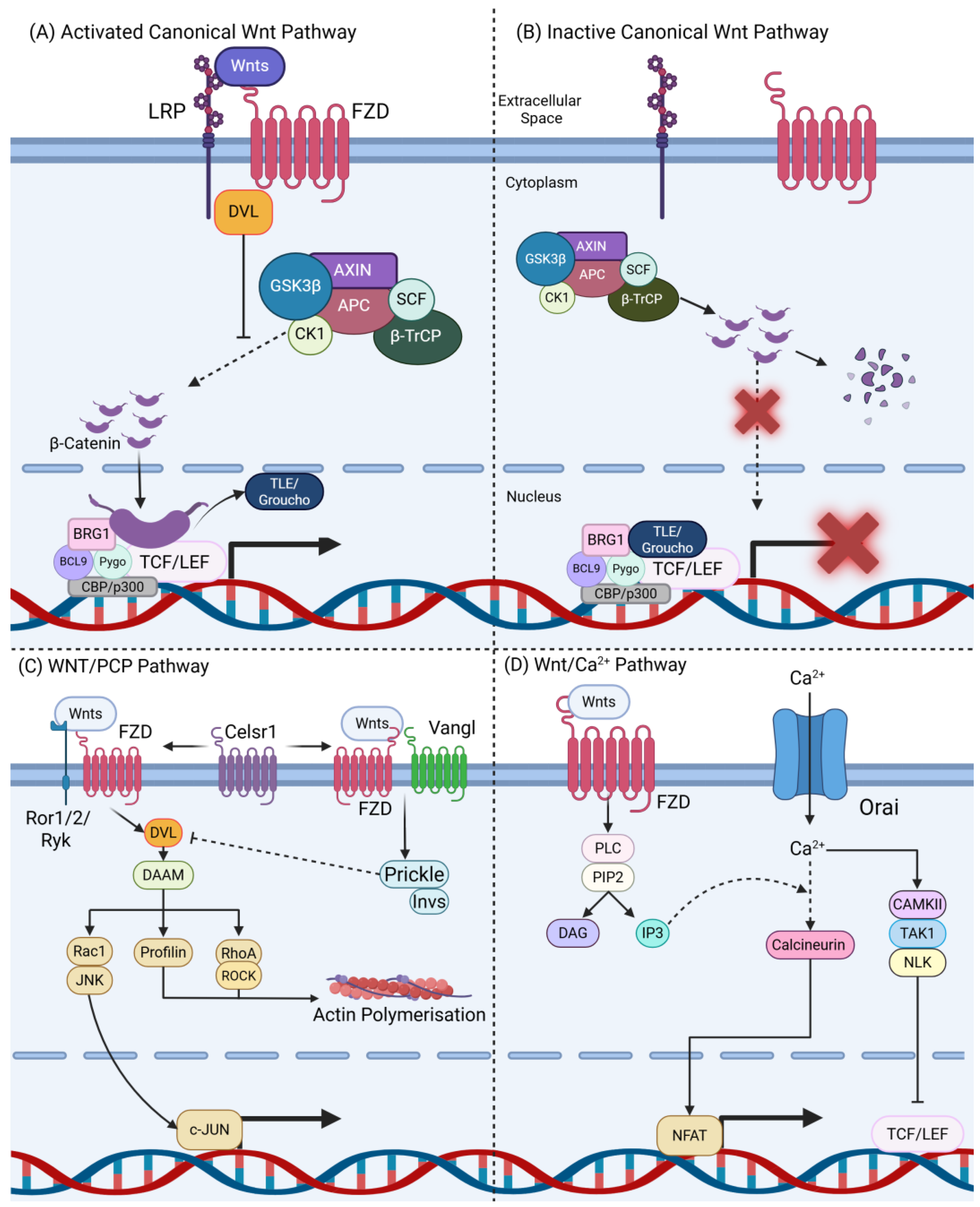
1.1. Overview of the Wnt Signalling Pathway

Wnt signalling is a highly conserved pathway found in all animals, playing a key role in cell differentiation and body axis polarity [1,2,3]. Once development has completed, Wnt signalling is required to conserve normal tissue homeostasis through the maintenance of cell renewal and stem cell compartments [1,2,3]. The deregulation of the Wnt signalling pathway is often associated with disease, specifically cancer [1,2], reflecting overlaps between the hallmarks of cancer and the function of the Wnt signalling pathway [1,2,4]. As the Wnt signalling pathway is often deregulated in cancer, it is an attractive avenue for therapeutic intervention [1,2,3,4].
The Wnt ligand family is composed of 19 glycoproteins in vertebrates [3,5]. These ligands are modified in the cytoplasm of the sending cell by O-acyltransferase porcupine (PORCN) which covalently attaches palmitoleic acid to a conserved serine residue [3,5]. The palmitoylation of Wnt proteins by PORCN is required for the secretion of all mammalian Wnts and is critical for interaction with Wntless (WLS) which transports the modified Wnt proteins to the plasma membrane [3,5]. There are several mechanisms by which Wnt ligands can be transmitted to receiving cells, including secretion as micelles, diffusion and association with glypicans, and attachment to extracellular vesicles or cytonemes [6,7,8].
The Wnt signalling pathway can be subdivided into two pathways: canonical and non-canonical (Figure 1) [1,3,5]. The canonical Wnt signalling pathway is made up of three major components: the ligand receptor complex, the destruction complex, and β-catenin transcriptional complex [1,2,3,4]. The ligand receptor complex is formed when the pathway is “activated” by a Wnt ligand, a family of 19 secreted glycoproteins [1,5]. This activation of canonical Wnt signalling is characterised by the dimerisation of FZD Wnt receptors and LDL-receptor-related protein 5/6 (LRP5/6) co-receptors, facilitated via the binding of a Wnt ligand to FZD’s cysteine-rich domain (CRD) (Figure 1A) [1,5]. A complete ligand/receptor complex leads to the recruitment of Dishevelled (Dvl) to the plasma membrane. Dvl is a phosphoprotein which phosphorylates LRP5/6 when the Wnt ligand/receptor complex is formed and leads to the dissociation of AXIN from the destruction complex (Figure 1A) [1,5]. The lack of destruction complex leads to the accumulation of β-catenin within the cytoplasm and its nuclear translocation. Once in the nucleus, β-catenin displaces Wnt transcriptional co-repressors such as Transducin-like enhancer of split (TLE) and Groucho, leading to the formation of the β-catenin transcriptional complex (Figure 1A) [1,5]. The β-catenin transcriptional complex consists of co-transcriptional factors such as T cell factor (TCF) and lymphoid enhancer factor (LEF) [1,5].
In the absence of Wnt ligand, the ligand/receptor complex is unable to form, which subsequently allows the compilation of the destruction complex (Figure 1B). The destruction complex consists of Axis Inhibition (AXIN), Glycogen Synthase Kinase 3 Beta (GSK3β), Adenomatous Polyposis Coli (APC), Casein Kinase 1 (CK1), Skp1-Cullin-F-box Containing Complex (SCF), Protein Phosphatase 2A (PP2A), and Beta-Transducin Repeat-Containing Protein (β-TrCP) [2,5]. CK1 delivers a phosphorylation modification to β-catenin. This modification at Seri45 allows GSK3β to phosphorylate β-catenin, creating a binding site for β-TrCP that facilitates ubiquitination and subsequent proteasomal degradation of β-catenin (Figure 1B) [1,5]. This prevents accumulation and nuclear translocation of β-catenin, thus inhibiting the formation of the β-catenin transcriptional complex (Figure 1B) [1,5].
Non-canonical Wnt signalling can be further subdivided into the Wnt/planar cell polarity (PCP) (Figure 1C) and Wnt/calcium (Ca2+) pathway (Figure 1D) [1,5]. In the Wnt/PCP pathway, FZD receptors work with several co-receptors including Protein Tyrosine Kinase 7 (PTK7), Muscle-Specific Kinase (MUSK), Receptor Tyrosine Kinase-Like Orphan Receptors 1 and 2 (ROR1/ROR2), Receptor like Tyrosine Kinase (RYK), Cadherin EGF LAG Seven-Pass G-Type Receptor 1 (CELSR1), and Van Gogh-Like Protein 2 (VANGL2). Downstream signalling is dependent on the co-receptor; however, all cascades begin with the localisation of Dvl to the plasma membrane (Figure 1C), which was reviewed further in Qin et al. [5].
The Wnt/Ca2+ pathway predominantly signals through Wnt5a and FZD2 (Figure 1D) [5]. The binding of the Wnt ligand to FZD2 results in PLC activation and the cleavage of PIP2 [5]. This results in the cytoplasmic influx of Ca2+, resulting in the cellular polarisation and activation of the downstream components of the Wnt/Ca2+ (Figure 1D) [5].
There are 10 frizzled Wnt receptors in vertebrates, which recognise and bind the fatty acyl groups on Wnt ligands, which mediates association with co-receptors to form the ligand/receptor complex to transmit Wnt signalling into the receiving cells [9]. Frizzled (Fzd) receptors have seven transmembrane domains, an extracellular N-terminal with a conserved cysteine rich domain (CRD), which is responsible for binding to Wnt ligands, and an intracellular C-terminal with a putative PDZ binding domain [1]. The interaction of specific Wnt ligands with specific Wnt receptors is poorly understood and is dependent on the availability or receptors, co-receptors, and other pathway components [10]. FZD7 has been reported to transmit both β-catenin/canonical and non-canonical Wnt signalling, although this is not unique to FZD7 [1].

1.2. Frizzled Receptor 7 (FZD7) in the Wnt Pathway

The frizzled (FZD) receptor family consists of 10 members (FZD1-FZD10) which belong to the F class of G coupled receptors [11]. FZD7 is highly expressed throughout embryonic development, playing a key role in PCP pathway-mediated prechordal-plate-progenitor-cell protrusion formation and migration/mesoderm differentiation during gastrulation [12,13]. FZD7 has been extensively studied in the gastrointestinal tract with genetic deletion demonstrating its key, non-redundant role for the function of intestinal and gastric stem cells [14,15,16,17]. In the mouse intestine, Lgr5+ stem cells require FZD7 to maintain normal homeostasis and regeneration [14]. Alternatively, in the stomach, FZD7 is not required to regulate the activity of stem cells in the gastric epithelium, and Fzd7-deficient Lgr5+ were able to lineage trace as per normal Fzd7-proficient Lgr5+ cells [1,14,15,16,17]. FZD7 differential expression has been shown in many cancer types when comparing tumours to patient-matched healthy tissue samples, where it transmits oncogenic Wnt signalling to promote tumorigenesis and malignant progression [1,18,19]. Although this review focusses on the growing body of work identifying FZD7 as an attractive target for cancer therapy, several other FZD Wnt receptors are also implicated in cancer [20,21], suggesting they may also be targets for therapy.
The inhibition of FZD7 has been used to demonstrate its requirement in several cellular functions that are important to cancer, including proliferation, stemness, differentiation, migration, invasion, and regulating the location of differentiated cells in normal tissue [15,22,23,24]. These studies, and many others described in this review, have helped identify FZD7 as a regulator of tumorigenesis and malignant progression, with increased FZD7 often associated with poor clinical outcomes [25,26,27]. Table 1 highlights the experiments investigating how FZD7 expression alters cancer function in vitro and in vivo. Table 2 displays the range of approaches taken to target FZD7 since 2016 identified in this review. Table 3 shows all cancer-relevant miRNA interactions with FZD7 identified since 2016 (Table 1, Table 2 and Table 3).

1.3. Wnt Signalling Dysregulation in Cancer

The Wnt signalling pathway is deregulated in multiple cancer types, due to mutations or the deregulation of Wnt signalling components [2,3,4]. As previously mentioned, the Wnt signalling pathway plays a key role in adult tissue homeostasis through regulating cellular function including proliferation, migration, and invasion [2,3,4]. Additionally, Wnt signalling is also involved in the regulation of stem cell compartments. The deregulation of both cellular function and stem cell compartments can result in aggressive disease with poor prognosis [2,3,4].

2. Gastrointestinal Cancers

Gastrointestinal (GI) cancers are reported to be attributed to approximately 26% of the global cancer burden and are responsible for up to 35% of all cancer-related mortalities worldwide [45,47,52].
Gastric tumours show an upregulation of FZD7 expression when compared to surrounding healthy tissue [45,47,53]. FZD7 is accumulated on the surface of cells containing mutations affecting genes involved in FZD receptor cell surface turnover such as RNF43 in ~54% of patients [17]. FZD7 expression has also been correlated with poor patient prognosis [52]. The genetic deletion of FZD7 in vivo inhibits tumour initiation and growth in Wnt-driven (mutant APC) and cytokine-driven (mutant GP130) mouse models of gastric cancer [17]. Similarly, the overexpression of the transcription factor B-cell lymphoma 6 (BCL6) results in the transcriptional repression of FZD7, and an associated decrease in proliferation, migration, and invasion in AGS and SGC-7901 gastric cancer cells [54].
Pharmacologically targeting FZD7 using OMP18R5 (Vantictumab), which binds FZD1/2/5/7/8, showed a reduction in the formation of both SGC-7901 and MKN-45 gastric cancer spheroids [45]. This was underpinned by a reduction in gastric cancer stemness markers CD44 and ALDH1, which re-sensitised SGC-7901 chemo-resistant cells to cisplatin [45]. Further in vivo studies have shown that OMP18R5 drastically reduces weight and the number of tumours in gp130F/F mice which develop spontaneous STAT3-dependant tumours [17]. Additionally, OMP18R5 reduced the expression of multiple Wnt target genes, including Myc and CD44, as well as Fzd6 and Fzd7 [17]. An upregulation of Fzd2 has also been identified in gastric tumours upon OMP18R5 treatment; however, Fzd2-elevated expression is unable to rescue Fzd7 loss [14,17]. This non-redundant role is consistent with Fzd7’s role during intestinal regeneration [14] and mesoderm differentiation [55]. However, Fzd7−/− mice are viable, whilst Fzd7−/−, Fzd2−/− mice are embryonic-lethal, indicating redundancy during development [56].
Colorectal cancer (CRC) is attributed to ~9.2% of cancer-related mortality in both sexes and 10.2% of total diagnosed cancer cases globally [57]. The Wnt signalling pathway in spontaneous CRC is often mutationally deregulated with ~60% of patients presenting with APC mutations [57,58,59]. Other Wnt regulators, including RNF43, CTNNB1, and AXIN2, are also mutated; however, the frequency is reported to be much lower at ~5% [58]. FZD7 is upregulated in ~37% of CRC tumours and positively correlates with poor patient survival [19,57]. To supplement the literature, we analysed publicly available RNAseq data, which show the correlation between FZD7 mRNA expression and CRC stage (Figure 2). Notably, the knockdown of FZD7 in CRC cells has recently been shown to reduce tumorigenic traits, including cell proliferation and migration [28]. Furthermore, FZD7 is upregulated via the circCSPP1-mediated downregulation of miR-944 in doxorubicin-resistant patient samples [28]. This implies FZD7 plays a role during chemotherapy resistance and presents an opportunity to re-sensitise tumours to chemotherapy agents by targeting FZD7 [28].
Targeting FZD7 in CRC cells using human single-chain fragment variable antibodies (scFVs) showed an 83% inhibition of growth via an MTT assay in SW480 cells when compared to FZD7-negative SKBR-3 cells [46]. However, FACS analysis revealed ~8% non-specific binding to the FZD7-negative cells [46].
Alternatively, to target the FZD7 receptor directly, recombinant soluble FZD7 decoy receptors can be used to inhibit ligand receptor complex formation [47]. A soluble version of the Wnt ligand binding CRD of FZD7 is reported to significantly increase apoptosis and decrease the expression of β-catenin and cyclin D1 in both AGS (gastric cancer) and SW480 (CRC) cells [47]. This approach leads to a reduction in the formation of the Wnt ligand/receptor complex and a subsequent reduction in canonical Wnt signalling downstream markers. However, this may prove a difficult therapeutic approach as previous in vivo studies have used FZD8 decoy receptors which were able to inhibit tumour growth; however, this was reversable when treatment was discontinued [60].

3. Breast Cancer

Breast cancer (BCa) is the most commonly diagnosed cancer, and the main cause of cancer-associated death in women with 2.3 million cases [61]. In 2020, there were 685,000 deaths associated with BCa worldwide [61]. In the UK, the incidence of BCa in 2020 was 53,889, and there were >11,800 BCa-associated deaths per year [62]. The current treatments for BCa cancer include surgery, endocrine therapy, bone-modifying therapies, chemotherapy, and immunotherapy [63]. Unfortunately, these treatments have several adverse effects and have been shown to reduce in efficacy due to therapeutic resistance; thus, novel therapeutic approaches for tackling BCa are required [63].
FZD7 mRNA expression is increased in human triple-negative breast cancer (TNBC) when compared to non-TNBC [29,35,48], suggesting FZD7 is an attractive target for therapy [29,30]. Using publicly available datasets, we presented FZD7 mRNA levels in a range of different subtypes of BCa, with the highest expression observed in TNBC (Figure 3). This presents a link between FZD7 and TNBC, which is associated with poor prognosis, although FZD7 levels were not significantly different between different stages of disease progression in each subtype.
FZD7 knockdown via shRNA in MDA-MB-231 and BT-20 TNBC BCa cells resulted in reduced migration, invasion, proliferation, colony formation, and stemness in vitro and blocked the growth of TNBC xenografts [29]. The analysis of the FZD7 knockdown in TNBC xenografts identified that β-catenin-dependent canonical Wnt signalling was reduced as several transcriptional Wnt target genes decreased, including AXIN2, CyclinD1 and MYC [29,30]. Interestingly, Yin et al. suggested that Wnt5a/5b (previously known to be associated with non-canonical Wnt signalling) facilitated BCa progression in a model of TNBC, confirmed by means of co-immunoprecipitation of Wnt5a/5b and FZD7 [31]. The knockdown of FZD7 reduced the expression of Wnt5a/b, which was rescued when FZD7 was upregulated. Further bioinformatic analysis associated Col6a1 with FZD7 and Wnt5b expression, which is considered to drive BCa stemness and metastasis [31]. The expression of Col6a1 was reduced in FZD7/Wnt5b knockdown tissues, while Col6a1 inhibition reduced the expression of Wnt5a/b in BCa cells [31]. In addition, the knockdown of Col6a1 impaired BCa migration, invasion, and mammosphere formation, suggesting that Col6a1 mediates FZD7/Wnt5b to regulate BCa cancer stem cells and progression [31].
In recent years, nanoparticles have been utilised to improve Fzd7-directed therapies in breast cancer [35]. The innovative technique of delivering doxorubicin (a chemotherapy agent used to treat BCa) in poly(lactic-co-glycolic acid) (PLGA) nanoparticles (NPs) covered with antibodies targeting FZD7, proved to be an efficient treatment [64]. When doxorubicin was delivered by anti-FZD7 nanoparticles in MDA-MB-231 cells, it was able to localise into the nucleus of FZD7-positive BCa cells, enhancing the treatment efficacy compared to freely delivered Doxorubicin [64]. Furthermore, an elegant technique combined anti-FZD7 antibody nanoparticles with β-catenin small interfering RNAs (siRNAs), which resulted in a significant decrease in the proliferation, migration, and spheroid formation of TNBC cells [35]. Tumour growth and metastasis were inhibited when both the FZD7 receptor and β-catenin were simultaneously targeted using nanoparticle technology in vivo [35].

4. Hepatocellular Carcinoma

Hepatocellular carcinoma (HCC) is the sixth and fourth highest cancer in both incidence and cancer-related mortality, respectively [65]. FZD7 is upregulated in HCC when compared to healthy liver tissue, suggesting it is a good target for therapy [23]. Indeed, the shRNA knockdown of FZD7 in HCC cell lines results in NF-κB induced apoptosis through the downregulation of BCL-2 and BCL-XL [23]. FZD7 expression in hepatitis B virus infection-associated HCC is no different to expression in spontaneous HCC [34]. These data suggest that FZD7 is a suitable target for both hepatitis B virus-infected and non-virus-induced HCC [34]. RNAseq data from patient samples further support a positive correlation between FZD7’s mRNA expression and disease progression in HCC (Figure 4).
Since 2016, few advances have been made on specifically targeting FZD7 using small molecule agents in HCC. However, FZD7 has since been identified to play a role in multi-drug resistance (MDR) in HCC [66]. siRNA-mediated FZD7 knockdown was able to sensitise HCC cells to several chemotherapy agents (including 5-FU, MMC, and ADR), significantly reducing their IC50 dosages [66], indicating that targeting FZD7 may sensitise HCC patients to chemotherapy.
Additionally, regulators of FZD7 gene transcription have been identified in HCC, specifically miRNA-542-3p [32]. miR-542-3p downregulation in HCC is associated with an increase in clonogenicity and increased FZD7 expression, suggesting miR-543-3p may negatively regulate FZD7 expression via an antagonistic mechanism of action [32]. miR-504 is also downregulated in HCC and is able to bind directly to the 3′ untranslated region of FZD7 mRNA, reducing its expression [33]. Indeed, reduced miR-504 expression leads to increased FZD7 expression and promotes colony formation in HCC cells [33]. These transcriptional inhibitors of FZD7 could potentially be utilised therapeutically to reverse cellular changes induced through an overexpression of FZD7 in HCC.

5. Lung Cancer

Lung cancer is the leading cause of cancer-related mortality worldwide [49]. It has been previously shown that Wnt signalling, specifically FZD7, plays a major role in both lung tumorigenesis and tumour progression [49]. Using publicly available RNAseq datasets, we have identified the upregulation of FZD7 mRNA in lung squamous cell carcinoma (LUSC) when compared to lung adenocarcinoma (LUAD) (Figure 5), with no significant difference between NSCLC and SCLC observed. These data suggest a potential link between FZD7’s mRNA expression and poor lung cancer prognosis.
The role of FZD7 within lung cancer has been extensively studied, and miR-27b-3p has been identified as a potential tool to directly downregulate the expression of FZD7 mRNA to promote apoptosis and suppress cancer cell viability [36]. Alternatively, a humanised FZD7 antibody, SHH002-hu1, has also been shown to be effective in both in vitro and in vivo studies [49]. SHH002-hu1 treatment can inhibit migration and invasion assays, in non-small cell lung cancer cell lines, to similar levels compared to FZD7 siRNA-mediated knockdown [49]. Additionally, SHH002-hu1 was more efficacious than XAV-939 (A Tankyrase inhibitor) in various lung cancer xenograft models [49]. These preclinical data suggest that targeting FZD7 with SHH002-hu1 could be a potent therapeutic approach for non-small cell lung cancer patients in the future, although further investigation into FZD7 expression levels across subtypes of lung cancer would aid in the translation of this compound.

6. Ovarian Cancer

In 2020, ovarian cancer (OC) was the third most common cancer globally with an incidence of 313,959 cases annually and a mortality rate of 207,252 cases per year [67]. Due to its high prevalence and mortality rate, considerable research efforts have gone into finding novel therapeutic targets [67]. FZD7 has been shown to have a role in driving ovarian cancer progression. Indeed, a number of studies have found that FZD7 is upregulated in ovarian cancer, and targeting this receptor results in reduced ovarian cancer progression, presenting FZD7 as an actionable target in ovarian cancer [39,68].
New findings suggest that FZD7 also plays a role in regulating the activity of ovarian cancer stem cells (OCSCs) and extracellular matrix (ECM) remodelling [37]. Tissue transglutaminase (TG2) is a protein that links epithelial cells into the ECM via fibronectins and integrins [38]. Recently, Condello et al. found an association between TG2, fibronectin, and integrin β1 as ECM contributors of ovarian cancer stemness via FZD7 [37]. FZD7 can also act as the receptor for TG2 to activate ovarian cancer stem cells in OC cells [37]. Integrin-linked kinase (ILK) is another ECM molecule that is reported to interact with FZD7 to support OCSCs [69]. In a study carried out by Atwani et al., ILK was associated with lower survival rate in patients with OC and increased expression of ILK was also found in metastatic samples from patients, suggesting its progression in OC [69]. OCSCs spheroids treated with the ILK inhibitor compound 22 (cpd-22), showed decreased expression of FZD7 and Myc. The direct link between ILK and FZD7 was later confirmed by OC spheroids obtained from patients and treated with Wnt3a, showing increased ILK and FZD7 expression by IF staining, co-IP, and a proximity ligation assay, which detects proteins localised within 40nm distance in tissues [69]. Moreover, when the ILK-FZD7 pathway was disrupted by cpd-22, it led to reduced OCSC spheroid formation and lower intraperitoneal metastasis in vivo, especially when combined with carboplatin [69].
FZD7 has also been implicated as a regulator of OC stem cell activity via epigenetic regulation of TWIST via H3K4me3 and H3K27ac at the TWIST1 proximal promoter. Similarly, FZD7 has been implicated in OC resistance to cell death via TWIST regulation of BCL2 [39]. These data strongly suggest that FZD7 promotes tumour initiation/growth/progression in OC, but there are no papers demonstrating that the direct inhibition of FZD7 can inhibit OC disease progression. However, recently, there has been a novel antibody drug conjugate (ADC) using a FZD7 specific antibody (septuximab vedotin), which has been shown to be able to specifically kill FZD7-positive OC cells in vitro, and dramatically regress OC xenograft tumours [50]. The use of a humanised mouse has also demonstrated that FZD7-ADC treatment did not cause any noticeable toxicity [50]. This is a substantial innovation in targeting FZD7-expressing cancer cells as its high specificity reduces toxicity concerns, which were raised previously due to OMP18R5′s translational issues [50,70].

7. Leukaemia

Chronic myeloid leukaemia (CML) is a myeloproliferative cancer originated from hematopoietic stem cells (HSCs) characterised by a mutation in the BCR-ABL oncogene [71]. CML can be treated by tyrosine kinase inhibitors (TKIs) such as Imatinib Mesylate (IM); however, most patients eventually develop resistance, and, therefore, new therapies are required [72].
FZD7 is upregulated in CML when in contact with bone mesenchymal stem cells (BMSCs) [40]. Co-culture studies showed that silencing FZD7 in BMSCs maintain a lower expression of FZD7 in CML cells and, conversely, increased FZD7 expression in CML cells when FZD7 was overexpressed in BMSCs [40]. Moreover, the knockdown of FZD7 in CML cells reduced the expression of Wnt target genes MDR1 and CD44, suggesting that FZD7 acted through the canonical Wnt/β-catenin pathway [40]. Silencing FZD7 in BMSCs sensitised co-cultured CML cells to IM treatment, opening a possible combination of IM and FZD7 inhibition for CML treatment [40]. This is one of the first papers to describe how FZD7 in a non-cancer cell can regulate the function of neighbouring cancer cells, suggesting FZD7 is an attractive target for this type of pro-tumour mechanism in CML and potentially other cancers.
The upregulation of FZD7 in human HSCs during the chronic phase of CML has also been detected by microarray, identifying FZD7 as a possible regulator of CML at different stages [73]. In acute lymphoblastic leukaemia (ALL), the role of long noncoding RNA (lncRNA) and FZD7 has been investigated by Wang et al., revealing a competing endogenous RNA (ceRNA) network that included differentially expressed mRNAs from the bone marrow of patients with ALL, and identified FZD7 as one of the main hub genes [74]. Additionally, FZD7 mRNA had a high correlation with the LncRNA Wilms tumour 1 homologue antisense RNA (WT1-AS). It has been suggested to act as a tumour suppressor in cervical cancer, gastric cancer, papillary thyroid carcinoma, non-small cell lung cancer, and hepatocellular carcinoma [75,76,77,78,79]. However, in patients with breast cancer and colorectal cancer, the upregulation of WT1-AS was associated with poor prognosis, suggesting a dichotomic role for WT1-AS depending on its expression status in individual cancer types [80,81]. Based on this evidence, further studies are needed to clarify the role of lncRNA WT1-AS and its association with FZD7 in ALL [74].

8. Prostate Cancer

Recently, a number of studies have highlighted the importance of FZD7 in prostate cancer (PCa) [41,82]. Ren et al. established that miR-613 has a binding site within the 3′-UTR of FZD7 [41]. The overexpression of miR-613 in PCa cells resulted in a significant decrease in FZD7 mRNA and protein levels. Conversely, the inhibition of miR-613 increased FZD7 expression, further validating FZD7 as a target for miR-613 [41]. The overexpression of FZD7 also led to increased invasion and proliferation in PCa cells, indicating FZD7 plays an oncogenic role during PCa [41].
Recent work has also identified a novel link between FZD7 and GIPC2 during PCa metastasis [82]. GIPC2 is part of the GAIP-interacting protein family containing a C-terminus domain (GIPC). GIPC2 comprises GIPC homology 1 (GH1), PDZ, and GH2 domains and is associated with familial hearing loss and cancer [82]. GIPC2 levels are increased in metastatic PCa clinical samples compared to primary PCa [82]. The overexpression of GIPC2 increased the migration, invasion, and cell adhesion hallmarks in metastatic PCa cells, while the metastatic potential of PCa cells in vivo was reduced when GIPC2 was inhibited [82]. GIPC2 overexpression reduced GSK3β levels in PCa cells and activated β-catenin signalling, and conversely, the inhibition of GIPC2 reduced β-catenin signalling [82]. Co-immunoprecipitation assays revealed that GIPC2 directly interacts with FZD7, specifically through the PDZ domain in GIPC2 [82]. The overexpression of GIPC2 resulted in increased PCa metastasis which was reduced when FZD7 was knocked down in PCa cells [82]. These studies identify and validate FZD7 as an attractive target for PCa.

9. Pancreatic Cancer

Pancreatic cancer is recognised as one of the most aggressive cancers with a 5-year survival rate, which is lower than 10% [83]. In 2020, pancreatic cancer accounted for an incidence of 495,773 new cases and 466,003 deaths worldwide [83]. The low survival rate of patients with pancreatic cancer is reflective of late diagnosis and ineffective therapeutic approaches. Analysis of publicly available RNAseq data suggests that FZD7′s expression levels are associated with increased histological grade, which, in tone, is correlated with poor disease prognosis (Figure 6).
Recently, FZD7 has been implicated in chemoresistance in pancreatic cancer [42]. ATP-binding cassette subfamily G member 2 (ABCG2) is one of the ABC transporter superfamilies related to multidrug resistance in pancreatic cancer by transporting anti-cancer drugs outside the cancer cells [42]. Tissue samples from pancreatic cancer patients showed increased expression for ABCG2 and Wnt5a compared to adjacent healthy tissue via immunohistochemistry (IHC) [42]. The survival rate was shown to be significantly lower in patients with high ABCG2 expression. Wnt5a is reported to regulate ABCG2 in capan-2 pancreatic cells in a dose-dependent manner [42]. The knockdown of FZD7 resulted in significantly lower β-catenin levels and ABCG2 in pancreatic cell lines in the presence of Wnt5a, suggesting FZD7 as the bridge between Wnt5a and ABCG2. Additionally, knocking down FZD7 led to increased apoptosis of capan-2 cells when treated with gemcitabine, highlighting the role of FZD7 in gemcitabine resistance in pancreatic cancer [42].
Analysis of publicly available databases as well as tissue from patients with pancreatic cancer, revealed FZD7 and WNT7b levels are increased in pancreatic tumours and positively correlate with poor clinical outcome [24]. The knockdown of FZD7 and WNT7b inhibited the proliferation of pancreatic cancer stem cells, reduced the levels of ABCG2 and made the cells more susceptible to gemcitabine treatment [24]. Conversely, when FZD7 and WNT7b were overexpressed, the opposite effects were observed [24]. The studies of Zhang et al. highlight how different Wnt ligands act through FZD7 to induce pancreatic cancer progression and chemoresistance [24]. Moreover, the high expression of FZD7 in pancreatic cancer has been associated with hepatic metastases in patients, and in vitro assays suggest that FZD7 promotes EMT as FZD7 silencing reduced invasion and migration linked to reduced Vimentin, Zeb1, and Slug [84]. Most recently, bioinformatics analysis from diverse databases suggest that FZD7 is upregulated in late stages of pancreatic cancer, a characteristic that could be associated with hepatic metastasis, suggesting FZD7 as a potential therapeutic target for pancreatic cancer and hepatic metastasis [85,86].

10. Melanoma

FZD7 is highly expressed in melanoma tissue when compared to healthy tissue [43]. Consistent with other cancer types, FZD7 has recently been shown to be regulated by miRNAs. miR-485-5p expression is decreased in human melanoma tissue compared to normal tissues [43]. Enforced miR-485-5p expression decreased melanoma cell proliferation and invasion, key features of cancer growth and progression that were restored when miR-485-5p was inhibited [43]. Bioinformatic analysis revealed that miR-485-5p can bind to the 3′-UTR region of FZD7, which was confirmed by a dual-luciferase assay [43]. Further studies confirmed that increased miR-485-5p expression led to reduced FZD7 expression, and when FZD7 was restored, the invasive and proliferative capacity of the melanoma cells also reverted [43]. These data identify FZD7 as a regulator of melanoma growth and progression, indicating that FZD7 presents a novel potential target for therapy against melanoma.
Most recently, it has been suggested that FZD7 supports melanoma melanosphere formation and amoeboid invasion via non-canonical Wnt/β-catenin pathway [44]. The Wnt11 ligand binds to FZD7 and its downstream effector DAAM1 to activate the non-canonical Rho-ROCK1/2–Myosin II signalling pathway in melanoma melanosphere [44]. When FZD7 was inhibited, there was a significant reduction in melanosphere formation, loss of cell rounding, lower Myosin II activity, and inhibited invasion capability [44], highlighting an important role for FZD7 in melanoma, and warranting further research.

11. Cholangiocarcinoma

Cholangiocarcinoma (cancer of the bile ducts) (CCa) is an extremely aggressive disease with poor prognosis and currently radical surgical resection remains the only curative treatment option [87]. The expression of FZD7 has been shown to be upregulated when comparing tumours to patient-matched healthy liver tissue [18]. Transcriptional upregulation of FZD7 within intrahepatic CCa has been associated with an upregulation of circACTN4, which co-localises with YBX1 to enhance FZD7 transcriptional expression [88]. Further in silico analysis has identified Receptor-Like Tyrosine Kinase (RYK) to be overexpressed in stage 4 CCa, significantly correlating with FZD7 expression and poor prognosis [89]. FZD7 has not been targeted as a therapeutic intervention for CCa in preclinical studies. However, the combination of these findings and the low rate of cytoplasmic Wnt component mutations observed in CCa [90] indicate FZD7 as an attractive therapeutic target for CCa in the near future.

12. Renal Cancer

Analysis of 53 human renal tumours revealed that FZD7 expression is upregulated when compared to healthy renal tissue [91]. Furthermore, the manipulation of FZD7 expression presented a positive correlation between FZD7 expression and proliferation in renal cell carcinoma (RCC) cell lines [91]. Additionally, Wnt3a-supplemented media were unable to rescue proliferation in FZD7 knockdown RCC cell lines, implicating FZD7 as a non-redundant transmitter of oncogenic Wnt signalling in RCC [91].
In RCC, miR-613 is downregulated and has been reported to directly interact with FZD7, binding to the 3′ UTR region, which is associated with decreased FZD7 expression [22]. The overexpression of miR-613 significantly reduced proliferation and invasion in ACHN and 786-O RCC cells, whereas artificially inducing miR-613 overexpression appears to counteract FZD7-dependant RCC cells [22]. Although FZD7 is yet to be targeted pharmacologically in renal cancer, current research evidence suggests FZD7 may prove to be an attractive therapeutic target. However, additional work is needed to establish the true status and predictive value of FZD7 in renal cancer.

13. Discussion

FZD7 is highly expressed in several cancer types with current data suggesting it is an attractive target for therapy [24,39,40,42,44,68,82,84]. Moreover, the specific targeting of FZD7 can inhibit many features of cancer from initiation to growth and metastasis [22,27,45,46,47,49,50,64,84].
FZD7’s expression and role in the gastrointestinal tract has been extensively studied in both healthy tissue and cancer [14,15,16,17]. Although the deletion of FZD7 in GI organoids results in organoid death, the deletion of FZD7 in vivo is well tolerated as the tissue has evolved a mechanism to repopulate the epithelium following acute loss of critical genes, including FZD7 [15,16] or MYC [92]. This is supported by a recent systemic knockout of FZD7, which again demonstrates that FZD7 loss is well tolerated in the GI epithelium with no adverse health effects to the mice [93]. Importantly, the deletion of FZD7 in gastric tumours did not trigger repopulation (since these highly regulated evolutionary mechanisms are lost in the tumour), but rather, the FZD7 deleted cells failed to proliferate, probably due to their lack of FZD7 that renders them unable to respond to the oncogenic Wnt signalling derived from the tumour microenvironment and niche [14,15,16,17]. FZD5 is also expressed in the intestinal epithelium; however, genetic deletion results in widespread crypt atrophy in the small intestine and colon, associated with severe weight loss of the mice [13]. These data further suggest that FZD7 is an attractive target for therapy, supported by recent work revealing the treatment of humanised-FZD7 mice that are able to respond to the new FZD7-ADC show no signs of ill health or intestinal pathology [50].
The deletion of FZD7 in normal, non-tumour gastric epithelium resulted in perturbed differentiation and the mis-localisation of differentiated cells before repopulation was triggered, in which homeostasis was restored within two weeks [15]. Together, these results support an evolutionarily conserved mechanism in the GI tract to tolerate and respond to deleterious genetic events such as loss of FZD7 and indicate FZD7 as an attractive target for therapy.
In the last eight years, research into targeting FZD7 has continued to be a promising approach to target Wnt-driven cancers. This is due to its differential and increased expression across many cancers compared to healthy tissues. Due to the superior specificity of novel humanised FZD7 antibodies currently being investigated (e.g., FZD7-ADC and SHH002-hu1), the on-target toxicity linked to the pan-FZD receptor inhibitor OMP-18R5 has potential to be mitigated if/when these compounds progress to clinical trials [49,50,70]. It will be important to further understand the molecular/cellular mechanism of how toxicity is avoided by these FZD7-directed therapies to help progress them towards clinical trials.
To advance the field of FZD7-targeted cancer therapies, a key next step is to investigate FZD7 protein expression in patient tissue samples using tissue microarray (TMA) studies. Many current investigations focus predominantly on mRNA expression, potentially overlooking important post-translational regulation by components such as RNF43 which is deregulated in several cancers, resulting in increased FZD proteins on the cell surface [94]. Protein studies via TMA can offer more direct insights into FZD7’s role in cancer progression and metastasis and potentially help identify which patients may benefit from FZD7-directed therapies. Moreover, the validation of a universally accepted, commercially available, FZD7-specific antibody would significantly enhance our ability to study FZD7’s functionality, not only within cancer cells but also within the metastatic niche, the extracellular matrix (ECM), and stromal cells, all critical in tumorigenesis and metastasis [95].
Antibody-drug conjugates are a novel cancer therapy comprising a monoclonal antibody aimed to target cancer cells linked to a cytotoxic payload by a chemical linker [96,97,98]. Such antibody-conjugated designs result in specific targeting of cancer cells, limiting the cytotoxicity to normal cells that have a lower expression of the antibody of interest [98]. FZD7 is an ideal candidate for an ADC target as it has several attractive features [96].
Firstly, FZD7 is highly expressed throughout embryonic development and then its expression is reduced in normal tissues [96]. In contrast, FZD7 expression is highly upregulated in a diverse range of cancer types, making it a good candidate to target cancer cells [96]. Secondly, FZD7 is a surface receptor, and not a secreted protein from the cancer cells, thus providing specific cancer cell targeting [96]. Finally, when the ADC compound binds FZD7 in the cancer cells, it is internalised in lysosomes of FZD7-expressing cells, resulting in apoptosis of FZD7 high cancer cells [50]. Indeed, our recently published FZD7-ADC compound shows promise to be an effective treatment for FZD7 expressing cancer types, opening a new window for cancer therapy [50].
Monoclonal antibody-based therapeutic approaches currently appear to be the most attractive method of targeting the differential expression of FZD7 in multiple cancer types [49,50]. However, the cost to produce these compounds could continue to limit the research field [99]. A possible solution to this approach would be adopting scFV antibodies, which are significantly cheaper and easier to produce [100]. Research on the efficacy of scFVs’ ability to inhibit FZD7 shows promising results; however, it requires further investigation [46]. A potential avenue for harnessing scFVs′ could be using them to deliver cytotoxic agents such as MMAE or MMAF as this has been shown to be efficacious using monoclonal antibodies [49,50,99,100]. This approach could reduce the cost of compound production and help progress on this approach to target FZD7 expressing cancer cells.
Nanoparticles also offer significant value in cancer therapy due to their ability to enhance drug delivery precision and minimise off-target effects. In the context of targeting FZD7, nanoparticles have been effectively engineered to deliver chemotherapeutic agents like doxorubicin directly to tumour cells [64]. By conjugating FZD7-specific antibodies to nanoparticles, researchers have achieved targeted drug delivery, reducing systemic toxicity. Furthermore, combining nanoparticles with siRNA-targeting β-catenin has demonstrated improved inhibition of tumour growth and metastasis [35]. These approaches underline nanoparticles’ potential to revolutionise cancer treatment by enhancing therapeutic efficacy while minimising adverse effects [35,64].
Targeting FZD7 specifically has the potential to be less toxic than pan-Wnt/FZD inhibitors. Indeed, our promising new FZD7-ADC displayed no toxicity in humanised-FZD7-mice [50], although it will be important to understand how toxicity is avoided to progress this new therapy along the translational pipeline. The FZD7-blocking antibody, SHH002-hu1, shows good anti-tumour effects in NSCLC xenografts and no cytotoxicity of bronchial epithelial cells in vitro; however, it is not clear if there was any toxicity in vivo [49], which will be important to identify to inform clinical trials. Similarly, the promising Fz7-21, a Fzd7-binding peptide, will need to be investigated in preclinical in vivo models of cancer to establish efficacy and toxicity for them to progress to clinic [51]. As FZD7 promotes cancer stem cell activity [17,31], future research should also focus on co-treatments with chemotherapy to target chemo-resistant cancer stem cell populations. Similarly, checkpoint inhibitor response can be improved with co-treatment with OMP-18R5 [101], suggesting FZD7-specific inhibitors may also be attractive to combine with modifiers of the tumour immune response for future research.

14. Conclusions

This review highlights the increasing body of work that strongly suggests that FZD7 is an attractive target for therapeutic intervention, due to its high expression in several cancers, its role in promoting tumour initiation, growth, and progression, and also the recent evidence that targeting FZD7 itself, or FZD7 expressing cells, is well tolerated, with no adverse health signs. Together, this information should encourage researchers and industrial partners to continue to develop new FZD7-directed therapies and propel current FZD7-targeting agents along the translational pipeline, to help improve cancer patient health whilst avoiding toxicity [50].

Author Contributions

Conceptualization, K.H., H.M.A., D.J.F., E.V. and T.J.P.; methodology, K.H. and H.M.A.; software, W.E.H.; validation, K.H. and H.M.A.; formal analysis, K.H., W.E.H. and T.J.P.; investigation, K.H., H.M.A. and W.E.H.; resources, K.H., H.M.A. and W.E.H.; data curation, K.H., W.E.H. and T.J.P.; writing—original draft preparation, K.H. and H.M.A.; writing—review and editing, K.H., H.B.P., K.W. and T.J.P.; visualisation, K.H. and W.E.H.; supervision, D.J.F., E.V., H.B.P. and T.J.P.; project administration, T.J.P. All authors have read and agreed to the published version of the manuscript.

Funding

TP/HP are funded by a Prostate Cancer Research Project Grant (6962); HP is funded by a CRUK Fellowship (#A27894).

Conflicts of Interest

The authors declare no conflicts of interest.

References

  1. Phesse, T.; Flanagan, D.; Vincan, E. Frizzled7: A Promising Achilles’ Heel for Targeting the Wnt Receptor Complex to Treat Cancer. Cancers 2016, 8, 50. [Google Scholar] [CrossRef] [PubMed]
  2. Zhan, T.; Rindtorff, N.; Boutros, M. Wnt signaling in cancer. Oncogene 2017, 36, 1461–1473. [Google Scholar] [CrossRef] [PubMed]
  3. Holzem, M.; Boutros, M.; Holstein, T.W. The origin and evolution of Wnt signalling. Nat. Rev. Genet. 2024, 25, 500–512. [Google Scholar] [CrossRef] [PubMed]
  4. Zhong, Z.; Yu, J.; Virshup, D.M.; Madan, B. Wnts and the hallmarks of cancer. Cancer Metastasis Rev. 2020, 39, 625–645. [Google Scholar] [CrossRef]
  5. Qin, K.; Yu, M.; Fan, J.; Wang, H.; Zhao, P.; Zhao, G.; Zeng, W.; Chen, C.; Wang, Y.; Wang, A.; et al. Canonical and noncanonical Wnt signaling: Multilayered mediators, signaling mechanisms and major signaling crosstalk. Genes Dis. 2024, 11, 103–134. [Google Scholar] [CrossRef]
  6. Stanganello, E.; Scholpp, S. Role of cytonemes in Wnt transport. J. Cell Sci. 2016, 129, 665–672. [Google Scholar] [CrossRef]
  7. Routledge, D.; Rogers, S.; Ono, Y.; Brunt, L.; Meniel, V.; Tornillo, G.; Ashktorab, H.; Phesse, T.J.; Scholpp, S. The scaffolding protein flot2 promotes cytoneme-based transport of wnt3 in gastric cancer. eLife 2022, 11, e77376. [Google Scholar] [CrossRef]
  8. Maurice, M.M.; Angers, S. Mechanistic insights into Wnt–β-catenin pathway activation and signal transduction. Nat. Rev. Mol. Cell Biol. 2025, 26, 371–388. [Google Scholar] [CrossRef]
  9. Nile, A.H.; Mukund, S.; Stanger, K.; Wang, W.; Hannoush, R.N. Unsaturated fatty acyl recognition by Frizzled receptors mediates dimerization upon Wnt ligand binding. Proc. Natl. Acad. Sci. USA 2017, 114, 4147–4152. [Google Scholar] [CrossRef]
  10. Dijksterhuis, J.P.; Baljinnyam, B.; Stanger, K.; Sercan, H.O.; Ji, Y.; Andres, O.; Rubin, J.S.; Hannoush, R.N.; Schulte, G. Systematic mapping of WNT-FZD protein interactions reveals functional selectivity by distinct WNT-FZD pairs. J. Biol. Chem. 2015, 290, 6789–6798. [Google Scholar] [CrossRef]
  11. Bous, J.; Kinsolving, J.; Grätz, L.; Scharf, M.M.; Voss, J.H.; Selcuk, B.; Adebali, O.; Schulte, G. Structural basis of frizzled 7 activation and allosteric regulation. Nat. Commun. 2024, 15, 7422. [Google Scholar] [CrossRef] [PubMed]
  12. Čapek, D.; Smutny, M.; Tichy, A.-M.; Morri, M.; Janovjak, H.; Heisenberg, C.-P. Light-activated Frizzled7 reveals a permissive role of non-canonical wnt signaling in mesendoderm cell migration. eLife 2019, 8, e42093. [Google Scholar] [CrossRef] [PubMed]
  13. Mu, Q.; Ha, A.; Santos, A.J.M.; Lo, Y.-H.; van Unen, V.; Miao, Y.; Tomaske, M.; Guzman, V.K.; Alwahabi, S.; Yuan, J.J.; et al. FZD5 controls intestinal crypt homeostasis and colonic Wnt surrogate agonist response. Dev. Cell 2024, 60, 342–351.e5. [Google Scholar] [CrossRef] [PubMed]
  14. Flanagan, D.J.; Phesse, T.J.; Barker, N.; Schwab, R.H.M.; Amin, N.; Malaterre, J.; Stange, D.E.; Nowell, C.J.; Currie, S.A.; Saw, J.T.S.; et al. Frizzled7 Functions as a Wnt Receptor in Intestinal Epithelial Lgr5+ Stem Cells. Stem Cell Rep. 2015, 4, 759–767. [Google Scholar] [CrossRef]
  15. Flanagan, D.J.; Barker, N.; Nowell, C.; Clevers, H.; Ernst, M.; Phesse, T.J.; Vincan, E. Loss of the Wnt receptor frizzled 7 in the mouse gastric epithelium is deleterious and triggers rapid repopulation in vivo. DMM Dis. Models Mech. 2017, 10, 971–980. [Google Scholar] [CrossRef]
  16. Flanagan, D.; Barker, N.; Ernst, M.; Vincan, E.; Phesse, T. The function of lgr5+ cells in the gastric antrum does not require fzd7 or myc in vivo. Biomedicines 2019, 7, 50. [Google Scholar] [CrossRef]
  17. Flanagan, D.J.; Barker, N.; Di Costanzo, N.S.; Mason, E.A.; Gurney, A.; Meniel, V.S.; Koushyar, S.; Austin, C.R.; Ernst, M.; Pearson, H.B.; et al. Frizzled-7 is required for Wnt signaling in gastric tumors with and without APC mutations. Cancer Res. 2019, 79, 970–981. [Google Scholar] [CrossRef]
  18. Boulter, L.; Guest, R.V.; Kendall, T.J.; Wilson, D.H.; Wojtacha, D.; Robson, A.J.; Ridgway, R.A.; Samuel, K.; Van Rooijen, N.; Barry, S.T.; et al. WNT signaling drives cholangiocarcinoma growth and can be pharmacologically inhibited. J. Clin. Investig. 2015, 125, 1269–1285. [Google Scholar] [CrossRef]
  19. Ye, C.; Xu, M.; Lin, M.; Zhang, Y.; Zheng, X.; Sun, Y.; Deng, Y.; Pan, J.; Xu, Z.; Lu, X.; et al. Overexpression of FZD7 is associated with poor survival in patients with colon cancer. Pathol. Res. Pract. 2019, 215, 152478. [Google Scholar] [CrossRef]
  20. Koushyar, S.; Meniel, V.S.; Phesse, T.J.; Pearson, H.B. Exploring the Wnt Pathway as a Therapeutic Target for Prostate Cancer. Biomolecules 2022, 12, 309. [Google Scholar] [CrossRef]
  21. Zeng, C.M.; Chen, Z.; Fu, L. Frizzled Receptors as Potential Therapeutic Targets in Human Cancers. Int. J. Mol. Sci. 2018, 19, 1543. [Google Scholar] [CrossRef] [PubMed]
  22. Song, H.; Nan, Y.; Wang, X.; Zhang, G.; Zong, S.; Kong, X. MicroRNA-613 inhibits proliferation and invasion of renal cell carcinoma cells through targeting FZD7. Mol. Med. Rep. 2017, 16, 4279–4286. [Google Scholar] [CrossRef] [PubMed]
  23. Xue, Y.; Chen, C.; Xu, W.; Xu, H.; Zheng, J.; Gu, Y. Downregulation of Frizzled-7 induces the apoptosis of hepatocellular carcinoma cells through inhibition of NF-κB. Oncol. Lett. 2018, 15, 7693–7701. [Google Scholar] [CrossRef]
  24. Zhang, Z.; Xu, Y.; Zhao, C. Fzd7/Wnt7b signaling contributes to stemness and chemoresistance in pancreatic cancer. Cancer Med. 2021, 10, 3332–3345. [Google Scholar] [CrossRef]
  25. Schiffgens, S.; Wilkens, L.; Brandes, A.A.; Meier, T.; Franceschi, E.; Ermani, M.; Hartmann, C.; Erol Sandalcioglu, I.; Dumitru, C.A. Sex-specific clinicopathological significance of novel (Frizzled-7) and established (MGMT, IDH1) biomarkers in glioblastoma. Oncotarget 2016, 7, 55169. [Google Scholar] [CrossRef]
  26. Cao, T.-T.; Xiang, D.; Liu, B.-L.; Huang, T.-X.; Tan, B.-B.; Zeng, C.-M.; Wang, Z.-Y.; Ming, X.-Y.; Zhang, L.-Y.; Jin, G.; et al. FZD7 is a novel prognostic marker and promotes tumor metastasis via WNT and EMT signaling pathways in esophageal squamous cell carcinoma. Oncotarget 2017, 8, 65957. [Google Scholar] [CrossRef]
  27. Li, G.; Su, Q.; Liu, H.; Wang, D.; Zhang, W.; Lu, Z.; Chen, Y.; Huang, X.; Li, W.; Zhang, C.; et al. Frizzled7 Promotes Epithelial-to-mesenchymal Transition and Stemness Via Activating Canonical Wnt/β-catenin Pathway in Gastric Cancer. Int. J. Biol. Sci. 2018, 14, 280–293. [Google Scholar] [CrossRef]
  28. Xi, L.; Liu, Q.; Zhang, W.; Luo, L.; Song, J.; Liu, R.; Wei, S.; Wang, Y. Circular RNA circCSPP1 knockdown attenuates doxorubicin resistance and suppresses tumor progression of colorectal cancer via miR-944/FZD7 axis. Cancer Cell Int. 2021, 21, 153. [Google Scholar] [CrossRef]
  29. Yang, L.; Wu, X.; Wang, Y.; Zhang, K.; Wu, J.; Yuan, Y.C.; Deng, X.; Chen, L.; Kim, C.C.H.; Lau, S.; et al. FZD7 has a critical role in cell proliferation in triple negative breast cancer. Oncogene 2011, 30, 4437–4446. [Google Scholar] [CrossRef]
  30. Simmons, G.E., Jr.; Pandey, S.; Nedeljkovic-Kurepa, A.; Saxena, M.; Wang, A.; Pruitt, K. Frizzled 7 Expression Is Positively Regulated by SIRT1 and β-Catenin in Breast Cancer Cells. PLoS ONE 2014, 9, e98861. [Google Scholar] [CrossRef]
  31. Yin, P.; Bai, Y.; Wang, Z.; Sun, Y.; Gao, J.; Na, L.; Zhang, Z.; Wang, W.; Zhao, C. Non-canonical Fzd7 signaling contributes to breast cancer mesenchymal-like stemness involving Col6a1. Cell Commun. Signal. 2020, 18, 143. [Google Scholar] [CrossRef] [PubMed]
  32. Wu, W.; Dang, S.; Feng, Q.; Liang, J.; Wang, Y.; Fan, N. MicroRNA-542-3p inhibits the growth of hepatocellular carcinoma cells by targeting FZD7/Wnt signaling pathway. Biochem. Biophys. Res. Commun. 2017, 482, 100–105. [Google Scholar] [CrossRef] [PubMed]
  33. Quan, H.; Li, B.; Yang, J. MicroRNA-504 functions as a tumor suppressor in hepatocellular carcinoma through inhibiting Frizzled-7-mediated-Wnt/β-catenin signaling. Biomed. Pharmacother. 2018, 107, 754–762. [Google Scholar] [CrossRef] [PubMed]
  34. Javanmard, D.; Karbalaie Niya, M.H.; Kalafkhany, D.; Najafi, M.; Ziaee, M.; Babaei, M.R.; Kiani, S.J.; Esghaei, M.; Jazayeri, S.M.; Panahi, M.; et al. Downregulation of gsk3β and upregulation of urg7 in hepatitis b-related hepatocellular carcinoma. Hepat. Mon. 2020, 20, e100899. [Google Scholar] [CrossRef]
  35. Dang, M.N.; Suri, S.; Li, K.; Gomez Casas, C.; Stigliano, G.; Riley, R.S.; Scully, M.A.; Hoover, E.C.; Aboeleneen, S.B.; Kramarenko, G.C.; et al. Antibody and siRNA Nanocarriers to Suppress Wnt Signaling, Tumor Growth, and Lung Metastasis in Triple-Negative Breast Cancer. Adv. Ther. 2024, 7, 2300426. [Google Scholar] [CrossRef]
  36. Sun, Y.; Xu, T.; Cao, Y.W.; Ding, X.Q. Antitumor effect of miR-27b-3p on lung cancer cells via targeting Fzd7. Eur. Rev. Med. Pharmacol. Sci. 2017, 21, 4113–4123. [Google Scholar]
  37. Condello, S.; Sima, L.; Ivan, C.; Cardenas, H.; Schiltz, G.; Mishra, R.K.; Matei, D. Tissue tranglutaminase regulates interactions between ovarian cancer stem cells and the tumor niche. Cancer Res. 2018, 78, 2990–3001. [Google Scholar] [CrossRef]
  38. Sima, L.E.; Yakubov, B.; Zhang, S.; Condello, S.; Grigorescu, A.A.; Nwani, N.G.; Chen, L.; Schiltz, G.E.; Arvanitis, C.; Zhang, Z.Y.; et al. Small molecules target the interaction between tissue transglutaminase and fibronectin. Mol. Cancer Ther. 2019, 18, 1057–1068. [Google Scholar] [CrossRef]
  39. Tan, M.; Asad, M.; Heong, V.; Wong, M.K.; Tan, T.Z.; Ye, J.; Kuay, K.T.; Thiery, J.P.; Scott, C.; Huang, R.Y.J. The FZD7-TWIST1 axis is responsible for anoikis resistance and tumorigenesis in ovarian carcinoma. Mol. Oncol. 2019, 13, 757–780. [Google Scholar] [CrossRef]
  40. Liu, N.; Zang, S.; Liu, Y.; Wang, Y.; Li, W.; Liu, Q.; Ji, M.; Ma, D.; Ji, C. FZD7 regulates BMSCs-mediated protection of CML cells. Oncotarget 2015, 7, 6175. [Google Scholar] [CrossRef]
  41. Ren, W.; Li, C.; Duan, W.; Du, S.; Yang, F.; Zhou, J.; Xing, J. MicroRNA-613 represses prostate cancer cell proliferation and invasion through targeting Frizzled7. Biochem. Biophys. Res. Commun. 2016, 469, 633–638. [Google Scholar] [CrossRef] [PubMed]
  42. Zhang, Z.; Gao, S.; Xu, Y.; Zhao, C. Regulation of ABCG2 expression by Wnt5a through FZD7 in human pancreatic cancer cells. Mol. Med. Rep. 2021, 23, 52. [Google Scholar] [CrossRef] [PubMed]
  43. Wu, J.; Li, J.; Ren, J.; Zhang, D. MicroRNA-485-5p represses melanoma cell invasion and proliferation by suppressing Frizzled7. Biomed. Pharmacother. 2017, 90, 303–310. [Google Scholar] [CrossRef] [PubMed]
  44. Rodriguez-Hernandez, I.; Maiques, O.; Kohlhammer, L.; Cantelli, G.; Perdrix-Rosell, A.; Monger, J.; Fanshawe, B.; Bridgeman, V.L.; Karagiannis, S.N.; Penin, R.M.; et al. WNT11-FZD7-DAAM1 signalling supports tumour initiating abilities and melanoma amoeboid invasion. Nat. Commun. 2020, 11, 5315. [Google Scholar] [CrossRef]
  45. Cheng, Y.; Li, L.; Pan, S.; Jiang, H.; Jin, H. Targeting Frizzled-7 decreases stemness and chemotherapeutic resistance in gastric cancer cells by suppressing MYC expression. Med. Sci. Monit. 2019, 25, 8637–8644. [Google Scholar] [CrossRef]
  46. Fazeli, M.; Zarei, N.; Moazen, B.; Nejatollahi, F. Anti-proliferative effects of human anti-FZD7 single chain antibodies on colorectal cancer cells. Shiraz E Med. J. 2017, 18, e45219. [Google Scholar] [CrossRef]
  47. Hafezi, N.; Valadan, R.; Asgarian, O.H.; Ajami, A. Anti-tumor activity of a recombinant soluble Fzd7 decoy receptor in human gastric and colon cancer cells. Iran. J. Basic Med. Sci. 2022, 25, 187–192. [Google Scholar] [CrossRef]
  48. Li, J.; Lin, N.; Zhang, S.; Weng, L.; Chen, C.; Ou, W.; Cao, Y. Characterization of the tumor microenvironment in breast cancer brain metastasis. Heliyon 2024, 10, e34876. [Google Scholar] [CrossRef]
  49. Li, K.; Mao, S.; Li, X.; Zhao, H.; Wang, J.; Wang, C.; Wu, L.; Zhang, K.; Yang, H.; Jin, M.; et al. Frizzled-7-targeting antibody (SHH002-hu1) potently suppresses non–small-cell lung cancer via Wnt/β-catenin signaling. Cancer Sci. 2023, 114, 2109–2122. [Google Scholar] [CrossRef]
  50. Do, M.; Wu, C.C.N.; Sonavane, P.R.; Juarez, E.F.; Adams, S.R.; Ross, J.; Baena, A.R.y.; Patel, C.; Mesirov, J.P.; Carson, D.A.; et al. A FZD7-specific Antibody–Drug Conjugate Induces Ovarian Tumor Regression in Preclinical Models. Mol. Cancer Ther. 2022, 21, 113–124. [Google Scholar] [CrossRef]
  51. Nile, A.H.; de Sousa, E.M.F.; Mukund, S.; Piskol, R.; Hansen, S.; Zhou, L.; Zhang, Y.; Fu, Y.; Gogol, E.B.; Kömüves, L.G.; et al. A selective peptide inhibitor of Frizzled 7 receptors disrupts intestinal stem cells. Nat. Chem. Biol. 2018, 14, 582–590. [Google Scholar] [CrossRef] [PubMed]
  52. Hafezi, N.; Alizadeh-Navaei, R.; Golpour, M.; Zafari, P.; Ajami, A. Role of Frizzled receptor expression on patients’ survival with gastrointestinal cancers: A systematic review with meta-analysis. Casp. J. Intern. Med. 2022, 13, 1. [Google Scholar] [CrossRef]
  53. Astudillo, P. A Non-canonical Wnt Signature Correlates with Lower Survival in Gastric Cancer. Front. Cell Dev. Biol. 2021, 9, 633675. [Google Scholar] [CrossRef] [PubMed]
  54. Guo, S.; Deng, J.; Wang, P.; Kou, F.; Wu, Z.; Zhang, N.; Zhao, Z.; Nie, Y.; Yang, L. The malignancy suppression and ferroptosis facilitation of BCL6 in gastric cancer mediated by FZD7 repression are strengthened by RNF180/RhoC pathway. Cell Biosci. 2023, 13, 73. [Google Scholar] [CrossRef]
  55. Chidiac, R.; Yang, A.; Kubarakos, E.; Mikolajewicz, N.; Han, H.; Almeida, M.P.; Thibeault, P.E.; Lin, S.; MacLeod, G.; Gratton, J.P.; et al. Selective activation of FZD2 and FZD7 reveals non-redundant function during mesoderm differentiation. Stem Cell Rep. 2025, 20, 102391. [Google Scholar] [CrossRef]
  56. Yu, H.; Ye, X.; Guo, N.; Nathans, J. Frizzled 2 and frizzled 7 function redundantly in convergent extension and closure of the ventricular septum and palate: Evidence for a network of interacting genes. Development 2012, 139, 4383–4394. [Google Scholar] [CrossRef]
  57. Michelli, M.; Zougros, A.; Chatziandreou, I.; Michalopoulos, N.V.; Lazaris, A.C.; Saetta, A.A. Concurrent Wnt pathway component expression in breast and colorectal cancer. Pathol. Res. Pract. 2020, 216, 153005. [Google Scholar] [CrossRef]
  58. Muzny, D.M.; Bainbridge, M.N.; Chang, K.; Dinh, H.H.; Drummond, J.A.; Fowler, G.; Kovar, C.L.; Lewis, L.R.; Morgan, M.B.; Newsham, I.F.; et al. Comprehensive molecular characterization of human colon and rectal cancer. Nature 2012, 487, 330–337. [Google Scholar] [CrossRef]
  59. Ferrell, M.; Guven, D.C.; Gomez, C.G.; Nasrollahi, E.; Giza, R.; Cheng, S.; Syed, M.P.; Magge, T.; Singhi, A.; Saeed, A.; et al. Investigating the WNT and TGF-beta pathways alterations and tumor mutation burden in young-onset colorectal cancer. Sci. Rep. 2024, 14, 17884. [Google Scholar] [CrossRef]
  60. DeAlmeida, V.I.; Miao, L.; Ernst, J.A.; Koeppen, H.; Polakis, P.; Rubinfeld, B. The Soluble Wnt Receptor Frizzled8CRD-hFc Inhibits the Growth of Teratocarcinomas In vivo. Cancer Res. 2007, 67, 5371–5379. [Google Scholar] [CrossRef]
  61. Sung, V.Y.C.; Knight, J.F.; Johnson, R.M.; Stern, Y.E.; Saleh, S.M.; Savage, P.; Monast, A.; Zuo, D.; Duhamel, S.; Park, M. Co-dependency for MET and FGFR1 in basal triple-negative breast cancers. NPJ Breast Cancer 2021, 7, 36. [Google Scholar] [CrossRef] [PubMed]
  62. Liu, C.; Li, M.; Dong, Z.X.; Jiang, D.; Li, X.; Lin, S.; Chen, D.; Zou, X.; Zhang, X.D.; Luker, G.D. Heterogeneous microenvironmental stiffness regulates pro-metastatic functions of breast cancer cells. Acta Biomater. 2021, 131, 326–340. [Google Scholar] [CrossRef] [PubMed]
  63. Trayes, K.P.; Cokenakes, S.E.H. Breast Cancer Treatment. Am. Fam. Physician 2021, 104, 171–178. [Google Scholar] [PubMed]
  64. Hoover, E.C.; Ruggiero, O.M.; Swingler, R.N.; Day, E.S. FZD7-Targeted Nanoparticles to Enhance Doxorubicin Treatment of Triple-Negative Breast Cancer. ACS Omega 2024, 9, 14323–14335. [Google Scholar] [CrossRef]
  65. Ma, K.; Wu, H.; Ji, L. Construction of HBV gene-related prognostic and diagnostic models for hepatocellular carcinoma. Front. Genet. 2023, 13, 1065644. [Google Scholar] [CrossRef]
  66. Chen, Z.; Huang, C.; Ma, T.; Jiang, L.; Tang, L.; Shi, T.; Zhang, S.; Zhang, L.; Zhu, P.; Li, J.; et al. Reversal effect of quercetin on multidrug resistance via FZD7/β-catenin pathway in hepatocellular carcinoma cells. Phytomedicine 2018, 43, 37–45. [Google Scholar] [CrossRef]
  67. Huang, J.; Chan, W.C.; Ngai, C.H.; Lok, V.; Zhang, L.; Lucero-Prisno, D.E.; Xu, W.; Zheng, Z.J.; Elcarte, E.; Withers, M.; et al. Worldwide Burden, Risk Factors, and Temporal Trends of Ovarian Cancer: A Global Study. Cancers 2022, 14, 2230. [Google Scholar] [CrossRef]
  68. Asad, M.; Wong, M.K.; Tan, T.Z.; Choolani, M.; Low, J.; Mori, S.; Virshup, D.; Thiery, J.P.; Huang, R.Y.J. FZD7 drives in vitro aggressiveness in stem-A subtype of ovarian cancer via regulation of non-canonical wnt/PCP pathway. Cell Death Dis. 2014, 5, e1346. [Google Scholar] [CrossRef]
  69. Atwani, R.; Nagare, R.; Rogers, A.; Prasad, M.; Lazar, V.; Sandusky, G.; Tong, Y.; Pin, F.; Condello, S. Integrin-linked kinase-frizzled 7 interaction maintains cancer stem cells to drive platinum resistance in ovarian cancer. J. Exp. Clin. Cancer Res. 2024, 43, 156. [Google Scholar] [CrossRef]
  70. Diamond, J.R.; Becerra, C.; Richards, D.; Mita, A.; Osborne, C.; O’Shaughnessy, J.; Zhang, C.; Henner, R.; Kapoun, A.M.; Xu, L.; et al. Phase Ib clinical trial of the anti-frizzled antibody vantictumab (OMP-18R5) plus paclitaxel in patients with locally advanced or metastatic HER2-negative breast cancer. Breast Cancer Res. Treat. 2020, 184, 53–62. [Google Scholar] [CrossRef]
  71. Jabbour, E.; Kantarjian, H. Chronic myeloid leukemia: 2020 update on diagnosis, therapy and monitoring. Am. J. Hematol. 2020, 95, 691–709. [Google Scholar] [CrossRef] [PubMed]
  72. Senapati, J.; Sasaki, K.; Issa, G.C.; Lipton, J.H.; Radich, J.P.; Jabbour, E.; Kantarjian, H.M. Management of chronic myeloid leukemia in 2023—Common ground and common sense. Blood Cancer J. 2023, 13, 58. [Google Scholar] [CrossRef] [PubMed]
  73. de Cássia Viu Carrara, R.; Fontes, A.M.; Abraham, K.J.; Orellana, M.D.; Haddad, S.K.; Palma, P.V.B.; Panepucci, R.A.; Zago, M.A.; Covas, D.T. Expression differences of genes in the PI3K/AKT, WNT/b-catenin, SHH, NOTCH and MAPK signaling pathways in CD34+ hematopoietic cells obtained from chronic phase patients with chronic myeloid leukemia and from healthy controls. Clin. Transl. Oncol. 2018, 20, 542–549. [Google Scholar] [CrossRef]
  74. Wang, W.; Lyu, C.; Wang, F.; Wang, C.; Wu, F.; Li, X.; Gan, S. Identification of Potential Signatures and Their Functions for Acute Lymphoblastic Leukemia: A Study Based on the Cancer Genome Atlas. Front. Genet. 2021, 12, 656042. [Google Scholar] [CrossRef]
  75. Lv, L.; Chen, G.; Zhou, J.; Li, J.; Gong, J. WT1-AS promotes cell apoptosis in hepatocellular carcinoma through down-regulating of WT1. J. Exp. Clin. Cancer Res. 2015, 34, 119. [Google Scholar] [CrossRef]
  76. Du, T.; Zhang, B.; Zhang, S.; Jiang, X.; Zheng, P.; Li, J.; Yan, M.; Zhu, Z.; Liu, B. Decreased expression of long non-coding RNA WT1-AS promotes cell proliferation and invasion in gastric cancer. Biochim. Biophys. Acta Mol. Basis Dis. 2016, 1862, 12–19. [Google Scholar] [CrossRef]
  77. Jiang, X.; Wang, J.; Fang, L. LncRNA WT1-AS over-expression inhibits non-small cell lung cancer cell stemness by down-regulating TGF-β1. BMC Pulm. Med. 2020, 20, 113. [Google Scholar] [CrossRef]
  78. Le, F.; Luo, P.; Ouyang, Q.; Zhong, X. LncRNA WT1-AS downregulates survivin by upregulating miR-203 in papillary thyroid carcinoma. Cancer Manag. Res. 2020, 12, 443–449. [Google Scholar] [CrossRef]
  79. Tong, W.; Zhang, H. Overexpression of long non-coding RNA WT1-AS or silencing of PIK3AP1 are inhibitory to cervical cancer progression. Cell Cycle 2021, 20, 2583–2596. [Google Scholar] [CrossRef]
  80. Sun, M.; Wu, D.; Zhou, K.; Li, H.; Gong, X.; Wei, Q.; Du, M.; Lei, P.; Zha, J.; Zhu, H.; et al. An eight-lncRNA signature predicts survival of breast cancer patients: A comprehensive study based on weighted gene co-expression network analysis and competing endogenous RNA network. Breast Cancer Res. Treat. 2019, 175, 59–75. [Google Scholar] [CrossRef]
  81. Zhang, H.; Wang, Z.; Wu, J.; Ma, R.; Feng, J. Long noncoding RNAs predict the survival of patients with colorectal cancer as revealed by constructing an endogenous RNA network using bioinformation analysis. Cancer Med. 2019, 8, 863–873. [Google Scholar] [CrossRef]
  82. Wang, L.; Wang, J.; Yin, X.; Guan, X.; Li, Y.; Xin, C.; Liu, J. GIPC2 interacts with Fzd7 to promote prostate cancer metastasis by activating WNT signaling. Oncogene 2022, 41, 2609–2623. [Google Scholar] [CrossRef] [PubMed]
  83. Ilic, I.; Ilic, M. International patterns in incidence and mortality trends of pancreatic cancer in the last three decades: A joinpoint regression analysis. World J. Gastroenterol. 2022, 28, 4698–4715. [Google Scholar] [CrossRef] [PubMed]
  84. Zhang, Z.; Xu, Y. FZD7 accelerates hepatic metastases in pancreatic cancer by strengthening EMT and stemness associated with TGF-β/SMAD3 signaling. Mol. Med. 2022, 28, 82. [Google Scholar] [CrossRef] [PubMed]
  85. Chen, X.; Liu, F.; Xue, Q.; Weng, X.; Xu, F. Metastatic pancreatic cancer: Mechanisms and detection (Review). Oncol. Rep. 2021, 46, 231. [Google Scholar] [CrossRef]
  86. Li, Y.; Liu, Z.; Zhang, Y. Expression and prognostic impact of FZDs in pancreatic adenocarcinoma. BMC Gastroenterol. 2021, 21, 79. [Google Scholar] [CrossRef]
  87. Jiang, H.; Gao, B.; Meng, Z.; Wang, Y.; Jiao, T.; Li, J.; Li, X.; Cao, Y.; Zhang, X.; Li, C.; et al. Integrative multi-omics analysis reveals the role of tumor-associated endothelial cells and their signature in prognosis of intrahepatic cholangiocarcinoma. J. Transl. Med. 2024, 22, 948. [Google Scholar] [CrossRef]
  88. Chen, Q.; Wang, H.; Li, Z.; Li, F.; Liang, L.; Zou, Y.; Shen, H.; Li, J.; Xia, Y.; Cheng, Z.; et al. Circular RNA ACTN4 promotes intrahepatic cholangiocarcinoma progression by recruiting YBX1 to initiate FZD7 transcription. J. Hepatol. 2022, 76, 135–147. [Google Scholar] [CrossRef]
  89. Zapata-García, J.A.; Jave-Suárez, L.F.; Aguilar-Lemarroy, A. Delving into the Role of Receptor-like Tyrosine Kinase (RYK) in Cancer: In Silico Insights into Its Diagnostic and Prognostic Utility. J. Mol. Pathol. 2024, 5, 66–80. [Google Scholar] [CrossRef]
  90. Tokumoto, N.; Ikeda, S.; Ishizaki, Y.; Kurihara, T.; Ozaki, S.; Iseki, M.; Shimizu, Y.; Itamoto, T.; Arihiro, K.; Okajima, M.; et al. Immunohistochemical and mutational analyses of Wnt signaling components and target genes in intrahepatic cholangiocarcinomas. Int. J. Oncol. 2005, 27, 973–980. [Google Scholar] [CrossRef]
  91. Xu, R.; Zeng, S.; Xie, W.; Sun, C.; Chen, Y.L.; Chen, M.J.; Zhang, L. The expression and function of Frizzled-7 in human renal cell carcinoma. Clin. Transl. Oncol. 2016, 18, 269–276. [Google Scholar] [CrossRef]
  92. Muncan, V.; Sansom, O.J.; Tertoolen, L.; Phesse, T.J.; Begthel, H.; Sancho, E.; Cole, A.M.; Gregorieff, A.; de Alboran, I.M.; Clevers, H.; et al. Rapid loss of intestinal crypts upon conditional deletion of the Wnt/Tcf-4 target gene c-Myc. Mol. Cell. Biol. 2006, 26, 8418–8426. [Google Scholar] [CrossRef] [PubMed]
  93. Gu, N.X.; Guo, Y.R.; Lin, S.E.; Wang, Y.H.; Lin, I.H.; Chen, Y.F.; Yen, Y. Frizzled 7 modulates goblet and Paneth cell fate, and maintains homeostasis in mouse intestine. Development 2023, 150, dev200932. [Google Scholar] [CrossRef] [PubMed]
  94. Bugter, J.M.; van Kerkhof, P.; Jordens, I.; Janssen, E.; Tran Ngoc Minh, T.; Iglesias van Montfort, D.; Jamieson, C.; Maurice, M.M. E3 ligases RNF43 and ZNRF3 display differential specificity for endocytosis of Frizzled receptors. Life Sci. Alliance 2024, 7, e202402575. [Google Scholar] [CrossRef] [PubMed]
  95. Nabhan, A.N.; Webster, J.D.; Adams, J.J.; Blazer, L.; Everrett, C.; Eidenschenk, C.; Arlantico, A.; Fleming, I.; Brightbill, H.D.; Wolters, P.J.; et al. Targeted alveolar regeneration with Frizzled-specific agonists. Cell 2023, 186, 2995–3012.e2915. [Google Scholar] [CrossRef]
  96. Ritchie, M.; Tchistiakova, L.; Scott, N. Implications of receptor-mediated endocytosis and intracellular trafficking dynamics in the development of antibody drug conjugates. mAbs 2013, 5, 13–21. [Google Scholar] [CrossRef]
  97. Diamantis, N.; Banerji, U. Antibody-drug conjugates—An emerging class of cancer treatment. Br. J. Cancer 2016, 114, 362–367. [Google Scholar] [CrossRef]
  98. Lambert, J.M.; Berkenblit, A. Antibody–Drug Conjugates for Cancer Treatment. Annu. Rev. Med. 2018, 69, 191–207. [Google Scholar] [CrossRef]
  99. Samaranayake, H.; Wirth, T.; Schenkwein, D.; Räty, J.K.; Ylä-Herttuala, S. Challenges in monoclonal antibody-based therapies. Ann. Med. 2009, 41, 322–331. [Google Scholar] [CrossRef]
  100. Gezehagn Kussia, G.; Tessema, T.S. The Potential of Single-Chain Variable Fragment Antibody: Role in Future Therapeutic and Diagnostic Biologics. J. Immunol. Res. 2024, 2024, 1804038. [Google Scholar] [CrossRef]
  101. DeVito, N.C.; Sturdivant, M.; Thievanthiran, B.; Xiao, C.; Plebanek, M.P.; Salama, A.K.S.; Beasley, G.M.; Holtzhausen, A.; Novotny-Diermayr, V.; Strickler, J.H.; et al. Pharmacological Wnt ligand inhibition overcomes key tumor-mediated resistance pathways to anti-PD-1 immunotherapy. Cell Rep. 2021, 35, 109071. [Google Scholar] [CrossRef]
Figure 1. The Wnt signalling pathway. Panel (A) shows the activated canonical Wnt signalling pathway. Wnt ligands bind to FZD receptors and LRP5/6 co-receptors, forming the ligand receptor complex. This leads to recruitment of Dishevelled (Dvl) to the plasma membrane which inhibits the function of the destruction complex. β-catenin accumulates in the cytoplasm and translocases into the nucleus, displacing TLE/Groucho, forming the β-catenin transcriptional complex. The formation of this transcriptional complex leads to the activation of canonical Wnt target genes which regulate cellular functions associated with tissue homeostasis. Panel (B) shows inactive canonical Wnt signalling pathway. In the absence of Wnt ligands, the destruction complex, which is composed of AXIN, APC, GSK3β, CK1, SCF, and β-TrCP, sequentially phosphorylates cytoplasmic β-catenin. This results in ubiquitination of β-catenin and subsequent proteasomal degradation. Inhibiting β-catenin cytoplasmic accumulation limits its nuclear translocation, ensuring canonical Wnt target gene remain transcriptionally repressed. Panels (C,D) show the non-canonical Wnt signalling pathway. Panel (C) shows the Wnt/Planar cell polarity (PCP) pathway. Activation of the Wnt/PCP pathway may occur via multiple receptors, including FZDs, CELSR1, and VANGL2. Binding of specific Wnt ligands to FZD/Ror1/2/Ryk complex leads to recruitment of Dvl. This begins a signalling cascade through DAAM which results in either transcriptional activation of Wnt/PCP target genes via c-JUN or activation of actin polymerisation through RhoA, ROCK, and Proflin. When Wnt ligands bind to FZD/Vangl complex Prickle is recruited to supress Dvl action. These complexes form on opposite ends of the cell surface, notating the Wnt/PCP anterior/posterior axis. Panel (D) shows the Wnt/Ca2+ pathway. Orai proteins allow calcium to enter the cell. Binding of specific Wnt ligands to FZD receptors results in G protein activation of PLC which cleaves PIP2 into DAG and IP3. Calcium enters the cell and accumulates within the endoplasmic reticulum, where IP3 binds to its corresponding receptor, inducing a release of calcium into the cytoplasm. DAG and cytoplasmic Ca2+ initiate a signalling cascade to activate actin polymerisation via PKC and CDC42. IP3 mediated cytoplasmic Ca2+ then induce transcriptional repression of β-catenin/TCF/LEF transcriptional complex and activates NFAT via Calcineurin. Created in BioRender, https://BioRender.com/0mvqg1w (Accessed on 28 March 2025).
Sci 07 00061 g001
Figure 2. FZD7 mRNA expression is increased in late-stage colorectal adenocarcinoma. FZD7 mRNA expression levels across American Joint Committee on Cancer (AJCC) neoplasm disease stages (Stages I, II, III, and IV). Bars represent mean expression ± standard error of the mean (S.E.M). Statistically significant differences were observed between Stage I and Stages III and IV (p < 0.05, denoted by *). Statistical analysis was performed using one-way ANOVA. Data included Stage 1 (n = 91), Stage 2 (n = 188), Stage 3 (n = 151), and Stage 4 (n = 68). Data on patient samples were obtained from the Colorectal Adenocarcinoma dataset (TCGA, PanCancer Atlas) and accessed on 2 December 2024 via https://www.cbioportal.org/.
Sci 07 00061 g002
Figure 3. FZD7 mRNA expression across breast cancer subtypes. FZD7 mRNA expression levels in different breast cancer subtypes: Luminal A (n = 379), Luminal B (n = 244), HER2-enriched (HER2) (n = 230), Basal (n = 363), and triple-negative breast cancer (TNBC) (n = 251). Bars represent mean expression ± standard error of the mean (S.E.M). Statistically significant differences were observed between Luminal A and Basal or TNBC (p < 0.0001, denoted by ****) Statistical analysis was performed using one-way ANOVA. Non-specific luminal disease group removed. Data were obtained from the following GEO series (GSE), 12276, 12763, 12777, 13787, 16446, 17907, 20685, 20713, 21653, 31448, 45827, 48391, 65216, 76275 on 02/12/2024.
Sci 07 00061 g003
Figure 4. FZD7 mRNA expression status in liver hepatocellular carcinoma. FZD7 mRNA expression across neoplasm disease stages (Stages I, II, and III) based on the American Joint Committee on Cancer (AJCC) classification. Bars represent mean expression ± standard error of the mean (SEM). Significant differences were observed between stages (p < 0.05, denoted by *; p < 0.01, denoted by **). Statistical analysis was performed using one-way ANOVA. Data included Stage I (n = 165), Stage II (n = 81), and Stage III (n = 78). Patient’s data were obtained from the Liver Hepatocellular Carcinoma dataset (TCGA, PanCancer Atlas) accessed on 2 December 2024 via https://www.cbioportal.org/.
Sci 07 00061 g004
Figure 5. FZD7 mRNA expression in lung adenocarcinoma (LUAD) and lung squamous cell carcinoma (LUSC). FZD7 mRNA expression levels in lung adenocarcinoma (LUAD) (n = 510) and lung squamous cell carcinoma (LUSC) (n = 484). Bars represent mean expression ± standard error of the mean (S.E.M). LUSC exhibited significantly higher FZD7 mRNA expression compared to LUAD (p < 0.0001, denoted by ****). Statistical analysis was performed using an unpaired t-test. Data were obtained from 994 patient samples in the lung adenocarcinoma (TCGA, PanCancer Atlas) and lung squamous cell carcinoma (TCGA, PanCancer Atlas) datasets and accessed on 2 December 2024 via https://www.cbioportal.org/.
Sci 07 00061 g005
Figure 6. FZD7 mRNA expression status in pancreatic adenocarcinoma. Bar graph showing FZD7 mRNA expression across neoplasm histologic grades (G1 (n = 31), G2 (n = 94), and G3 (n = 48)). Bars represent mean expression ± standard error of the mean (SEM). Statistically significant differences were observed between grades (p < 0.05, denoted by *; p < 0.01, denoted by **). Statistical analysis was performed using one-way ANOVA. Data include 173 patient samples and were obtained from the Pancreatic Adenocarcinoma dataset (TCGA, PanCancer Atlas) accessed on 2 December 2024 via https://www.cbioportal.org/.
Sci 07 00061 g006
Table 1. FZD7 mechanism in cancer.
Cancer TypesInvestigating FZD7 Role in Cancer FunctionReference
In VitroIn Vivo
GastricFZD7 knockdown reduces colony-forming ability, c-MYC and cyclinD1 expression in MKN28 and MKN24.FZD7 knock out reduces tumour burden, c-MYC, CD44, and cyclinD1 expression in gastric GEMM (gp130F/F).[17]
ColorectalcircCSPP1/miRNA-944-mediated knockdown of FZD7 in CRC cells has been shown to reduce tumorigenic traits and reduces chemoresistance.shRNA-targeting cricCSPP1 (a positive regulator of FZD7) reduced tumour size in subcutaneous CRC model.[28]
BreastFZD7 knockdown via shRNA in MDA-MB-231 and BT-20 TNBC BCa cells resulted in reduced migration, invasion, proliferation, and colony formation.FZD7 shRNA knockdown in MDA-MB-231 reduced xenograft tumour size in subcutaneous model. Additionally, Axin2, Cyclin D1 and MYC expression were all downregulated.[29,30,31]
Hepatocellular CarcinomashRNA knockdown of FZD7 in HCC cell lines results in NF-κB-induced apoptosis through downregulation of Bcl-2 and Bcl-XL.
siRNA-mediated FZD7 knockdown was able to sensitise HCC cells to several chemo-therapy agents (including 5-FU, MMC, and ADR) significantly reducing their IC50 dosages.
Increased FZD7 expression promotes colony formation in HCC cells.
[23,32,33,34,35]
LungDownregulation of FZD7 mRNA via miR-27b-3p promotes apoptosis and suppress cancer cell viability. [36]
OvarianshRNA downregulation of FZD7 results in epithelial-like phenotype and reduction in EMT-associated gene expression.FZD7 knockout in MA-148 cells was unable to prevent tumour formation in a subcutaneous xenograft model of OC. However, direct comparisons of FZD7 proficient and deficient cells were not made.[37,38,39]
LeukaemiaChanges in FZD7 expression in CML cells mirror those in BMSCs. Manipulation of FZD7 expression (knockdown or overexpression) in BMSCs influences FZD7 expression in surrounding CML cells.
Direct knockdown of FZD7 in CML cells reduced the expression of Wnt target genes MDR1 and CD44, suggesting that FZD7 acts through the canonical Wnt/β-catenin pathway.
[40]
ProstateOverexpression of FZD7 led to increased invasion and proliferation in PCa cells, indicating FZD7 plays an oncogenic role in PCa. [41]
PancreaticKnockdown of FZD7 resulted in significantly lower β-catenin levels and ABCG2 in pancreatic cell lines in the presence of Wnt5a, suggesting FZD7 as the bridge between Wnt5a and ABCG2. Additionally, knocking down FZD7 led to increased apoptosis of capan-2 cells when treated with gemcitabine, highlighting the role of FZD7 in gemcitabine resistance in pancreatic cancer. [24,42]
MelanomaFZD7 knockdown via miR-486-5p expression resulted in a reversible loss of invasion and proliferation capacity in melanoma cell line.
Furthermore, FZD7 is required for melanoma melanosphere formation and invasion via the non-canonical Wnt signalling pathway.
[43,44]
RenalInducing overexpression of miR-613 (negative regulator of FZD7) significantly reduced proliferation and invasion of FZD7 dependant RCC cells [22]
Table 2. List of approaches targeting FZD7 since 2016.
Compound NameMechanism of ActionCancer TypesReference
OMP18R5Noncompetitive inhibitor of FZD1, 2, 5, 7, and 8.Gastric[17,45]
FZD7 specific single-chain fragment variable antibodiesBinds and inhibits FZD7 mediated signalling
(binding region not mentioned).
CRC[46]
Soluble recombinant FZD7 decoy receptorBinds to ligands associated with FZD7, limiting the formation of the Wnt ligand/receptor complex.Gastric and CRC[47]
FZD7 targeting nanoparticlesDeliver doxorubicin or beta-catenin siRNA directly to FZD7 expressing cells.Breast[35,48]
SHH002-hu1Humanised antibody which competitively inhibit FZD7 mediated signalling.Lung[49]
FZD7-ADCHumanised antibody which noncompetitively binds to FZD7 and delivers a microtubule inhibitor.Ovarian[50]
Fz7-21Small peptide inhibitor of FZD7 mediated signalling.N/A[51]
Table 3. List of miRNA interactions with FZD7.
miRNAMechanismCancer TypesReference
miR-27b-3pDirectly downregulates FZD7 expression in lung cancer.Lung[36]
miR-485-5pBinds to the 3′-untranslated region of FZD7, increased miR-485-5p expression led to reduced FZD7 expression and when FZD7 was restored, the invasive and proliferative capacity of the melanoma cells was restored.Melanoma[43]
miR-504Binds directly to the 3′ untranslated region of FZD7 mRNA, reducing its expression. Downregulated in HCC.HCC[33]
miR-542-3pDownregulation in HCC is associated with an increase in clonogenicity and increased FZD7 expression.HCC[32]
miR-613Binds within the 3′-UTR of FZD7, reduces FZD7 expression. Overexpression of miR-613 significantly reduced proliferation and invasion in ACHN and 786-O RCC cells, whereas artificially inducing miR-613 overexpression appears to counteract FZD7-dependant tumour cells.Prostate and RCC[22,41]
miR-944FZD7 is upregulated via circCSPP1-mediated downregulation of miR-944 in doxorubicin resistant patient samples.CRC[28]
Disclaimer/Publisher’s Note: The statements, opinions and data contained in all publications are solely those of the individual author(s) and contributor(s) and not of MDPI and/or the editor(s). MDPI and/or the editor(s) disclaim responsibility for any injury to people or property resulting from any ideas, methods, instructions or products referred to in the content.

