
Eccentric Exercise and Muscle Damage: An Introductory Guide

 ,
,  ,
,  ,
,  and
and
Abstract
1. Introduction
The Approach of the Present Review
2. Mechanisms Involved in Eccentric Exercise-Induced Muscle Damage
2.1. Contemporary Theories About the Mechanisms Involved in Muscle Damage
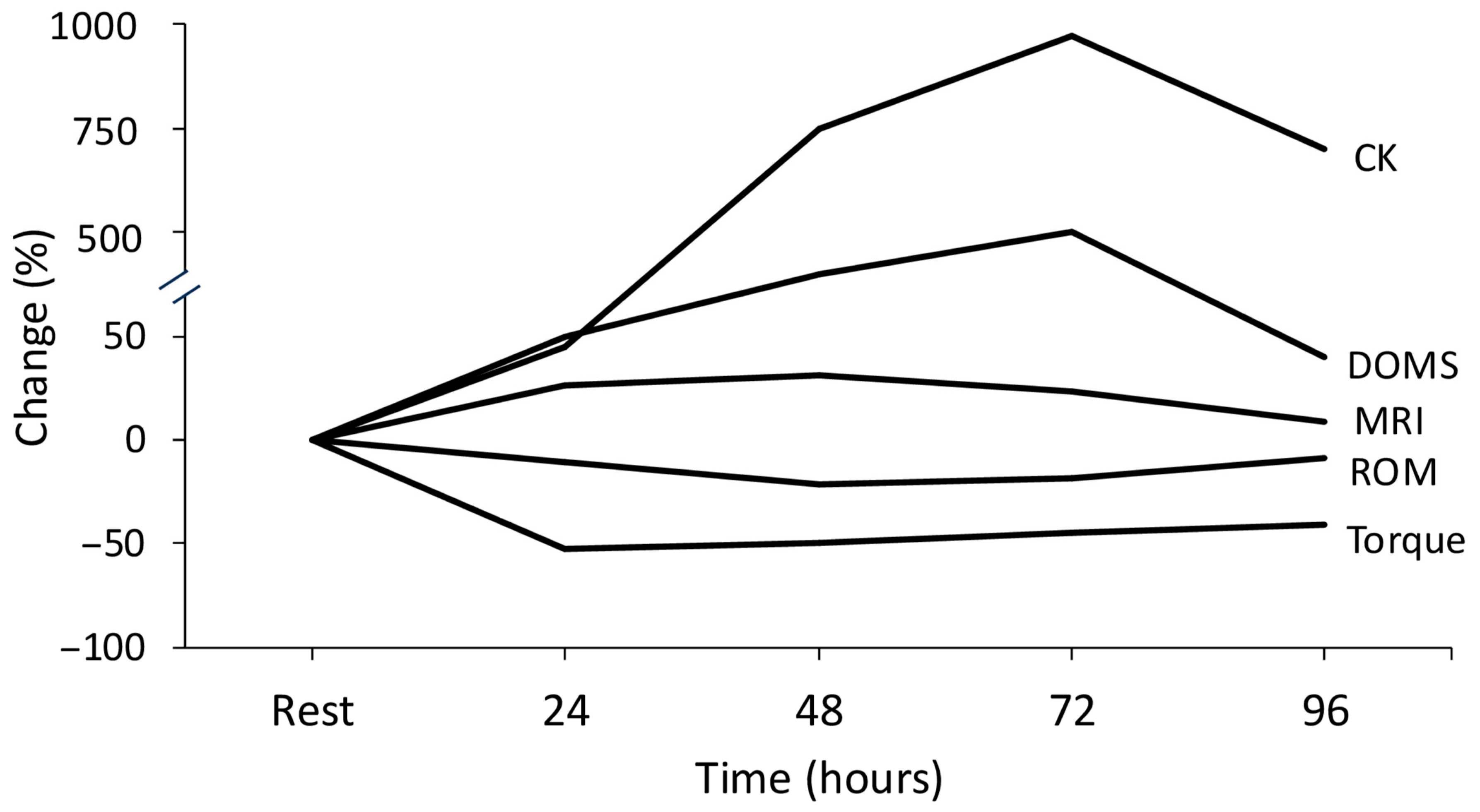
2.2. Biomarkers for the Assessment of Muscle Damage
2.3. Take Home Message
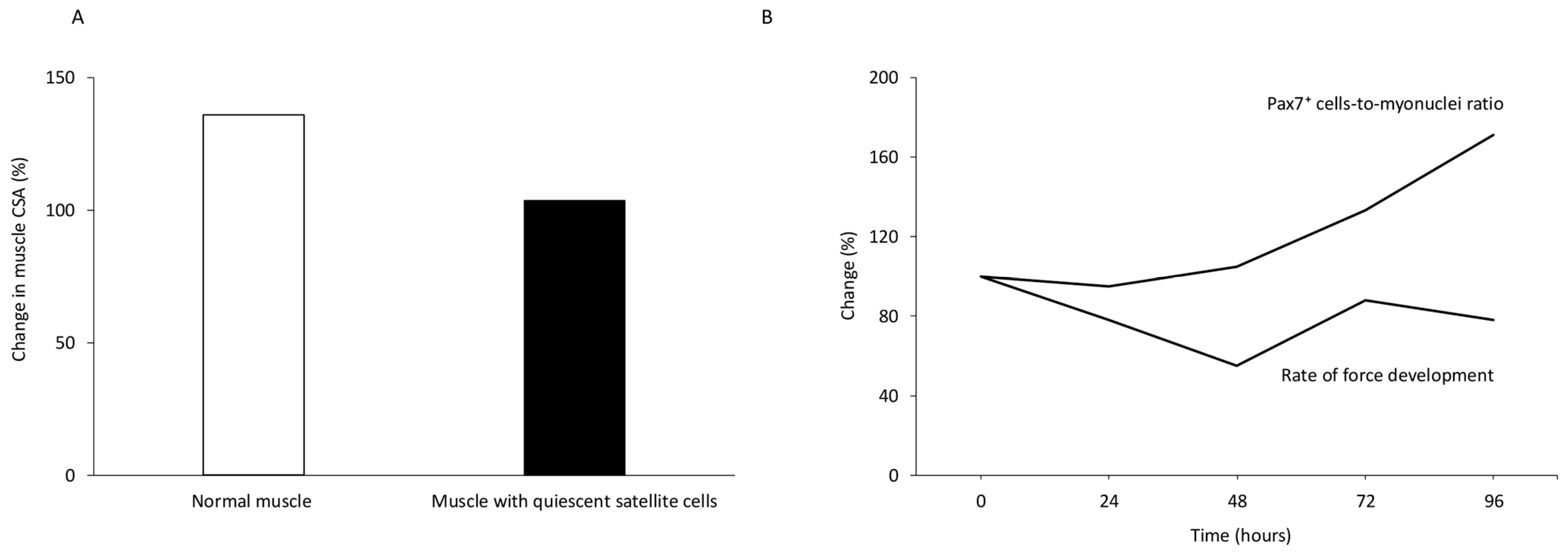
3. Skeletal Muscle Repair
Take Home Message
4. Energy Expenditure
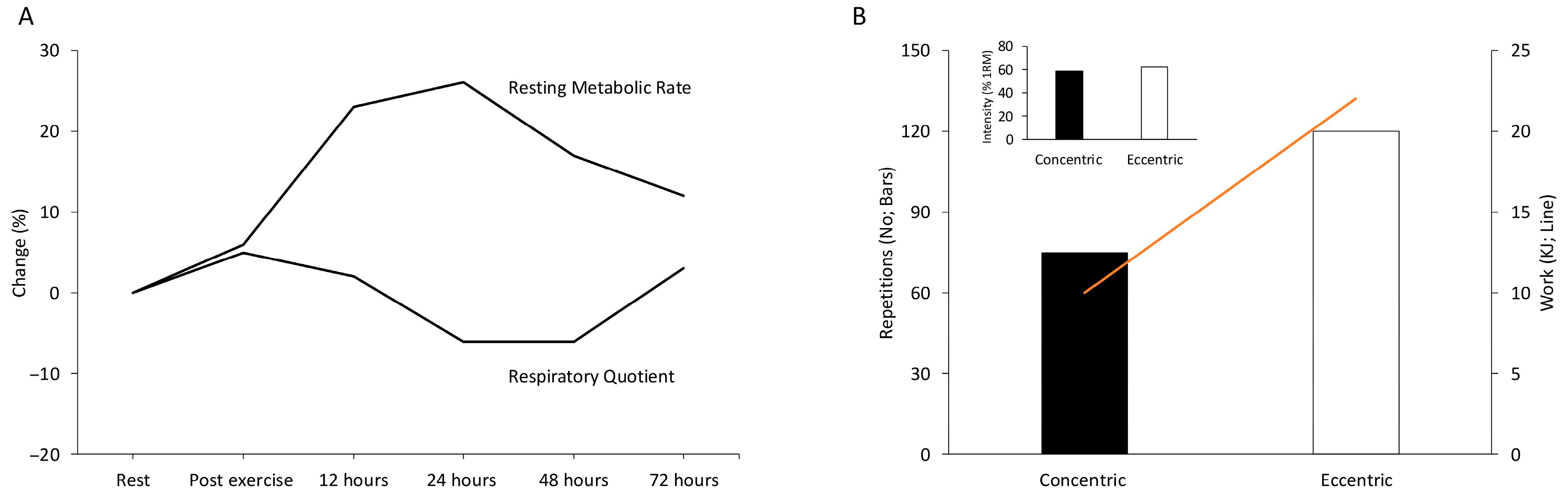
4.1. Energy Expenditure During Eccentric Exercise
4.2. Energy Expenditure During the Recovery Period (Days) Post Eccentric Exercise
4.3. Take Home Message
5. Repeated Sessions of Eccentric Exercise
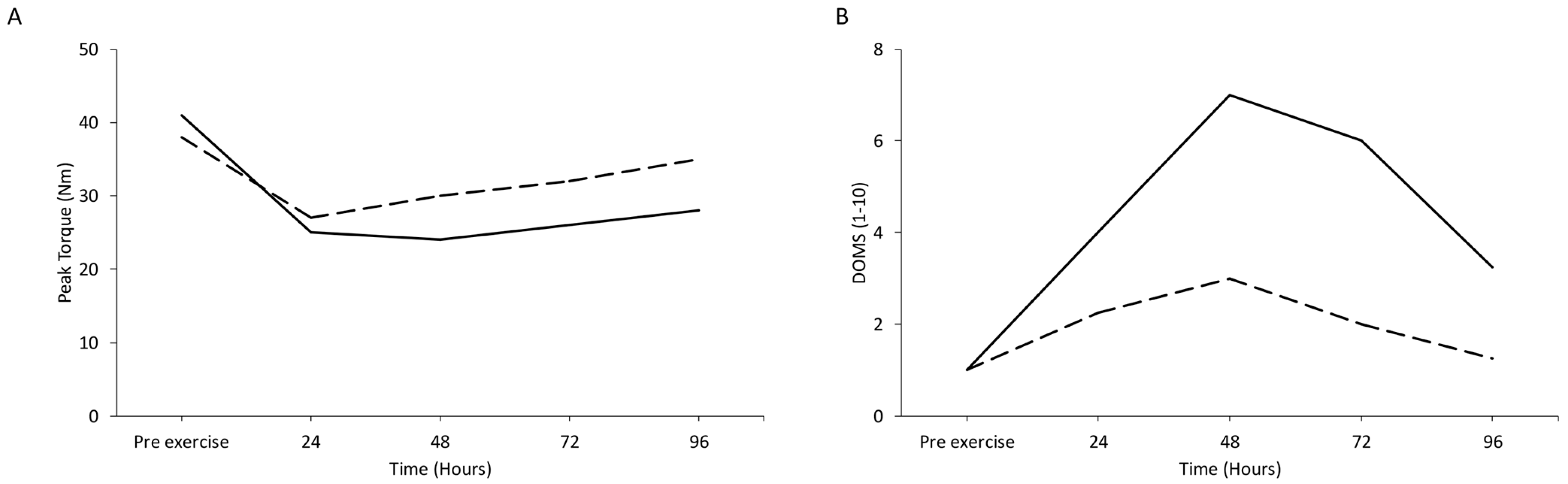
5.1. The Repeated Bout Effect Phenomenon
5.2. Chronic Eccentric Exercise: Eccentric Exercise per se Does Not Induce Muscle Damage
5.3. Take Home Message
6. Architectural and Morphological Characteristics of Adaptation to Eccentric Training
6.1. Challenges and Insights from Animal Models
6.2. Long-Term Muscle Adaptations
6.3. Take Home Message
7. Neural Responses and Adaptations to Eccentric Exercise
7.1. The Effect of Acute Eccentric Exercise on Neural Control
7.2. Transcranial Magnetic Stimulation (TMS)
7.3. Neural Adaptations to Chronic Eccentric Exercise
7.4. Cross-Educational Effect
7.5. Eccentric Exercise and Proprioception
7.6. Take Home Message
8. Exercise-Induced Muscle Damage, Mitochondria and Microstructural Adaptations
8.1. Acute Effects of Eccentric Exercise on Mitochondrial Function
8.2. Oxidative Stress and Mitochondrial Function
8.3. Chronic Effects of Eccentric Training on Mitochondrial Function
8.4. Microstructural and Sarcoplasmic Reticulum Adaptations in Response to Eccentric Exercise
8.5. Take Home Message
9. The Role of Connective Tissue in Exercise-Induced Muscle Damage
Take Home Message
10. Nutritional Manipulation of Exercise-Induced Muscle Damage
Take Home Message
11. Future Research and Advanced Methods in Eccentric Exercise
12. Conclusions
- Eccentric exercise-induced muscle damage is mainly the result of unaccustomed high mechanical tension during lengthening contractions, which is further influenced by long muscle length and high contraction velocity. Importantly, the extent of muscle damage also depends on the eccentric methodology employed, as different exercise models impose different mechanical and physiological demands. Indirect biomarkers of exercise induced muscle damage do not describe the true extent of structural myofiber damage but instead capture changes in distinct physiological processes.
- Following eccentric exercise, skeletal muscle repair is achieved through a coordinated response in which satellite cells are key, but not always indispensable, players, working alongside inflammatory, and intrinsic membrane–repair mechanisms. The severity of exercise induced muscle damage and the nature of the exercise performed influence the extent to which regeneration depends on satellite cells. However, the role of satellite cells in long-term adaptations and in responses to non-damaging exercise remains unresolved.
- Eccentric exercise uniquely combines high force output performed with low energy demands. At the same time, resting energy expenditure remains elevated for several days after eccentric exercise due to muscle repair processes, protein synthesis and increased fat oxidation. These characteristics make eccentric exercise a potent tool for both performance enhancement and metabolic conditioning.
- After an initial bout of damaging eccentric exercise session, subsequent sessions of the same nature progressively cause less muscle damage, a phenomenon known as the repeated bout effect. This adaptive “memory” of skeletal muscle involves neural, architectural, connective tissue and molecular adaptations. Over time, eccentric training becomes no more injurious than other modes of resistance training and, when strategically alternated and combined with concentric work, may optimally enhance muscle function, resilience and health, particularly in populations such as older adults.
- Eccentric training shares distinct, often region-specific architectural adaptations, most notably increased fascicle length and altered morphology achieved via a mix of sarcomere- and tendon-level changes. Interestingly, eccentric training shares similar hypertrophic signaling with concentric training, so its efficient and high-force benefits should be leveraged within carefully designed programs.
- Eccentric exercise is controlled by the nervous system in a fundamentally different way than concentric exercise characterized by lower neural activation for the same or greater force, enhanced cortical drive with concurrent spinal inhibition, and a planned down-weighting of spindle input. Acute eccentric exercise may cause impairments in neuromuscular performance and proprioception, yet chronic eccentric exercise induces neural adaptations, including altered motor unit recruitment, improved proprioception and cross-education effects, that in turn enhance control, protection, and strength over time.
- Acute eccentric exercise disrupts mitochondrial and sarcoplasmic reticulum structure, by elevating calcium and reactive oxygen species levels, thereby triggering mitophagy. However, during repeated bouts of eccentric exercise, these same stress signals drive targeted remodeling of mitochondria, the sarcoplasmic reticulum and the cytoskeleton, restoring function, enhancing excitation–contraction coupling, and improving the muscle’s resilience to future mechanical loads.
- Acute eccentric exercise causes perturbations in connective tissue, namely fascia, tendon and extracellular matrix, which likely contribute to the development of DOMS. With chronic eccentric exercise, connective tissue adapts via collagen turnover and structural remodeling, enhancing stiffness, force transmission and joint stability. However, the precise role of these adaptations in exercise-induced muscle damage and recovery remains difficult to define, as they occur slowly and are challenging to capture with available research tools.
- Nutritional strategies to limit exercise-induced muscle damage, especially antioxidant and polyphenol supplementation, have shown inconsistent benefits and may even blunt training adaptations. These supplements should therefore be used sparingly, targeted to true deficiencies or tight competition schedules rather than applied routinely.
Author Contributions
Funding
Data Availability Statement
Conflicts of Interest
References
- Fenn, W.O. The relation between the work performed and the energy liberated in muscular contraction. J. Physiol. 1924, 58, 373–395. [Google Scholar] [CrossRef] [PubMed]
- Fick, A. Neue Beitrage zur Kenntnis von der Warme Entwicklung im Muskel; Verlag von Emil Strauss: Bonn, Germany, 1892; Volume 51, pp. 541–569. [Google Scholar]
- Katz, B. The relation between force and speed in muscular contraction. J. Physiol. 1939, 96, 45–64. [Google Scholar] [CrossRef] [PubMed]
- Hough, T. Ergographic Studies in Muscular Fatigue and Soreness. J. Boston Soc. Med. Sci. 1900, 5, 81–92. [Google Scholar] [PubMed]
- Hough, T. Ergographic studies in muscular soreness. Am. J. Physiol. 1902, 7, 76–92. [Google Scholar] [CrossRef]
- Hoppeler, H. Molecular networks in skeletal muscle plasticity. J. Exp. Biol. 2016, 219, 205–213. [Google Scholar] [CrossRef]
- Holloszy, J.O. Biochemical adaptations in muscle. Effects of exercise on mitochondrial oxygen uptake and respiratory enzyme activity in skeletal muscle. J. Biol. Chem. 1967, 242, 2278–2282. [Google Scholar] [CrossRef]
- Jamurtas, A.Z.; Theocharis, V.; Tofas, T.; Tsiokanos, A.; Yfanti, C.; Paschalis, V.; Koutedakis, Y.; Nosaka, K. Comparison between leg and arm eccentric exercises of the same relative intensity on indices of muscle damage. Eur. J. Appl. Physiol. 2005, 95, 179–185. [Google Scholar] [CrossRef]
- Gasser, H.S.; Hill, A.V. The dynamics of muscular contraction. Proc. R. Soc. London B Biol. Sci. 1924, 96, 368–437. [Google Scholar] [CrossRef]
- Helweg, J. Om Funktionsmyopathien Som Srnertearsag; Gyldendal: Copenhagen, Denmark, 1934. [Google Scholar]
- Hartree, W.; Hill, A.V. The anaerobic delayed heat production after a tetanus. Proc. R. Soc. London B Biol. Sci. 1928, 103, 207–217. [Google Scholar] [CrossRef]
- Hill, A.V.; Long, C.N.; Lupton, H. The effect of fatigue on the relation between work and speed, in contraction of human arm muscles. J. Physiol. 1924, 58, 334–337. [Google Scholar] [CrossRef]
- Hill, A.V.; Long, C.N.; Lupton, H. Muscular exercise, lactic acid, and the supply and utilisation of oxygen. Parts I–III. Proc. R. Soc. London B Biol. Sci. 1924, 96, 438–475. [Google Scholar] [CrossRef]
- Abbott, B.C.; Aubert, X.M.; Hill, A.V. The absorption of work by a muscle stretched during a single twitch or a short tetanus. Proc. R. Soc. London B Biol. Sci. 1951, 139, 86–104. [Google Scholar] [CrossRef] [PubMed]
- Hill, A.V. The heat of activation and the heat of shortening in a muscle twitch. Proc. R. Soc. London B Biol. Sci. 1949, 136, 195–211. [Google Scholar] [CrossRef]
- Hill, A.V. The mechanics of voluntary muscle. Lancet 1951, 2, 947–951. [Google Scholar] [CrossRef]
- Asmussen, E. Observations on experimental muscular soreness. Acta Rheumatol. Scand. 1956, 2, 109–116. [Google Scholar] [CrossRef] [PubMed]
- Komi, P.V.; Buskirk, E.R. Effect of eccentric and concentric muscle conditioning on tension and electrical activity of human muscle. Ergonomics 1972, 15, 417–434. [Google Scholar] [CrossRef]
- de Vries, H. Electromyographic observations on the effects of static stretching upon muscular distress. Res. Q. 1961, 32, 468–479. [Google Scholar] [CrossRef]
- Abraham, W.M. Factors in delayed muscle soreness. Med. Sci. Sports 1977, 9, 11–20. [Google Scholar] [CrossRef]
- Friden, J.; Sjostrom, M.; Ekblom, B. A morphological study of delayed muscle soreness. Experientia 1981, 37, 506–507. [Google Scholar] [CrossRef]
- Armstrong, R.B. Mechanisms of exercise-induced delayed onset muscular soreness: A brief review. Med. Sci. Sports Exerc. 1984, 16, 529–538. [Google Scholar] [CrossRef]
- Lieber, R.L.; Friden, J. Selective damage of fast glycolytic muscle fibres with eccentric contraction of the rabbit tibialis anterior. Acta Physiol. Scand. 1988, 133, 587–588. [Google Scholar] [CrossRef]
- Smith, L.L. Acute inflammation: The underlying mechanism in delayed onset muscle soreness? Med. Sci. Sports Exerc. 1991, 23, 542–551. [Google Scholar] [CrossRef]
- Jones, D.A.; Round, J.M.; de Haan, A. Skeletal Muscle from Molecules to Movement; Churchill Livingstone: London, UK, 2004. [Google Scholar]
- Jones, D.A.; Jackson, M.J.; McPhail, G.; Edwards, R.H. Experimental mouse muscle damage: The importance of external calcium. Clin. Sci. 1984, 66, 317–322. [Google Scholar] [CrossRef] [PubMed]
- MacIntosh, B.R.; Gardiner, P.F.; McComas, A.J. Skeletal Muscle: Form and Function; Human Kinetics: Champaign, IL, USA, 2006. [Google Scholar]
- Raastad, T.; Owe, S.G.; Paulsen, G.; Enns, D.; Overgaard, K.; Crameri, R.; Kiil, S.; Belcastro, A.; Bergersen, L.; Hallen, J. Changes in calpain activity, muscle structure, and function after eccentric exercise. Med. Sci. Sports Exerc. 2010, 42, 86–95. [Google Scholar] [CrossRef]
- Friden, J.; Lieber, R.L. Ultrastructural evidence for loss of calcium homeostasis in exercised skeletal muscle. Acta Physiol. Scand. 1996, 158, 381–382. [Google Scholar] [CrossRef] [PubMed]
- Lieber, R.L.; Schmitz, M.C.; Mishra, D.K.; Friden, J. Contractile and cellular remodeling in rabbit skeletal muscle after cyclic eccentric contractions. J. Appl. Physiol. 1994, 77, 1926–1934. [Google Scholar] [CrossRef] [PubMed]
- Yu, J.G.; Malm, C.; Thornell, L.E. Eccentric contractions leading to DOMS do not cause loss of desmin nor fibre necrosis in human muscle. Histochem. Cell Biol. 2002, 118, 29–34. [Google Scholar] [CrossRef]
- Hellsten, Y.; Ahlborg, G.; Jensen-Urstad, M.; Sjodin, B. Indication of in vivo xanthine oxidase activity in human skeletal muscle during exercise. Acta Physiol. Scand. 1988, 134, 159–160. [Google Scholar] [CrossRef]
- Gomez-Cabrera, M.C.; Pallardo, F.V.; Sastre, J.; Vina, J.; Garcia-del-Moral, L. Allopurinol and markers of muscle damage among participants in the Tour de France. JAMA 2003, 289, 2503–2504. [Google Scholar] [CrossRef]
- Vina, J.; Gomez-Cabrera, M.C.; Lloret, A.; Marquez, R.; Minana, J.B.; Pallardo, F.V.; Sastre, J. Free radicals in exhaustive physical exercise: Mechanism of production, and protection by antioxidants. IUBMB Life 2000, 50, 271–277. [Google Scholar] [CrossRef]
- Hansen, J.M.; Zhang, H.; Jones, D.P. Differential oxidation of thioredoxin-1, thioredoxin-2, and glutathione by metal ions. Free Radic. Biol. Med. 2006, 40, 138–145. [Google Scholar] [CrossRef] [PubMed]
- Sanchis-Gomar, F.; Pareja-Galeano, H.; Gomez-Cabrera, M.C.; Candel, J.; Lippi, G.; Salvagno, G.L.; Mann, G.E.; Vina, J. Allopurinol prevents cardiac and skeletal muscle damage in professional soccer players. Scand. J. Med. Sci. Sports 2015, 25, e110–e115. [Google Scholar] [CrossRef] [PubMed]
- Hill, A.V. The mechanics of active muscle. Proc. R. Soc. London B Biol. Sci. 1953, 141, 104–117. [Google Scholar] [CrossRef]
- Morgan, D.L. New insights into the behavior of muscle during active lengthening. Biophys. J. 1990, 57, 209–221. [Google Scholar] [CrossRef]
- Proske, U.; Morgan, D.L. Muscle damage from eccentric exercise: Mechanism, mechanical signs, adaptation and clinical applications. J. Physiol. 2001, 537, 333–345. [Google Scholar] [CrossRef] [PubMed]
- Yeung, E.W.; Balnave, C.D.; Ballard, H.J.; Bourreau, J.P.; Allen, D.G. Development of T-tubular vacuoles in eccentrically damaged mouse muscle fibres. J. Physiol. 2002, 540, 581–592. [Google Scholar] [CrossRef]
- Crameri, R.M.; Aagaard, P.; Qvortrup, K.; Langberg, H.; Olesen, J.; Kjaer, M. Myofibre damage in human skeletal muscle: Effects of electrical stimulation versus voluntary contraction. J. Physiol. 2007, 583, 365–380. [Google Scholar] [CrossRef] [PubMed]
- Paulsen, G.; Crameri, R.; Benestad, H.B.; Fjeld, J.G.; Morkrid, L.; Hallen, J.; Raastad, T. Time course of leukocyte accumulation in human muscle after eccentric exercise. Med. Sci. Sports Exerc. 2010, 42, 75–85. [Google Scholar] [CrossRef]
- Herzog, W.; Leonard, T.R. Force enhancement following stretching of skeletal muscle: A new mechanism. J. Exp. Biol. 2002, 205, 1275–1283. [Google Scholar] [CrossRef]
- Herzog, W. Mechanisms of enhanced force production in lengthening (eccentric) muscle contractions. J. Appl. Physiol. 2014, 116, 1407–1417. [Google Scholar] [CrossRef]
- Joumaa, V.; Herzog, W. Calcium sensitivity of residual force enhancement in rabbit skinned fibers. Am. J. Physiol. Cell Physiol. 2014, 307, C395–C401. [Google Scholar] [CrossRef] [PubMed]
- Joumaa, V.; Rassier, D.E.; Leonard, T.R.; Herzog, W. The origin of passive force enhancement in skeletal muscle. Am. J. Physiol. Cell Physiol. 2008, 294, C74–C78. [Google Scholar] [CrossRef]
- Warren, G.L.; Lowe, D.A.; Armstrong, R.B. Measurement tools used in the study of eccentric contraction-induced injury. Sports Med. 1999, 27, 43–59. [Google Scholar] [CrossRef]
- Prokopiou, N.; Mandalidis, D.; Terzis, G.; Paschalis, V. Eccentric exercise-induced muscle damage is independent of limb dominance in young women. Appl. Sci. 2025, 15, 10466. [Google Scholar] [CrossRef]
- Hyldahl, R.D.; Hubal, M.J. Lengthening our perspective: Morphological, cellular, and molecular responses to eccentric exercise. Muscle Nerve 2014, 49, 155–170. [Google Scholar] [CrossRef] [PubMed]
- Deyhle, M.R.; Tiede, D.; Xin, L.; Hyldahl, R.D.; Hubal, M.J. Common Markers of Muscle Damage Are Associated with Divergent Gene Expression Patterns after Eccentric Contractions. Med. Sci. Sports Exerc. 2024, 56, 1108–1117. [Google Scholar] [CrossRef]
- Lyu, X.; Gao, Y.; Liu, Q.; Zhao, H.; Zhou, H.; Pan, S. Exercise-induced muscle damage: Multi-parametric MRI quantitative assessment. BMC Musculoskelet. Disord. 2021, 22, 239. [Google Scholar] [CrossRef]
- Lavender, A.P.; Nosaka, K. Changes in markers of muscle damage of middle-aged and young men following eccentric exercise of the elbow flexors. J. Sci. Med. Sport 2008, 11, 124–131. [Google Scholar] [CrossRef]
- Margaritelis, N.V.; Theodorou, A.A.; Chatzinikolaou, P.N.; Kyparos, A.; Nikolaidis, M.G.; Paschalis, V. Eccentric exercise per se does not affect muscle damage biomarkers: Early and late phase adaptations. Eur. J. Appl. Physiol. 2021, 121, 549–559. [Google Scholar] [CrossRef]
- Theodorou, A.A.; Nikolaidis, M.G.; Paschalis, V.; Sakellariou, G.K.; Fatouros, I.G.; Koutedakis, Y.; Jamurtas, A.Z. Comparison between glucose-6-phosphate dehydrogenase-deficient and normal individuals after eccentric exercise. Med. Sci. Sports Exerc. 2010, 42, 1113–1121. [Google Scholar] [CrossRef] [PubMed]
- Clarkson, P.M.; Hubal, M.J. Are women less susceptible to exercise-induced muscle damage? Curr. Opin. Clin. Nutr. Metab. Care 2001, 4, 527–531. [Google Scholar] [CrossRef]
- Byrne, C.; Twist, C.; Eston, R. Neuromuscular function after exercise-induced muscle damage: Theoretical and applied implications. Sports Med. 2004, 34, 49–69. [Google Scholar] [CrossRef]
- Sorichter, S.; Puschendorf, B.; Mair, J. Skeletal muscle injury induced by eccentric muscle action: Muscle proteins as markers of muscle fiber injury. Exerc. Immunol. Rev. 1999, 5, 5–21. [Google Scholar]
- Kuby, S.A.; Noda, L.; Lardy, H.A. Adenosinetriphosphate-creatine transphosphorylase. I. Isolation of the crystalline enzyme from rabbit muscle. J. Biol. Chem. 1954, 209, 191–201. [Google Scholar] [CrossRef] [PubMed]
- Vejjajiva, A.; Teasdale, G.M. Serum Creatine Kinase and Physical Exercise. Br. Med. J. 1965, 1, 1653–1654. [Google Scholar] [CrossRef] [PubMed][Green Version]
- Nosaka, K.; Clarkson, P.M. Variability in serum creatine kinase response after eccentric exercise of the elbow flexors. Int. J. Sports Med. 1996, 17, 120–127. [Google Scholar] [CrossRef]
- Malm, C.; Nyberg, P.; Engstrom, M.; Sjodin, B.; Lenkei, R.; Ekblom, B.; Lundberg, I. Immunological changes in human skeletal muscle and blood after eccentric exercise and multiple biopsies. J. Physiol. 2000, 529, 243–262. [Google Scholar] [CrossRef] [PubMed]
- Vissing, K.; Andersen, J.L.; Schjerling, P. Are exercise-induced genes induced by exercise? FASEB J. 2005, 19, 94–96. [Google Scholar] [CrossRef]
- Allen, G.M.; McKenzie, D.K.; Gandevia, S.C. Twitch interpolation of the elbow flexor muscles at high forces. Muscle Nerve 1998, 21, 318–328. [Google Scholar] [CrossRef]
- Malm, C. Exercise-induced muscle damage and inflammation: Fact or fiction? Acta Physiol. Scand. 2001, 171, 233–239. [Google Scholar] [CrossRef]
- Newham, D.J.; Jones, D.A.; Edwards, R.H. Plasma creatine kinase changes after eccentric and concentric contractions. Muscle Nerve 1986, 9, 59–63. [Google Scholar] [CrossRef]
- Newham, D.J.; Jones, D.A.; Ghosh, G.; Aurora, P. Muscle fatigue and pain after eccentric contractions at long and short length. Clin. Sci. 1988, 74, 553–557. [Google Scholar] [CrossRef]
- Friden, J.; Lieber, R.L. Serum creatine kinase level is a poor predictor of muscle function after injury. Scand. J. Med. Sci. Sports 2001, 11, 126–127. [Google Scholar] [CrossRef] [PubMed]
- Mizumura, K.; Taguchi, T. Neurochemical mechanism of muscular pain: Insight from the study on delayed onset muscle soreness. J. Physiol. Sci. 2024, 74, 4. [Google Scholar] [CrossRef]
- Woolf, C.J.; Ma, Q.P.; Allchorne, A.; Poole, S. Peripheral cell types contributing to the hyperalgesic action of nerve growth factor in inflammation. J. Neurosci. 1996, 16, 2716–2723. [Google Scholar] [CrossRef]
- Andersen, H.; Arendt-Nielsen, L.; Svensson, P.; Danneskiold-Samsoe, B.; Graven-Nielsen, T. Spatial and temporal aspects of muscle hyperalgesia induced by nerve growth factor in humans. Exp. Brain Res. 2008, 191, 371–382. [Google Scholar] [CrossRef]
- Pezet, S.; McMahon, S.B. Neurotrophins: Mediators and modulators of pain. Annu. Rev. Neurosci. 2006, 29, 507–538. [Google Scholar] [CrossRef]
- Paschalis, V.; Koutedakis, Y.; Baltzopoulos, V.; Mougios, V.; Jamurtas, A.Z.; Giakas, G. Short vs. long length of rectus femoris during eccentric exercise in relation to muscle damage in healthy males. Clin. Biomech. 2005, 20, 617–622. [Google Scholar] [CrossRef] [PubMed][Green Version]
- Karyofyllidou, A.V.; Terzis, G.; Mandalidis, D.; Margaritelis, N.V.; Paschalis, V. The effects of concentric and eccentric exercise performed at long muscle length on muscle damage. J. Sports Sci. 2025, 43, 3160–3168. [Google Scholar] [CrossRef] [PubMed]
- Chapman, D.; Newton, M.; Sacco, P.; Nosaka, K. Greater muscle damage induced by fast versus slow velocity eccentric exercise. Int. J. Sports Med. 2006, 27, 591–598. [Google Scholar] [CrossRef]
- Chapman, D.W.; Newton, M.; McGuigan, M.; Nosaka, K. Effect of lengthening contraction velocity on muscle damage of the elbow flexors. Med. Sci. Sports Exerc. 2008, 40, 926–933. [Google Scholar] [CrossRef] [PubMed]
- Ueda, H.; Tsuchiya, Y.; Ochi, E. Fast-Velocity Eccentric Cycling Exercise Causes Greater Muscle Damage Than Slow Eccentric Cycling. Front. Physiol. 2020, 11, 596640. [Google Scholar] [CrossRef]
- Handford, M.J.; Bright, T.E.; Mundy, P.; Lake, J.; Theis, N.; Suchomel, T.J.; Hughes, J.D. The Current Knowledge and Future Direction into a New Era of Eccentric Training, A Delphi Study. Int. J. Strength Cond. 2025, 5, 1–19. [Google Scholar] [CrossRef]
- Armstrong, R.B.; Ogilvie, R.W.; Schwane, J.A. Eccentric exercise-induced injury to rat skeletal muscle. J. Appl. Physiol. Respir. Environ. Exerc. Physiol. 1983, 54, 80–93. [Google Scholar] [CrossRef] [PubMed]
- Yin, H.; Price, F.; Rudnicki, M.A. Satellite cells and the muscle stem cell niche. Physiol. Rev. 2013, 93, 23–67. [Google Scholar] [CrossRef] [PubMed]
- Mauro, A. Satellite cell of skeletal muscle fibers. J. Biophys. Biochem. Cytol. 1961, 9, 493–495. [Google Scholar] [CrossRef]
- Carlson, B.M.; Faulkner, J.A. The regeneration of skeletal muscle fibers following injury: A review. Med. Sci. Sports Exerc. 1983, 15, 187–198. [Google Scholar] [CrossRef]
- Laguens, R. Satellite Cells of Skeletal Muscle Fibers in Human Progressive Muscular Dystrophy. Virchows Arch. Pathol. Anat. Physiol. Klin. Med. 1963, 336, 564–569. [Google Scholar] [CrossRef]
- Reger, J.F.; Craig, A.S. Studies on the fine structure of muscle fibers and associated satellite cells in hypertrophic human deltoid muscle. Anat. Rec. 1968, 162, 483–500. [Google Scholar] [CrossRef]
- Gros, J.; Manceau, M.; Thome, V.; Marcelle, C. A common somitic origin for embryonic muscle progenitors and satellite cells. Nature 2005, 435, 954–958. [Google Scholar] [CrossRef]
- Tzu, J.; Marinkovich, M.P. Bridging structure with function: Structural, regulatory, and developmental role of laminins. Int. J. Biochem. Cell Biol. 2008, 40, 199–214. [Google Scholar] [CrossRef] [PubMed]
- Mackey, A.L.; Kjaer, M. Connective tissue regeneration in skeletal muscle after eccentric contraction-induced injury. J. Appl. Physiol. 2017, 122, 533–540. [Google Scholar] [CrossRef]
- Kuang, S.; Gillespie, M.A.; Rudnicki, M.A. Niche regulation of muscle satellite cell self-renewal and differentiation. Cell Stem Cell 2008, 2, 22–31. [Google Scholar] [CrossRef]
- Vracko, R. Basal lamina scaffold-anatomy and significance for maintenance of orderly tissue structure. Am. J. Pathol. 1974, 77, 314–346. [Google Scholar]
- Fry, C.S.; Lee, J.D.; Jackson, J.R.; Kirby, T.J.; Stasko, S.A.; Liu, H.; Dupont-Versteegden, E.E.; McCarthy, J.J.; Peterson, C.A. Regulation of the muscle fiber microenvironment by activated satellite cells during hypertrophy. FASEB J. 2014, 28, 1654–1665. [Google Scholar] [CrossRef]
- Seale, P.; Sabourin, L.A.; Girgis-Gabardo, A.; Mansouri, A.; Gruss, P.; Rudnicki, M.A. Pax7 is required for the specification of myogenic satellite cells. Cell 2000, 102, 777–786. [Google Scholar] [CrossRef]
- Allbrook, D. Skeletal muscle regeneration. Muscle Nerve 1981, 4, 234–245. [Google Scholar] [CrossRef]
- Ishikawa, H. Electron microscopic observations of satellite cells with special reference to the development of mammalian skeletal muscles. Z. Anat. Entwicklungsgesch. 1966, 125, 43–63. [Google Scholar] [CrossRef] [PubMed]
- Kelly, A.M. Satellite cells and myofiber growth in the rat soleus and extensor digitorum longus muscles. Dev. Biol. 1978, 65, 1–10. [Google Scholar] [CrossRef] [PubMed]
- Hanzlikova, V.; Mackova, E.V.; Hnik, P. Satellite cells of the rat soleus muscle in the process of compensatory hypertrophy combined with denervation. Cell Tissue Res. 1975, 160, 411–421. [Google Scholar] [CrossRef]
- Darr, K.C.; Schultz, E. Exercise-induced satellite cell activation in growing and mature skeletal muscle. J. Appl. Physiol. 1987, 63, 1816–1821. [Google Scholar] [CrossRef]
- O’Reilly, C.; McKay, B.; Phillips, S.; Tarnopolsky, M.; Parise, G. Hepatocyte growth factor (HGF) and the satellite cell response following muscle lengthening contractions in humans. Muscle Nerve 2008, 38, 1434–1442. [Google Scholar] [CrossRef]
- Fernando, C.A.; Pangan, A.M.; Cornelison, D.; Segal, S.S. Recovery of blood flow regulation in microvascular resistance networks during regeneration of mouse gluteus maximus muscle. J. Physiol. 2019, 597, 1401–1417. [Google Scholar] [CrossRef] [PubMed]
- Jacobsen, N.L.; Norton, C.E.; Shaw, R.L.; Cornelison, D.D.W.; Segal, S.S. Myofibre injury induces capillary disruption and regeneration of disorganized microvascular networks. J. Physiol. 2022, 600, 41–60. [Google Scholar] [CrossRef] [PubMed]
- Nederveen, J.P.; Joanisse, S.; Snijders, T.; Thomas, A.C.Q.; Kumbhare, D.; Parise, G. The influence of capillarization on satellite cell pool expansion and activation following exercise-induced muscle damage in healthy young men. J. Physiol. 2018, 596, 1063–1078. [Google Scholar] [CrossRef]
- Brown, A.; Thomas, A.C.Q.; Hatt, A.A.; McGlory, C.; Phillips, S.M.; Kumbhare, D.; Parise, G.; Joanisse, S. Aerobic conditioning alters the satellite cell and ribosome response to acute eccentric contractions in young men and women. Am. J. Physiol. Cell Physiol. 2022, 323, C1577–C1585. [Google Scholar] [CrossRef]
- Mendias, C.L.; Tatsumi, R.; Allen, R.E. Role of cyclooxygenase-1 and -2 in satellite cell proliferation, differentiation, and fusion. Muscle Nerve 2004, 30, 497–500. [Google Scholar] [CrossRef]
- Hawke, T.J.; Garry, D.J. Myogenic satellite cells: Physiology to molecular biology. J. Appl. Physiol. 2001, 91, 534–551. [Google Scholar] [CrossRef] [PubMed]
- Young, L.V.; Wakelin, G.; Cameron, A.W.R.; Springer, S.A.; Ross, J.P.; Wolters, G.; Murphy, J.P.; Arsenault, M.G.; Ng, S.; Collao, N.; et al. Muscle injury induces a transient senescence-like state that is required for myofiber growth during muscle regeneration. FASEB J. 2022, 36, e22587. [Google Scholar] [CrossRef]
- Behmoaras, J.; Gil, J. Similarities and interplay between senescent cells and macrophages. J. Cell Biol. 2021, 220, e202010162. [Google Scholar] [CrossRef]
- Finaud, J.; Lac, G.; Filaire, E. Oxidative stress: Relationship with exercise and training. Sports Med. 2006, 36, 327–358. [Google Scholar] [CrossRef]
- Esposito, K.; Nappo, F.; Marfella, R.; Giugliano, G.; Giugliano, F.; Ciotola, M.; Quagliaro, L.; Ceriello, A.; Giugliano, D. Inflammatory cytokine concentrations are acutely increased by hyperglycemia in humans: Role of oxidative stress. Circulation 2002, 106, 2067–2072. [Google Scholar] [CrossRef]
- Nikolaidis, M.G.; Paschalis, V.; Giakas, G.; Fatouros, I.G.; Koutedakis, Y.; Kouretas, D.; Jamurtas, A.Z. Decreased blood oxidative stress after repeated muscle-damaging exercise. Med. Sci. Sports Exerc. 2007, 39, 1080–1089. [Google Scholar] [CrossRef] [PubMed]
- Westerblad, H.; Allen, D.G. Emerging roles of ROS/RNS in muscle function and fatigue. Antioxid. Redox Signal. 2011, 15, 2487–2499. [Google Scholar] [CrossRef]
- Alaranta, A.; Alaranta, H.; Heliovaara, M.; Airaksinen, M.; Helenius, I. Ample use of physician-prescribed medications in Finnish elite athletes. Int. J. Sports Med. 2006, 27, 919–925. [Google Scholar] [CrossRef]
- Vane, J.R.; Botting, R.M. Anti-inflammatory drugs and their mechanism of action. Inflamm. Res. 1998, 47, S78–S87. [Google Scholar] [CrossRef] [PubMed]
- Trappe, T.A.; White, F.; Lambert, C.P.; Cesar, D.; Hellerstein, M.; Evans, W.J. Effect of ibuprofen and acetaminophen on postexercise muscle protein synthesis. Am. J. Physiol. Endocrinol. Metab. 2002, 282, E551–E556. [Google Scholar] [CrossRef] [PubMed]
- Mackey, A.L.; Kjaer, M.; Dandanell, S.; Mikkelsen, K.H.; Holm, L.; Dossing, S.; Kadi, F.; Koskinen, S.O.; Jensen, C.H.; Schroder, H.D.; et al. The influence of anti-inflammatory medication on exercise-induced myogenic precursor cell responses in humans. J. Appl. Physiol. 2007, 103, 425–431. [Google Scholar] [CrossRef]
- Paulsen, G.; Mikkelsen, U.R.; Raastad, T.; Peake, J.M. Leucocytes, cytokines and satellite cells: What role do they play in muscle damage and regeneration following eccentric exercise? Exerc. Immunol. Rev. 2012, 18, 42–97. [Google Scholar] [PubMed]
- Folker, E.S.; Baylies, M.K. Nuclear positioning in muscle development and disease. Front. Physiol. 2013, 4, 363. [Google Scholar] [CrossRef]
- Robertson, T.A.; Papadimitriou, J.M.; Grounds, M.D. Fusion of myogenic cells to the newly sealed region of damaged myofibres in skeletal muscle regeneration. Neuropathol. Appl. Neurobiol. 1993, 19, 350–358. [Google Scholar] [CrossRef]
- Goh, Q.; Millay, D.P. Requirement of myomaker-mediated stem cell fusion for skeletal muscle hypertrophy. eLife 2017, 6, 20007. [Google Scholar] [CrossRef] [PubMed]
- McCarthy, J.J.; Dupont-Versteegden, E.E.; Fry, C.S.; Murach, K.A.; Peterson, C.A. Methodological issues limit interpretation of negative effects of satellite cell depletion on adult muscle hypertrophy. Development 2017, 144, 1363–1365. [Google Scholar] [CrossRef]
- Sambasivan, R.; Yao, R.; Kissenpfennig, A.; Van Wittenberghe, L.; Paldi, A.; Gayraud-Morel, B.; Guenou, H.; Malissen, B.; Tajbakhsh, S.; Galy, A. Pax7-expressing satellite cells are indispensable for adult skeletal muscle regeneration. Development 2011, 138, 3647–3656. [Google Scholar] [CrossRef] [PubMed]
- Kinney, M.C.; Dayanidhi, S.; Dykstra, P.B.; McCarthy, J.J.; Peterson, C.A.; Lieber, R.L. Reduced skeletal muscle satellite cell number alters muscle morphology after chronic stretch but allows limited serial sarcomere addition. Muscle Nerve 2017, 55, 384–392. [Google Scholar] [CrossRef]
- Mikkelsen, U.R.; Langberg, H.; Helmark, I.C.; Skovgaard, D.; Andersen, L.L.; Kjaer, M.; Mackey, A.L. Local NSAID infusion inhibits satellite cell proliferation in human skeletal muscle after eccentric exercise. J. Appl. Physiol. 2009, 107, 1600–1611. [Google Scholar] [CrossRef]
- Carlson, B. (Ed.) Muscle injury and regeneration. In Muscle Biology, the Life History of a Muscle; Academic Press: Cambridge, MA, USA, 2022; pp. 117–141. [Google Scholar]
- Bansal, D.; Miyake, K.; Vogel, S.S.; Groh, S.; Chen, C.C.; Williamson, R.; McNeil, P.L.; Campbell, K.P. Defective membrane repair in dysferlin-deficient muscular dystrophy. Nature 2003, 423, 168–172. [Google Scholar] [CrossRef]
- Barthelemy, F.; Defour, A.; Levy, N.; Krahn, M.; Bartoli, M. Muscle Cells Fix Breaches by Orchestrating a Membrane Repair Ballet. J. Neuromuscul. Dis. 2018, 5, 21–28. [Google Scholar] [CrossRef]
- Demonbreun, A.R.; Quattrocelli, M.; Barefield, D.Y.; Allen, M.V.; Swanson, K.E.; McNally, E.M. An actin-dependent annexin complex mediates plasma membrane repair in muscle. J. Cell Biol. 2016, 213, 705–718. [Google Scholar] [CrossRef] [PubMed]
- Wehrstein, M.; Schoffel, A.; Weiberg, N.; Gwechenberger, T.; Betz, T.; Rittweg, M.; Parstorfer, M.; Pilz, M.; Friedmann-Bette, B. Eccentric Overload during Resistance Exercise: A Stimulus for Enhanced Satellite Cell Activation. Med. Sci. Sports Exerc. 2022, 54, 388–398. [Google Scholar] [CrossRef]
- Roman, W.; Pinheiro, H.; Pimentel, M.R.; Segales, J.; Oliveira, L.M.; Garcia-Dominguez, E.; Gomez-Cabrera, M.C.; Serrano, A.L.; Gomes, E.R.; Munoz-Canoves, P. Muscle repair after physiological damage relies on nuclear migration for cellular reconstruction. Science 2021, 374, 355–359. [Google Scholar] [CrossRef]
- Debattisti, V.; Horn, A.; Singh, R.; Seifert, E.L.; Hogarth, M.W.; Mazala, D.A.; Huang, K.T.; Horvath, R.; Jaiswal, J.K.; Hajnoczky, G. Dysregulation of Mitochondrial Ca2+ Uptake and Sarcolemma Repair Underlie Muscle Weakness and Wasting in Patients and Mice Lacking MICU1. Cell Rep. 2019, 29, 1274–1286.e6. [Google Scholar] [CrossRef] [PubMed]
- Murach, K.A.; Fry, C.S.; Dupont-Versteegden, E.E.; McCarthy, J.J.; Peterson, C.A. Fusion and beyond: Satellite cell contributions to loading-induced skeletal muscle adaptation. FASEB J. 2021, 35, e21893. [Google Scholar] [CrossRef] [PubMed]
- Chauveau, A. La depence energetique qu’entrainent respectivement le travail moteur et le travail resistant de l’homme qui s’eleve ou dscend sur la roue de Hirm. Evaluation d’apres l’oxygene absorbe dans les echanges respiratoires. CR Acad. Sci. 1901, 132, 194–201. [Google Scholar]
- Abbott, B.C.; Bigland, B.; Ritchie, J.M. The physiological cost of negative work. J. Physiol. 1952, 117, 380–390. [Google Scholar] [CrossRef]
- Abbott, B.C.; Aubert, X.M.; Hill, A.V. The absorption of work when a muscle is stretched. J. Physiol. 1950, 111, 41p–42p. [Google Scholar]
- Hill, A.V. Production and absorption of work by muscle. Science 1960, 131, 897–903. [Google Scholar] [CrossRef]
- Hill, A.V. Mechanics of the contractile element of muscle. Nature 1950, 166, 415–419. [Google Scholar] [CrossRef] [PubMed]
- Gordon, A.M.; Huxley, A.F.; Julian, F.J. The variation in isometric tension with sarcomere length in vertebrate muscle fibres. J. Physiol. 1966, 184, 170–192. [Google Scholar] [CrossRef]
- Huxley, A.F.; Niedergerke, R. Structural changes in muscle during contraction; interference microscopy of living muscle fibres. Nature 1954, 173, 971–973. [Google Scholar] [CrossRef]
- Huxley, A.F.; Simmons, R.M. Proposed mechanism of force generation in striated muscle. Nature 1971, 233, 533–538. [Google Scholar] [CrossRef]
- Huxley, H.; Hanson, J. Changes in the cross-striations of muscle during contraction and stretch and their structural interpretation. Nature 1954, 173, 973–976. [Google Scholar] [CrossRef]
- Kilgas, M.A.; Elmer, S.J. Back to the future! Revisiting the physiological cost of negative work as a team-based activity for exercise physiology students. Adv. Physiol. Educ. 2017, 41, 120–129. [Google Scholar] [CrossRef] [PubMed]
- Nishikawa, K.C.; Monroy, J.A.; Uyeno, T.E.; Yeo, S.H.; Pai, D.K.; Lindstedt, S.L. Is titin a ‘winding filament’? A new twist on muscle contraction. Proc. Biol. Sci. 2012, 279, 981–990. [Google Scholar] [CrossRef]
- Flitney, F.W.; Hirst, D.G. Cross-bridge detachment and sarcomere ‘give’ during stretch of active frog’s muscle. J. Physiol. 1978, 276, 449–465. [Google Scholar] [CrossRef]
- Menard, M.R.; Penn, A.M.; Lee, J.W.; Dusik, L.A.; Hall, L.D. Relative metabolic efficiency of concentric and eccentric exercise determined by 31P magnetic resonance spectroscopy. Arch. Phys. Med. Rehabil. 1991, 72, 976–983. [Google Scholar] [PubMed]
- Hill, A.V. The heat of shortening and the dynamic constants of muscle. Proc. R. Soc. London B Biol. Sci. 1938, 126, 136–195. [Google Scholar] [CrossRef]
- Huxley, A.F. Muscle structure and theories of contraction. Prog. Biophys. Biophys. Chem. 1957, 7, 255–318. [Google Scholar] [CrossRef]
- Rome, L.C.; Cook, C.; Syme, D.A.; Connaughton, M.A.; Ashley-Ross, M.; Klimov, A.; Tikunov, B.; Goldman, Y.E. Trading force for speed: Why superfast crossbridge kinetics leads to superlow forces. Proc. Natl. Acad. Sci. USA 1999, 96, 5826–5831. [Google Scholar] [CrossRef] [PubMed]
- Amiridis, I.G.; Martin, A.; Morlon, B.; Martin, L.; Cometti, G.; Pousson, M.; van Hoecke, J. Co-activation and tension-regulating phenomena during isokinetic knee extension in sedentary and highly skilled humans. Eur. J. Appl. Physiol. Occup. Physiol. 1996, 73, 149–156. [Google Scholar] [CrossRef]
- Lombardi, V.; Piazzesi, G. The contractile response during steady lengthening of stimulated frog muscle fibres. J. Physiol. 1990, 431, 141–171. [Google Scholar] [CrossRef] [PubMed]
- Cherouveim, E.D.; Margaritelis, N.V.; Koulouvaris, P.; Tsolakis, C.; Malliou, V.J.; Chatzinikolaou, P.N.; Franchi, M.V.; Porcelli, S.; Kyparos, A.; Vrabas, I.S.; et al. Skeletal muscle and cerebral oxygenation levels during and after submaximal concentric and eccentric isokinetic exercise. J. Sports Sci. 2021, 40, 195–202. [Google Scholar] [CrossRef]
- Paschalis, V.; Nikolaidis, M.G.; Giakas, G.; Theodorou, A.A.; Sakellariou, G.K.; Fatouros, I.G.; Koutedakis, Y.; Jamurtas, A.Z. Beneficial changes in energy expenditure and lipid profile after eccentric exercise in overweight and lean women. Scand. J. Med. Sci. Sports 2010, 20, e103–e111. [Google Scholar] [CrossRef] [PubMed]
- Herzog, W.; Leonard, T.R.; Joumaa, V.; Mehta, A. Mysteries of muscle contraction. J. Appl. Biomech. 2008, 24, 1–13. [Google Scholar] [CrossRef] [PubMed]
- Linke, W.A.; Ivemeyer, M.; Mundel, P.; Stockmeier, M.R.; Kolmerer, B. Nature of PEVK-titin elasticity in skeletal muscle. Proc. Natl. Acad. Sci. USA 1998, 95, 8052–8057. [Google Scholar] [CrossRef]
- Maruyama, K. Connectin, an elastic protein from myofibrils. J. Biochem. 1976, 80, 405–407. [Google Scholar] [CrossRef]
- Monroy, J.A.; Powers, K.L.; Gilmore, L.A.; Uyeno, T.A.; Lindstedt, S.L.; Nishikawa, K.C. What is the role of titin in active muscle? Exerc. Sport Sci. Rev. 2012, 40, 73–78. [Google Scholar] [CrossRef]
- Joumaa, V.; Herzog, W. Energy cost of force production is reduced after active stretch in skinned muscle fibres. J. Biomech. 2013, 46, 1135–1139. [Google Scholar] [CrossRef]
- Nishikawa, K.C.; Monroy, J.A.; Powers, K.L.; Gilmore, L.A.; Uyeno, T.A.; Lindstedt, S.L. A molecular basis for intrinsic muscle properties: Implications for motor control. In Progress in Motor Control; Advances in Experimental Medicine and Biology; Springer: Berlin/Heidelberg, Germany, 2013; Volume 782, pp. 111–125. [Google Scholar]
- Monte, A.; Tecchio, P.; Nardello, F.; Bachero-Mena, B.; Ardigo, L.P.; Zamparo, P. Influence of muscle-belly and tendon gearing on the energy cost of human walking. Scand. J. Med. Sci. Sports 2022, 32, 844–855. [Google Scholar] [CrossRef]
- Azizi, E.; Roberts, T.J. Geared up to stretch: Pennate muscle behavior during active lengthening. J. Exp. Biol. 2014, 217, 376–381. [Google Scholar] [CrossRef]
- Shortreed, K.; Johnston, A.; Hawke, T.J. Satellite cells and muscle repair. In Skeletal Muscle Damage and Repair; Tiidus, P.M., Ed.; Human Kinetics: Champaign, IL, USA, 2008. [Google Scholar]
- Dolezal, B.A.; Potteiger, J.A.; Jacobsen, D.J.; Benedict, S.H. Muscle damage and resting metabolic rate after acute resistance exercise with an eccentric overload. Med. Sci. Sports Exerc. 2000, 32, 1202–1207. [Google Scholar] [CrossRef] [PubMed]
- Welle, S.; Nair, K.S. Relationship of resting metabolic rate to body composition and protein turnover. Am. J. Physiol. 1990, 258, E990–E998. [Google Scholar] [CrossRef]
- Fielding, R.A.; Meredith, C.N.; O’Reilly, K.P.; Frontera, W.R.; Cannon, J.G.; Evans, W.J. Enhanced protein breakdown after eccentric exercise in young and older men. J. Appl. Physiol. 1991, 71, 674–679. [Google Scholar] [CrossRef]
- Burleson, M.A.; O’Bryant, H.S., Jr.; Stone, M.H.; Collins, M.A.; Triplett-McBride, T. Effect of weight training exercise and treadmill exercise on post-exercise oxygen consumption. Med. Sci. Sports Exerc. 1998, 30, 518–522. [Google Scholar] [CrossRef] [PubMed]
- Duncan, C.J. The role of phospholipase A2 in calcium-induced damage in cardiac and skeletal muscle. Cell Tissue Res. 1988, 253, 457–462. [Google Scholar] [CrossRef]
- Asp, S.; Daugaard, J.R.; Richter, E.A. Eccentric exercise decreases glucose transporter GLUT4 protein in human skeletal muscle. J. Physiol. 1995, 482, 705–712. [Google Scholar] [CrossRef]
- Kirwan, J.P.; Hickner, R.C.; Yarasheski, K.E.; Kohrt, W.M.; Wiethop, B.V.; Holloszy, J.O. Eccentric exercise induces transient insulin resistance in healthy individuals. J. Appl. Physiol. 1992, 72, 2197–2202. [Google Scholar] [CrossRef]
- Egner, I.M.; Bruusgaard, J.C.; Eftestol, E.; Gundersen, K. A cellular memory mechanism aids overload hypertrophy in muscle long after an episodic exposure to anabolic steroids. J. Physiol. 2013, 591, 6221–6230. [Google Scholar] [CrossRef]
- Clarkson, P.M.; Litchfield, P.; Graves, J.; Kirwan, J.; Byrnes, W.C. Serum creatine kinase activity following forearm flexion isometric exercise. Eur. J. Appl. Physiol. Occup. Physiol. 1985, 53, 368–371. [Google Scholar] [CrossRef]
- Byrnes, W.C.; Clarkson, P.M.; White, J.S.; Hsieh, S.S.; Frykman, P.N.; Maughan, R.J. Delayed onset muscle soreness following repeated bouts of downhill running. J. Appl. Physiol. 1985, 59, 710–715. [Google Scholar] [CrossRef] [PubMed]
- Clarkson, P.M.; Tremblay, I. Exercise-induced muscle damage, repair, and adaptation in humans. J. Appl. Physiol. 1988, 65, 1–6. [Google Scholar] [CrossRef]
- Ebbeling, C.B.; Clarkson, P.M. Muscle adaptation prior to recovery following eccentric exercise. Eur. J. Appl. Physiol. Occup. Physiol. 1990, 60, 26–31. [Google Scholar] [CrossRef]
- Newham, D.J.; Jones, D.A.; Clarkson, P.M. Repeated high-force eccentric exercise: Effects on muscle pain and damage. J. Appl. Physiol. 1987, 63, 1381–1386. [Google Scholar] [CrossRef]
- Margaritelis, N.V.; Theodorou, A.A.; Baltzopoulos, V.; Maganaris, C.N.; Paschalis, V.; Kyparos, A.; Nikolaidis, M.G. Muscle damage and inflammation after eccentric exercise: Can the repeated bout effect be removed? Physiol. Rep. 2015, 3, e12648. [Google Scholar] [CrossRef] [PubMed]
- Mair, J.; Mayr, M.; Muller, E.; Koller, A.; Haid, C.; Artner-Dworzak, E.; Calzolari, C.; Larue, C.; Puschendorf, B. Rapid adaptation to eccentric exercise-induced muscle damage. Int. J. Sports Med. 1995, 16, 352–356. [Google Scholar] [CrossRef] [PubMed]
- McHugh, M.P.; Connolly, D.A.; Eston, R.G.; Gleim, G.W. Exercise-induced muscle damage and potential mechanisms for the repeated bout effect. Sports Med. 1999, 27, 157–170. [Google Scholar] [CrossRef]
- Nikolaidis, M.G.; Jamurtas, A.Z.; Paschalis, V.; Fatouros, I.G.; Koutedakis, Y.; Kouretas, D. The effect of muscle-damaging exercise on blood and skeletal muscle oxidative stress: Magnitude and time-course considerations. Sports Med. 2008, 38, 579–606. [Google Scholar] [CrossRef]
- Nagahisa, H.; Ikezaki, K.; Yamada, R.; Yamada, T.; Miyata, H. Preconditioning Contractions Suppress Muscle Pain Markers after Damaging Eccentric Contractions. Pain Res. Manag. 2018, 2018, 3080715. [Google Scholar] [CrossRef] [PubMed]
- Hyldahl, R.D.; Nelson, B.; Xin, L.; Welling, T.; Groscost, L.; Hubal, M.J.; Chipkin, S.; Clarkson, P.M.; Parcell, A.C. Extracellular matrix remodeling and its contribution to protective adaptation following lengthening contractions in human muscle. FASEB J. 2015, 29, 2894–2904. [Google Scholar] [CrossRef] [PubMed]
- Mackey, A.L.; Brandstetter, S.; Schjerling, P.; Bojsen-Moller, J.; Qvortrup, K.; Pedersen, M.M.; Doessing, S.; Kjaer, M.; Magnusson, S.P.; Langberg, H. Sequenced response of extracellular matrix deadhesion and fibrotic regulators after muscle damage is involved in protection against future injury in human skeletal muscle. FASEB J. 2011, 25, 1943–1959. [Google Scholar] [CrossRef]
- Margaritelis, N.V.; Theodorou, A.A.; Kyparos, A.; Nikolaidis, M.G.; Paschalis, V. Effect of body composition on redox homeostasis at rest and in response to exercise: The case of underfat women. J. Sports Sci. 2019, 37, 1630–1637. [Google Scholar] [CrossRef] [PubMed]
- Saclier, M.; Cuvellier, S.; Magnan, M.; Mounier, R.; Chazaud, B. Monocyte/macrophage interactions with myogenic precursor cells during skeletal muscle regeneration. FEBS J. 2013, 280, 4118–4130. [Google Scholar] [CrossRef]
- Michailidis, Y.; Karagounis, L.G.; Terzis, G.; Jamurtas, A.Z.; Spengos, K.; Tsoukas, D.; Chatzinikolaou, A.; Mandalidis, D.; Stefanetti, R.J.; Papassotiriou, I.; et al. Thiol-based antioxidant supplementation alters human skeletal muscle signaling and attenuates its inflammatory response and recovery after intense eccentric exercise. Am. J. Clin. Nutr. 2013, 98, 233–245. [Google Scholar] [CrossRef]
- Fatouros, I.G.; Jamurtas, A.Z. Insights into the molecular etiology of exercise-induced inflammation: Opportunities for optimizing performance. J. Inflamm. Res. 2016, 9, 175–186. [Google Scholar] [CrossRef]
- Paschalis, V.; Nikolaidis, M.G.; Theodorou, A.A.; Panayiotou, G.; Fatouros, I.G.; Koutedakis, Y.; Jamurtas, A.Z. A weekly bout of eccentric exercise is sufficient to induce health-promoting effects. Med. Sci. Sports Exerc. 2011, 43, 64–73. [Google Scholar] [CrossRef]
- Cevenini, E.; Monti, D.; Franceschi, C. Inflamm-ageing. Curr. Opin. Clin. Nutr. Metab. Care 2013, 16, 14–20. [Google Scholar] [CrossRef]
- Franceschi, C.; Bonafe, M.; Valensin, S.; Olivieri, F.; De Luca, M.; Ottaviani, E.; De Benedictis, G. Inflamm-aging. An evolutionary perspective on immunosenescence. Ann. N.Y. Acad. Sci. 2000, 908, 244–254. [Google Scholar] [CrossRef]
- Salvioli, S.; Monti, D.; Lanzarini, C.; Conte, M.; Pirazzini, C.; Bacalini, M.G.; Garagnani, P.; Giuliani, C.; Fontanesi, E.; Ostan, R.; et al. Immune system, cell senescence, aging and longevity--inflamm-aging reappraised. Curr. Pharm. Des. 2013, 19, 1675–1679. [Google Scholar]
- Franceschi, C.; Capri, M.; Monti, D.; Giunta, S.; Olivieri, F.; Sevini, F.; Panourgia, M.P.; Invidia, L.; Celani, L.; Scurti, M.; et al. Inflammaging and anti-inflammaging: A systemic perspective on aging and longevity emerged from studies in humans. Mech. Ageing Dev. 2007, 128, 92–105. [Google Scholar] [CrossRef] [PubMed]
- Stoboy, H.; Friedebold, G. Changes in muscle function in atrophied muscles due to isometric training. Bull. N.Y. Acad. Med. 1968, 44, 553–559. [Google Scholar] [CrossRef] [PubMed][Green Version]
- Friden, J.; Seger, J.; Sjostrom, M.; Ekblom, B. Adaptive response in human skeletal muscle subjected to prolonged eccentric training. Int. J. Sports Med. 1983, 4, 177–183. [Google Scholar] [CrossRef]
- Cook, C.S.; McDonagh, M.J. Force responses to controlled stretches of electrically stimulated human muscle-tendon complex. Exp. Physiol. 1995, 80, 477–490. [Google Scholar] [CrossRef]
- Westing, S.H.; Seger, J.Y.; Thorstensson, A. Isoacceleration: A new concept of resistive exercise. Med. Sci. Sports Exerc. 1991, 23, 631–635. [Google Scholar] [CrossRef]
- Roig, M.; O’Brien, K.; Kirk, G.; Murray, R.; McKinnon, P.; Shadgan, B.; Reid, W.D. The effects of eccentric versus concentric resistance training on muscle strength and mass in healthy adults: A systematic review with meta-analysis. Br. J. Sports Med. 2009, 43, 556–568. [Google Scholar] [CrossRef]
- Wernbom, M.; Augustsson, J.; Thomee, R. The influence of frequency, intensity, volume and mode of strength training on whole muscle cross-sectional area in humans. Sports Med. 2007, 37, 225–264. [Google Scholar] [CrossRef]
- Franchi, M.V.; Reeves, N.D.; Narici, M.V. Skeletal Muscle Remodeling in Response to Eccentric vs. Concentric Loading: Morphological, Molecular, and Metabolic Adaptations. Front. Physiol. 2017, 8, 447. [Google Scholar] [CrossRef]
- Flann, K.L.; LaStayo, P.C.; McClain, D.A.; Hazel, M.; Lindstedt, S.L. Muscle damage and muscle remodeling: No pain, no gain? J. Exp. Biol. 2011, 214, 674–679. [Google Scholar] [CrossRef] [PubMed]
- Lastayo, P.C.; Reich, T.E.; Urquhart, M.; Hoppeler, H.; Lindstedt, S.L. Chronic eccentric exercise: Improvements in muscle strength can occur with little demand for oxygen. Am. J. Physiol. 1999, 276, R611–R615. [Google Scholar] [CrossRef]
- Nosaka, K.; Newton, M. Concentric or eccentric training effect on eccentric exercise-induced muscle damage. Med. Sci. Sports Exerc. 2002, 34, 63–69. [Google Scholar] [CrossRef] [PubMed]
- Folland, J.P.; Irish, C.S.; Roberts, J.C.; Tarr, J.E.; Jones, D.A. Fatigue is not a necessary stimulus for strength gains during resistance training. Br. J. Sports Med. 2002, 36, 370–373; discussion 374. [Google Scholar] [CrossRef] [PubMed]
- Smith, H.K.; Plyley, M.J.; Rodgers, C.D.; McKee, N.H. Expression of developmental myosin and morphological characteristics in adult rat skeletal muscle following exercise-induced injury. Eur. J. Appl. Physiol. Occup. Physiol. 1999, 80, 84–91. [Google Scholar] [CrossRef]
- LaStayo, P.C.; Ewy, G.A.; Pierotti, D.D.; Johns, R.K.; Lindstedt, S. The positive effects of negative work: Increased muscle strength and decreased fall risk in a frail elderly population. J. Gerontol. A Biol. Sci. Med. Sci. 2003, 58, M419–M424. [Google Scholar] [CrossRef]
- Schoenfeld, B.J.; Ogborn, D.I.; Vigotsky, A.D.; Franchi, M.V.; Krieger, J.W. Hypertrophic Effects of Concentric vs. Eccentric Muscle Actions: A Systematic Review and Meta-analysis. J. Strength Cond. Res. 2017, 31, 2599–2608. [Google Scholar] [CrossRef] [PubMed]
- Hather, B.M.; Tesch, P.A.; Buchanan, P.; Dudley, G.A. Influence of eccentric actions on skeletal muscle adaptations to resistance training. Acta Physiol. Scand. 1991, 143, 177–185. [Google Scholar] [CrossRef]
- Hortobagyi, T.; Dempsey, L.; Fraser, D.; Zheng, D.; Hamilton, G.; Lambert, J.; Dohm, L. Changes in muscle strength, muscle fibre size and myofibrillar gene expression after immobilization and retraining in humans. J. Physiol. 2000, 524, 293–304. [Google Scholar] [CrossRef]
- Vikne, H.; Refsnes, P.E.; Ekmark, M.; Medbo, J.I.; Gundersen, V.; Gundersen, K. Muscular performance after concentric and eccentric exercise in trained men. Med. Sci. Sports Exerc. 2006, 38, 1770–1781. [Google Scholar]
- Theodorou, A.A.; Panayiotou, G.; Paschalis, V.; Nikolaidis, M.G.; Kyparos, A.; Mademli, L.; Grivas, G.V.; Vrabas, I.S. Stair descending exercise increases muscle strength in elderly males with chronic heart failure. BMC Res. Notes 2013, 6, 87. [Google Scholar] [CrossRef]
- Bigland-Ritchie, B.; Woods, J.J. Integrated electromyogram and oxygen uptake during positive and negative work. J. Physiol. 1976, 260, 267–277. [Google Scholar] [CrossRef] [PubMed]
- Barnett, J.G.; Holly, R.G.; Ashmore, C.R. Stretch-induced growth in chicken wing muscles: Biochemical and morphological characterization. Am. J. Physiol. 1980, 239, C39–C46. [Google Scholar] [CrossRef]
- Williams, P.E.; Goldspink, G. Changes in sarcomere length and physiological properties in immobilized muscle. J. Anat. 1978, 127, 459–468. [Google Scholar] [PubMed]
- Tabary, J.C.; Tabary, C.; Tardieu, C.; Tardieu, G.; Goldspink, G. Physiological and structural changes in the cat’s soleus muscle due to immobilization at different lengths by plaster casts. J. Physiol. 1972, 224, 231–244. [Google Scholar] [CrossRef] [PubMed]
- Williams, P.E.; Goldspink, G. Longitudinal growth of striated muscle fibres. J. Cell Sci. 1971, 9, 751–767. [Google Scholar] [CrossRef]
- Bishop, S.P.; Cole, C.R. Ultrastructural changes in the canine myocardium with right ventricular hypertrophy and congestive heart failure. Lab. Investig. 1969, 20, 219–229. [Google Scholar]
- Lieber, R.L.; Friden, J. Functional and clinical significance of skeletal muscle architecture. Muscle Nerve 2000, 23, 1647–1666. [Google Scholar] [CrossRef]
- Blazevich, A.J.; Cannavan, D.; Coleman, D.R.; Horne, S. Influence of concentric and eccentric resistance training on architectural adaptation in human quadriceps muscles. J. Appl. Physiol. 2007, 103, 1565–1575. [Google Scholar] [CrossRef]
- Seynnes, O.R.; de Boer, M.; Narici, M.V. Early skeletal muscle hypertrophy and architectural changes in response to high-intensity resistance training. J. Appl. Physiol. 2007, 102, 368–373. [Google Scholar] [CrossRef]
- Hinks, A.; Jacob, K.; Mashouri, P.; Medak, K.D.; Franchi, M.V.; Wright, D.C.; Brown, S.H.M.; Power, G.A. An increase in serial sarcomere number induced via weighted downhill running improves work loop performance in the rat soleus. bioRxiv 2022. [Google Scholar] [CrossRef]
- Franchi, M.V.; Atherton, P.J.; Reeves, N.D.; Fluck, M.; Williams, J.; Mitchell, W.K.; Selby, A.; Beltran Valls, R.M.; Narici, M.V. Architectural, functional and molecular responses to concentric and eccentric loading in human skeletal muscle. Acta Physiol. 2014, 210, 642–654. [Google Scholar] [CrossRef]
- Kawakami, Y.; Abe, T.; Fukunaga, T. Muscle-fiber pennation angles are greater in hypertrophied than in normal muscles. J. Appl. Physiol. 1993, 74, 2740–2744. [Google Scholar] [CrossRef]
- Reeves, N.D.; Maganaris, C.N.; Longo, S.; Narici, M.V. Differential adaptations to eccentric versus conventional resistance training in older humans. Exp. Physiol. 2009, 94, 825–833. [Google Scholar] [CrossRef] [PubMed]
- Butterfield, T.A.; Leonard, T.R.; Herzog, W. Differential serial sarcomere number adaptations in knee extensor muscles of rats is contraction type dependent. J. Appl. Physiol. 2005, 99, 1352–1358. [Google Scholar] [CrossRef]
- Koh, T.J.; Herzog, W. Eccentric training does not increase sarcomere number in rabbit dorsiflexor muscles. J. Biomech. 1998, 31, 499–501. [Google Scholar] [CrossRef]
- Gillis, G.B.; Biewener, A.A. Effects of surface grade on proximal hindlimb muscle strain and activation during rat locomotion. J. Appl. Physiol. 2002, 93, 1731–1743. [Google Scholar] [CrossRef]
- Hinks, A.; Patterson, M.A.; Njai, B.S.; Power, G.A. Age-related blunting of serial sarcomerogenesis and mechanical adaptations following 4 wk of maximal eccentric resistance training. J. Appl. Physiol. 2024, 136, 1209–1225. [Google Scholar] [CrossRef]
- Andrews, M.H.; Pai, S.A.; Gurchiek, R.D.; Pincheira, P.A.; Chaudhari, A.S.; Hodges, P.W.; Lichtwark, G.A.; Delp, S.L. Multiscale hamstring muscle adaptations following 9 weeks of eccentric training. J. Sport Health Sci. 2024, 14, 100996. [Google Scholar] [CrossRef]
- Pincheira, P.A.; Boswell, M.A.; Franchi, M.V.; Delp, S.L.; Lichtwark, G.A. Biceps femoris long head sarcomere and fascicle length adaptations after 3 weeks of eccentric exercise training. J. Sport Health Sci. 2022, 11, 43–49. [Google Scholar] [CrossRef]
- Blazevich, A.J.; Herzog, W.; Nunes, J.P. Triggering sarcomerogenesis: Examining key stimuli and the role attributed to eccentric training-Historical, systematic, and meta-analytic review. J. Sport Health Sci. 2025, 14, 101073. [Google Scholar] [CrossRef]
- Chen, J.; Mashouri, P.; Fontyn, S.; Valvano, M.; Elliott-Mohamed, S.; Noonan, A.M.; Brown, S.H.M.; Power, G.A. The influence of training-induced sarcomerogenesis on the history dependence of force. J. Exp. Biol. 2020, 223, 218776. [Google Scholar] [CrossRef] [PubMed]
- Lynn, R.; Morgan, D.L. Decline running produces more sarcomeres in rat vastus intermedius muscle fibers than does incline running. J. Appl. Physiol. 1994, 77, 1439–1444. [Google Scholar] [CrossRef] [PubMed]
- Williams, P.E.; Catanese, T.; Lucey, E.G.; Goldspink, G. The importance of stretch and contractile activity in the prevention of connective tissue accumulation in muscle. J. Anat. 1988, 158, 109–114. [Google Scholar] [PubMed]
- Power, G.A.; Franchi, M.V.; Hinks, A. The mechanical loading environment associated with eccentric exercise is one of the key stimuli to trigger sarcomerogenesis: It’s a stretch to say eccentric exercise does not promote serial sarcomerogenesis. J. Sport Health Sci. 2025, 15, 101089. [Google Scholar] [CrossRef]
- Noorkoiv, M.; Nosaka, K.; Blazevich, A.J. Neuromuscular adaptations associated with knee joint angle-specific force change. Med. Sci. Sports Exerc. 2014, 46, 1525–1537. [Google Scholar] [CrossRef]
- Franchi, M.V.; Ruoss, S.; Valdivieso, P.; Mitchell, K.W.; Smith, K.; Atherton, P.J.; Narici, M.V.; Fluck, M. Regional regulation of focal adhesion kinase after concentric and eccentric loading is related to remodelling of human skeletal muscle. Acta Physiol. 2018, 223, e13056. [Google Scholar] [CrossRef]
- Benford, J.; Hughes, J.; Waldron, M.; Theis, N. Concentric versus eccentric training: Effect on muscle strength, regional morphology, and architecture. Transl. Sports Med. 2021, 4, 46–55. [Google Scholar] [CrossRef]
- Huxley, A.F.; Peachey, L.D. The maximum length for contraction in vertebrate straiated muscle. J. Physiol. 1961, 156, 150–165. [Google Scholar] [CrossRef] [PubMed]
- Lichtwark, G.A.; Farris, D.J.; Chen, X.; Hodges, P.W.; Delp, S.L. Microendoscopy reveals positive correlation in multiscale length changes and variable sarcomere lengths across different regions of human muscle. J. Appl. Physiol. 2018, 125, 1812–1820. [Google Scholar] [CrossRef] [PubMed]
- Douglas, J.; Pearson, S.; Ross, A.; McGuigan, M. Eccentric Exercise: Physiological Characteristics and Acute Responses. Sports Med. 2017, 47, 663–675. [Google Scholar] [CrossRef]
- Kehat, I.; Davis, J.; Tiburcy, M.; Accornero, F.; Saba-El-Leil, M.K.; Maillet, M.; York, A.J.; Lorenz, J.N.; Zimmermann, W.H.; Meloche, S.; et al. Extracellular signal-regulated kinases 1 and 2 regulate the balance between eccentric and concentric cardiac growth. Circ. Res. 2011, 108, 176–183. [Google Scholar] [CrossRef] [PubMed]
- Wretman, C.; Lionikas, A.; Widegren, U.; Lannergren, J.; Westerblad, H.; Henriksson, J. Effects of concentric and eccentric contractions on phosphorylation of MAPKerk1/2 and MAPKp38 in isolated rat skeletal muscle. J. Physiol. 2001, 535, 155–164. [Google Scholar] [CrossRef]
- Morais, G.P.; da Rocha, A.L.; Neave, L.M.; Lucas, G.A.; Leonard, T.R.; Carvalho, A.; da Silva, A.S.R.; Herzog, W. Chronic uphill and downhill exercise protocols do not lead to sarcomerogenesis in mouse skeletal muscle. J. Biomech. 2020, 98, 109469. [Google Scholar] [CrossRef]
- Lieber, R.L.; Roberts, T.J.; Blemker, S.S.; Lee, S.S.M.; Herzog, W. Skeletal muscle mechanics, energetics and plasticity. J. Neuroeng. Rehabil. 2017, 14, 108. [Google Scholar] [CrossRef] [PubMed]
- Garma, T.; Kobayashi, C.; Haddad, F.; Adams, G.R.; Bodell, P.W.; Baldwin, K.M. Similar acute molecular responses to equivalent volumes of isometric, lengthening, or shortening mode resistance exercise. J. Appl. Physiol. 2007, 102, 135–143. [Google Scholar] [CrossRef]
- Ato, S.; Makanae, Y.; Kido, K.; Fujita, S. Contraction mode itself does not determine the level of mTORC1 activity in rat skeletal muscle. Physiol. Rep. 2016, 4, e12976. [Google Scholar] [CrossRef] [PubMed]
- Komi, P.V.; Viitasalo, J.T. Changes in motor unit activity and metabolism in human skeletal muscle during and after repeated eccentric and concentric contractions. Acta Physiol. Scand. 1977, 100, 246–254. [Google Scholar] [CrossRef]
- Bigland, B.; Lippold, O.C. The relation between force, velocity and integrated electrical activity in human muscles. J. Physiol. 1954, 123, 214–224. [Google Scholar] [CrossRef]
- Cavagna, G.A.; Kaneko, M. Mechanical work and efficiency in level walking and running. J. Physiol. 1977, 268, 467–481. [Google Scholar] [CrossRef]
- Colliander, E.B.; Tesch, P.A. Effects of eccentric and concentric muscle actions in resistance training. Acta Physiol. Scand. 1990, 140, 31–39. [Google Scholar] [CrossRef] [PubMed]
- Komi, P.V. Measurement of the force–velocity relationship in human muscle under concentric and eccentric contractions. In Biomechanics III: 3rd International Seminar on Biomechanics, Rome, September 1971; Medicine and Sport; Karger Publishers: Basel, Switzerland, 1973; Volume 8, pp. 224–229. [Google Scholar]
- Nardone, A.; Romano, C.; Schieppati, M. Selective recruitment of high-threshold human motor units during voluntary isotonic lengthening of active muscles. J. Physiol. 1989, 409, 451–471. [Google Scholar] [CrossRef]
- Enoka, R.M. Eccentric contractions require unique activation strategies by the nervous system. J. Appl. Physiol. 1996, 81, 2339–2346. [Google Scholar] [CrossRef]
- Semmler, J.G.; Tucker, K.J.; Allen, T.J.; Proske, U. Eccentric exercise increases EMG amplitude and force fluctuations during submaximal contractions of elbow flexor muscles. J. Appl. Physiol. 2007, 103, 979–989. [Google Scholar] [CrossRef]
- Duchateau, J.; Baudry, S. Insights into the neural control of eccentric contractions. J. Appl. Physiol. 2014, 116, 1418–1425. [Google Scholar] [CrossRef]
- Duchateau, J.; Enoka, R.M. Neural control of lengthening contractions. J. Exp. Biol. 2016, 219, 197–204. [Google Scholar] [CrossRef]
- Fang, Y.; Siemionow, V.; Sahgal, V.; Xiong, F.; Yue, G.H. Distinct brain activation patterns for human maximal voluntary eccentric and concentric muscle actions. Brain Res. 2004, 1023, 200–212. [Google Scholar] [CrossRef] [PubMed]
- Bobotas, V.; Chatzinikolaou, P.N.; Methenitis, S.; Doika, E.; Spiliopoulou, P.; Malliou, V.J.; Mpampoulis, T.; Terzis, G.; Geladas, N.; Paschalis, V. The Effect of Exercise-Induced Muscle Damage on Muscle and Cerebral Oxygenation and Performance. Int. J. Sports Med. 2025, 46, 1049–1060. [Google Scholar] [CrossRef]
- Fang, Y.; Siemionow, V.; Sahgal, V.; Xiong, F.; Yue, G.H. Greater movement-related cortical potential during human eccentric versus concentric muscle contractions. J. Neurophysiol. 2001, 86, 1764–1772. [Google Scholar] [CrossRef] [PubMed]
- Moritani, T.; deVries, H.A. Neural factors versus hypertrophy in the time course of muscle strength gain. Am. J. Phys. Med. 1979, 58, 115–130. [Google Scholar]
- Yue, G.H.; Liu, J.Z.; Siemionow, V.; Ranganathan, V.K.; Ng, T.C.; Sahgal, V. Brain activation during human finger extension and flexion movements. Brain Res. 2000, 856, 291–300. [Google Scholar] [CrossRef]
- Lee, K.; Kouzaki, K.; Ochi, E.; Kobayashi, K.; Tsutaki, A.; Hiranuma, K.; Kami, K.; Nakazato, K. Eccentric contractions of gastrocnemius muscle-induced nerve damage in rats. Muscle Nerve 2014, 50, 87–94. [Google Scholar] [CrossRef] [PubMed]
- Ochi, E.; Ueda, H.; Tsuchiya, Y.; Kouzaki, K.; Nakazato, K. Eccentric contraction-induced muscle damage in human flexor pollicis brevis is accompanied by impairment of motor nerve. Scand. J. Med. Sci. Sports 2020, 30, 462–471. [Google Scholar] [CrossRef]
- Ochi, E.; Ueda, H.; Tsuchiya, Y.; Nakazato, K. Eccentric exercise causes delayed sensory nerve conduction velocity but no repeated bout effect in the flexor pollicis brevis muscles. Eur. J. Appl. Physiol. 2021, 121, 3069–3081. [Google Scholar] [CrossRef]
- Pratt, S.J.P.; Shah, S.B.; Ward, C.W.; Inacio, M.P.; Stains, J.P.; Lovering, R.M. Effects of in vivo injury on the neuromuscular junction in healthy and dystrophic muscles. J. Physiol. 2013, 591, 559–570. [Google Scholar] [CrossRef] [PubMed]
- Vasilaki, A.; Pollock, N.; Giakoumaki, I.; Goljanek-Whysall, K.; Sakellariou, G.K.; Pearson, T.; Kayani, A.; Jackson, M.J.; McArdle, A. The effect of lengthening contractions on neuromuscular junction structure in adult and old mice. Age 2016, 38, 259–272. [Google Scholar] [CrossRef] [PubMed]
- Paul, G.J.; Acharyya, P.; Dwyer, R.M.; Deschenes, M.R. Aging reduces mechanotoxin system sensitivity and prolongs neuromuscular junction degradation and muscle regeneration after injury in mice. GeroScience 2023, 45, 1181–1204. [Google Scholar]
- Deschenes, M.R.; Tufts, H.L.; Oh, J.; Li, S.; Noronha, A.L.; Adan, M.A. Effects of exercise training on neuromuscular junctions and their active zones in young and aged muscles. Neurobiol. Aging 2020, 95, 1–8. [Google Scholar] [CrossRef]
- Piitulainen, H.; Bottas, R.; Komi, P.; Linnamo, V.; Avela, J. Impaired action potential conduction at high force levels after eccentric exercise. J. Electromyogr. Kinesiol. 2010, 20, 879–887. [Google Scholar] [CrossRef]
- Isner-Horobeti, M.E.; Dufour, S.P.; Vautravers, P.; Geny, B.; Coudeyre, E.; Richard, R. Eccentric exercise training: Modalities, applications and perspectives. Sports Med. 2013, 43, 483–512. [Google Scholar] [CrossRef]
- Kouzaki, K.; Kobayashi, M.; Nakamura, K.I.; Ohta, K.; Nakazato, K. Repeated bouts of fast eccentric contraction produce sciatic nerve damage in rats. Muscle Nerve 2016, 54, 936–942. [Google Scholar] [CrossRef]
- Goodall, S.; Thomas, K.; Barwood, M.; Keane, K.; Gonzalez, J.T.; St Clair Gibson, A.; Howatson, G. Neuromuscular changes and the rapid adaptation following a bout of damaging eccentric exercise. Acta Physiol. 2017, 220, 486–500. [Google Scholar] [CrossRef]
- Gandevia, S.C. Spinal and supraspinal factors in human muscle fatigue. Physiol. Rev. 2001, 81, 1725–1789. [Google Scholar] [CrossRef]
- Howatson, G.; van Someren, K.A. Evidence of a contralateral repeated bout effect after maximal eccentric contractions. Eur. J. Appl. Physiol. 2007, 101, 207–214. [Google Scholar] [CrossRef] [PubMed]
- Todd, G.; Taylor, J.L.; Gandevia, S.C. Measurement of voluntary activation of fresh and fatigued human muscles using transcranial magnetic stimulation. J. Physiol. 2003, 551, 661–671. [Google Scholar] [CrossRef] [PubMed]
- Hallett, M. Transcranial magnetic stimulation: A primer. Neuron 2007, 55, 187–199. [Google Scholar] [CrossRef]
- Rossini, P.M.; Burke, D.; Chen, R.; Cohen, L.G.; Daskalakis, Z.; Di Iorio, R.; Di Lazzaro, V.; Ferreri, F.; Fitzgerald, P.B.; George, M.S.; et al. Non-invasive electrical and magnetic stimulation of the brain, spinal cord, roots and peripheral nerves: Basic principles and procedures for routine clinical and research application. An updated report from an I.F.C.N. Committee. Clin. Neurophysiol. 2015, 126, 1071–1107. [Google Scholar] [CrossRef]
- Rossi, S.; Antal, A.; Bestmann, S.; Bikson, M.; Brewer, C.; Brockmoller, J.; Carpenter, L.L.; Cincotta, M.; Chen, R.; Daskalakis, J.D.; et al. Safety and recommendations for TMS use in healthy subjects and patient populations, with updates on training, ethical and regulatory issues: Expert Guidelines. Clin. Neurophysiol. 2021, 132, 269–306. [Google Scholar] [CrossRef]
- Spampinato, D.; Celnik, P. Multiple Motor Learning Processes in Humans: Defining Their Neurophysiological Bases. Neuroscientist 2021, 27, 246–267. [Google Scholar] [CrossRef]
- Hedayatpour, N.; Falla, D. Delayed onset of vastii muscle activity in response to rapid postural perturbations following eccentric exercise: A mechanism that underpins knee pain after eccentric exercise? Br. J. Sports Med. 2014, 48, 429–434. [Google Scholar] [CrossRef]
- Methenitis, S.; Theodorou, A.A.; Chatzinikolaou, P.N.; Margaritelis, N.V.; Nikolaidis, M.G.; Paschalis, V. The effects of chronic concentric and eccentric training on position sense and joint reaction angle of the knee extensors. Eur. J. Sport Sci. 2023, 23, 1164–1174. [Google Scholar] [CrossRef]
- Dartnall, T.J.; Nordstrom, M.A.; Semmler, J.G. Adaptations in biceps brachii motor unit activity after repeated bouts of eccentric exercise in elbow flexor muscles. J. Neurophysiol. 2011, 105, 1225–1235. [Google Scholar] [CrossRef]
- Howatson, G.; Van Someren, K.; Hortobagyi, T. Repeated bout effect after maximal eccentric exercise. Int. J. Sports Med. 2007, 28, 557–563. [Google Scholar] [CrossRef] [PubMed]
- Falvo, M.J.; Schilling, B.K.; Bloomer, R.J.; Smith, W.A. Repeated bout effect is absent in resistance trained men: An electromyographic analysis. J. Electromyogr. Kinesiol. 2009, 19, e529–e535. [Google Scholar] [CrossRef] [PubMed]
- Warren, G.L.; Hermann, K.M.; Ingalls, C.P.; Masselli, M.R.; Armstrong, R.B. Decreased EMG median frequency during a second bout of eccentric contractions. Med. Sci. Sports Exerc. 2000, 32, 820–829. [Google Scholar] [CrossRef]
- Pincheira, P.A.; Hoffman, B.W.; Cresswell, A.G.; Carroll, T.J.; Brown, N.A.T.; Lichtwark, G.A. The repeated bout effect can occur without mechanical and neuromuscular changes after a bout of eccentric exercise. Scand. J. Med. Sci. Sports 2018, 28, 2123–2134. [Google Scholar] [CrossRef]
- Adamson, M.; Macquaide, N.; Helgerud, J.; Hoff, J.; Kemi, O.J. Unilateral arm strength training improves contralateral peak force and rate of force development. Eur. J. Appl. Physiol. 2008, 103, 553–559. [Google Scholar] [CrossRef]
- Carroll, T.J.; Herbert, R.D.; Munn, J.; Lee, M.; Gandevia, S.C. Contralateral effects of unilateral strength training: Evidence and possible mechanisms. J. Appl. Physiol. 2006, 101, 1514–1522. [Google Scholar] [CrossRef] [PubMed]
- Zhou, S. Chronic neural adaptations to unilateral exercise: Mechanisms of cross education. Exerc. Sport Sci. Rev. 2000, 28, 177–184. [Google Scholar] [PubMed]
- Connolly, D.A.; McHugh, M.P.; Padilla-Zakour, O.I.; Carlson, L.; Sayers, S.P. Efficacy of a tart cherry juice blend in preventing the symptoms of muscle damage. Br. J. Sports Med. 2006, 40, 679–683; discussion 683. [Google Scholar] [CrossRef]
- Chen, T.C.; Chen, H.L.; Lin, M.J.; Yu, H.I.; Nosaka, K. Contralateral Repeated Bout Effect of Eccentric Exercise of the Elbow Flexors. Med. Sci. Sports Exerc. 2016, 48, 2030–2039. [Google Scholar] [CrossRef] [PubMed]
- Chen, T.C.; Lin, M.J.; Chen, H.L.; Yu, H.I.; Nosaka, K. Contralateral Repeated Bout Effect of the Knee Flexors. Med. Sci. Sports Exerc. 2018, 50, 542–550. [Google Scholar] [CrossRef]
- Starbuck, C.; Eston, R.G. Exercise-induced muscle damage and the repeated bout effect: Evidence for cross transfer. Eur. J. Appl. Physiol. 2012, 112, 1005–1013. [Google Scholar] [CrossRef]
- Tsuchiya, Y.; Nakazato, K.; Ochi, E. Contralateral repeated bout effect after eccentric exercise on muscular activation. Eur. J. Appl. Physiol. 2018, 118, 1997–2005. [Google Scholar] [CrossRef]
- Xin, L.; Hyldahl, R.D.; Chipkin, S.R.; Clarkson, P.M. A contralateral repeated bout effect attenuates induction of NF-kappaB DNA binding following eccentric exercise. J. Appl. Physiol. 2014, 116, 1473–1480. [Google Scholar] [CrossRef] [PubMed]
- Farthing, J.P. Cross-education of strength depends on limb dominance: Implications for theory and application. Exerc. Sport Sci. Rev. 2009, 37, 179–187. [Google Scholar] [CrossRef]
- Hortobagyi, T.; Lambert, N.J.; Hill, J.P. Greater cross education following training with muscle lengthening than shortening. Med. Sci. Sports Exerc. 1997, 29, 107–112. [Google Scholar] [CrossRef]
- Brockett, C.; Warren, N.; Gregory, J.E.; Morgan, D.L.; Proske, U. A comparison of the effects of concentric versus eccentric exercise on force and position sense at the human elbow joint. Brain Res. 1997, 771, 251–258. [Google Scholar] [CrossRef] [PubMed]
- Paschalis, V.; Nikolaidis, M.G.; Giakas, G.; Jamurtas, A.Z.; Owolabi, E.O.; Koutedakis, Y. Position sense and reaction angle after eccentric exercise: The repeated bout effect. Eur. J. Appl. Physiol. 2008, 103, 9–18. [Google Scholar] [CrossRef]
- Paschalis, V.; Nikolaidis, M.G.; Giakas, G.; Jamurtas, A.Z.; Pappas, A.; Koutedakis, Y. The effect of eccentric exercise on position sense and joint reaction angle of the lower limbs. Muscle Nerve 2007, 35, 496–503. [Google Scholar] [CrossRef]
- Proske, U.; Gandevia, S.C. The proprioceptive senses: Their roles in signaling body shape, body position and movement, and muscle force. Physiol. Rev. 2012, 92, 1651–1697. [Google Scholar] [CrossRef]
- Duclay, J.; Martin, A. Evoked H-reflex and V-wave responses during maximal isometric, concentric, and eccentric muscle contraction. J. Neurophysiol. 2005, 94, 3555–3562. [Google Scholar] [CrossRef]
- Allen, T.J.; Proske, U. Effect of muscle fatigue on the sense of limb position and movement. Exp. Brain Res. 2006, 170, 30–38. [Google Scholar] [CrossRef]
- Carson, R.G.; Riek, S.; Shahbazpour, N. Central and peripheral mediation of human force sensation following eccentric or concentric contractions. J. Physiol. 2002, 539, 913–925. [Google Scholar] [CrossRef] [PubMed]
- Hadjisavvas, S.; Efstathiou, M.A.; Themistocleous, I.C.; Daskalaki, K.; Malliou, P.; Giannaki, C.D.; Lewis, J.; Stefanakis, M. Effect of concentric exercise-induced fatigue on proprioception, motor control and performance of the upper limb in handball players. PLoS ONE 2024, 19, e0315103. [Google Scholar] [CrossRef]
- Grabiner, M.D.; Owings, T.M. EMG differences between concentric and eccentric maximum voluntary contractions are evident prior to movement onset. Exp. Brain Res. 2002, 145, 505–511. [Google Scholar] [CrossRef] [PubMed]
- Canepa, P.; Papaxanthis, C.; Bisio, A.; Biggio, M.; Paizis, C.; Faelli, E.; Avanzino, L.; Bove, M. Motor Cortical Excitability Changes in Preparation to Concentric and Eccentric Movements. Neuroscience 2021, 475, 73–82. [Google Scholar] [CrossRef]
- Papaioannou, S.; Dimitriou, M. Goal-dependent tuning of muscle spindle receptors during movement preparation. Sci. Adv. 2021, 7, eabe0401. [Google Scholar] [CrossRef]
- Baar, K. Using molecular biology to maximize concurrent training. Sports Med. 2014, 44, S117–S125. [Google Scholar] [CrossRef]
- Hood, D.A.; Memme, J.M.; Oliveira, A.N.; Triolo, M. Maintenance of Skeletal Muscle Mitochondria in Health, Exercise, and Aging. Annu. Rev. Physiol. 2019, 81, 19–41. [Google Scholar] [CrossRef] [PubMed]
- Friden, J.; Sjostrom, M.; Ekblom, B. Myofibrillar damage following intense eccentric exercise in man. Int. J. Sports Med. 1983, 4, 170–176. [Google Scholar] [CrossRef] [PubMed]
- Friden, J. Muscle soreness after exercise: Implications of morphological changes. Int. J. Sports Med. 1984, 5, 57–66. [Google Scholar] [CrossRef]
- Su, Q.S.; Zhang, J.G.; Dong, R.; Hua, B.; Sun, J.Z. Comparison of changes in markers of muscle damage induced by eccentric exercise and ischemia/reperfusion. Scand. J. Med. Sci. Sports 2010, 20, 748–756. [Google Scholar] [CrossRef]
- Crouser, E.D.; Julian, M.W.; Joshi, M.S.; Bauer, J.A.; Wewers, M.D.; Hart, J.M.; Pfeiffer, D.R. Cyclosporin A ameliorates mitochondrial ultrastructural injury in the ileum during acute endotoxemia. Crit. Care Med. 2002, 30, 2722–2728. [Google Scholar] [CrossRef]
- Rattray, B.; Caillaud, C.; Ruell, P.A.; Thompson, M.W. Heat exposure does not alter eccentric exercise-induced increases in mitochondrial calcium and respiratory dysfunction. Eur. J. Appl. Physiol. 2011, 111, 2813–2821. [Google Scholar] [CrossRef]
- Choksi, K.B.; Boylston, W.H.; Rabek, J.P.; Widger, W.R.; Papaconstantinou, J. Oxidatively damaged proteins of heart mitochondrial electron transport complexes. Biochim. Biophys. Acta 2004, 1688, 95–101. [Google Scholar] [CrossRef]
- Brookes, P.S.; Yoon, Y.; Robotham, J.L.; Anders, M.W.; Sheu, S.S. Calcium, ATP, and ROS: A mitochondrial love-hate triangle. Am. J. Physiol. Cell Physiol. 2004, 287, C817–C833. [Google Scholar] [CrossRef]
- He, F.; Li, J.; Liu, Z.; Chuang, C.C.; Yang, W.; Zuo, L. Redox Mechanism of Reactive Oxygen Species in Exercise. Front. Physiol. 2016, 7, 486. [Google Scholar] [CrossRef]
- Touron, J.; Costes, F.; Coudeyre, E.; Perrault, H.; Richard, R. Aerobic Metabolic Adaptations in Endurance Eccentric Exercise and Training: From Whole Body to Mitochondria. Front. Physiol. 2020, 11, 596351. [Google Scholar] [CrossRef] [PubMed]
- Close, G.L.; Ashton, T.; Cable, T.; Doran, D.; MacLaren, D.P. Eccentric exercise, isokinetic muscle torque and delayed onset muscle soreness: The role of reactive oxygen species. Eur. J. Appl. Physiol. 2004, 91, 615–621. [Google Scholar] [CrossRef] [PubMed]
- Davies, K.J.; Quintanilha, A.T.; Brooks, G.A.; Packer, L. Free radicals and tissue damage produced by exercise. Biochem. Biophys. Res. Commun. 1982, 107, 1198–1205. [Google Scholar] [CrossRef] [PubMed]
- Powers, S.K.; Nelson, W.B.; Hudson, M.B. Exercise-induced oxidative stress in humans: Cause and consequences. Free Radic. Biol. Med. 2011, 51, 942–950. [Google Scholar] [CrossRef]
- Duan, C.; Delp, M.D.; Hayes, D.A.; Delp, P.D.; Armstrong, R.B. Rat skeletal muscle mitochondrial [Ca2+] and injury from downhill walking. J. Appl. Physiol. 1990, 68, 1241–1251. [Google Scholar] [CrossRef]
- Sonobe, T.; Inagaki, T.; Poole, D.C.; Kano, Y. Intracellular calcium accumulation following eccentric contractions in rat skeletal muscle in vivo: Role of stretch-activated channels. Am. J. Physiol. Regul. Integr. Comp. Physiol. 2008, 294, R1329–R1337. [Google Scholar] [CrossRef]
- Isner-Horobeti, M.E.; Rasseneur, L.; Lonsdorfer-Wolf, E.; Dufour, S.P.; Doutreleau, S.; Bouitbir, J.; Zoll, J.; Kapchinsky, S.; Geny, B.; Daussin, F.N.; et al. Effect of eccentric versus concentric exercise training on mitochondrial function. Muscle Nerve 2014, 50, 803–811. [Google Scholar] [CrossRef]
- Madsen, K.; Ertbjerg, P.; Djurhuus, M.S.; Pedersen, P.K. Calcium content and respiratory control index of skeletal muscle mitochondria during exercise and recovery. Am. J. Physiol. 1996, 271, E1044–E1050. [Google Scholar] [CrossRef]
- Molnar, A.M.; Servais, S.; Guichardant, M.; Lagarde, M.; Macedo, D.V.; Pereira-Da-Silva, L.; Sibille, B.; Favier, R. Mitochondrial H2O2 production is reduced with acute and chronic eccentric exercise in rat skeletal muscle. Antioxid. Redox Signal. 2006, 8, 548–558. [Google Scholar] [CrossRef] [PubMed]
- Laker, R.C.; Drake, J.C.; Wilson, R.J.; Lira, V.A.; Lewellen, B.M.; Ryall, K.A.; Fisher, C.C.; Zhang, M.; Saucerman, J.J.; Goodyear, L.J.; et al. Ampk phosphorylation of Ulk1 is required for targeting of mitochondria to lysosomes in exercise-induced mitophagy. Nat. Commun. 2017, 8, 548. [Google Scholar] [CrossRef] [PubMed]
- Romanello, V.; Sandri, M. The connection between the dynamic remodeling of the mitochondrial network and the regulation of muscle mass. Cell. Mol. Life Sci. 2021, 78, 1305–1328. [Google Scholar] [CrossRef]
- Otera, H.; Mihara, K. Molecular mechanisms and physiologic functions of mitochondrial dynamics. J. Biochem. 2011, 149, 241–251. [Google Scholar] [CrossRef]
- Vainshtein, A.; Desjardins, E.M.; Armani, A.; Sandri, M.; Hood, D.A. PGC-1alpha modulates denervation-induced mitophagy in skeletal muscle. Skelet. Muscle 2015, 5, 9. [Google Scholar] [CrossRef]
- Chen, H.; Chan, D.C. Mitochondrial dynamics—Fusion, fission, movement, and mitophagy—In neurodegenerative diseases. Hum. Mol. Genet. 2009, 18, R169–R176. [Google Scholar] [CrossRef]
- Kim, K.H.; Lee, M.S. Autophagy--a key player in cellular and body metabolism. Nat. Rev. Endocrinol. 2014, 10, 322–337. [Google Scholar] [CrossRef]
- Anding, A.L.; Baehrecke, E.H. Cleaning House: Selective Autophagy of Organelles. Dev. Cell 2017, 41, 10–22. [Google Scholar] [CrossRef] [PubMed]
- Kuma, A.; Komatsu, M.; Mizushima, N. Autophagy-monitoring and autophagy-deficient mice. Autophagy 2017, 13, 1619–1628. [Google Scholar] [CrossRef] [PubMed]
- Maejima, I.; Takahashi, A.; Omori, H.; Kimura, T.; Takabatake, Y.; Saitoh, T.; Yamamoto, A.; Hamasaki, M.; Noda, T.; Isaka, Y.; et al. Autophagy sequesters damaged lysosomes to control lysosomal biogenesis and kidney injury. EMBO J. 2013, 32, 2336–2347. [Google Scholar] [CrossRef] [PubMed]
- Garcia, D.; Shaw, R.J. AMPK: Mechanisms of Cellular Energy Sensing and Restoration of Metabolic Balance. Mol. Cell 2017, 66, 789–800. [Google Scholar] [CrossRef] [PubMed]
- Chen, C.C.W.; Erlich, A.T.; Hood, D.A. Role of Parkin and endurance training on mitochondrial turnover in skeletal muscle. Skelet. Muscle 2018, 8, 10. [Google Scholar] [CrossRef]
- Mueller, M.; Breil, F.A.; Lurman, G.; Klossner, S.; Fluck, M.; Billeter, R.; Dapp, C.; Hoppeler, H. Different molecular and structural adaptations with eccentric and conventional strength training in elderly men and women. Gerontology 2011, 57, 528–538. [Google Scholar] [CrossRef]
- Liu, J.; Kam, K.W.; Borchert, G.H.; Kravtsov, G.M.; Ballard, H.J.; Wong, T.M. Further study on the role of HSP70 on Ca2+ homeostasis in rat ventricular myocytes subjected to simulated ischemia. Am. J. Physiol. Cell Physiol. 2006, 290, C583–C591. [Google Scholar] [CrossRef]
- Orsini, F.; Migliaccio, E.; Moroni, M.; Contursi, C.; Raker, V.A.; Piccini, D.; Martin-Padura, I.; Pelliccia, G.; Trinei, M.; Bono, M.; et al. The life span determinant p66Shc localizes to mitochondria where it associates with mitochondrial heat shock protein 70 and regulates trans-membrane potential. J. Biol. Chem. 2004, 279, 25689–25695. [Google Scholar] [CrossRef]
- Franzini-Armstrong, C. The sarcoplasmic reticulum and the control of muscle contraction. FASEB J. 1999, 13, S266–S270. [Google Scholar] [CrossRef]
- Ortenblad, N.; Lunde, P.K.; Levin, K.; Andersen, J.L.; Pedersen, P.K. Enhanced sarcoplasmic reticulum Ca2+ release following intermittent sprint training. Am. J. Physiol. Regul. Integr. Comp. Physiol. 2000, 279, R152–R160. [Google Scholar] [CrossRef]
- Lamb, G.D.; Westerblad, H. Acute effects of reactive oxygen and nitrogen species on the contractile function of skeletal muscle. J. Physiol. 2011, 589, 2119–2127. [Google Scholar] [CrossRef]
- Bellinger, A.M.; Reiken, S.; Carlson, C.; Mongillo, M.; Liu, X.; Rothman, L.; Matecki, S.; Lacampagne, A.; Marks, A.R. Hypernitrosylated ryanodine receptor calcium release channels are leaky in dystrophic muscle. Nat. Med. 2009, 15, 325–330. [Google Scholar] [CrossRef]
- Glancy, B.; Hartnell, L.M.; Combs, C.A.; Femnou, A.; Sun, J.; Murphy, E.; Subramaniam, S.; Balaban, R.S. Power Grid Protection of the Muscle Mitochondrial Reticulum. Cell Rep. 2017, 19, 487–496. [Google Scholar] [CrossRef]
- MacMillan, N.J.; Kapchinsky, S.; Konokhova, Y.; Gouspillou, G.; de Sousa Sena, R.; Jagoe, R.T.; Baril, J.; Carver, T.E.; Andersen, R.E.; Richard, R.; et al. Eccentric Ergometer Training Promotes Locomotor Muscle Strength but Not Mitochondrial Adaptation in Patients with Severe Chronic Obstructive Pulmonary Disease. Front. Physiol. 2017, 8, 114. [Google Scholar] [CrossRef]
- Powers, S.K.; Lategan-Potgieter, R.; Goldstein, E. Exercise-induced Nrf2 activation increases antioxidant defenses in skeletal muscles. Free Radic. Biol. Med. 2024, 224, 470–478. [Google Scholar] [CrossRef]
- Volkman, R. Uber die Regeneration des quergestreiften ‘Muskelgewebes beim Menschen und Saugethier. Beitr. Pathol. Anat. Allg. Pathol. 1893, 12, 233. [Google Scholar]
- Schmalbruch, H. The morphology of regeneration of skeletal muscles in the rat. Tissue Cell 1976, 8, 673–692. [Google Scholar] [CrossRef] [PubMed]
- Gulati, A.K.; Reddi, A.H.; Zalewski, A.A. Changes in the basement membrane zone components during skeletal muscle fiber degeneration and regeneration. J. Cell Biol. 1983, 97, 957–962. [Google Scholar] [CrossRef]
- Howell, J.N.; Chila, A.G.; Ford, G.; David, D.; Gates, T. An electromyographic study of elbow motion during postexercise muscle soreness. J. Appl. Physiol. 1985, 58, 1713–1718. [Google Scholar] [CrossRef]
- Fritz, V.K.; Stauber, W.T. Characterization of muscles injured by forced lengthening. II. Proteoglycans. Med. Sci. Sports Exerc. 1988, 20, 354–361. [Google Scholar] [CrossRef] [PubMed]
- Stauber, W.T.; Clarkson, P.M.; Fritz, V.K.; Evans, W.J. Extracellular matrix disruption and pain after eccentric muscle action. J. Appl. Physiol. 1990, 69, 868–874. [Google Scholar] [CrossRef]
- Maganaris, C.N.; Paul, J.P. In vivo human tendon mechanical properties. J. Physiol. 1999, 521, 307–313. [Google Scholar] [CrossRef]
- Maganaris, C.N.; Paul, J.P. Tensile properties of the in vivo human gastrocnemius tendon. J. Biomech. 2002, 35, 1639–1646. [Google Scholar] [CrossRef]
- Bloch, R.J.; Gonzalez-Serratos, H. Lateral force transmission across costameres in skeletal muscle. Exerc. Sport Sci. Rev. 2003, 31, 73–78. [Google Scholar] [CrossRef]
- Ramaswamy, K.S.; Palmer, M.L.; van der Meulen, J.H.; Renoux, A.; Kostrominova, T.Y.; Michele, D.E.; Faulkner, J.A. Lateral transmission of force is impaired in skeletal muscles of dystrophic mice and very old rats. J. Physiol. 2011, 589, 1195–1208. [Google Scholar] [CrossRef] [PubMed]
- Li, R.; Narici, M.V.; Erskine, R.M.; Seynnes, O.R.; Rittweger, J.; Pisot, R.; Simunic, B.; Fluck, M. Costamere remodeling with muscle loading and unloading in healthy young men. J. Anat. 2013, 223, 525–536. [Google Scholar] [CrossRef] [PubMed]
- Carlsson, E.; Thornell, L.E. Diversification of the myofibrillar M-band in rat skeletal muscle during postnatal development. Cell Tissue Res. 1987, 248, 169–180. [Google Scholar] [CrossRef]
- Lange, S.; Pinotsis, N.; Agarkova, I.; Ehler, E. The M-band: The underestimated part of the sarcomere. Biochim. Biophys. Acta Mol. Cell Res. 2020, 1867, 118440. [Google Scholar] [CrossRef]
- Sjostrom, M.; Kidman, S.; Larsen, K.H.; Angquist, K.A. Z- and M-band appearance in different histochemically defined types of human skeletal muscle fibers. J. Histochem. Cytochem. 1982, 30, 1–11. [Google Scholar] [CrossRef]
- Wilke, J.; Schwiete, C.; Behringer, M. Effects of Maximal Eccentric Exercise on Deep Fascia Stiffness of the Knee Flexors: A Pilot Study using Shear-Wave Elastography. J. Sports Sci. Med. 2022, 21, 419–425. [Google Scholar] [CrossRef] [PubMed]
- Gans, C.; de Vree, F. Functional bases of fiber length and angulation in muscle. J. Morphol. 1987, 192, 63–85. [Google Scholar] [CrossRef]
- Eng, C.M.; Roberts, T.J. Aponeurosis influences the relationship between muscle gearing and force. J. Appl. Physiol. 2018, 125, 513–519. [Google Scholar] [CrossRef] [PubMed]
- Reeves, N.D.; Narici, M.V.; Maganaris, C.N. Strength training alters the viscoelastic properties of tendons in elderly humans. Muscle Nerve 2003, 28, 74–81. [Google Scholar] [CrossRef] [PubMed]
- Kubo, K.; Kanehisa, H.; Ito, M.; Fukunaga, T. Effects of isometric training on the elasticity of human tendon structures in vivo. J. Appl. Physiol. 2001, 91, 26–32. [Google Scholar] [CrossRef] [PubMed]
- Crossland, H.; Brook, M.S.; Quinlan, J.I.; Franchi, M.V.; Phillips, B.E.; Wilkinson, D.J.; Maganaris, C.N.; Greenhaff, P.L.; Szewczyk, N.J.; Smith, K.; et al. Metabolic and molecular responses of human patellar tendon to concentric- and eccentric-type exercise in youth and older age. GeroScience 2023, 45, 331–344. [Google Scholar] [CrossRef] [PubMed]
- Bontemps, B.; Gruet, M.; Louis, J.; Owens, D.J.; Miric, S.; Vercruyssen, F.; Erskine, R.M. Patellar Tendon Adaptations to Downhill Running Training and Their Relationships with Changes in Mechanical Stress and Loading History. J. Strength Cond. Res. 2024, 38, 21–29. [Google Scholar] [CrossRef]
- Heinemeier, K.M.; Olesen, J.L.; Haddad, F.; Langberg, H.; Kjaer, M.; Baldwin, K.M.; Schjerling, P. Expression of collagen and related growth factors in rat tendon and skeletal muscle in response to specific contraction types. J. Physiol. 2007, 582, 1303–1316. [Google Scholar] [CrossRef]
- Heinemeier, K.M.; Olesen, J.L.; Schjerling, P.; Haddad, F.; Langberg, H.; Baldwin, K.M.; Kjaer, M. Short-term strength training and the expression of myostatin and IGF-I isoforms in rat muscle and tendon: Differential effects of specific contraction types. J. Appl. Physiol. 2007, 102, 573–581. [Google Scholar] [CrossRef]
- Quinlan, J.I.; Franchi, M.V.; Gharahdaghi, N.; Badiali, F.; Francis, S.; Hale, A.; Phillips, B.E.; Szewczyk, N.; Greenhaff, P.L.; Smith, K.; et al. Muscle and tendon adaptations to moderate load eccentric vs. concentric resistance exercise in young and older males. GeroScience 2021, 43, 1567–1584. [Google Scholar] [CrossRef]
- Farup, J.; Rahbek, S.K.; Vendelbo, M.H.; Matzon, A.; Hindhede, J.; Bejder, A.; Ringgard, S.; Vissing, K. Whey protein hydrolysate augments tendon and muscle hypertrophy independent of resistance exercise contraction mode. Scand. J. Med. Sci. Sports 2014, 24, 788–798. [Google Scholar] [CrossRef]
- Arampatzis, A.; Karamanidis, K.; Morey-Klapsing, G.; De Monte, G.; Stafilidis, S. Mechanical properties of the triceps surae tendon and aponeurosis in relation to intensity of sport activity. J. Biomech. 2007, 40, 1946–1952. [Google Scholar] [CrossRef]
- Holm, L.; van Hall, G.; Rose, A.J.; Miller, B.F.; Doessing, S.; Richter, E.A.; Kjaer, M. Contraction intensity and feeding affect collagen and myofibrillar protein synthesis rates differently in human skeletal muscle. Am. J. Physiol. Endocrinol. Metab. 2010, 298, E257–E269. [Google Scholar] [CrossRef]
- Kjaer, M.; Langberg, H.; Heinemeier, K.; Bayer, M.L.; Hansen, M.; Holm, L.; Doessing, S.; Kongsgaard, M.; Krogsgaard, M.R.; Magnusson, S.P. From mechanical loading to collagen synthesis, structural changes and function in human tendon. Scand. J. Med. Sci. Sports 2009, 19, 500–510. [Google Scholar] [CrossRef]
- Malliaras, P.; Kamal, B.; Nowell, A.; Farley, T.; Dhamu, H.; Simpson, V.; Morrissey, D.; Langberg, H.; Maffulli, N.; Reeves, N.D. Patellar tendon adaptation in relation to load-intensity and contraction type. J. Biomech. 2013, 46, 1893–1899. [Google Scholar] [CrossRef]
- Heinemeier, K.M.; Skovgaard, D.; Bayer, M.L.; Qvortrup, K.; Kjaer, A.; Kjaer, M.; Magnusson, S.P.; Kongsgaard, M. Uphill running improves rat Achilles tendon tissue mechanical properties and alters gene expression without inducing pathological changes. J. Appl. Physiol. 2012, 113, 827–836. [Google Scholar] [CrossRef] [PubMed]
- Mikkelsen, U.R.; Schjerling, P.; Helmark, I.C.; Reitelseder, S.; Holm, L.; Skovgaard, D.; Langberg, H.; Kjaer, M.; Heinemeier, K.M. Local NSAID infusion does not affect protein synthesis and gene expression in human muscle after eccentric exercise. Scand. J. Med. Sci. Sports 2011, 21, 630–644. [Google Scholar] [CrossRef]
- Christensen, B.; Dandanell, S.; Kjaer, M.; Langberg, H. Effect of anti-inflammatory medication on the running-induced rise in patella tendon collagen synthesis in humans. J. Appl. Physiol. 2011, 110, 137–141. [Google Scholar] [CrossRef] [PubMed]
- Andersen, M.B.; Pingel, J.; Kjaer, M.; Langberg, H. Interleukin-6: A growth factor stimulating collagen synthesis in human tendon. J. Appl. Physiol. 2011, 110, 1549–1554. [Google Scholar] [CrossRef]
- Magnusson, S.P.; Kjaer, M. The impact of loading, unloading, ageing and injury on the human tendon. J. Physiol. 2019, 597, 1283–1298. [Google Scholar] [CrossRef]
- Hart, W.; van den Hamer, C.J.; van der Sluys Veer, J. The use of hydroxy-DL-proline-2-14C in the investigation of hydroxyproline metabolism in normal subjects and in patients with renal insufficiency. Clin. Nephrol. 1976, 6, 379–387. [Google Scholar]
- Weiss, P.H.; Klein, L. The quantitative relationship of urinary peptide hydroxyproline excretion to collagen degradation. J. Clin. Investig. 1969, 48, 1–10. [Google Scholar] [CrossRef] [PubMed]
- Brown, M.A.; Stevenson, E.J.; Howatson, G. Montmorency tart cherry (Prunus cerasus L.) supplementation accelerates recovery from exercise-induced muscle damage in females. Eur. J. Sport Sci. 2019, 19, 95–102. [Google Scholar] [CrossRef]
- Mavropalias, G.; Calapre, L.; Morici, M.; Koeda, T.; Poon, W.C.K.; Barley, O.R.; Gray, E.; Blazevich, A.J.; Nosaka, K. Changes in plasma hydroxyproline and plasma cell-free DNA concentrations after higher- versus lower-intensity eccentric cycling. Eur. J. Appl. Physiol. 2021, 121, 1087–1097. [Google Scholar] [CrossRef]
- Tofas, T.; Jamurtas, A.Z.; Fatouros, I.; Nikolaidis, M.G.; Koutedakis, Y.; Sinouris, E.A.; Papageorgakopoulou, N.; Theocharis, D.A. Plyometric exercise increases serum indices of muscle damage and collagen breakdown. J. Strength Cond. Res. 2008, 22, 490–496. [Google Scholar] [CrossRef]
- Hansen, M.; Trappe, T.; Crameri, R.M.; Qvortrup, K.; Kjaer, M.; Langberg, H. Myofibrillar proteolysis in response to voluntary or electrically stimulated muscle contractions in humans. Scand. J. Med. Sci. Sports 2009, 19, 75–82. [Google Scholar] [CrossRef]
- Langberg, H.; Skovgaard, D.; Petersen, L.J.; Bulow, J.; Kjaer, M. Type I collagen synthesis and degradation in peritendinous tissue after exercise determined by microdialysis in humans. J. Physiol. 1999, 521, 299–306. [Google Scholar] [CrossRef] [PubMed]
- Miller, B.F.; Ellis, D.; Robinson, M.M.; Rivera, J.D.; Kjaer, M.; Langberg, H. Measurement of skeletal muscle collagen breakdown by microdialysis. Scand. J. Med. Sci. Sports 2011, 21, e1–e8. [Google Scholar] [CrossRef] [PubMed]
- Miller, B.F.; Olesen, J.L.; Hansen, M.; Dossing, S.; Crameri, R.M.; Welling, R.J.; Langberg, H.; Flyvbjerg, A.; Kjaer, M.; Babraj, J.A.; et al. Coordinated collagen and muscle protein synthesis in human patella tendon and quadriceps muscle after exercise. J. Physiol. 2005, 567, 1021–1033. [Google Scholar] [CrossRef] [PubMed]
- de Boer, M.D.; Maganaris, C.N.; Seynnes, O.R.; Rennie, M.J.; Narici, M.V. Time course of muscular, neural and tendinous adaptations to 23 day unilateral lower-limb suspension in young men. J. Physiol. 2007, 583, 1079–1091. [Google Scholar] [CrossRef]
- Nielsen, R.H.; Holm, L.; Malmgaard-Clausen, N.M.; Reitelseder, S.; Heinemeier, K.M.; Kjaer, M. Increase in tendon protein synthesis in response to insulin-like growth factor-I is preserved in elderly men. J. Appl. Physiol. 2014, 116, 42–46. [Google Scholar] [CrossRef]
- Allen, D.G.; Lamb, G.D.; Westerblad, H. Skeletal muscle fatigue: Cellular mechanisms. Physiol. Rev. 2008, 88, 287–332. [Google Scholar] [CrossRef]
- Reid, M.B. Redox interventions to increase exercise performance. J. Physiol. 2016, 594, 5125–5133. [Google Scholar] [CrossRef]
- Howatson, G.; Hoad, M.; Goodall, S.; Tallent, J.; Bell, P.G.; French, D.N. Exercise-induced muscle damage is reduced in resistance-trained males by branched chain amino acids: A randomized, double-blind, placebo controlled study. J. Int. Soc. Sports Nutr. 2012, 9, 20. [Google Scholar] [CrossRef] [PubMed]
- Wojcik, J.R.; Walber-Rankin, J.; Smith, L.L.; Gwazdauskas, F.C. Comparison of carbohydrate and milk-based beverages on muscle damage and glycogen following exercise. Int. J. Sport Nutr. Exerc. Metab. 2001, 11, 406–419. [Google Scholar] [CrossRef]
- Northeast, B.; Clifford, T. The Effect of Creatine Supplementation on Markers of Exercise-Induced Muscle Damage: A Systematic Review and Meta-Analysis of Human Intervention Trials. Int. J. Sport Nutr. Exerc. Metab. 2021, 31, 276–291. [Google Scholar] [CrossRef]
- McAnulty, S.R.; McAnulty, L.S.; Nieman, D.C.; Quindry, J.C.; Hosick, P.A.; Hudson, M.H.; Still, L.; Henson, D.A.; Milne, G.L.; Morrow, J.D.; et al. Chronic quercetin ingestion and exercise-induced oxidative damage and inflammation. Appl. Physiol. Nutr. Metab. 2008, 33, 254–262. [Google Scholar] [CrossRef]
- Wangdi, J.T.; O’Leary, M.F.; Kelly, V.G.; Jackman, S.R.; Tang, J.C.Y.; Dutton, J.; Bowtell, J.L. Tart Cherry Supplement Enhances Skeletal Muscle Glutathione Peroxidase Expression and Functional Recovery after Muscle Damage. Med. Sci. Sports Exerc. 2021, 54, 609–621. [Google Scholar] [CrossRef]
- Schouten, M.; Dalle, S.; Costamagna, D.; Ramaekers, M.; Bogaerts, S.; Van Thienen, R.; Peers, K.; Thomis, M.; Koppo, K. Palmitoylethanolamide Does Not Affect Recovery from Exercise-Induced Muscle Damage in Healthy Males. Med. Sci. Sports Exerc. 2024, 56, 2372–2384. [Google Scholar] [CrossRef]
- Beaton, L.J.; Allan, D.A.; Tarnopolsky, M.A.; Tiidus, P.M.; Phillips, S.M. Contraction-induced muscle damage is unaffected by vitamin E supplementation. Med. Sci. Sports Exerc. 2002, 34, 798–805. [Google Scholar] [CrossRef] [PubMed]
- Childs, A.; Jacobs, C.; Kaminski, T.; Halliwell, B.; Leeuwenburgh, C. Supplementation with vitamin C and N-acetyl-cysteine increases oxidative stress in humans after an acute muscle injury induced by eccentric exercise. Free Radic. Biol. Med. 2001, 31, 745–753. [Google Scholar] [CrossRef]
- Close, G.L.; Ashton, T.; Cable, T.; Doran, D.; Holloway, C.; McArdle, F.; MacLaren, D.P. Ascorbic acid supplementation does not attenuate post-exercise muscle soreness following muscle-damaging exercise but may delay the recovery process. Br. J. Nutr. 2006, 95, 976–981. [Google Scholar] [CrossRef] [PubMed]
- Corr, L.D.; Field, A.; Pufal, D.; Clifford, T.; Harper, L.D.; Naughton, R.J. The effects of cocoa flavanols on indices of muscle recovery and exercise performance: A narrative review. BMC Sports Sci. Med. Rehabil. 2021, 13, 90. [Google Scholar] [CrossRef]
- Deli, C.K.; Fatouros, I.G.; Paschalis, V.; Tsiokanos, A.; Georgakouli, K.; Zalavras, A.; Avloniti, A.; Koutedakis, Y.; Jamurtas, A.Z. Iron Supplementation Effects on Redox Status following Aseptic Skeletal Muscle Trauma in Adults and Children. Oxidative Med. Cell. Longev. 2017, 2017, 4120421. [Google Scholar] [CrossRef]
- Krokidas, A.; Gakis, A.G.; Aktypi, O.; Antonopoulou, S.; Nomikos, T. Effect of Spirulina Nigrita® Supplementation on Indices of Exercise-Induced Muscle Damage after Eccentric Protocol of Upper Limbs in Apparently Healthy Volunteers. Nutrients 2024, 16, 1651. [Google Scholar] [CrossRef]
- Owens, D.J.; Sharples, A.P.; Polydorou, I.; Alwan, N.; Donovan, T.; Tang, J.; Fraser, W.D.; Cooper, R.G.; Morton, J.P.; Stewart, C.; et al. A systems-based investigation into vitamin D and skeletal muscle repair, regeneration, and hypertrophy. Am. J. Physiol. Endocrinol. Metab. 2015, 309, E1019–E1031. [Google Scholar] [CrossRef] [PubMed]
- Phillips, T.; Childs, A.C.; Dreon, D.M.; Phinney, S.; Leeuwenburgh, C. A dietary supplement attenuates IL-6 and CRP after eccentric exercise in untrained males. Med. Sci. Sports Exerc. 2003, 35, 2032–2037. [Google Scholar] [CrossRef] [PubMed]
- Rawson, E.S.; Gunn, B.; Clarkson, P.M. The effects of creatine supplementation on exercise-induced muscle damage. J. Strength Cond. Res. 2001, 15, 178–184. [Google Scholar]
- Clifford, T.; Hayes, E.J.; Scragg, J.H.; Taylor, G.; Smith, K.; Bowden Davies, K.A.; Stevenson, E.J. The Effects of a High-Protein Diet on Markers of Muscle Damage Following Exercise in Active Older Adults: A Randomized, Controlled Trial. Int. J. Sport Nutr. Exerc. Metab. 2020, 30, 323–329. [Google Scholar] [CrossRef]
- Bell, P.G.; Walshe, I.H.; Davison, G.W.; Stevenson, E.; Howatson, G. Montmorency cherries reduce the oxidative stress and inflammatory responses to repeated days high-intensity stochastic cycling. Nutrients 2014, 6, 829–843. [Google Scholar] [CrossRef]
- Doma, K.; Gahreman, D.; Connor, J. Fruit supplementation reduces indices of exercise-induced muscle damage: A systematic review and meta-analysis. Eur. J. Sport Sci. 2021, 21, 562–579. [Google Scholar] [CrossRef]
- Owens, D.J.; Twist, C.; Cobley, J.N.; Howatson, G.; Close, G.L. Exercise-induced muscle damage: What is it, what causes it and what are the nutritional solutions? Eur. J. Sport Sci. 2019, 19, 71–85. [Google Scholar] [CrossRef] [PubMed]
- Abbott, W.; Brashill, C.; Brett, A.; Clifford, T. Tart Cherry Juice: No Effect on Muscle Function Loss or Muscle Soreness in Professional Soccer Players After a Match. Int. J. Sports Physiol. Perform. 2020, 15, 249–254. [Google Scholar] [CrossRef] [PubMed]
- Lamb, K.L.; Ranchordas, M.K.; Johnson, E.; Denning, J.; Downing, F.; Lynn, A. No Effect of Tart Cherry Juice or Pomegranate Juice on Recovery from Exercise-Induced Muscle Damage in Non-Resistance Trained Men. Nutrients 2019, 11, 1593. [Google Scholar] [CrossRef]
- Gomez-Cabrera, M.C.; Domenech, E.; Romagnoli, M.; Arduini, A.; Borras, C.; Pallardo, F.V.; Sastre, J.; Vina, J. Oral administration of vitamin C decreases muscle mitochondrial biogenesis and hampers training-induced adaptations in endurance performance. Am. J. Clin. Nutr. 2008, 87, 142–149. [Google Scholar] [CrossRef] [PubMed]
- Paulsen, G.; Hamarsland, H.; Cumming, K.T.; Johansen, R.E.; Hulmi, J.J.; Borsheim, E.; Wiig, H.; Garthe, I.; Raastad, T. Vitamin C and E supplementation alters protein signalling after a strength training session, but not muscle growth during 10 weeks of training. J. Physiol. 2014, 592, 5391–5408. [Google Scholar] [CrossRef]
- Ristow, M.; Zarse, K.; Oberbach, A.; Kloting, N.; Birringer, M.; Kiehntopf, M.; Stumvoll, M.; Kahn, C.R.; Bluher, M. Antioxidants prevent health-promoting effects of physical exercise in humans. Proc. Natl. Acad. Sci. USA 2009, 106, 8665–8670. [Google Scholar] [CrossRef]
- Bongiovanni, T.; Genovesi, F.; Nemmer, M.; Carling, C.; Alberti, G.; Howatson, G. Nutritional interventions for reducing the signs and symptoms of exercise-induced muscle damage and accelerate recovery in athletes: Current knowledge, practical application and future perspectives. Eur. J. Appl. Physiol. 2020, 120, 1965–1996. [Google Scholar] [CrossRef]
- Tidball, J.G. Inflammatory processes in muscle injury and repair. Am. J. Physiol. Regul. Integr. Comp. Physiol. 2005, 288, R345–R353. [Google Scholar] [CrossRef] [PubMed]
- Tidball, J.G.; Villalta, S.A. Regulatory interactions between muscle and the immune system during muscle regeneration. Am. J. Physiol. Regul. Integr. Comp. Physiol. 2010, 298, R1173–R1187. [Google Scholar] [CrossRef] [PubMed]
- Margaritelis, N.V.; Theodorou, A.A.; Paschalis, V.; Veskoukis, A.S.; Dipla, K.; Zafeiridis, A.; Panayiotou, G.; Vrabas, I.S.; Kyparos, A.; Nikolaidis, M.G. Adaptations to endurance training depend on exercise-induced oxidative stress: Exploiting redox interindividual variability. Acta Physiol. 2018, 222, 12898. [Google Scholar] [CrossRef] [PubMed]
- Halliwell, B. The antioxidant paradox. Lancet 2000, 355, 1179–1180. [Google Scholar] [CrossRef]
- Dolopikou, C.F.; Kourtzidis, I.A.; Margaritelis, N.V.; Vrabas, I.S.; Koidou, I.; Kyparos, A.; Theodorou, A.A.; Paschalis, V.; Nikolaidis, M.G. Acute nicotinamide riboside supplementation improves redox homeostasis and exercise performance in old individuals: A double-blind cross-over study. Eur. J. Nutr. 2020, 59, 505–515. [Google Scholar] [CrossRef]
- Margaritelis, N.V.; Paschalis, V.; Theodorou, A.A.; Kyparos, A.; Nikolaidis, M.G. Antioxidant supplementation, redox deficiencies and exercise performance: A falsification design. Free Radic. Biol. Med. 2020, 158, 44–52. [Google Scholar] [CrossRef] [PubMed]
- Paschalis, V.; Theodorou, A.A.; Kyparos, A.; Dipla, K.; Zafeiridis, A.; Panayiotou, G.; Vrabas, I.S.; Nikolaidis, M.G. Low vitamin C values are linked with decreased physical performance and increased oxidative stress: Reversal by vitamin C supplementation. Eur. J. Nutr. 2016, 55, 45–53. [Google Scholar] [CrossRef] [PubMed]
- Paschalis, V.; Theodorou, A.A.; Margaritelis, N.V.; Kyparos, A.; Nikolaidis, M.G. N-acetylcysteine supplementation increases exercise performance and reduces oxidative stress only in individuals with low levels of glutathione. Free Radic. Biol. Med. 2018, 115, 288–297. [Google Scholar] [CrossRef]
- Houtvast, D.C.J.; Betz, M.W.; Van Hooren, B.; Vanbelle, S.; Verdijk, L.B.; van Loon, L.J.C.; Trommelen, J. Underpowered studies in muscle metabolism research: Determinants and considerations. Clin. Nutr. ESPEN 2024, 64, 334–343. [Google Scholar] [CrossRef] [PubMed]


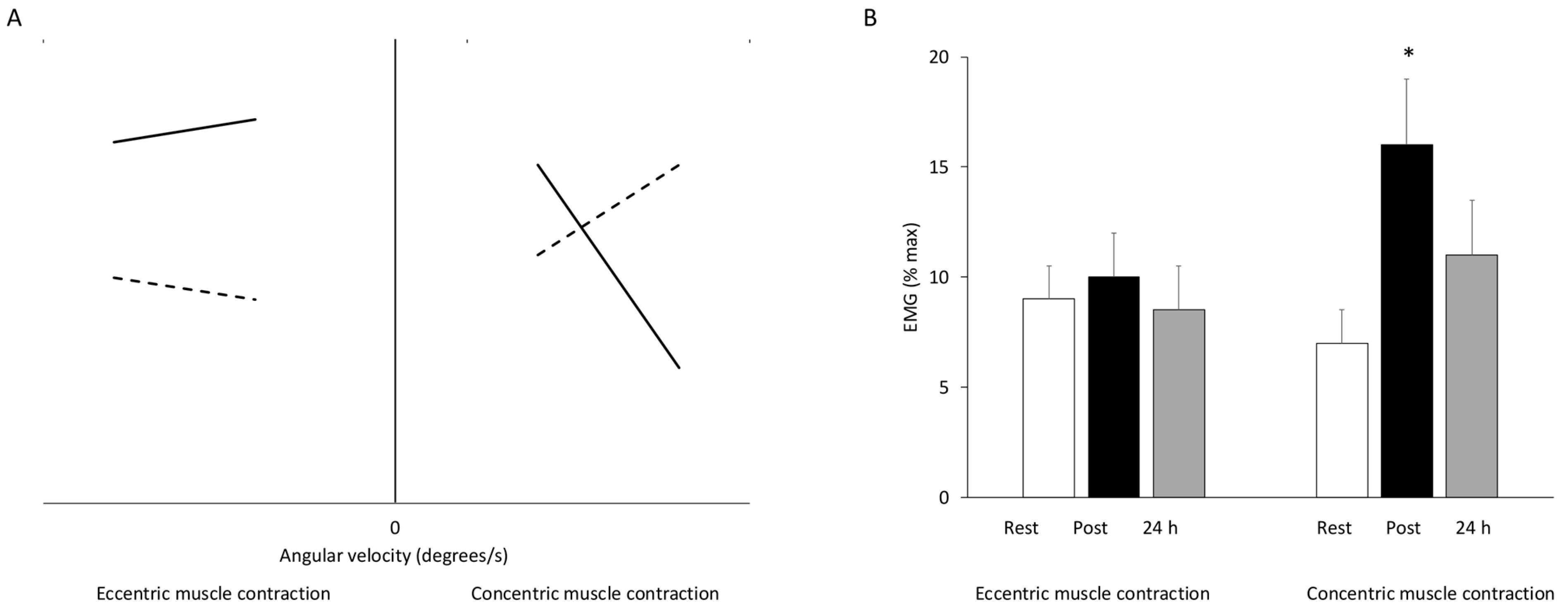


Disclaimer/Publisher’s Note: The statements, opinions and data contained in all publications are solely those of the individual author(s) and contributor(s) and not of MDPI and/or the editor(s). MDPI and/or the editor(s) disclaim responsibility for any injury to people or property resulting from any ideas, methods, instructions or products referred to in the content. |
© 2026 by the authors. Licensee MDPI, Basel, Switzerland. This article is an open access article distributed under the terms and conditions of the Creative Commons Attribution (CC BY) license.
Share and Cite
Paschalis, V.; Margaritelis, N.V.; Chatzinikolaou, P.N.; Theodorou, A.A.; Nikolaidis, M.G. Eccentric Exercise and Muscle Damage: An Introductory Guide. J. Funct. Morphol. Kinesiol. 2026, 11, 139. https://doi.org/10.3390/jfmk11020139
Paschalis V, Margaritelis NV, Chatzinikolaou PN, Theodorou AA, Nikolaidis MG. Eccentric Exercise and Muscle Damage: An Introductory Guide. Journal of Functional Morphology and Kinesiology. 2026; 11(2):139. https://doi.org/10.3390/jfmk11020139
Chicago/Turabian StylePaschalis, Vassilis, Nikos V. Margaritelis, Panagiotis N. Chatzinikolaou, Anastasios A. Theodorou, and Michalis G. Nikolaidis. 2026. "Eccentric Exercise and Muscle Damage: An Introductory Guide" Journal of Functional Morphology and Kinesiology 11, no. 2: 139. https://doi.org/10.3390/jfmk11020139
APA StylePaschalis, V., Margaritelis, N. V., Chatzinikolaou, P. N., Theodorou, A. A., & Nikolaidis, M. G. (2026). Eccentric Exercise and Muscle Damage: An Introductory Guide. Journal of Functional Morphology and Kinesiology, 11(2), 139. https://doi.org/10.3390/jfmk11020139