Abstract
A novel solid-state neutron and gamma radiation monitor-dosimeter based on biopolymer polylactic acid (PLA) is presented. The resulting detector (PLAD) technology takes advantage of property changes of the renewable PLA resin when subject to ionizing nuclear radiation. A simple yet rapid and accurate (±10%) low-cost (<$0.01/detector) mass loss upon dissolution (MLD) technique was successfully developed; MLD is based on a simple mass balance for discerning neutron and/or gamma doses using small (40 mg, ~4 mm diameter) ultra-low-cost (<$0.01) resin beads via dissolution in acetone. The GammaCellTM Co-60 irradiator, and the PUR-1 12 kW fission nuclear research reactor were utilized, respectively. Irradiation absorbed doses ranged from 1 to 100 kGy. Acetone bath temperature was varied from ~40 °C to ~54 °C. Results revealed a strong dependence of MLD on acetone bath temperature between neutron and gamma photon dose components; this allowed for the unique ability of PLAD to potentially perform as both a neutron-cum-gamma or as a gamma or neutron radiation dosimeter and intensity level detector. A linear trend is found for combined neutron and gamma radiation doses from 0 to 40 kGy when dissolution is conducted above 50 °C. The important potential ability to distinguish neutron from gamma radiation fields was scoped and found to be feasible by determining MLD at 45 °C. The potential was studied for simultaneous use as an in-core neutron and gamma monitor of an operating 3 GWt light-water reactor (LWR). Scoping tests were conducted with the pre-irradiated (@ 20 °C) PLAD resin beads followed by heating to in-core LWR coolant (300 °C) conditions for ~30 s corresponding to the time to reach ~40 kGy total doses in a typical 3 GWt LWR. MLD results were unaffected, indicating the exciting and unique potential for in situ (low-cost, accurate and rapid) simultaneous mapping of neutron and gamma radiation fluxes, related dosimetry, and fission power level monitoring.
Keywords:
radiation detection; dosimetry; neutron; gamma; reactor; PLAD; MLD; MLR; nuclear reactor; PUR-1; Co-60; GammaCellTM irradiator 1. Introduction
Neutron and/or gamma radiation monitoring and related dosimetry are of paramount importance, covering a myriad of arenas ranging from nuclear power reactor monitoring (under extreme conditions), personnel radiation safety, nuclear medicine/therapy, food preservation, sterilization, etc. A wide range of differing instrumentation techniques have been developed over the past century and have largely remained the same [1]; these past approaches rely on sensors based on counting charge buildup in ionized gases/solids (such as fission chambers or ion chambers) or monitoring light flashes from induced scintillaton and thermoluminescence. Existing systems range from complex, bulky $M range spectrometers down towards $1–10 K portable survey meters or in the $10–100 range for personnel dosimeters. Importantly, different techniques and instruments are deployed for neutron (neutral particles) and gamma radiation monitoring. While gamma radiation detectors and dosimeters are commonplace, neutron detection and dosimetry is far more complex—especially for monitoring fission power generation in commercial power reactors. This paper presents a novel technology that addresses the need for devising a low-cost, rapid turnaround, simple to deploy sensor system that can be used to detect and monitor both the neutron and gamma radiation fields even under extreme radiation and thermal fields—as may be encountered in nuclear power reactors. The technology discussed in this paper is based on the PLA renewable biopolymer introduced below.
Ionizing radiation interaction is well-known to lead to chain scission of polymers, including for the biopolymer PLA; this is followed by physio-chemical property changes, including degradation of key properties such as strength, optical transmissivity, and dissolution [2].
A key positive feature of PLA is its “green” characteristic and ability to be tailored for a wide range of potential applications via ionizing radiation or other means, such as thermal treatment [3,4,5]. Purdue University’s Metastable Fluids Advanced Research Laboratory (MFARL) has conducted research into tailoring PLA resins via photon-electron radiation to advance the field of radiation instrumentation—leading to the PolyLactic-acid based solid-state radiation detector/dosimeter (PLAD) [6]. PLAD, as a monitoring device for discerning radiation dose received from a combination of neutron and photon radiation fields (e.g., for nuclear fission energy reactor systems), remained a significant gap.
For the results presented in this paper, PLA was hypothesized to represent a promising material for use even in harsh nuclear environments, such as within the core of operating light water reactors (LWRs) and accelerator-based systems. Our goal is to advance PLAD towards deriving a novel, nonpowered solid-state, ultra-lightweight-scalable [e.g., ~0.1+ g (4 mm)/detector], affordable (<$0.1/unit), environmentally-friendly, easy-to-use, general purpose and real-time gamma-beta-alpha-fission-neutron monitor that is also readily deployable in extreme, e.g., 106+ R/h, mixed radiation fields as in LWRs. A novel approach was warranted—one that is simple to execute using standard laboratory equipment, able to decipher for both neutron and photon radiation types, also effective over a practically extensive range of radiation dose levels (1–100 kGy), and capable of being deployed under harsh thermal–hydraulic conditions such as in ~300 °C within the coolant field of LWRs.
The specific PLAD technique presented in this paper arose out of our general observations at MFARL that one of the most common solvents, acetone, readily dissolves PLA resin in an accelerated fashion with increasing solvent temperature; noteworthy is that we have also used acetone under ambient conditions to smoothen out the surfaces of 3D printed PLA samples; other approaches, notwithstanding [7]. This motivated a scoping study to examine the fractional dissolution of PLA resin beads when placed within an acetone bath at ~50 °C (which was chosen to be at a level a few degrees below its boiling point). Surprisingly, scoping trials revealed a reproducible linear increase over the 0–100 Gy dose range using PLA resin beads irradiated with 1.2–1.3 MeV gamma photons. Details are presented in later sections. This motivated more systematic studies, including assessing the exciting potential for monitoring neutron radiation (an aspect of paramount importance) in mixed neutron–gamma radiation fields of a fission nuclear reactor.
This paper discusses an advanced PLAD technique that measures the mass–loss ratio of PLA by dissolution (MLD) in acetone, aiming for photon and neutron dosimetry monitoring. It complements our earlier work on mass–loss and porosity induced via heat and pressure, which to date has shown validity for photon (not neutron) dosimetry assessments [6]. This advanced PLAD–MLD technique was developed by irradiating PLA resin in a gamma-only radiation irradiator and separately within an operating fission nuclear research reactor which offered a complex mixture of neutrons and gamma radiation fields spanning an extensive range of energies (10−2 eV to 20 MeV). The following sections discuss details on experiments, results, and analyses.
2. Experimentation—Irradiation of PLA Samples with Gamma Radiation and Mixed-Field Gamma–Neutron Radiation
This effort aimed to assess the combined and individual effects of absorbed gamma and neutron radiation types, respectively. As such, two types of irradiation sources were utilized: (1) Co-60 irradiator emitting ~1.1 MeV and ~1.2 MeV gamma photons; and (2) nuclear fission reactor—which provided a complex energy spectrum combination of neutrons and gamma photon radiation types.
2.1. Irradiation—Co-60 Gamma Source
Purdue University’s Nordion GammaCell 220TM Co-60 irradiator [8] performed γ photon irradiation of PLA resin samples procured from NatureWorks, LLC (Nordion Intl. Inc., Kanata, Ontario, Canada). The average dose in the GammaCellTM was initially calibrated [9] with well-established Fricke Dosimetry [10] in 1993. The dose rates extending to the usage time were evaluated based on the well-known half-life decay of the Co-60 source. The accuracy of the estimated dose rate is ±0.56% at the 95% confidence limit. By the summer of 2021, when the irradiations were performed, the achievable dose rates were ~2 kGy/day. The dose map within the irradiator based on data provided by the manufacturer is plotted in Figure 1.
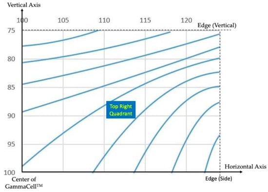
Figure 1.
Isodose curves (relative to dose at center) for Nordion GammaCell 220TM Co-60 irradiator (data from Ref. [9]).
The well-established and widely used Monte Carlo Nuclear Particle (MCNP) code was used to simulate the irradiator internals [2] and to characterize the irradiator core for its spatial radiation dose rate profile, the results of which were used to guide the positioning of samples used for this current study. Figure 2 provides the results of dose variation with radial (from the centerline) and axial locations within the irradiator. PLA resins were irradiated in the presence of room air. The maximum dose was 114.4 kGy.
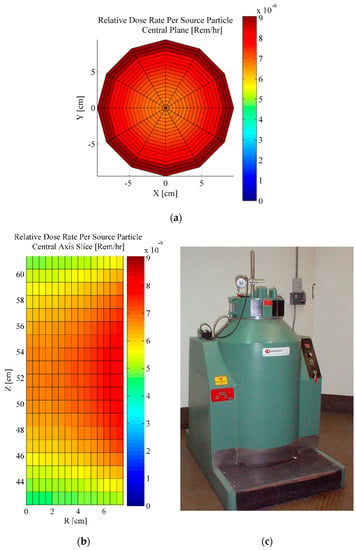
Figure 2.
MCNP code model results [3] of dose rate variations in Purdue University’s Gammacell irradiator, (a) mid-section, axial, (b) R-Z slice, radial, (c) irradiator [8].
Both evaluations found that the dose rate at the wall (close to the Co-60 capsules) is about 20% higher than at the center. For these studies, the PLA resin bearing samples were always kept in the central region of the chamber.
Considering the 5.3 y half-life for Co-60 decay over time, the gamma doses were evaluated by multiplying the time-averaged dose rate over the irradiation duration by the irradiation time. The dose evaluated from Fricke dosimetry was then converted to the real-time gamma radiation dose absorbed by PLA. This is deemed to be appropriate since the mass absorption coefficient (provided by the National Institute of Standards and Technology (NIST) database [10]) of ferrous sulfate, the main component of standard Fricke dosimetry, is very close to that for which PLA exposed to 1.25 MeV photons (the average of 1.17 and 1.33 MeV energies for Co-60 photons): 0.02955 cm2/g for ferrous sulfate, and 0.02816 cm2/g for PLA.
Using the GammaCellTM irradiator, PLA resin beads were subjected to accumulated dose levels ranging from 1 to ~100 kGy.
2.2. Irradiation—Mixed Gamma and Neutron Source in PUR-1 Reactor
Nuclear reactors present a complex irradiation environment, including the predominant neutron and gamma radiation fields and the short-range fission fragments-alpha-beta-neutrino particles from nuclear reactions and secondary radiation. When samples are placed in research reactor ports, the primary radiation types are neutrons and gamma photons. For our studies involving mixed neutron–gamma radiation fields, we conducted irradiations in Purdue University’s 12-kW pool-type (PUR-1) research nuclear reactor [11], which operates as a pool-type reactor under ~0.1 MPa/20 °C type thermal-hydraulic conditions and offers a neutron–gamma flux field in the range of ~1010/cm2-s.
Four sample irradiation ports in the PUR-1 were utilized for this study, two around the central area and two at the edge, each containing seven aluminum capsules. The PUR-1 operational power during these irradiations was ~8 kW. The PUR-1 organization has developed a core simulation tool based on the well-known MCNP code to predict the reactor’s neutron/gamma energy flux distributions and estimate the absorbed dose.
The PUR-1 power level was maintained at 8 kW for the entire range of time in the reactor from when the capsules were first loaded to when they were removed. Additionally, it was noticed that the aluminum capsules within which the samples were irradiated exhibited negligibly low neutron activation dose rates of <10−3 Gy/h (0.1 R/h) upon removal of the capsules after in-reactor irradiation.
PLA 4043D® (crystalline form) resin beads (see inset) supplied by NatureWorks, LLC were filled into individual aluminum capsules; the capsules were then stacked in a sample cylinder tube which is then inserted into the irradiation port holder tube of the reactor core. The schematic for a sample capsule assemblage (along the X/Y-Z plane) is shown in Figure 3. The irradiation port rests on the grid plate at the bottom of the core.
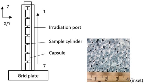
Figure 3.
Schematic of PLA resin bead (inset) bearing capsules assemblage.
The relevant dimensions for the capsules and irradiation port are as follows:
Capsule Dimensions: height (interior) = 5.4737 cm; diameter (interior)= 2.2098 cm; height (external) = 7.8486 cm; diameter (exterior) = 2.54 cm.
Irradiation Port Dimensions: height = 70.644 cm; diameter (interior) = 2.66 cm; diameter (exterior) = 2.87 cm.
Although seven capsules (capsules 1–7) are irradiated in total, only six of them were filled with PLA resin since the one on the very top (#1) is right adjacent to the upper free boundary surface, and for which the neutron–gamma fluxes and dose estimations are difficult to estimate with good accuracy. Consequently, the first (#1) capsule was kept empty. The flux profile and dose evaluations were done by MCNP code-based modeling. The X-Y midplane’s thermal and total neutron flux (n/cm2/s) profiles are color-coded (1010–1011 n/cm2/s) in Figure 4 and Figure 5, respectively.
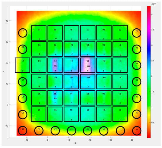
Figure 4.
Spatial thermal neutron flux at the X-Y midplane.
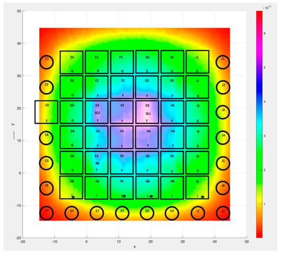
Figure 5.
Spatial total neutron flux at the X-Y midplane.
From practical considerations, the PUR-1 irradiation was conducted over 6 h, during which the maximum total (gamma and neutron) absorbed dose was ~40 kGy.
Figure 4 and Figure 5 indicate that the average fast-to-thermal neutron flux ratio is about 2:1. Radial port locations D6, F6, G6, and I6 (marked with ★ in Figure 5 were available and used for irradiation. As mentioned, seven capsules were loaded in each location (D, F, G, and I, respectively).
The calculated neutron and gamma radiation absorbed doses evaluated for each PLA resin-bearing capsule are listed in Table 1. It is noted that for the irradiation port locations D and I towards the left and right of the center of the core, the total dose levels range from ~9 kGy to ~17 kGy, respectively. The total dose levels are higher for the central irradiation port locations G and F, ranging from ~20 kGy to ~40 kG, respectively. The neutron dose is derived primarily from fast (MeV), not thermal (eV) energy neutrons; this is to be expected since the PLA resin beads were not doped (e.g., with B) in this study. The gamma-to-neutron dose ratio is ~2:1 for total doses <20 kGy, and gradually approaches ~1:1 for capsules where the total dose is >20 kGy. This is expected due to neutron leakage at edges and from the increased neutron-based fission rate in the central regions of the core.

Table 1.
Calculated neutron, gamma, and total dose for each capsule.
3. Novel Experimental Methodology—Ratio of Mass Dissolution (RMD) Figure of Merit for Detection and Dosimetry of Absorbed Gamma/Neutron Energy in PLA Resin
Having performed irradiations for gamma-only fields in the Co-60 irradiator and combined neutron–gamma fields in the PUR-1, for the irradiated PLA samples, we now needed a novel detection technique to rapidly use simple tools to measure not only the total absorbed dose but also to investigate for the potential to differentiate between and identify the dose received from neutron versus gamma photon radiation. Our past approaches focused primarily on gamma–electron radiation dosimetry.
Experiments at MFARL involving PLA and acetone (a standard laboratory cleaning solvent) proved that acetone (C3H6O) dissolves PLA resins—an effect of increased acetone temperature. This observation was examined for utility for gamma radiation dosimetry through scoping tests to assess the degree of PLA mass dissolution when placed in an acetone-bearing glass beaker in a temperature-controlled water bath for ~20 min at 50 °C (chosen arbitrarily to be close to but under the well-established boiling point of 56.2 °C at 0.1 MPa). The ratio of mass dissolved to the initial starting group (RMD) was derived from,
where ∆md refers to the mass difference between the initial ten PLA beads and the remaining PLA after dissolution, and m0d refers to the initial mass of the ten PLA beads before dissolution.
RMD = ∆md/m0d
The results of these (pristine) scoping tests with Co-60 gamma irradiated PLA resin over a wide 0–114 kGy dose range are plotted in Figure 6. We note that 0 kGy dose implies un-irradiated PLA at which, as well, experiments for RMD determination were conducted. At each dose level, three experiments were conducted—the error bars depict the range of data from the mean value. In order to check for reproducibility, limited experiments were conducted independently (by another co-author after a gap of several months) with ~38 kGy and ~117 kGy irradiated PLA samples using the same overall setup as for the earlier (pristine) tests. Results of the repeat tests (open circles) indicate that the pristine data are generally reproducible and overlap the original data—albeit with a slight bias towards a lower RMD (which is attributed to a likely acetone bath temperature undershoot by about 0.5 °C based on more detailed studies reported in this paper later on). Overall, the data exhibit a linear trend for gamma dose levels ranging between 0 and 117 kGy, as depicted in Figure 6.
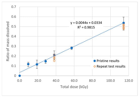
Figure 6.
RMD for gamma irradiated PLA resin dissolution in acetone at 50 °C—scoping and repeat test results. (Note: error bars where shown to depict the range of RMDs for that experiment).
Following the scoping test findings of Figure 6, a systematic protocol and approaches were examined to detect gamma-only and more complex mixed neutron–gamma irradiation doses. The various steps leading to a testing protocol are discussed below.
3.1. PLA Sample Preparation
PLA resin beads supplied by NatureWorksTM, LLC can vary in size and mass. Since mass dissolution can be expected to depend on the surface area of resin in contact with acetone, it was deemed logical to minimize RMD-related errors by utilizing (per availability of irradiated samples) a practical sample mass of ~0.4 g, using resin beads as close to being identical to each other. Consequently, ten PLA resin beads (each ~0.04 g, ~4 mm OD) were selected for each RMD-related experiment.
3.2. Thermal Dissolution Apparatus and Protocol
Two different (temperature-controlled) water bath devices were used for the experiments: OaktonTM StableTemp® and Joanlab®, respectively. Each water bath was characterized for stable attainment of the temperature of the water bath with the temperature of the acetone-bearing containers for the duration of each RMD measurement.
About 40 mL of acetone was utilized for each RMD experiment. The acetone was placed in a beaker and preheated to the desired temperature in a water bath positioned within a fume hood. To avoid significant evaporative loss of acetone, the temperature was necessarily kept below the boiling point of acetone, 56 °C (at 0.1 MPa). In total, 3–4 beakers are heated together each time, meaning 3–4 PLA resin samples (each ~0.4 g total) can be prepared simultaneously during each experimental run.
Before putting in the PLA beads, and also before removing the beakers, the temperature inside the acetone (in every single beaker) is measured with a Digi-Sense® Type J/K thermocouple meter (combined with an OmegaTM type-K probe) and averaged from among the beakers. According to the specifications provided by the manufacturer, the probe’s precision is between 0.5 °C and 2.2 °C, while the meter is 0.5 °C. The overall uncertainty from the thermocouple is then taken as 2.2 °C. Regardless, it was noted that the indicated temperature stays relatively constant (less than 0.5 °C variation), even considering that as time passes and the acetone evaporates, the temperature in the acetone may change. The setup of the experiment is shown in Figure 7a,b (actual) and Figure 7c (schematic).
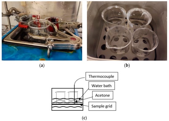
Figure 7.
Setups for MLD studies: (a) StableTemp® water bath, where clamps were used to hold the beakers to avoid direct contact between the beakers and the bottom of the water bath; (b) Joanlab® water bath, where a sample grid was used to prevent direct contact of the beakers and the bottom of the water bath; (c) schematic (for Joanlab® water bath setup) of how the temperature was measured in each beaker. The thermocouple was inserted directly into the acetone instead of the water bath.
Note that for a StableTemp® water bath, the actual temperature in the water is ~1 °C above the setup value measured at the same level where the beakers are held, and the average temperature in acetone among beakers is 0.6 ± 0.3 °C below the setup value; while for the Joanlab® water bath, the actual temperature in the water is ~2 °C above the setup value and the average temperature in acetone among beakers is 0.5 ± 0.5 °C above the setup value—as measured with the aforementioned thermocouple. The inserted thermocouple in the acetone measures the temperatures quoted in subsequent sections.
PLA beads are subjected to dissolution in the preheated acetone-bearing beaker for 20 min (a value chosen from experiential learning—while yet amenable for optimization). After the 20 min dissolution, the beakers are removed from the water bath, followed by drainage of the acetone and separation of the remaining PLA resin.
3.3. The Rationale for Selecting Temperature Range for Discriminating Gamma vs. Neutron Radiation Effects in RMD Experiments
From experience, it was noted that PLA dissolution in acetone is minimal at ~20 °C. It was already found (as noted from scoping study results shown in Figure 6) that significant mass dissolution is readily attainable over the 10–100 kGy range (for gamma irradiation dose) when the acetone temperature reaches ~50 °C, i.e., gets close to its 56.2 °C boiling point. Therefore, the useful temperature range was deemed to lie between 20+ °C and below 56 °C—to study the effect of temperature-based activation of dissolution from neutron and/or gamma radiation-induced absorbed energy (i.e., dose) in PLA.
It is non-obvious and was yet unclear as to what level of effect a neutron radiation dose would have on mass dissolution compared to the effects from a gamma radiation dose. Gamma radiation (at least for photon energies below 1.2 MeV) interactions with atoms are known [1] to mainly be governed by interaction with the 10,000× larger (~10−11 m) size electron cloud surrounding the ~10−15 m size individual nuclei of atoms in the bulk of materials—i.e., via photoelectric and Compton scattering phenomena. On the other hand, neutrons (uncharged neutral particles) remain blind to electrons surrounding the nuclei of atoms and interact only with the nucleus of atoms in a variety of ways ranging from elastic/inelastic scattering, photon producing, charged particle/neutron generating to nuclear fission. In this way, neutron radiation can create heavy charged ion recoils on a localized (sub-nm) basis, creating ion–electron pairs within molecules over short ranges of a few microns. Since chemistry-related phenomena (including dissolution) are governed via electron exchange, it is reasonable to assume that for the same total radiation dose, the effect on mass dissolution will be greater for gamma photons vs. neutrons. It was hypothesized that the activation thermal energy (tied to the temperature of acetone) required for initiation of mass dissolution should logically be lower for gamma radiation-dosed samples. For the same radiation energy-based absorbed dose and acetone temperature, earlier onset of mass dissolution should occur for PLA samples subject to gamma radiation. If this hypothesis proved correct, it could allow us to discriminate the neutron and gamma flux fields within a nuclear reactor—besides being of value to discriminate neutron versus gamma radiation fields.
The temperature range for this study varied from a low of ~40 °C to a high of ~54 °C.
3.4. Post-Dissolution Treatment and RMD Determination
After being subject to acetone for 20 min, the PLA resin sample was next placed in a preheated oven at ~77 °C (170 °F) for ~30 min for drying, before being weighed again to derive RMD using Equation (1) discussed earlier.
4. Results and Discussion
Due to resource (irradiator and reactor irradiation) and time constraints it was not possible to conduct several tens of tests for each combination of dose and the desired range of acetone bath temperature. For select combinations of dose and temperature the RMD values were obtained with 3–4 samples. Otherwise, a single sample was examined for gaging trends and threshold values of onset of effects on RMD for neutron vs. gamma radiation effects. Results of RMD for the range of combination of parameters are plotted in the five figures in Section 4. Where multiple samples were examined, the mean value and spread of data about the mean value are shown. Table 2 summarizes the testing range of experimental combinations.

Table 2.
Test Matrix of Experiment Combinations for RMD Determinations.
Results of experiments over the 1–40 kGy range with Co-60 gamma irradiation and PUR-1 neutron and gamma irradiation are plotted in Figure 8. As noted from Figure 8, the RMD values over the 1–40 kGy range from mass dissolution tests conducted with acetone at 53.5 °C are generally compatible with each other, regardless of the radiation source or even their respective energy spectra. This indicates that acetone at 53.5 °C provides activation energy for mass dissolution that is far above the threshold energy required for the onset of PLA dissolution regardless of the type of radiation used or even without any irradiation. While this finding permits one to correlate radiation dose with RMD and perform dosimetry within ±10% accuracy over a wide range of radiation doses, it does not by itself allow discrimination between the contribution from neutron and gamma radiation components.
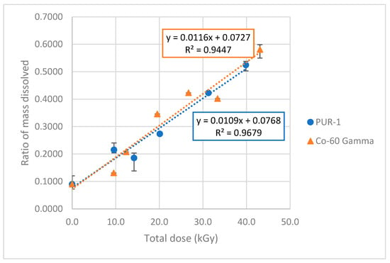
Figure 8.
RMDs for MLD in acetone at 53.5 °C. (Note: error bars where shown depict the range of RMDs for that experiment).
In order to affirm the above-discussed conclusion pertaining to the activation energy for dissolution via temperature state, experiments were conducted at an even higher temperature of 54.3 °C—closer to the boiling point. In order to assess for reproducibility, several tests were conducted with 0 and 43 kGy Co-60 γ source irradiated resin samples; the 1 σ value remained < 0.05. The results of experiments at 54.3 °C are shown in Figure 9. Once again, we find that the RMD values for irradiated PLA with either Co-60 gammas or PUR-1 neutrons and gammas are compatible with each other over the 40 kGy range.
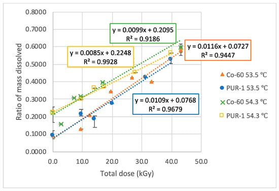
Figure 9.
RMDs for MLD in acetone at 53.5 °C and 54.3 °C. (Note: error bars where shown depict the range of RMDs for that experiment).
An interesting trend is observed. While the RMD values for 40 kGy total dose are similar to experiments at 53.5 °C and 54.3 °C, the spread widens with a reduced dose. At the extreme low end (i.e., 0 kGy), a mere 1 °C difference in acetone temperature leads to a doubling of the RMD. Experiments with acetone at temperatures above 54.3 °C were not attempted to date but may be useful to conduct in sealed (pressurized) vessels.
4.1. Discrimination of Neutron vs. Gamma Irradiation
Instead, it was deemed particularly interesting to assess threshold temperatures for activation of dissolution for allowing discrimination of neutron versus gamma radiation. It was ascertained that at room temperature (~21.5 °C), the RMD value remains close to zero. However, interesting changes became apparent when the acetone temperature rose at and above ~40.5 °C. The results of scoping tests are shown in Figure 10. As seen from Figure 10, for total dose levels below ~30 kGy, the RMD values are actually below zero, i.e., indicative of acetone mass uptake into PLA resin versus dissolution for acetone temperatures below 45.5 °C. An inflection point occurs for dose levels above 30 kGy for acetone temperatures at/above 40.5 °C. For the dose level of ~40 kGy, a sharp increase in RMD occurs at ~40.5 °C—but only for the Co-60 irradiated sample, not for the combined PUR-1 sample. This constitutes an important finding in relation to the activation of dissolution via irradiation from gamma photons. While the Co-60 irradiation is mainly from gamma photons, the PUR-1 irradiation dose at 40 kGy is split roughly 50/50 between gamma and neutron doses, respectively. Effectively, at a total PUR-1 dose of 40 kGy, the gamma dose component is only ~20 kGy (as noted in Table 1). The intermolecular bond damage caused at 20 kGy dose coupled with thermal energy activation at 45.5 °C is simply not sufficient to lead to significant mass dissolution. This would explain why the RMD rises to ~0.17 at a total dose of 40 kGy deposited from Co-60 gamma rays. Only once the acetone temperature rises towards 45 °C does the RMD for the 40 kGy (PUR-1) irradiated sample rise to RMD ~0.15, and the ~43 kGy (Co-60) irradiated sample RMD rises towards ~0.32, which is ~2.2× greater than for the PUR-1 sample. This difference in RMD at ~40 kGy (total dose) between the Co-60 and PUR-1 cases correlates directly to the ability to discriminate between and discern the neutron versus gamma radiation-induced dose components, respectively. That is, even at 45.5 °C, the activation (thermal) energy is sufficient to lead to mass dissolution of PLA as caused only by gamma irradiation, but not yet from a neutron radiation dose. Only when the temperature rises to/above 50 °C does the activation energy for dissolution exceed the level necessary for both neutron and gamma irradiation.
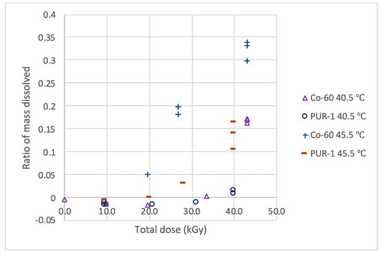
Figure 10.
RMDs for MLD in acetone at 40.5 °C and 45.5 °C. The existence of such a “threshold dose” tied to the temperature of the acetone bath for irradiated PLA is a highly interesting phenomenon for enabling not just total dose determination but also for separating the two underlying radiation types. A composite plot showing the RMD variation with total radiation dose and temperature is shown in Figure 11. (Note: actual data shown-error bars are not needed).
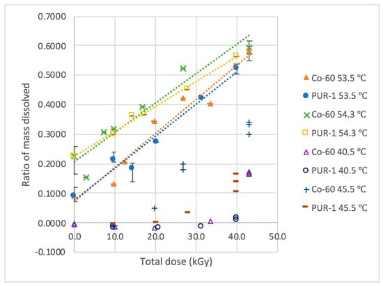
Figure 11.
RMDs for MLD in acetone at 40.5 °C, 45.5 °C, 53.5 °C, and 54.3 °C. (Note: error bars where shown depict the range of RMDs for that experiment).
Especially striking is the influence of acetone temperature on RMD variation at the total (mixed field neutron and gamma radiation) dose of 40 kGy from the PUR-1: leaping suddenly from ~0.01 at 40.5 °C to ~0.15 at 45.5 °C, and then from there to ~0.5 at 53.5 °C before leveling off to ~0.55 at 54.3 °C.
4.2. Nuclear Power Reactor Application Potential—Neutron-Gamma Dosimetry and Flux Monitoring
The irradiation dose of up to ~40 kGy required about 6 h irradiation time in the PUR-1 (operating at a power of 8 kWt), wherein the neutron flux was ~1010 n/cm2/s and the coolant temperature was ~20 °C. In contrast, the average neutron flux in a typical 3 GWt LWR is 1000× higher at ~1013 n/cm2/s and the core averaged coolant temperature is about 300 °C. Assuming a linear relationship with neutron flux, the equivalent amount of irradiation time to attain ~40 kGy doses in a power reactor core scales to about 21 s and could be readily achieved by moving PLA resin samples in–out of the core via instrument guide tubes. The key question to consider is what is the impact on RMD when the irradiated PLA resin is subject to a 300 °C temperature air environment for a 20–30 s timeframe? In order to shed light on this question, PLA samples at total irradiation dose levels of 0 and 40 kGy (via Co-60 and PUR-1) were kept in a preheated oven at an air temperature of ~300 °C air environment for 30 s, after which they were assessed for RMD via subject to mass dissolution testing in an acetone bath temperature of 54.3 °C. The results are shown in Figure 12, together with earlier results obtained with irradiations conducted at ~20 °C. As noted from Figure 12, the RMD data obtained with 300 °C post-heating for 30 s are in close proximity of the data obtained via irradiation at 20 °C.
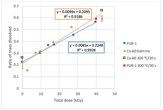
Figure 12.
RMD for MLD in acetone at 54.3 °C. (Note: error bars where shown depict the range of RMDs for that experiment).
This finding is encouraging in terms of the potential for monitoring the neutron and gamma flux fields in an operating LWR.
5. Summary and Concluding Remarks
This paper presents a novel biomaterial-based solid-state gamma/neutron radiation dose-monitoring metric—mass loss upon dissolution (MLD), as a new technique developed for PLA resin-based neutron and gamma radiation monitoring. The technique derives the ratio of mass dissolved (RMD) as a metric for determining the radiation dose from neutron and gamma radiation fields, in addition to offering the possibility for discriminating between the relative components of the two radiation types. Irradiations were conducted using a Co-60 bearing GammaCellTM for the gamma dose, and by using the PUR-1 for nuclear fission neutron-cum-gamma radiation dose. The total dose varied from 0 to 40 kGy for samples irradiated in the PUR-1 and from 0 to 130 kGy for samples irradiated in the GammaCellTM, respectively.
RMD values were determined by subjecting the irradiated PLA samples to dissolution in an acetone bath for 20 min at temperatures ranging from 20 °C, 40.5 °C, 45.5 °C, 53.5 °C, and 54.5 °C, respectively. Little to no RMD variation was noted for acetone bath temperatures below 40.5 °C, after which a sudden threshold activation energy effect manifested itself, offering the potential for not only monitoring but also to separately allow discrimination and determination of the neutron and gamma dose components, respectively, at the acetone bath temperature of 45.5 °C. For tests conducted at/above 50 °C, a linear trend was obtained for RMD vs. total dose irrespective of radiation type, neutron, or gamma.
The extension of the results obtained for RMD variations via PUR-1 irradiations at 20 °C was assessed in scoping fashion for application to the possibility of monitoring and mapping the neutron and gamma radiation field intensities in a 3 GWt power reactor coolant environment of 300 °C. The equivalent RMD values over the radiation dose range of 0 to 40 kGy were generally compatible with each other. The PLAD-based approach based on RMD conducting dissolution for about 20 min. in acetone at ~53 °C, the in-core power and neutron–gamma flux fields (and associated dosimetry) appears feasible to be conducted within ~0.5 h. In such a scenario, it may be possible to utilize the PLAD for short duration (e.g., <1 min.) core-wide irradiation using instrument guide tubes. More research is clearly desirable in this area.
It will also be interesting to try the effects of other solvent alternatives to acetone for studying relative dissolution of PLA per experiences cited in the public domain [11,12,13]. Our own past PLAD studies [2,3] for monitoring gamma–electron beam dose vs. relative viscosity examined irradiated PLA dissolved in chloroform. However, chloroform is far less user-friendly and more expensive than acetone. Corrosive alkaline solutions (e.g., NaOH-water) are also effective at dissolving PLA, but the solvent temperature needs to be much higher (i.e., ~80 °C) to derive results within one hour. In the extreme if only water is chosen as a solvent, the temperature needs to be even higher, and much longer times (e.g., days) should be expected for significant hydrolysis.
Author Contributions
Conceptualization, R.P.T.; Methodology, N.B., W.J. and R.P.T.; Software, T.M.; Validation, W.J., T.B. and R.P.T.; Formal analysis, W.J. and R.P.T.; Investigation, W.J. and R.P.T.; Resources, T.M and R.P.T.; Data curation, W.J. and R.P.T.; Writing-original draft, W.J. and R.P.T.; Writing-review and editing, W.J., T.M. and R.P.T.; Supervision, R.P.T.; Project administration, R.P.T.; Funding acquisition, R.P.T. All authors have read and agreed to the published version of the manuscript.
Funding
This research was partly sponsored by Purdue University, West Lafayette, USA, the Bilsland Ph.D. Fellowship, and in part by the United States Department of Energy.
Data Availability Statement
Upon request from the corresponding author Rusi Taleyarkhan.
Acknowledgments
The cooperation and past research of several MFARL colleagues are deeply appreciated, as is assistance from the staff of Purdue University’s REMS and PUR-1 organizations.
Conflicts of Interest
The authors declare no conflict of interest.
References
- Knoll, G.F. Radiation Detection and Measurement, 3rd ed.; John Wiley and Sons, Inc.: Hoboken, NJ, USA, 2000. [Google Scholar]
- Loo, S.C.J.; Ooi, C.P.; Boey, Y.C.F. Radiation effects on poly(lactide-co-glycolide)(PLGA) and poly(l-lactide)(PLLA). Polym. Degrad. Stab. 2004, 83, 259–265. [Google Scholar] [CrossRef]
- Bakken, A. Tailoring and Assessment of PLA BioPolymers for Use as VOC-Free Adhesives. Ph.D. Thesis (Major Professor: Dr. Rusi Taleyarkhan), Purdue University, West Lafayette, IN, USA, December 2017. [Google Scholar]
- Bakken, A.; Boyle, N.; Archambault, B.; Hagen, A.; Kostry, N.; Fischer, K.; Taleyarkhan, R.P. Thermal and ionizing radiation induced degradation and resulting formulation and performance of tailored PLA hot melt adhesives. Int. J. Adhes. Adhes. 2016, 71, 66–83. [Google Scholar] [CrossRef]
- Jiang, W.; Bakken, A.; Taleyarkhan, R.P. Irradiation induced Crosslinking in “Green” Polylactic-acid (PLA) Polymers for Enhanced Strength and Elevated Temperature Applications. In Proceedings of the 2020 International Conference on Nuclear Engineering collocated with the American Society of Mechanical Engineers, SME 2020 Power Conference, Virtual, 4–5 August 2020; Volume 3, p. 16767. [Google Scholar]
- Jiang, W.; DiPrete, D.; Taleyarkhan, R.P. PLA Renewable Bio Polymer Based Solid-State Gamma Radiation Detector-Dosimeter for Biomedical and Nuclear Industry Applications. Sensors 2022, 22, 8265. [Google Scholar] [CrossRef] [PubMed]
- MFARL Research Based Experiences—2012 to 2023. Available online: https://www.wevolver.com/article/dissolving-pla-how-to-melt-pla-and-smooth-3d-prints (accessed on 1 July 2023).
- Nordion Website. Available online: https://www.nordion.com/products/irradiation-systems/ (accessed on 4 August 2022).
- Nordion Gamma Irradiator Certificate of Measurement for Gamma Cell 220 No. 235; Communicated from Nordion Int. Inc.: Kanata, ON, Canada, 1994.
- National Institute of Standards and Technology. Available online: https://physics.nist.gov/PhysRefData/XrayMassCoef/ComTab/fricke.html (accessed on 8 August 2022).
- Available online: https://engineering.purdue.edu/NE/research/facilities/reactor (accessed on 1 July 2023).
- National Center for Biotechnology Information. PubChem Compound Summary for CID 6212, Chloroform. Retrieved 13 March 2023. 2023. Available online: https://pubchem.ncbi.nlm.nih.gov/compound/Chloroform (accessed on 1 July 2023).
- National Center for Biotechnology Information. PubChem Compound Summary for CID 180, Acetone. Retrieved 13 March 2023. 2023. Available online: https://pubchem.ncbi.nlm.nih.gov/compound/Acetone (accessed on 1 July 2023).
Disclaimer/Publisher’s Note: The statements, opinions and data contained in all publications are solely those of the individual author(s) and contributor(s) and not of MDPI and/or the editor(s). MDPI and/or the editor(s) disclaim responsibility for any injury to people or property resulting from any ideas, methods, instructions or products referred to in the content. |
© 2023 by the authors. Licensee MDPI, Basel, Switzerland. This article is an open access article distributed under the terms and conditions of the Creative Commons Attribution (CC BY) license (https://creativecommons.org/licenses/by/4.0/).