Article

Developmental Toxicity and Teratogenic Effects of Dicarboximide Fungicide Iprodione on Zebrafish (Danio rerio) Embryos

by
Chang-Young Yoon
1,
Kyongmi Chon
1,
Bala Murali Krishna Vasamsetti
1,*,
Sojeong Hwang
1,
Kyeong-Hun Park
1 and
Kee Sung Kyung
2
1
Toxicity and Risk Assessment Division, Department of Agro-Food Safety and Crop Protection, National Institute of Agricultural Sciences, Rural Development Administration, Wanju-gun 55365, Republic of Korea
2
Department of Environmental and Biological Chemistry, College of Agriculture, Life and Environment Sciences, Chungbuk National University, Cheongju 28644, Republic of Korea
*
Author to whom correspondence should be addressed.
Fishes 2024, 9(11), 425; https://doi.org/10.3390/fishes9110425
Submission received: 24 August 2024 / Revised: 30 September 2024 / Accepted: 21 October 2024 / Published: 23 October 2024

Abstract

Iprodione (IDN) is a broad-spectrum fungicide used to treat various fungal infections in plants. Despite its extensive use, assessment of its toxicity in aquatic organisms remains incomplete. This study investigated the deleterious effects of IDN using zebrafish (ZF) as a model organism. ZF embryos, beginning at 2 h post-fertilization (hpf), were exposed to IDN (3.75–40 mg/L), and both mortality and deformities were assessed. The impact of IDN on mortality was concentration-dependent and significant from 14 mg/L. Importantly, IDN induced several deformities at sublethal concentrations, including abnormal somites, reduced retinal pigment accumulation, yolk sac edema, hatching failure, abnormal swim bladders, and spinal curvature. The EC50 values for IDN-induced deformities were 3.44 ± 0.74 to 21.42 ± 6.00 mg/L. The calculated teratogenic index values for all deformities were above 1, indicating that IDN is teratogenic to ZF. IDN-exposed ZF also displayed abnormalities in touch-evoked escape responses. IDN significantly affected heart rate and blood flow, and induced pericardial edema and hyperemia in a concentration-dependent manner, suggesting its influence on cardiac development and the function of ZF. In conclusion, these results suggest that IDN exerts toxic effects on ZF embryos, affecting mortality, development, and behavior.
Key Contribution: The study shows that IDN poses a significant risk to zebrafish embryos, leading to malformations, increased mortality, and altered behavior. These results highlight the importance of careful monitoring and further investigation on the potential environmental and health hazards of IDN.

Graphical Abstract

1. Introduction

Iprodione (IDN) is a dicarboximide-based fungicide widely utilized in agriculture for its ability to inhibit spore germination and fungal growth in plants [1]. It is commonly applied to various crops, including grapes, peaches, and onions [2]. In 2022, IDN consumption in Korea amounted to 30,242 kg [3], and the global IDN market is projected to reach USD 2.1 million by 2026 [4]. IDN residues have been detected in various crops, including tomatoes [5], tobacco leaves [6], and grapes [7]. Furthermore, IDN persists and accumulates in the soil over extended periods [8] and has been detected in groundwater [9], raising concerns that runoff may contain high concentrations of IDN, which could pose risks to both soil and aquatic organisms [10,11]. Given the extensive use of IDN and its environmental persistence, there is a critical need to assess its non-target toxicity to better understand its ecological impact.
The US Environmental Protection Agency has classified IDN as possibly carcinogenic to humans and moderately toxic to animals [12]. IDN is also an endocrine disruptor that decreases the number of sperm progenitor cells in rats [13] and reduces the expression of androgen-related genes by binding to androgen receptors [14]. Furthermore, IDN disrupts oocyte maturation and exhibits cardiotoxicity and hepatotoxicity in zebrafish (ZF) [15,16,17]. IDN has also been linked to reproductive toxicity and neurotoxicity in animals [18,19]. Therefore, environmental exposure to IDN poses substantial risks to non-target organisms and humans, highlighting the necessity for comprehensive toxicity assessments.
ZF has been used as an experimental animal in environmental toxicity screening for pesticides and chemicals [20]. Their high fecundity, large numbers of eggs, and short lifespans make them ideal experimental animal models [21,22]. ZF embryos are transparent and develop rapidly, enabling the observation of all stages of embryonic development and organ formation [23]. This transparency and rapid development facilitate easier assessment of abnormal morphology and behavior when organisms are exposed to pesticides or chemicals [24,25,26,27]. Considering the known toxicity of IDN to aquatic organisms, it is crucial to elucidate its specific toxic effects on ZF to better understand the broader ecological implications of IDN contamination in aquatic environments. While previous research has focused on the cardiotoxic and hepatotoxic effects of IDN in ZF [16,17], the broader teratogenic potential of IDN—specifically, its ability to cause developmental deformities—remains poorly understood.
Therefore, this study aims to address this critical gap by investigating the toxicological effects of IDN on ZF, with a specific emphasis on developmental deformities. By exploring the adverse outcomes of IDN exposure in this model organism, this study aims to provide valuable insights into the ecological risks posed by IDN, ultimately informing strategies for better environmental protection and public health safety.

2. Materials and Methods

2.1. ZF Brood Maintenance and Embryo Collection

This study was conducted in accordance with the Guidelines for The Care and Use of Laboratory Animals and was approved by the Animal Ethics Committee of the National Institute of Agricultural Sciences, Rural Development Administration, Republic of Korea (Protocol Code: NAS-202305 [30 January 2023]).
The ZF brood (Danio rerio) was kept in 50 L aquariums filled with dechlorinated water, maintained at a constant temperature of 25 ± 1 °C, and subjected to a 12 h light and 12 h dark photoperiod. Brine shrimp (INVE Aquaculture, Dendermonde, Belgium) or frozen bloodworms (Hikari Bio-Pure, Sacramento, CA, USA) were fed to ZF twice daily.
ZF stocks of less than one year of age were used for mating to ensure the collection of healthy embryos. ZF mating, embryo collection, and cleaning procedures were conducted, as previously described [26,27]. Briefly, well-fed male and female ZF (up to 20 randomly selected fish) were placed in a pre-aerated tank containing 10–12 L of dechlorinated water at a temperature of 27 ± 1 °C to collect embryos. The mating tanks were continuously aerated and lined with nylon netting to prevent the fish from eating their embryos. The next day, embryos were collected 30 min after the light was turned on and rinsed with E3 medium to remove any surface debris. The embryos were incubated at 26 ± 1 °C until 2 h post-fertilization (hpf), and the healthy fertilized embryos were selected for the toxicity assessment experiments.

2.2. ZF Embryo Development Toxicity Test

2.2.1. Embryo Exposure

Embryo toxicity tests were performed according to the OECD Test Guideline No. 236 [28]. Additionally, several developmental deformities were assessed at 24, 48, 72, 96, and 144 hpf. An IDN stock (purity: 99.8%, Sigma-Aldrich, St. Louis, MO, USA) was prepared in dimethylsulfoxide (DMSO; Sigma-Aldrich). The required test concentrations (3.79, 5.31, 7.43, 10.41, 14.57, 20.40, 28.57, and 40.00 mg/L) were prepared by dissolving the appropriate amount of IDN stock solution in E3 medium (290 mg of NaCl, 8.3 mg of KCl, 48 mg of CaCl2, 81.5 mg of MgCl2, 0.1% methylene blue, pH 7.2 per 1 L of distilled water). The control group received the E3 medium containing 0.05% DMSO, and this DMSO concentration was consistently maintained across all other treatment groups. A concentration of 4.0 mg/L 3,4-dichloroaniline dissolved in the E3 medium was used as a positive control. Toxicity tests were performed using 24-well plates (SPL Life Sciences, Daejeon, Republic of Korea), and IDN treatment was initiated at the 2 hpf embryonic stage. In each 24-well plate, 20 wells were allocated to the test solution, filled with 2 mL per well, whereas the remaining four wells contained 2 mL of E3 medium, serving as an internal plate control. Healthy embryos were added to each well (one embryo per well) and incubated in an incubator at 26 ± 1 °C in the dark. The test solution was replaced every 24 h for up to 96 h, after which the embryos were incubated for 144 h without changing the solution. The toxicity evaluation experiment was performed three times, with each experiment consisting of two 24-well plates.

2.2.2. Deformity Assessment

Embryos that turned black and had no heartbeat were classified as dead. Mortality was documented at 24, 48, 72, and 96 hpf using a microscope (Stemi 508, Zeiss, Oberkochen, Germany). The embryo mortality percentage was calculated by comparing the number of dead embryos or larvae at the time of observation with the initial number of embryos at the start of exposure.
All phenotypic scoring conducted after IDN exposure was based on previous reports [25,26,27]. Deformities were scored at least twice for each phenotype to ensure data reliability. Somite deformities were observed at 24 hpf, and retinal pigment accumulation, tail blood flow, and hyperemia were observed at 48 hpf. Fish exhibiting no, intermittent, or slower blood flow in the dorsal aorta region than those observed in the control fish were identified as those with abnormal blood circulation. Cardiac and yolk sac edema was measured at 72 hpf, hatching success was assessed at 72 and 96 hpf, and swim bladder and spinal deformities were evaluated at 144 hpf. The percentage of deformities was calculated as the ratio of embryos with malformations to the total number of live embryos at that time point.

2.3. Cardiac Function Assessment

The heart rates of the embryos or larvae were assessed at 48, 72, and 96 hpf. These measurements were obtained in an air-conditioned room at a constant temperature of 26 ± 1 °C under anesthesia using 10 mg/L tricaine (Sigma-Aldrich) and a Stemi 508 microscope (Zeiss). For each experimental condition, the heart rates of ten embryos (five embryos from each 24-well plate) were recorded (n = 3). Heartbeats were measured twice for 10 s, and the average of these measurements was used to calculate the number of heartbeats per minute (hpm).

2.4. Body Length Measurement

Body length measurements were observed at 144 hpf under anesthesia with 10 mg/L tricaine (Sigma-Aldrich). The chorion of the embryos was gently opened using forceps for IDN treatment concentrations wherein hatching failure was severe. Larval images were captured under all test conditions using a stereomicroscope (Stemi 508, Zeiss). The length between the tip of the caudal fin and the tip of the mouth was considered as the body length. Numerical data from the images were obtained using the OptiView 3.7 software (Korealabtech, Seongnam, Republic of Korea). In each experiment, the body lengths of 10 larvae were measured under each test condition, and the experiment was repeated thrice.

2.5. Behavioral Analysis (Touch-Evoked Escape Response)

Touch-Evoked Escape Response measurements were conducted at 144 hpf, as described previously [24]. The assessment involved gently touching the heads and tails of the ZF larvae using a thin (1 mm) flexible nylon wire. Larvae that did not respond to touch after four attempts, or those that traveled a shorter distance than the control larvae, were classified as having an abnormal response to touch. The chorion of the embryos was gently opened using forceps at 96 hpf for IDN treatment concentrations wherein hatching failure was severe. Embryos with severe developmental abnormalities were excluded from the analysis, but larvae with minimal deformities were included, as their inclusion was unavoidable, particularly at higher IDN concentrations. The experiment was repeated thrice, with each assessment evaluating 10 embryos.

2.6. Statistical Analysis

LC50 and EC50 values were calculated using Prism 5.0 (GraphPad, La Jolla, CA, USA) and percentage data from three independent experiments. Statistical significance between the control and treatment groups was assessed using the Kruskal–Wallis test, followed by Mann–Whitney U test to determine the significance between the groups. The teratogenic index (TI) of each observed deformity was calculated by dividing the LC50 (96 h) by the EC50 value of each deformity. The statistical significance of the data was set at p < 0.05.

3. Results

3.1. IDN Is Toxic to ZF

At 96 hpf, the embryo mortality in the internal plate control group was below 10%, whereas the positive control group exhibited mortality rates exceeding 60% (Table 1). Both results met the criteria for test validity according to OECD Test Guideline 236. The solvent control group and groups treated with 3.79 and 5.31 mg/L IDN displayed no embryo mortality up to 96 hpf (Table 1). Embryonic lethality increased in a concentration-dependent manner, starting with 7.43 mg/L of IDN. The cumulative mortality percentages and LC50 values at 24, 48, 72, and 96 hpf for the groups treated with different IDN concentrations are shown in Table 1.

3.2. IDN Is Teratogenic to ZF

The embryos and larvae in the negative control group exhibited no abnormalities throughout the study period (Figure S1). The embryos or larvae in the solvent control group exhibited normal growth and phenotypes during the testing period; however, the IDN-treated group displayed several developmental defects, most of which increased in a concentration-dependent manner (Table 2). Table 3 shows the LC50, EC50, and TI values for the scored deformities after IDN treatment. The TI values for all phenotypes assessed during the testing period were above 1, indicating that IDN was teratogenic to ZF and affected its overall growth and development.
IDN did not influence tail detachment, a phenotype that must be assessed according to the OECD Test Guideline 236. The control and IDN-treated groups exhibited normal tail detachment (Figure 1). Abnormalities in somite formation were noticeable after exposure to 5.31 mg/L IDN, with a significant difference in somite formation observed between the 28.57 mg/L IDN treatment and control groups (p < 0.05; Figure 1 and Table 2).
Delayed retinal pigmentation was observed at 48 hpf in the IDN-treated groups when compared to the control groups (Figure 1). Although delayed retinal pigmentation was evident from a concentration of 3.79 mg/L IDN (12.50% (IQR: 0.00–36.25)), significant differences were observed when compared to the control group starting from 10.41 mg/L IDN (p < 0.05; 60.53% (IQR: 49.47–75.99)) (Table 2 and Figure 1). Variations in retinal pigment accumulation were clearly visible at 36 hpf (Figure S2).
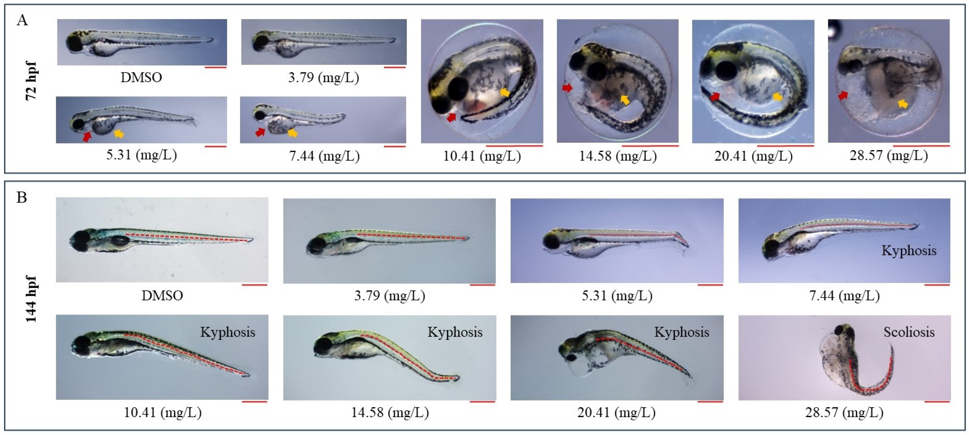
The frequency of yolk sac edema increased in a concentration-dependent manner after IDN exposure (Table 2). Compared to the control group, a significant difference in the incidence of yolk sac edema was observed from a concentration of 10.41 mg/L IDN (p < 0.05; 52.64% (IQR: 18.42–75.99)) (Table 2 and Figure 2).
In the IDN-treated groups, hatching failure occurred in a concentration-dependent manner at 72 and 96 hpf when compared to the control group (Table 2 and Figure 2). At 14.57 mg/L IDN, the hatching failure rate was 100% at 72 hpf. At 96 hpf, 100% hatching was observed in the control and the 3.79 mg/L IDN groups. Hatching failure increased in a concentration-dependent manner from IDN concentrations above 3.79 mg/L, with a significant difference observed starting from 10.41 mg/L (p < 0.05).
IDN severely affected swim bladder formation in ZF in a concentration-dependent manner (Figure S3). At a dosage of 3.79 mg/L of IDN, 60.00% (IQR: 35.00–85.00) of ZF exhibited partially inflated swim bladders, which was a significant difference when compared to the control group (p < 0.05; Table 2). At a concentration of 10.41 mg/L IDN, swim bladder abnormalities reached 100%.

3.3. IDN Affects the Cardiac Development and Function of ZF

IDN affected cardiac development and function in ZF embryos and larvae (Table 2 and Figure 3). At 48 hpf, hyperemia and abnormal tail blood flow were observed in IDN-treated ZF. At 7.43 mg/L IDN, 17.50% (IQR: 10.00–25.79) of the ZF showed hyperemia, which significantly differed from that of the control group (p < 0.05). From 28.57 mg/L IDN, hyperemia reached 100% (Table 2 and Figure 1 and Figure 2). Abnormalities in tail blood flow were not observed in the control group or at 3.79 mg/L IDN; however, 10.40% (IQR: 0.00–22.50) of ZF exhibited abnormal blood flow starting at 7.43 mg/L IDN. The blood flow abnormalities reached 100% at 28.57 mg/L IDN (Table 2). The abnormalities in the blood of the IDN-exposed ZF larvae are shown in Video S1. While the control and 3.79 mg/L IDN groups exhibited normal cardiac development, starting at 5.31 mg/L IDN, 25.00% (IQR: 0.00–35.00) of ZF displayed edema in the cardiac region (Table 2 and Figure 3). Beginning at a concentration of 7.43 mg/L IDN, IDN exerted significant effects on cardiac edema formation when compared to those in the control group (p < 0.05). The heart rate decreased with increasing IDN exposure at all tested time points. The median heart rates of the control group were 168.0 (IQR: 162.0–169.5), 180.0 (IQR: 174.0–186.0), and 204.0 (IQR: 192.0–210.0) beats per minute (bpm) at 48, 72, and 96 hpf, respectively. At 48 hpf, the heart rates for IDN exposure groups at concentrations of 3.79, 5.31, 7.43, 10.41, 14.57, 20.40, and 28.57 mg/L were 162.0 (IQR: 162.0–168.0), 156.0 (IQR: 150.0–168.0), 144.0 (IQR: 138.0–150.0), 126.0 (IQR: 126.0–133.0), 102.0 (IQR: 96.0–114.0), 96.0 (IQR: 94.5–108.0), and 102.0 (IQR: 88.5–108.0) bpm, respectively. At 72 hpf, the heart rates were 180.0 (IQR: 174.0–186.0), 171.0 (IQR: 168.0–175.0), 156.0 (IQR: 142.5–162.0), 153.0 (IQR: 150.0–162.0), 138.0 (IQR: 114.0–144.0), 126.0 (IQR: 114.0–132.0), and 111.0 (IQR: 90.0–120.0) bpm. At 96 hpf, the heart rates further reduced, with values of 189.0 (IQR: 180.0–192.0), 174.0 (IQR: 162.0–175.5), 162.0 (IQR: 148.5–168.0), 144.0 (IQR: 136.5–150.0), 138.0 (IQR: 132.0–150.0), 108.0 (IQR: 96.0–120.0), and 90.0 (IQR: 60.0–109.5) bpm at the same respective concentrations of Iprodione. The differences in heart rate between the control and IDN-treated groups are shown in Video S2. These results indicate that IDN significantly affected cardiac development and function in ZF embryos or larvae.

3.4. IDN Affects Normal ZF Growth

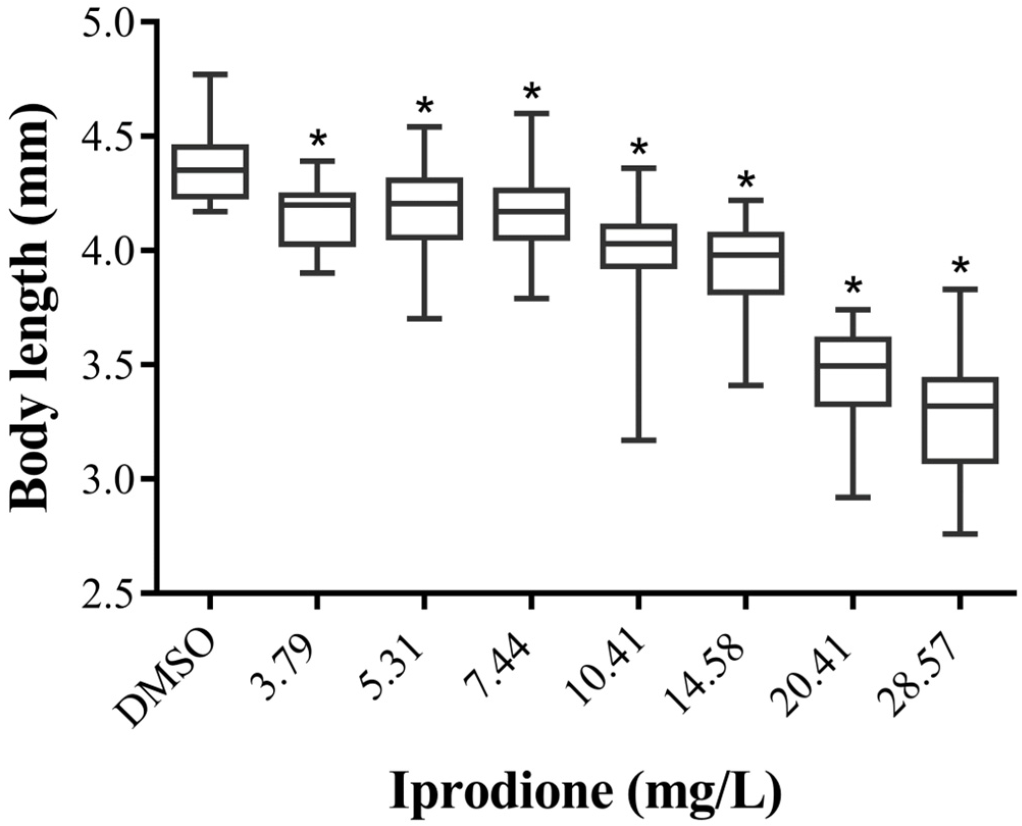
The body length of zebrafish (ZF) decreased with increasing concentrations of Iprodione (IDN), as shown in Figure 4. The control group had a median body length of 4.350 mm (IQR: 4.238–4.453 mm). The body lengths at IDN concentrations of 3.79, 5.31, 7.43, 10.41, 14.57, 20.40, 28.57, and 40.00 mg/L were 4.200 mm (IQR: 4.028–4.243 mm), 4.205 mm (IQR: 4.058–4.308 mm), 4.170 mm (IQR: 4.055–4.263 mm), 4.030 mm (IQR: 3.930–4.105 mm), 3.980 mm (IQR: 3.818–4.070 mm), 3.495 mm (IQR: 3.328–3.613 mm), and 3.320 mm (IQR: 3.078–3.435 mm), respectively. A statistically significant reduction in body length was observed beginning at the lowest concentration of 3.79 mg/L IDN (p < 0.05).
IDN exposure significantly increased the occurrence of spinal deformities in ZF (Table 2 and Figure 2) when compared with control ZF. The control group displayed normal spines, whereas the groups treated with IDN exhibited spinal deformities, including lordosis, kyphosis, and scoliosis. Approximately 50% of the ZF larvae exhibited spinal curvature at a concentration of 10.41 mg/L IDN.

3.5. IDN Affects ZF Behavior

The behavioral responses of ZF larvae exposed to IDN were evaluated at 144 hpf using the touch-evoked response assay. Video S3 illustrates the abnormal swimming behavior after IDN exposure. These results demonstrate a significant difference in the touch response of larvae exposed to IDN from a concentration of 10.41 mg/L when compared to the control group (p < 0.05; Table 2). Nearly all controls responded to the first or second touch and immediately reached the corner of the Petri dish. Although the response at 3.79 mg/L was similar to that of the controls, larvae exposed to 5.31 mg/L and higher concentrations exhibited different behaviors. They required three or four attempts to respond and traveled a much shorter distance than the controls. This effect became more prominent as the concentration increased. These findings suggest that exposure to IDN significantly impairs the sensory and motor functions of ZF.

4. Discussion

This study revealed that IDN treatment induces various developmental defects, including hatching abnormalities, cardiac developmental and functional anomalies, reduced growth, and behavioral abnormalities, particularly at sublethal concentrations. These observations strongly suggest that IDN has teratogenic effects on ZF and poses considerable risks to normal growth and survival.
Hatching failure was one of the most notable effects observed in IDN-exposed ZF, consistent with findings from a previous study indicating a decrease in hatchability after IDN exposure [16]. Reduced hatchability was also observed when ZF embryos were exposed to procymidone, a dicarboximide-based fungicide, even at a low concentration of 100 ng/L [29], suggesting that dicarboximide-based fungicides commonly affect the hatching success of ZF embryos. Under optimal conditions, ZF embryos typically start hatching at approximately 35 hpf, with nearly all healthy embryos hatching at 96 hpf [23]. During early developmental stages, ZF embryos predominantly rely on egg yolk as a nutrient source, which is crucial for supporting embryonic growth and development. Therefore, a delay in hatching can affect the growth and development of ZF [30] and lead to death. The results of this study showed that many embryos exposed to a sublethal concentration of IDN did not hatch at 96 hpf. When forced open, the larvae exhibited drastically reduced growth, severely malformed bodies, and did not survive. Reduced hatchability can partially be attributed to abnormal protease secretion [31], which is essential for chorion digestion, or to the lack of twitching movements due to the neurotoxic effects of IDN; however, further in-depth investigation into these specific aspects is required. Nonetheless, these observations underscore the significance of hatching during normal ZF growth.
Pesticides from different families induce abnormal blood flow, pericardial edema, and altered heart rate [24,25,26,27]. The heart is the first organ that is formed and becomes functional in ZF, thereby making it the primary organ of interest in pesticide toxicity studies [32]. During ZF embryonic development, the incidence of cardiac edema indicates abnormal cardiac development, whereas abnormalities in blood flow and heart rate can indicate functional cardiac abnormalities [33]. In this study, these cardiac-related abnormalities were evident after IDN exposure; therefore, IDN can be classified as a cardiac toxin to ZF that has detrimental effects on cardiac development and functionality. Furthermore, IDN exposure also results in alterations in cardiomyocyte counts [16]. Additionally, the exposure of ZF embryos to procymidone resulted in cardiac edema, arrhythmias, and atrial and ventricular deformations [29], supporting the notion that dicarboximide-based fungicides induce cardiac toxicity in ZF. The expressions of transcription factors, such as GATA-binding protein, T-box 5b, and NK2 homeobox 5, and proteins, such as atrial natriuretic peptide, ventricular myosin heavy chain, and myosin heavy chain, which are essential for the structural integrity and functional efficiency of the heart, are also affected by IDN exposure in ZF [16]. These observations highlight the need to investigate how environmental consequences such as IDN affect cardiac development and function.
Abnormal swimming in response to touch was one of the abnormalities observed after IDN exposure, suggesting that IDN impacts the swimming behavior of ZF. ZF larvae can swim actively by 72 hpf when they usually start to respond to external stimuli [34]. In fish, swimming depends on the proper functioning of the spine, swim bladder, vision, and brain function [34]. Abnormalities in any of these can impair swimming ability, impacting crucial survival behaviors, such as feeding and predator avoidance, which can lead to death. One potential explanation for the swimming impairments observed in ZF exposed to IDN is its impact on the development of the swim bladder. Even at low concentrations, such as 3.79 mg/L, ZF exposed to IDN had swim bladders that were partially or completely uninflated. Any malformations in the swim bladder can substantially affect the ability of a fish to swim properly because this organ plays a crucial role in maintaining buoyancy [26,34]. Another potential cause of swimming difficulties observed in ZF exposed to IDN may be related to its harmful impact on spinal development. The spine plays a crucial role in the swimming mechanics of fish and is a fundamental structural component that facilitates effective movement [35]. Consequently, spinal deformities in ZF treated with IDN could be a contributing factor to swimming impairments because ZF with abnormal spines cannot maintain their normal posture [26,27]. Furthermore, IDN induces oxidative stress in ZF [17] and causes neurobehavioral changes in rat brain tissue [19]. IDN exposure leads to excessive reactive oxygen species generation and mitochondrial dysfunction in porcine cells [36], both of which affect the nervous system [37,38]. Thus, swimming disabilities observed after IDN exposure may be caused by uninflated swim bladders, defective spine shape, or the effect of IDN on brain function.
Fish living in aquatic environments are susceptible to ocular toxicity as a result of exposure to pollutants, which can cause changes in the retina, leading to morphological changes and irregularities in electrical signals [39]. IDN exposure delayed retinal pigment accumulation, suggesting that IDN induced ocular toxicity in ZF. Reduced retinal pigment accumulation is associated with visual acuity in ZF [40]. Retinal pigment epithelial cells are crucial for transporting retinol to support the neural retina and form the blood–retinal barrier that maintains the retinal structure, and protect the neural retina from light-induced damage [41,42]. Thus, IDN influences eye development and vision in ZF. ZF with delayed retinal pigment accumulation exhibits abnormalities in vision-mediated escape responses and defects in the phototransduction signaling pathway, which are crucial for vision [24,43,44]. This impairment suggests that IDN-exposed ZF will struggle to escape from predators, potentially leading to death.
ZF exposed to IDN showed spinal structural abnormalities, even at low IDN concentrations, with the severity and frequency of spinal curvature increasing with increasing IDN concentrations. Spinal deformities were also observed when ZF embryos were exposed to procymidone [29], indicating that spinal deformities constitute one of the primary teratogenic responses to dicarboximide-based fungicide toxicity. Spinal malformations have been observed in ZF embryos exposed to various pesticides, such as phosmet [27], glyphosate [45] pendimethalin [46], dithiocarbamates [47], and etridiazole [26], suggesting that exposure to pesticides during embryonic development may negatively impact spinal development in ZF. Spinal deformities can occur as a result of Wnt signaling dysregulation or collagen reduction in the spine [48]. These deformities may also be associated with deficiencies or calcium and phosphate ion dysregulation, which are essential for the normal development of ZF [49].
This study primarily examined acute exposure scenarios, which may not capture the potential long-term or chronic effects of IDN at lower concentrations—critical factors for a comprehensive understanding of its ecological and health impacts. Despite selecting animals with minimal deformities for behavioral analysis, some deformities were still present, which potentially introduced bias and was a limitation of the study. Nevertheless, the ZF model is widely recognized for its relevance in toxicology research as it provides valuable insights that can guide future studies across different species and provide a solid foundation for further research in these critical aspects.

5. Conclusions

This study elucidated the significant toxicological effects of IDN on ZF and revealed its teratogenic potential. IDN exposure results in substantial developmental impairments, including hatching failure, cardiac anomalies, reduced growth, behavioral deficits, swimming disabilities, and ocular toxicity, even at sublethal concentrations. These findings emphasize the severe risks that IDN poses to normal ZF growth and survival. The observed behavioral abnormalities and teratogenic effects highlight the urgent need for further research into the mechanisms underlying IDN toxicity and its long-term impact on aquatic ecosystems. These investigations will be necessary for developing rigorous environmental assessments and regulatory measures to mitigate the adverse effects of IDN and similar pesticides on aquatic life.

Supplementary Materials

The following supporting information can be downloaded at: https://www.mdpi.com/article/10.3390/fishes9110425/s1, Figure S1: Representative images showing development of zebrafish embryos/larvae at 24, 48-, 72-, 96-, and 144-h post-fertilization (hpf). The embryos were untreated controls (E3 medium), and the images display normal developmental progress. Scale bar = 0.5 mm; Figure S2: Representative images showing retinal pigment accumulation (indicated by the red dotted line) observed in Iprodione-treated zebrafish at the indicated concentrations. Scale bar = 0.5 mm; Figure S3: Representative images showing deformities in swim bladders (indicated by the red dotted line) observed in Iprodione-treated zebrafish at the indicated concentrations; Video S1: Video showing the blood flow differences between control and Iprodione-treated zebrafish larvae observed at 48 hpf; Video S2: Video showing the heartbeat differences between control and Iprodione-treated zebrafish larvae observed at 48 hpf; Video S3: Video showing the abnormal swimming behavior observed in control and Iprodione-treated zebrafish larvae observed at 144 hpf.

Author Contributions

Conceptualization, B.M.K.V. and K.C.; methodology, B.M.K.V. and C.-Y.Y.; software, C.-Y.Y.; validation, B.M.K.V. and C.-Y.Y.; formal analysis, B.M.K.V. and C.-Y.Y.; investigation, C.-Y.Y., S.H. and B.M.K.V.; data curation, C.-Y.Y.; writing—original draft preparation, C.-Y.Y. and B.M.K.V.; writing—review and editing, B.M.K.V. and K.C.; supervision, K.C., K.-H.P. and K.S.K.; project administration, C.-Y.Y.; funding acquisition, C.-Y.Y. All authors have read and agreed to the published version of the manuscript.

Funding

This research was funded by the Research Program for Agriculture Science and Technology Development (Project No. PJ01683502), National Institute of Agricultural Sciences, Rural Development Administration, Republic of Korea. It was also partially supported by 2023 the RDA Fellowship Program of the National Institute of Agricultural Sciences, Rural Development Administration, Republic of Korea.

Institutional Review Board Statement

The study was conducted in accordance with Guidelines for Care and Use of Laboratory Animals as approved by the Animal Ethics Committee of the National Institute of Agricultural Sciences, Rural Development Administration, Republic of Korea (protocol code NAS-202305 (31 January 2023)).

Informed Consent Statement

Not applicable.

Data Availability Statement

Data are contained within the article or Supplementary Materials.

Conflicts of Interest

The authors declare no conflicts of interest.

References

  1. Ahmed, A.; Shamsi, A.; Bano, B. Deciphering the toxic effects of iprodione, a fungicide and malathion, an insecticide on thiol protease inhibitor isolated from yellow Indian mustard seeds. Environ. Toxicol. Pharmacol. 2018, 61, 52–60. [Google Scholar] [CrossRef] [PubMed]
  2. US EPA (1998) EPA R.E.D. FACTS; EPA-738-F-98-017: IPRODIONE. Available online: https://archive.epa.gov/pesticides/reregistration/web/pdf/2335.pdf (accessed on 3 February 2023).
  3. Korea Crop Protection Association. Agrochemical Year Book 2023; Korea Crop Protection Association: Seoul, Republic of Korea, 2023; p. 115. [Google Scholar]
  4. Industry ARC. Iprodione Market-Forecast (2023–2028); Iprodione Market Size Report, 2021–2026; Industry ARC: Hyderabad, India, 2021; Available online: https://www.industryarc.com/ (accessed on 21 March 2023).
  5. Stensvand, A.; Christiansen, A. Investigation on fungicide residues in greenhouse-grown strawberries. J. Agric. Food Chem. 2000, 48, 917–920. [Google Scholar] [CrossRef] [PubMed]
  6. Wang, X.; Xu, G.; Wang, F.; Sun, H.; Li, Y. Iprodione residues and dissipation rates in tobacco leaves and soil. Bull. Environ. Contam. Toxicol. 2012, 89, 877–881. [Google Scholar] [CrossRef] [PubMed]
  7. Loutfy, N.; Malhat, F.; Kamel, E.; Saber, A. Residual pattern and dietary intake of iprodione on grapes under Egyptian field conditions: A prelude to risk assessment profile. Hum. Ecol. Risk Assess. Int. J. 2015, 21, 265–279. [Google Scholar] [CrossRef]
  8. Campos, M.; Perruchon, C.; Karas, P.; Karavasilis, D.; Diez, M.; Karpouzas, D. Bioaugmentation and rhizosphere-assisted biodegradation as strategies for optimization of the dissipation capacity of biobeds. J. Environ. Manag. 2017, 187, 103–110. [Google Scholar] [CrossRef]
  9. Vidal, J.M.; García, M.G.; Galera, M.M.; Frenich, A.G. Comparison of multicomponent determination of iprodione, procymidone and chlorothalonil by partial least squares modelling using spectrophotometric and high-performance liquid chromatography data. Anal. Lett. 1997, 30, 2409–2432. [Google Scholar] [CrossRef]
  10. Katsoula, A.; Vasileiadis, S.; Sapountzi, M.; Karpouzas, D.G. The response of soil and phyllosphere microbial communities to repeated application of the fungicide iprodione: Accelerated biodegradation or toxicity? FEMS Microbiol. Ecol. 2020, 96, fiaa056. [Google Scholar] [CrossRef]
  11. Radice, S.; Ferraris, M.; Marabini, L.; Grande, S.; Chiesara, E. Effect of iprodione, a dicarboximide fungicide, on primary cultured rainbow trout (Oncorhynchus mykiss) hepatocytes. Aquat. Toxicol. 2001, 54, 51–58. [Google Scholar] [CrossRef]
  12. EFSA. Peer Review of the Pesticide Risk Assessment of the Active Substance Iprodione; EFSA: Parma, Italy, 2016.
  13. Durand, P.; Martin, G.; Blondet, A.; Gilleron, J.; Carette, D.; Janczarski, S.; Christin, E.; Pointis, G.; Perrard, M.-H. Effects of low doses of carbendazim or iprodione either separately or in mixture on the pubertal rat seminiferous epithelium: An ex vivo study. Toxicol. Vitr. 2017, 45, 366–373. [Google Scholar] [CrossRef]
  14. Blystone, C.R.; Lambright, C.S.; Cardon, M.C.; Furr, J.; Rider, C.V.; Hartig, P.C.; Wilson, V.S.; Gray, L.E., Jr. Cumulative and antagonistic effects of a mixture of the antiandrogens vinclozolin and iprodione in the pubertal male rat. Toxicol. Sci. 2009, 111, 179–188. [Google Scholar] [CrossRef]
  15. Maskey, E.; Crotty, H.; Wooten, T.; Khan, I.A. Disruption of oocyte maturation by selected environmental chemicals in zebrafish. Toxicol. Vitr. 2019, 54, 123–129. [Google Scholar] [CrossRef]
  16. Wei, Y.; Meng, Y.; Huang, Y.; Liu, Z.; Zhong, K.; Ma, J.; Zhang, W.; Li, Y.; Lu, H. Development toxicity and cardiotoxicity in zebrafish from exposure to iprodione. Chemosphere 2021, 263, 127860. [Google Scholar] [CrossRef]
  17. Hu, W.; Chen, G.; Yuan, W.; Guo, C.; Liu, F.; Zhang, S.; Cao, Z. Iprodione induces hepatotoxicity in zebrafish by mediating ROS generation and upregulating p53 signalling pathway. Ecotoxicol. Environ. Saf. 2024, 270, 115911. [Google Scholar] [CrossRef]
  18. Hassan, M.A.; El Bohy, K.M.; El Sharkawy, N.I.; Imam, T.S.; El-Metwally, A.E.; Hamed Arisha, A.; Mohammed, H.A.; Abd-Elhakim, Y.M. Iprodione and chlorpyrifos induce testicular damage, oxidative stress, apoptosis and suppression of steroidogenic-and spermatogenic-related genes in immature male albino rats. Andrologia 2021, 53, e13978. [Google Scholar] [CrossRef]
  19. Abd-Elhakim, Y.M.; El Sharkawy, N.I.; Gharib, H.S.; Hassan, M.A.; Metwally, M.M.; Elbohi, K.M.; Hassan, B.A.; Mohammed, A.T. Neurobehavioral responses and toxic brain reactions of juvenile rats exposed to iprodione and chlorpyrifos, alone and in a mixture. Toxics 2023, 11, 431. [Google Scholar] [CrossRef]
  20. He, J.-H.; Gao, J.-M.; Huang, C.-J.; Li, C.-Q. Zebrafish models for assessing developmental and reproductive toxicity. Neurotoxicol. Teratol. 2014, 42, 35–42. [Google Scholar] [CrossRef]
  21. Nagel, R. DarT: The embryo test with the Zebrafish Danio rerio—A general model in ecotoxicology and toxicology. Altex 2002, 19, 38–48. [Google Scholar]
  22. Lammer, E.; Carr, G.; Wendler, K.; Rawlings, J.; Belanger, S.; Braunbeck, T. Is the fish embryo toxicity test (FET) with the zebrafish (Danio rerio) a potential alternative for the fish acute toxicity test? Comp. Biochem. Physiol. Part C Toxicol. Pharmacol. 2009, 149, 196–209. [Google Scholar] [CrossRef]
  23. Kimmel, C.B.; Ballard, W.W.; Kimmel, S.R.; Ullmann, B.; Schilling, T.F. Stages of embryonic development of the zebrafish. Dev. Dyn. 1995, 203, 253–310. [Google Scholar] [CrossRef]
  24. Vasamsetti, B.M.K.; Chon, K.; Kim, J.; Oh, J.-A.; Yoon, C.-Y.; Park, H.-H. Transcriptome-based identification of genes responding to the organophosphate pesticide phosmet in Danio rerio. Genes 2021, 12, 1738. [Google Scholar] [CrossRef]
  25. Vasamsetti, B.M.K.; Chon, K.; Kim, J.; Oh, J.-A.; Yoon, C.-Y.; Park, H.-H. Developmental toxic effects of thiram on developing zebrafish (Danio rerio) embryos. Toxics 2022, 10, 369. [Google Scholar] [CrossRef] [PubMed]
  26. Vasamsetti, B.M.K.; Kim, N.-S.; Chon, K.; Park, H.-H. Teratogenic and developmental toxic effects of etridiazole on zebrafish (Danio rerio) embryos. Appl. Biol. Chem. 2020, 63, 80. [Google Scholar] [CrossRef]
  27. Vasamsetti, B.M.K.; Kim, N.S.; Chon, K.; Park, H.-H. Developmental toxic effects of phosmet on zebrafish (Danio rerio) embryos. Korean J. Pestic. Sci. 2020, 24, 343–351. [Google Scholar] [CrossRef]
  28. OECD. Test No. 236: Fish Embryo Acute Toxicity (FET) Test; OECD: Paris, France, 2013. [Google Scholar]
  29. Wu, A.; Yu, Q.; Lu, H.; Lou, Z.; Zhao, Y.; Luo, T.; Fu, Z.; Jin, Y. Developmental toxicity of procymidone to larval zebrafish based on physiological and transcriptomic analysis. Comp. Biochem. Physiol. Part C Toxicol. Pharmacol. 2021, 248, 109081. [Google Scholar] [CrossRef]
  30. Sant, K.E.; Timme-Laragy, A.R. Zebrafish as a model for toxicological perturbation of yolk and nutrition in the early embryo. Curr. Environ. Health Rep. 2018, 5, 125–133. [Google Scholar] [CrossRef]
  31. Schoots, A.F.; Stikkelbroeck, J.J.; Bekhuis, J.F.; Denucé, J.M. Hatching in teleostean fishes: Fine structural changes in the egg envelope during enzymatic breakdown in vivo and in vitro. J. Ultrastruct. Res. 1982, 80, 185–196. [Google Scholar] [CrossRef]
  32. Hill, A.J.; Teraoka, H.; Heideman, W.; Peterson, R.E. Zebrafish as a model vertebrate for investigating chemical toxicity. Toxicol. Sci. 2005, 86, 6–19. [Google Scholar] [CrossRef]
  33. Yalcin, H.C.; Amindari, A.; Butcher, J.T.; Althani, A.; Yacoub, M. Heart function and hemodynamic analysis for zebrafish embryos. Dev. Dyn. 2017, 246, 868–880. [Google Scholar] [CrossRef]
  34. Planas, J.V.; Palstra, A.P.; Magnoni, L.J. Physiological Adaptations to Swimming in Fish; Frontiers Media SA: Lausanne, Switzerland, 2017. [Google Scholar] [CrossRef]
  35. Lauder, G.V. Patterns of evolution in the feeding mechanism of actinopterygian fishes. Am. Zool. 1982, 22, 275–285. [Google Scholar] [CrossRef]
  36. Park, W.; An, G.; Lim, W.; Song, G. Exposure to iprodione induces ROS production and mitochondrial dysfunction in porcine trophectoderm and uterine luminal epithelial cells, leading to implantation defects during early pregnancy. Chemosphere 2022, 307, 135894. [Google Scholar] [CrossRef]
  37. Islam, M.T. Oxidative stress and mitochondrial dysfunction-linked neurodegenerative disorders. Neurol. Res. 2017, 39, 73–82. [Google Scholar] [CrossRef]
  38. Oswald, M.C.; Garnham, N.; Sweeney, S.T.; Landgraf, M. Regulation of neuronal development and function by ROS. FEBS Lett. 2018, 592, 679–691. [Google Scholar] [CrossRef]
  39. Chen, X.-F.; Lin, Z.-C.; Qi, Z.; Cai, Z.; Chen, Z.-F. Effects of pollutant toxicity on the eyes of aquatic life monitored by visual dysfunction in zebrafish: A review. Environ. Chem. Lett. 2023, 21, 1177–1201. [Google Scholar] [CrossRef]
  40. Schonthaler, H.B.; Lampert, J.M.; von Lintig, J.; Schwarz, H.; Geisler, R.; Neuhauss, S.C. A mutation in the silver gene leads to defects in melanosome biogenesis and alterations in the visual system in the zebrafish mutant fading vision. Dev. Biol. 2005, 284, 421–436. [Google Scholar] [CrossRef]
  41. Zhao, S.; Rizzolo, L.J.; Barnstable, C.J. Differentiation and transdifferentiation of the retinal pigment epithelium. Int. Rev. Cytol. 1997, 171, 225–266. [Google Scholar] [CrossRef]
  42. Neuhauss, S.C.; Biehlmaier, O.; Seeliger, M.W.; Das, T.; Kohler, K.; Harris, W.A.; Baier, H. Genetic disorders of vision revealed by a behavioral screen of 400 essential loci in zebrafish. J. Neurosci. 1999, 19, 8603–8615. [Google Scholar] [CrossRef]
  43. Vasamsetti, B.M.K.; Chon, K.; Yoon, C.-Y.; Kim, J.; Choi, J.-Y.; Hwang, S.; Park, K.-H. Transcriptome profiling of etridiazole-exposed zebrafish (Danio rerio) embryos reveals pathways associated with cardiac and ocular toxicities. Int. J. Mol. Sci. 2023, 24, 15067. [Google Scholar] [CrossRef]
  44. Vasamsetti, B.M.K.; Chon, K.; Choi, J.-Y.; Kim, J.; Yoon, C.-Y. Transcriptome analysis of thiram-treated zebrafish (Danio rerio) embryos reveals disruption of reproduction signaling pathways. Biology 2023, 12, 156. [Google Scholar] [CrossRef]
  45. Díaz-Martín, R.D.; Carvajal-Peraza, A.; Yáñez-Rivera, B.; Betancourt-Lozano, M. Short exposure to glyphosate induces locomotor, craniofacial, and bone disorders in zebrafish (Danio rerio) embryos. Environ. Toxicol. Pharmacol. 2021, 87, 103700. [Google Scholar] [CrossRef]
  46. Merola, C.; Fabrello, J.; Matozzo, V.; Faggio, C.; Iannetta, A.; Tinelli, A.; Crescenzo, G.; Amorena, M.; Perugini, M. Dinitroaniline herbicide pendimethalin affects development and induces biochemical and histological alterations in zebrafish early-life stages. Sci. Total Environ. 2022, 828, 154414. [Google Scholar] [CrossRef]
  47. Staal, Y.C.; Meijer, J.; van der Kris, R.J.; de Bruijn, A.C.; Boersma, A.Y.; Gremmer, E.R.; Zwart, E.P.; Beekhof, P.K.; Slob, W.; van der Ven, L.T. Head skeleton malformations in zebrafish (Danio rerio) to assess adverse effects of mixtures of compounds. Arch. Toxicol. 2018, 92, 3549–3564. [Google Scholar] [CrossRef]
  48. Çelik, E.S.; Kaya, H.; Yılmaz, S. Effects of phosalone on mineral contents and spinal deformities in common carp (Cyprinus carpio, L. 1758). Turk. J. Fish. Aquat. Sci. 2012, 12, 259–264. [Google Scholar] [CrossRef]
  49. Muramoto, S. Vertebral column damage and decrease of calcium concentration in fish exposed experimentally to cadmium. Environ. Pollut. Ser. A Ecol. Biol. 1981, 24, 125–133. [Google Scholar] [CrossRef]
Figure 1. Representative images showing abnormalities observed at 24 (A) and 48 hpf (B) after iprodione exposure at the indicated concentrations. Yellow dotted rectangles represent somites; red arrows indicate hyperemia. Scale, 0.5 mm. hpf, hours post-fertilization; DMSO, dimethylsulfoxide.
Fishes 09 00425 g001
Figure 2. Representative images showing abnormalities observed at 72 hpf (A). Yellow arrows indicate yolk sac edema; red arrows represent pericardial edema. Representative images showing spinal abnormalities (red dotted lines) were observed at 144 hpf (B) after IDN exposure at the indicated concentrations. Scale, 0.5 mm.
Fishes 09 00425 g002
Figure 3. Heart rate response to varying concentrations of iprodione (IDN) at different developmental stages. The box plots illustrate the heartbeats per minute at 48, 72, and 96 hpf in response to different IDN concentrations (mg/L). Data are presented as the median with IQR. * p ≤ 0.05.
Fishes 09 00425 g003
Figure 4. The box plot illustrates the body length (mm) at 144 hpf after exposure to different concentrations of Iprodione. Each box represents the interquartile range (IQR) with the line inside indicating the median body length. Data are presented as the median with IQR. * p ≤ 0.05.
Fishes 09 00425 g004
Table 1. Mortality of zebrafish embryos or larvae at 24, 48, 72, and 96 h post-fertilization (hpf) across various iprodione exposure concentrations.
Concentration (mg/L)Mortality (%) (n = 6)
24 hpf48 hpf72 hpf96 hpf
DMSO0.00 (0.00–0.00)0.00 (0.00–0.00)0.00 (0.00–0.00)0.00 (0.00–0.00)
3.790.00 (0.00–0.00)0.00 (0.00–0.00)0.00 (0.00–0.00)0.00 (0.00–0.00)
5.310.00 (0.00–0.00)0.00 (0.00–0.00)0.00 (0.00–0.00)0.00 (0.00–0.00)
7.430.00 (0.00–0.00)0.00 (0.00–1.25)0.00 (0.00–1.25)0.00 (0.00–1.25)
10.410.00 (0.00–5.00)5.00 (0.00–5.00)5.00 (3.75–5.00) *5.00 (3.75–5.00) *
14.570.00 (0.00–6.25)15.00 (5.00–21.25) *17.50 (8.75–21.25) *17.50 (8.75–21.25) *
20.407.50 (5.00–22.50) *42.50 (23.75–46.25) *42.50 (36.25–61.25) *45.00 (36.25–61.25) *
28.5717.50 (5.00–26.25) *62.50 (42.50–70.00) *70.00 (56.25–75.00) *67.50 (60.00–75.00) *
40.00100.00 (100.00–100.00) *100.00 (100.00–100.00) *100.00 (100.00–100.00) *100.00 (100.00–100.00) *
LC50 (Mean ± SD)29.38 ± 0.3824.53 ± 4.0723.05 ± 3.8123.05 ± 3.84
Negative control0.00 (0.00–0.00)0.00 (0.00–0.00)0.00 (0.00–0.00)0.00 (0.00–0.00)
Positive control38.93 (33.81–51.72) *60.94 (58.88–65.73) *78.03 (70.03–84.90) *80.15 (77.68–82.70) *
Data are presented as the median, accompanied by the 25th and 75th percentiles in parentheses. The Mann–Whitney U test was used for statistical analysis. * p-value less than 0.05.
Table 2. Percentage of deformities observed at 24, 48, 72, 96, and 144 hpf after iprodione treatment.
Iprodione (mg/L)Abnormal Somite (%)Delayed Retina Pigment (%)Hyperemia (%)Abnormal Tail Blood Flow (%)
24 hpfDMSO0.00 (0.00–0.00)
3.790.00 (0.00–0.00)
5.312.50 (0.00–10.00)
7.432.50 (0.00–15.00)
10.4127.50 (0.00–37.50)
14.5739.45 (0.00–58.75)
20.4044.74 (0.00–60.53)
28.57100 (66.08–100.00) *
40.00-
48 hpfDMSO 0.00 (0.00–0.00)0.00 (0.00–0.00)0.00 (0.00–0.00)
3.79 12.50 (0.00–36.25)0.00 (0.00–2.50)0.00 (0.00–0.00)
5.31 12.50 (0.00–25.00)5.00 (0.00–16.25)0.00 (0.00–12.50)
7.43 18.29 (0.00–37.50)17.50 (10.00–25.79) *10.40 (0.00–22.50)
10.41 60.53 (49.47–75.99) *69.34 (56.58–75.99) *73.68 (63.75–100.00) *
14.57 81.14 (77.96–88.96) *94.45 (77.63–100.00) *83.75 (73.36–100.00) *
20.40 95.84 (90.68–100.00) *95.46 (85.32–100.00) *95.00 (85.46–100.00) *
28.57 100.00 (100.00–100.00) *100.00 (100.00–100.00) *100.00 (100.00–100.00) *
40.00 ---
Iprodione
(mg/L)
Pericardial Edema (%)Yolk Sac Edema (%)Unhatched Egg (%)
72 hpfDMSO0.00 (0.00–0.00)0.00 (0.00–0.00)32.50 (13.75–35.00)
3.790.00 (0.00–0.00)0.00 (0.00–0.00)37.50 (20.00–56.25)
5.3125.00 (0.00–35.00)17.50 (0.00–22.50)27.50 (20.00–91.25)
7.4338.56 (13.75–45.00) *27.50 (0.00–36.78)62.50 (48.03–100.00) *
10.4168.42 (53.95–81.05) *52.64 (18.42–75.99) *100.00 (87.04–100.00) *
14.5794.59 (90.31–100.00) *87.29 (42.54–100.00) *100.00 (100.00–100.00) *
20.40100.00 (69.70–100.00) *100.00(69.70–100.00) *100.00 (100.00–100.00) *
28.57100.00 (100.00–100.00) *100.00 (100.00–100.00) *100.00 (100.00–100.00) *
40.00---
96 hpfDMSO 0.00 (0.00–0.00)
3.79 0.00 (0.00–0.00)
5.31 0.00 (0.00–17.50)
7.43 2.50 (0.00–100.00)
10.41 36.06 (21.06–100.00) *
14.57 100.00 (87.65–100.00) *
20.40 100.00 (100.00–100.00) *
28.57 100.00 (100.00–100.00) *
40.00 -
Iprodione
(mg/L)
Abnormal Swim Bladder (%)Spine Curve (%)Abnormal Touch Response (%)
144 hpfDMSO0.00 (0.00–0.00)0.00 (0.00–0.00)0.00 (0.00–10.00)
3.7960.00 (35.00–85.00) *0.00 (0.00–5.00)10.00 (0.00–10.00)
5.31100.00 (80.00–100.00) *10.00 (0.00–20.00)5.00 (0.00–12.50)
7.43100.00 (95.00–100.00) *20.00 (0.00–40.00)10.00 (7.50–10.00)
10.41100.00 (100.00–100.00) *50.00 (40.00–65.00) *55.00 (27.50–82.50) *
14.57100.00 (100.00–100.00) *60.00 (60.00–80.00) *90.00 (67.50–90.00) *
20.40100.00 (100.00–100.00) *80.00 (75.00–100.00) *100.00 (100.00–100.00) *
28.57100.00 (100.00–100.00) *100.00 (100.00–100.00) *100.00 (100.00–100.00) *
40.00--
Data are presented as the median, accompanied by the 25th and 75th percentiles in parentheses. The Mann–Whitney U test was used for statistical analysis. * p-value less than 0.05.
Table 3. LC50, EC50, and teratogenic index values of iprodione for zebrafish at the indicated times.
DeformityTime (hpf)LC50 (mg/L)EC50 (mg/L)Teratogenic Index
(96 hpf LC50/EC50)
Mortality2429.38 ± 0.38--
Mortality4824.53 ± 4.07--
Mortality7223.05 ± 3.81--
Mortality9623.05 ± 3.84--
Abnormal somite24-21.42 ± 6.001.20 ± 0.47
Delayed retina pigment48-9.05 ± 1.392.61 ± 0.40
Hyperemia48-9.66 ± 0.942.41 ± 0.28
Abnormal tail blood flow48-9.69 ± 0.552.39 ± 0.14
Unhatched egg72-6.57 ± 2.294.05 ± 1.61
Pericardial edema72-8.94 ± 1.422.64 ± 0.42
Yolk sac edema72-11.91 ± 4.582.19 ± 0.69
Unhatched egg96-9.70 ± 2.952.65 ± 0.91
Abnormal swim bladder144-3.44 ± 0.747.15 ± 2.14
Spine curve144-9.97 ± 1.072.34 ± 0.31
Abnormal touch response144-10.64 ± 1.762.22 ± 0.33
Data are shown as mean ± SD.
Disclaimer/Publisher’s Note: The statements, opinions and data contained in all publications are solely those of the individual author(s) and contributor(s) and not of MDPI and/or the editor(s). MDPI and/or the editor(s) disclaim responsibility for any injury to people or property resulting from any ideas, methods, instructions or products referred to in the content.