Article

Effect of Polystyrene Microplastic Exposure on Individual, Tissue, and Gene Expression in Juvenile Crucian Carp (Carassius auratus)

1
College of Environmental Science and Engineering, Guilin University of Technology, 319 Yanshan Street, Guilin 541006, China
2
Guangxi Collaborative Innovation Center for Water Pollution Control and Safety in Karst Area, Guilin University of Technology, Guilin 541006, China
3
Guangxi Key Laboratory of Theory and Technology for Environmental Pollution Control, Guilin 541006, China
4
Guangxi Engineering Research Center of Comprehensive Treatment for Agricultural Non-Point Source Pollution, Guilin 541006, China
5
Modern Industry College of Ecology and Environmental Protection, Guilin University of Technology, Guilin 541006, China
6
Urban Vocational College of Sichuan, Mindong Avenue, Dongpo District, Meishan 620036, China
*
Authors to whom correspondence should be addressed.
Fishes 2024, 9(10), 385; https://doi.org/10.3390/fishes9100385
Submission received: 27 August 2024 / Revised: 17 September 2024 / Accepted: 18 September 2024 / Published: 27 September 2024

Abstract

:
Exposure to an environment containing microplastics can cause adverse effects on creatures through respiratory and digestive systems. In this paper, 50–500 μm polystyrene microplastics (exposure concentrations were 200 μg/L, 800 μg/L, and 3200 μg/L concentrations) were selected to study the distribution of polystyrene microplastics (PS-MPs) and the effects on the growth, development, tissue damage and gene expression of crucian carp juveniles. The results showed that PS-MPs were enriched in the intestinal tract (GIT) and gill tissue of crucian carp, and the average number of PS-MPs was between 0 to 2.33 items per individual. It was found that the average number of MPs in the intestine was more than in the gills, and it was independent of the PS-MP concentration. However, the specific gravity of PS-MPs in excreta was concentration-dependent. In addition, it was found that the exposure of the medium concentration group promoted the weight of the crucian carp larvae, inhibited the growth rate, and reduced the weight in the low and high concentration groups. The histopathological results indicated that the intestinal, gill, brain, and liver tissues all showed different degrees of damage, and the higher the concentration of PS-MPs, the more severe damage to the tissue cells. This experiment evaluated 15 genes in three treatments, which found that PS-MPs had different effects on gene expression in the liver, intestine, and gill tissues, and the tested genes were involved in different response pathways associated with virulence.
Key Contribution: Enrichment and distribution of PS-MPs and their influence on the growth of crucian carp; Combined individual, cellular, and molecular levels reflect the response mechanism of PS-MPs to juvenile crucian carp.

Graphical Abstract

1. Introduction

Microplastic particles are small in diameter, chemically stable, non-degradable, and have the characteristic of adsorption of other pollutants, which can be ingested by organisms, enriched in the body, and transferred across trophic levels of the food chain. Through the action of enzymes, MPs (microplastics) release toxic substances and adsorbed pollutants themselves, which affect biological growth, development, oxidative stress responses, and immune function [1,2]. Microplastics can inhibit the growth rate of fish, alter body length and weight and their feeding preferences, and even cause physical damage, inflammation, and vacuolization in fish tissues [3,4]. In addition, long-term or short-term exposure to microplastics and associated contaminants can cause endocrine disruption to aquatic organisms, increase the oxidative stress response, and the generation of genotoxicity in aquatic animals. Crucian carp is one of the most economically valuable aquaculture species in China. It is an omnivorous species, easily reared, and highly sensitive to various environmental pollutants. It is commonly used as a test organism in toxicological analysis and is considered a promising species to assess plastic pollution [5,6].
In this paper, PS-MPs were used as the exposure source, which can realistically reflect the effects of microplastics on fish in the natural environment by simulating the microplastic ingestion behavior of fish. Through a 32 d exposure experiment, we analyzed the enrichment and distribution characteristics of MPs in juvenile crucian carp and their effects on fish growth and development, histopathology, and immune function. We examined the response mechanism of juvenile crucian carp to PS-MPs and the intrinsic correlation of their physiological activities at the individual, cellular, and molecular levels.

2. Materials and Methods

2.1. Domestication of Experimental Fish

The test fish were healthy juvenile crucian carp with a body length of 3.03 ± 0.22 cm and weight of 0.80 ± 0.26 g (purchased from Huizhou Ecological Farm, Guangdong Province, China), which were bred in a glass aquarium (30 cm × 18 cm × 20 cm; L × W × H) for 14 days prior to the exposure test. They were fed dehulled shrimp egg feed once daily, with 24 h uninterrupted aeration to keep the water dissolved oxygen > 8.0 mg/L, the temperature was 23 ± 1 °C, with a light cycle of 14 h (light):10 h (dark).

2.2. Experimental Design

Four blank groups were set as control groups, fed food without microplastics, and low concentration (200 μg/L), medium concentration (800 μg/L), and high concentration (3200 μg/L) groups served as test groups, with four replicate tanks for each test group (n = 4), with 12 carp in each tank. The water after 24 h of de-chlorination by aeration was changed once at 16:00 each day by a full water change containing microplastics. Feeding was performed once each day at 09:00 (0.54 g per tank).

2.3. Detection Method and Data Processing

2.3.1. PS-MPs Characterization

The microplastics used in the experiment were obtained from polystyrene microplastic foam lunch boxes. After crushing the meal box through the foam box grinder, the experimental microplastics were selected through the 50–500 μm steel screen. The surface morphology of PS-MPs was examined using a scanning electron microscope, and the particle size distribution of PS-MPs was evaluated using a laser particle size meter with a particle size detection range of 0~2000 μm. In situ infrared spectroscopy of solid samples was carried out by potassium bromide pressing method with a scanning range of 450–4000 cm−1, 4 times, 4 cm−1. MPs measuring 50–500 μm were detected by a 514 nm Ar ion laser light source. The spectral range ranged from 200 cm−1 to 4300 cm−1, spectral resolution of 1 cm−1, spatial distribution rate of 0.5 μm transverse and 2 μm longitudinal, and 10× scan time. Spectrometer data were obtained by asymmetric least squares smoothing.

2.3.2. Sample Processing Method

After 32 days of exposure, six fish were chosen from each concentration (n = 6) and sedated with MS-222. After measuring body length and body weight, fish tissues were dissected on a clean table with normal saline and put into a conical flask containing 10% KOH for digestion that was sealed with foil (the ratio of solution volume to tissue volume is 6:1). Oscillations in a thermostatic water bath oscillator at temperature 60 °C and speed of 90 r/min for 48 h and use stainless steel sieve with pore sizes of 50 μm and 500 μm to extract microplastics in the tissue and the content of PS-MPs in each tissue in fish bodies was observed under a microscope. Fish excreta at the bottom of the tank were removed using a glass pipette, and the distribution of PS-MPs in the excreta was observed under a microscope after suction filtration. The experiment was approved by the Institutional Animal Care and Use Ethics Committee of Guilin University of Technology (GLUT-ACUEC-2024–9).

2.3.3. Growth Analysis

Before exposure, 10 carp in each concentration were selected at random to measure their body length and weight, and 10 fish were selected in each concentration for dissection after 32 d of exposure. Growth factors, an important indicator of the energy status of fish, were used to assess the impact of pollutants. Growth inhibition of crucian carp was judged by growth factors to observe the growth change of crucian carp, using the following Equation (1):
K = W/L3 × 100
where K is the growth factor, W is body weight (g), and L is body length (cm).

2.3.4. Histopathological Analyses

After 32 d of exposure, 9 fish per concentration were selected for histopathological analyses. The gills, liver, brain, and intestinal tissues of each group of crucian carp were stored in sterile centrifuge tubes containing 10% formaldehyde at room temperature. The embedding freezing was performed using a Cot embedding agent, and sectioning was performed using a Leica ice cutter with a thickness of 2 mm.

2.3.5. Gene Expression Analysis

After 32 d of exposure, 9 crucian carp were selected for analyses of gene expression. The liver, intestine, and gill tissues were analyzed by real-time fluorescence PCR. β-actin was selected as the housekeeping gene; the target genes included: IL-1β, IL-6, IL-8, TNFα, INF-γ for intestine, ChgH, Vtg1, CYP1A, GSTpi, GSTa for liver, IL-1β, S100A1, IFN-Y, SAA, IL-8 for gill. The sequencing information for each gene is shown in Table 1.
For the liver the following genes were tested: Choriogenin-H (ChgH), vitellogenin (Vtg1) is a yolk precursor protein produced in the liver. Encodes cytochrome P450 enzyme 1A (CYP1A), a detoxification phase I enzyme involved in the metabolism of biological endogenous and exogenous substances. Glutathione S-transferase (GST) is an important phase II enzyme in fish, and GSTpi and GSTa have key roles in catalyzing the addition of toxins to reduced glutathione for detoxification metabolism.
For intestinal tissues, the following genes were tested: interleukin 1β (IL-1β), a major inflammatory factor secreted by inflammatory synovial tissues, binds to the corresponding receptors on tissue cell membranes and exerts pro-inflammatory functions. Interleukin-6 (IL-6) is a cytokine with multiple activities, which has the function of regulating the immune system and the nervous system. Interleukin-8 (IL-8) is a multidimensional chemokine, synthesized and released by monocytes, endothelial cells, and other cells, and plays an important role in the inflammatory process and also participates in angiogenesis, which is closely related to the infiltration and metastasis of tumors. Tumor necrosis factor-α (TNFα) is produced by endotoxin activation of macrophages and is a non-species specific cytokine with a killing effect on cancer cells. Interferon-γ (INF-γ) is a detoxification phase II enzyme involved in the induction of histocompatibility antigen expression and immunomodulatory effects.
For gill tissues, the following genes were tested: Interleukin 1β (IL-1β) promotes inflammation and plays an important role in a variety of physiological processes, such as the regulation of cellular metabolism, hematopoiesis, and the modulation of the expression of other cytokines. The S100 family of proteins is a family of calcium-binding proteins with an EF helical structure that are widely distributed in the cytoplasm and nucleus of human cells, and the S100A1 gene is involved in numerous activities, such as cell cycle activity and cell differentiation. Interferons (IFN) are a class of cell-secreted proteins with a variety of biological functions, including antiviral activity. IFN-γ is a type II interferon (immunomodulatory interferon), which plays a major role in immunomodulation. Serum amyloid A (SAA) is an acute-phase protein that binds to plasma high-density lipoproteins and is a sensitive indicator for the diagnosis of viral and bacterial infections.

2.3.6. Quality Assurance and Quality Control

All experiments were undertaken on a sterile workbench, external microplastic contamination was strictly controlled, and the containers and samples were covered with tin foil to avoid airborne microplastics from entering the samples. The experimental equipment was washed more than three times with deionized or ultra-pure water and sterilized at high temperatures. To ensure the accuracy of the experiment, a blank group was set for each experiment for comparison.

2.3.7. Data Processing and Analysis

All data were expressed as mean ± standard deviation and significant differences between treated and control groups were analyzed using one-way ANOVA in the statistical analysis software IBM SPSS 24.0 software, and p-value < 0.05 (*) indicates a significant difference.

3. Results

3.1. Characterization of PS-MPs and Distribution Characteristics

PS-MP particle size ranged from 50 to 520 μm (Figure 1a). The d10 and d90 were the main measures of powder size distribution, and these indicated that about 10% of the PS-MPs particle size were <64 μm and 90% of microplastics were <352 μm. PS-MPs were irregularly shaped fragments with a uniform size distribution (Figure 1b).
Suspected microplastics were found by digestion and filtration experiments (Figure 2), and the microplastics in carp tissues and excreta of the low, medium, and high concentration groups were confirmed as PS-MPs by in situ infrared spectroscopy (Figure 3). The average number of PS-MPs in the intestinal and gill tissues ranged from 0 to 2.33 (Table 2); no PS-MPs were found in the muscle of any concentration group, and the average number of microplastics found in GIT was more than in the gills. The number of microplastics in each tissue did not change with concentration (Table 2), but the proportion of PS-MPs in excreta was positively correlated with concentration (Figure 4).

3.2. Effect of Polystyrene Microplastics on the Growth and Development

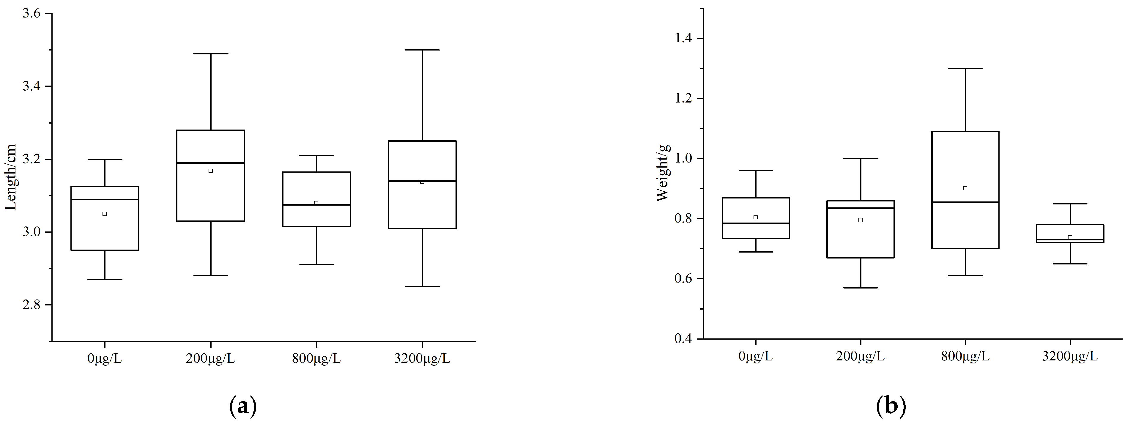
Prior to the experiment, the average body length of crucian carp was 3.03 cm, and their body length increased in each experimental group to 3.14 cm, 3.17 cm, 3.18 cm, and 3.15 cm for control, low, medium, and high exposure groups, respectively (Table 3), representing increases 0.95% (low), 1.27% (medium) and 0.31% (high), respectively, relative to the control group (Figure 5a).
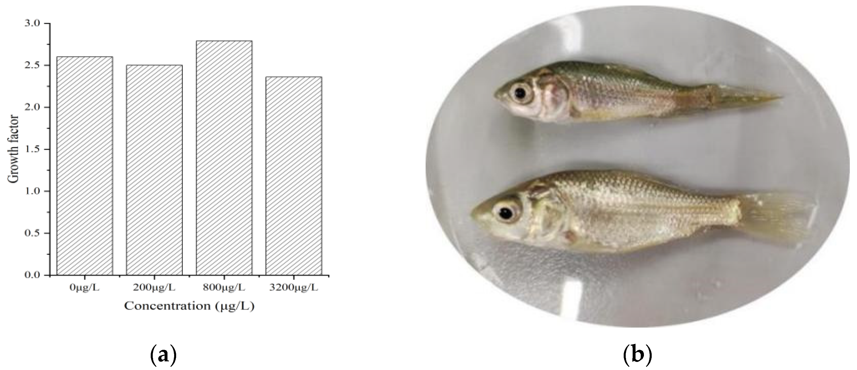
The average weight of crucian carp was 0.825 g before the exposure experiment to PS-MPs, and there was no significant difference in weight after exposure to PS-MPs compared to before the exposure experiment. However, compared with the control group, the average weight of crucian carp decreased by 1.25% and 7.5% in the low and high-concentration groups, respectively, and the average weight of the medium-concentration group increased by 12.5% (Figure 5b). The growth of crucian carp in the other concentration groups was not inhibited, except for the growth of crucian carp exposed in the medium concentration group (Figure 6a). The bodies of carp without microplastic exposure were mellow and full, the epidermis color was bright, and they appeared healthy. However, in the group exposed to high concentrations of PS-MPs, the epidermal color was dull, the body was wizened, and their appearance was pathological (Figure 6b).

3.3. Histopathological Analysis of GIT, Gill, Brain, and Liver

In the blank control group, the structure of the intestinal layers was normal, the mucosa of the intestinal tissue had developed a folded structure with an orderly arrangement, and peripheral tissues were smooth and stretched (Figure 7a). The gill, liver, and brain tissues were preserved intact, the cells were tightly arranged and regular, the structure was clear and normal, the nearby muscle tissue was smooth and stretched without abnormal pathological changes, and the cells presented a normal state; the nucleus was obviously located in the middle of the cells with a distinct boundary (Figure 8a, Figure 9a and Figure 10a).
In the low concentration group, compared with the control group, some epithelial cells were detached and lost, a few villi were divided, and the microvilli structure of small intestinal epithelial cells had some vacuole-like structures, but the overall structure was closely arranged and clearly outlined, with normal cytoplasmic distribution of cells (Figure 7a). The gills and brain cells exhibited an intact structure, orderly arrangement, and smooth nearby muscle tissue with no obvious abnormal pathological changes (Figure 8a and Figure 9a). Hepatocyte parts showed cell congestion, and the nuclei were off-center (Figure 10a). In comparison to the control group, there was a loss of partial epithelial cell detachment, shortened and partially split villi, and congestion in some cells in the medium concentration (Figure 7c). The pathological changes in the gill tissue showed lysis of the nucleus and lysis of a part of the peripheral structure of the gill filament (Figure 8c). In the brain tissue, some nuclei were off-center but with no serious pathological change (Figure 9c).
The intestinal lesions in the group exposed to a high concentration were the most serious, with a large number of epithelial cells shed and lost, a large number of divided and shortened villi, leukocyte infiltration and congestion, a scattered overall structural morphology, microvilli of small intestinal epithelial cells were damaged, exhibited a blurred outline, and a large number of cells were necrotic (Figure 7d). The gill cells were partially necrotic, lysed, and detached, with swollen and deformed cells, off-center nuclei, and a disorganized distribution of mucus cells and columnar cells (Figure 8d). Brain cells were loosely arranged, with a large number of off-center nuclei and vacuolated necrotic cells (Figure 9d). The hepatocytes in the medium and high-concentration groups showed different degrees of cellular congestion, off-center nuclei, and inflammatory pathologies, including vacuolation and swelling necrosis (Figure 10c,d).

3.4. Effect of Polystyrene Microplastics on Gene Expression

Different concentrations of PS-MPs had different effects on gene expression in the liver, GIT, and gill. Interleukin 1β (IL-1β) and S100A1 genes were up-regulated in the medium concentration and down-regulated at low and high exposure concentrations, as shown in Figure 11. The expression of the IFN-γ gene was up-regulated at low concentrations and down-regulated at medium and high exposure concentrations. The expression of the SAA gene was up-regulated at medium and high exposure concentrations but not significantly changed in the low concentration; IL-8 gene expression was up-regulated in all three experimental groups.
In intestinal tissues, IL-1βgene expression was down-regulated in the high concentration, with no significant changes in the remaining two concentrations. Interleukin-6 (IL-6) gene expression was significantly down-regulated in all three experimental groups (p < 0.05), and their expression levels were approximately the same. In the three experimental groups, the expression of interleukin-8 (IL-8) gene and TNFα gene was up-regulated with approximately the same expression levels. INF-γ gene was up-regulated in the L-GIT group compared with the control group, and there was no significant change in other concentration groups.
In liver tissue, the expression of the ChgH gene was significantly up-regulated in the low-concentration group (p < 0.05) and significantly down-regulated in medium and high-concentration groups (p < 0.05). The CYP1A gene was up-regulated in the medium concentration group, but there was no significant change in the remaining two concentrations. GSTa gene was significantly up-regulated in low and medium-concentration and down-regulated in high-concentration (p < 0.05) exposure groups. The GSTpi gene was down-regulated in low-concentration and up-regulated in medium and high-concentration exposure groups, and the expression level was approximately the same. The Vtg1 gene was up-regulated in the low-concentration group and significantly down-regulated in the medium and high-concentration exposure groups (p < 0.05).

4. Discussion

4.1. PS-MPs Distribution Enrichment

The presence of PS-MPs was only found in GIT and gill tissues because of the inability of PS-MPs of 50–500 μm particle size to enter the liver, muscle, and other tissues. This indicates that fish will retain microplastics in the digestive and respiratory systems through feeding. Microplastics enter the organism through feeding and respiration, and these microplastics are transferred and accumulated in different tissues [7,8]. The number of PS-MPs in the excrement of crucian carp in the present study increased with increased concentration of pollutants, indicating that the rate of uptake was directly proportional to the concentration of microplastics. In addition, crucian carp can expel microplastics through defecation, but during the period of exposure, microplastics will impose significant harm on its intestinal tract.

4.2. Growth

The research on the effect of microplastics on fish growth is mainly undertaken by evaluating body length and body weight, with growth parameters used to determine whether microplastics can affect fish growth [9,10,11]. Some studies found that microplastics did not significantly affect fish growth [9,12]. However, the present study found that PS-MP exposure had an effect on body weight and body length of juvenile crucian carp, whereby body length increased at medium exposure concentration group, while the growth rate decreased at low and high exposure concentrations, and body weight decreased significantly. Thus, exposure to medium concentrations of PS-MPs can promote growth because PS-MPs can act as an environmental fertilizer agent to regulate lipid metabolism, and this led to lipid accumulation, but as the concentration of PS-MPs increased, the growth of young carp was reduced. Zhang et al. demonstrated that exposure to 200 μm PS-MPs significantly increased fish body weight, adipocyte size, and hepatic lipid content associated with lipid metabolism [13]. PS-MPs can inhibit the growth of crucian fish because fish tend to eat microplastics similar in size to feed, and microplastics do not decompose in the body and remain in the digestive tract, resulting in gastrointestinal blockage and leading to a decline in feeding rate and eventually lead to growth inhibition and death [14]. In addition, crucian carp larvae eating microplastic will experience false satiety and consequently reduce food intake, but microplastics do not provide the nutrients needed for growth and development, affecting lipid metabolism and other metabolic processes and eventually leading to the inhibition of growth and development.

4.3. Histopathology

PS-MPs caused lesions in biological tissue, which were related to the size and concentration of MPs [15,16]. The results of this study showed that 50–500 μm PS-MPs caused serious damage to the intestine, and the severity of the lesions was significantly increased with increasing exposure dose. Dissection demonstrated congestion of the intestinal tract of the high-concentration group, and a large number of MPs had accumulated. Intestinal blockage was one of the main reasons for the serious lesions of intestinal tissue in the high-concentration group. The gill and liver tissues showed different degrees of damage, mainly characterized by an off-center nucleus, loose cell arrangement, cell vacuolation, congestion, and swelling, which were associated with the concentration of PS-MPs. Previous studies of microplastics on fish brain tissue found that microplastics can destroy the blood and brain barrier, leading to bleeding of cerebellar tissue and aggregation of a layer of red blood cells, forming micro-thrombosis and reduced neurotransmission of the cerebellum, and this damage was dependent on dose [17]. In contrast, the brain tissue cells of Carassius auratus in this experiment were less damaged than other tissues, and brain tissue damage was also dependent on the concentration of PS-MPs. Hamed demonstrated that kidney, liver, gill, intestine, muscle, and brain tissues all showed different degrees of damage in a concentration-dependent manner [18]. Our study found that the exposure of PS-MPs in the low concentration group did not produce significant damage to the tissues of crucian carp, but other tissues at medium and high exposure concentration groups had severe pathological changes, and the higher concentration of PS-MPs, the more severe the damage to tissues.

4.4. Gene Expression Analysis of Organization

4.4.1. Gene Expression Analysis of Gastrointestinal Tract

Transformation and detoxification of intracellular xenobiotics include stages I, II, and III, and some of the important enzymes involved in these stages include the cytochrome P450 family, glutathione-S-transferase, and the ABC transporter [19,20]. IL-1β and IL-6 are inflammatory factors that regulate the immune and nervous systems. The expression of IL-1β and IL-6 were down-regulated, which indicated that microplastic particles can induce oxidative stress and affect immune function in crucian carp. The decrease in expression was mainly due to the fact that PS-MPs affected the anti-bacterial ability of crucian carp and the gradual reduction of disease resistance. IL-8 and TNF-α are two kinds of genes related to tumor metastasis and anti-cancer cells, both of which were up-regulated, indicating that PS-MPs in the intestine may induce some cancers and through various types of cytokines to immunomodulate and kill cancer cells. Teles sought endotoxin upregulation of the cellular genes IL-1β, TNF-α, and IL-6, indicating that the toxin had a stimulatory effect on the pro-inflammatory process, leading to an increased inflammatory response [21].
IFN-γ is a pro-inflammatory cytokine, and this cytokine is primarily involved in lymphocyte immune responses to intracellular pathogens, including viruses and tumor control. This study also found that IFN-γ was only up-regulated and then downregulated at low exposure concentrations. Upregulation showed that inflammation occurred when crucian carp were exposed to a low concentration of PS-MPs. Downregulation occurred when the limit of its anti-bacterial ability was reached, and immune cells were reduced, indicating that crucian carp would become more inflamed with an increase in concentration.

4.4.2. Gene Expression Analysis of Liver

The liver is the main site for the synthesis of vitellogenin and ovalbumin in fish [22]. Long-term exposure to pollutants produces endocrine-disrupting effects on organisms through activation of hepatic sex hormone receptors regulating the expression of Chg and Vtg and peroxide proliferation-activated receptors [23]. Chg and Vtg genes were significantly expressed in the low concentration group but significantly down-regulated at the middle and high exposure concentrations, indicating that exposure to low concentrations of PS-MPs resulted in more vitellogenin and egg membrane proteogen and suggests that exposure to low concentrations of PS-MPs may increase steroid hormone biosynthesis and increase formation of estrogen receptor–ligand complexes, thereby increasing binding to Chg and Vtg estrogen-responsive elements, leading to increased transcription of Chg and Vtg.
Cytochrome P450 enzyme 1A (CYP1A) and glutathione S-transferase (GST) are important detoxification phase I and II enzymes of fish. GSTpi and GSTa have key roles in the metabolism of toxins and reduced glutathione. The expression of CYP1A, GSTa, and GSTpi genes were significantly up-regulated in the group exposed to medium concentrations, which indicated that PS-MPs had a toxic effect on crucian carp, and through detoxification enzymes to regulate the body to achieve balance. However, the expression of each gene was downregulated or did not change significantly in the group exposed to a high concentration, indicating that this exposure reached the limit value of the detoxification effect, and the fish entered a state where the impaired mechanism could not be recovered. Granby has previously proposed that in the first stage of bioaccumulation, the down-regulation of CYP1A and GSTa indicated an inhibition of detoxification [24]. The downregulation of CYP1A was associated with the presence of estrogen compounds, and the downregulation of GSTa in the present study indicated that oxidative stress occurred in crucian carp, consistent with the findings of Lu and Celander [25,26,27,28].

4.4.3. Gene Expression Analysis of Gill

In the inflammatory response, proinflammatory cytokine IL-1β acts to induce neutrophils to stimulate S100A1 generation, allowing leukocytes to infiltrate [29]. The slight up-regulation of IL-1β and S100A1 in the medium concentration group indicated that PS-MPs produced inflammation and triggered immune responses. However, IL-1β and S100A1 were down-regulated in the high-concentration group, indicating that the attachment of microplastic particles to the gills of crucian carp led to the reduction of their immune cells, mainly due to the age and sex differences of the fish [30]. The up-regulation of IFN-γ in the L-G group indicated that at low concentrations, gill tissue cells secreted proteins with various biological functions, including antiviral activity, which played a role in immune regulation. In addition, the expression pattern of Serum amyloid A (SAA) was significantly correlated with the immunohistochemical analysis of infected fry [31]. SAA was slightly up-regulated in the experimental groups, indicating that crucian carp would be immune reactive or acute when the body was infected with viruses and bacteria. In addition, IL-8 was up-regulated in all experimental groups and reached the maximum at medium exposure concentration, which represented the limit of its ability to resist pathogens.

5. Conclusions

In this study, PS-MPs were distributed and accumulated in crucian fish’s digestive and respiratory systems when exposed to microplastics, and this negatively affected the growth of juvenile crucian carp. At the cellular level, different concentrations of PS-MPs caused different degrees of histopathologic lesions in the brain, liver, intestine, and gill tissues of juvenile crucian carp. The higher the concentration of PS-MPs, the more serious the tissue cell damage. At the molecular level, different concentrations of PS-MPs had different effects on some selected genes, changing their levels of gene expression. In particular, in liver tissue, the Chg and Vtg genes were significantly expressed in the low-concentration group and significantly down-regulated in the medium- and high-concentration groups, in marked contrast to the trends for the other genes. In the natural environment, microplastics present various sizes, shapes, and concentrations and are associated with attached microorganisms (bacteria and viruses) and contaminants, which can lead to different degrees of physical damage to fish tissues at cellular and molecular levels. The toxic effects on fish in the natural environment may be more severe than revealed by indoor experiments. Therefore, later experiments will combine multifactorial variables from the field environment to explore the effects of microplastics on the physiology of fish.

Author Contributions

Conceptualization, D.W. and K.D.; methodology, W.L. (Wenjing Li) and D.W.; formal analysis, Y.H. and K.D.; investigation, K.D., W.L. (Wenrong Li) and D.W.; resources, K.D.; data curation, K.D. and Y.H.; writing—original draft, W.L. (Wenrong Li) and D.W.; writing—review and editing, K.D. and X.L.; visualization, Y.H. and K.D.; supervision, Y.H.; project administration, Y.H.; funding acquisition, K.D. All authors have read and agreed to the published version of the manuscript.

Funding

This research was funded by the Research funds of The Guangxi Key Laboratory of Theory and Technology for Environmental Pollution Control (guikeneng 2101Z013 and 2301Z003) and the Research Start-up Fund of Guilin University of Technology (GUQDJJ2001012).

Institutional Review Board Statement

All protocols in this study were approved by the Institutional Animal Care and Use Ethics Committee of Guilin University of Technology (GLUT-ACUEC-2024–9).

Data Availability Statement

The original contributions presented in the study are included in the article, further inquiries can be directed to the corresponding authors.

Conflicts of Interest

The authors declare no conflicts of interest.

References

  1. Zhao, X.; Xu, Q. Toxic effects of microplastics and nanoplastics on freshwater organisms. J. Jilin Norm. Univ. 2020, 41, 93–99. [Google Scholar] [CrossRef]
  2. Rafa, N.; Ahmed, B.; Zohora, F.; Bakya, J.; Ahmed, S.; Ahmed, S.F.; Mofijur, M.; Chowdhury, A.A.; Almomani, F. Microplastics as carriers of toxic pollutants: Source, transport, and toxicological effects. Environ. Pollut. 2024, 343, 123190. [Google Scholar] [CrossRef]
  3. Chen, Q.; Gundlach, M.; Yang, S.; Jiang, J.; Velki, M.; Yin, D.; Hollert, H. Quantitative investigation of the mechanisms of microplastics and nanoplastics toward zebrafish larvae locomotor activity. Sci. Total Environ. 2017, 584–585, 1022–1031. [Google Scholar] [CrossRef]
  4. Varó, I.; Osorio, K.; Estensoro, I.; Naya-Català, F.; Sitjà-Bobadilla, A.; Carlos Navarro, J.; Pérez-Sánchez, J.; Torreblanca, A.; Carla Piazzon, M. Effect of virgin low density polyethylene microplastic ingestion on intestinal histopathology and microbiota of gilthead sea bream. Aquaculture 2021, 545, 737245. [Google Scholar] [CrossRef]
  5. Hao, Y.; Sun, Y.; Li, M.; Fang, X.; Wang, Z.; Zuo, J.; Zhang, C. Adverse effects of polystyrene microplastics in the freshwater commercial fish, grass carp (Ctenopharyngodon idella): Emphasis on physiological response and intestinal microbiome. Sci. Total Environ. 2023, 856, 159270. [Google Scholar] [CrossRef]
  6. Hu, J.; Zuo, J.; Li, J.; Zhang, Y.; Ai, X.; Zhang, J.; Gong, D.; Sun, D. Effects of secondary polyethylene microplastic exposure on crucian (Carassius carassius) growth, liver damage, and gut microbiome composition. Sci. Total Environ. 2022, 802, 149736. [Google Scholar] [CrossRef]
  7. Jabeen, K.; Li, B.; Chen, Q.; Su, L.; Wu, C.; Hollert, H.; Shi, H. Effects of virgin microplastics on goldfish (Carassius auratus). Chemosphere 2018, 213, 323–332. [Google Scholar] [CrossRef]
  8. Lu, Y. Accumulation and distribution of microplastic particles with different characteristics in Zebrafish and related toxic effects. Master’s thesis, Nanjing University, Nanjing, China, 2017. [Google Scholar] [CrossRef]
  9. Feng, S.; Zeng, Y.; Cai, Z.; Wu, J.; Chan, L.; Zhu, J.; Zhou, J. Polystyrene microplastics alter the intestinal microbiota function and the hepatic metabolism status in marine medaka (Oryzias melastigma). Sci. Total Environ. 2021, 759, 143558. [Google Scholar] [CrossRef]
  10. Yin, L.; Chen, B.; Xia, B.; Shi, X.; Qu, K. Polystyrene microplastics alter the behavior, energy reserve and nutritional composition of marine jacopever (Sebastes schlegelii). J. Hazard. Mater. 2018, 360, 97–105. [Google Scholar] [CrossRef]
  11. Hu, J.; Zuo, J.; Li, J.; Zhang, Y.; Ai, X.; Gong, D.; Zhang, J.; Sun, D. Effects of microplastic exposure on crucian growth, liver damage, and gut microbiome composition. Environ. Sci. 2022, 43, 3664–3671. [Google Scholar] [CrossRef]
  12. Lemoine, C.; Kelleher, B.; Lagarde, R.; Northam, C.; Elebute, O.; Cassone, B. Transcriptional effects of polyethylene microplastics ingestion in developing zebrafish (Danio rerio). Environ. Pollut. 2018, 243, 591–600. [Google Scholar] [CrossRef]
  13. Zhang, X.; Wen, K.; Ding, D.; Liu, J.; Lei, Z.; Chen, X.; Ye, G.; Zhang, J.; Shen, H.; Yan, C.; et al. Size-dependent adverse effects of microplastics on intestinal microbiota and metabolic homeostasis in the marine medaka (Oryzias melastigma). Environ. Int. 2021, 151, 106452. [Google Scholar] [CrossRef]
  14. Horton, A.; Jürgens, M.; Lahive, E.; Bodegom, P.; Vijver, M. The influence of exposure and physiology on microplastic ingestion by the freshwater fish Rutilus rutilus (roach) in the River Thames, UK. Environ. Pollut. 2018, 236, 188–194. [Google Scholar] [CrossRef]
  15. Abarghouei, S.; Hedayati, A.; Raeisi, M.; Hadavand, B.; Rezaei, H.; Abed-Elmdoust, A. Size-dependent effects of microplastic on uptake, immune system, related gene expression and histopathology of goldfish (Carassius auratus). Chemosphere 2021, 276, 129977. [Google Scholar] [CrossRef]
  16. Yang, H.; Xiong, H.; Mi, K.; Xue, W.; Wei, W.; Zhang, Y. Toxicity comparison of nano-sized and micron-sized microplastics to Goldfish Carassius auratus Larvae. J. Hazard. Mater. 2020, 388, 122058. [Google Scholar] [CrossRef]
  17. Yin, K.; Wang, D.; Zhao, H.; Wang, Y.; Zhang, Y.; Liu, Y.; Li, B.; Xing, M. Polystyrene microplastics up-regulates liver glutamine and glutamate synthesis and promotes autophagy-dependent ferroptosis and apoptosis in the cerebellum through the liver-brain axis. Environ. Pollut. 2022, 307, 119449. [Google Scholar] [CrossRef]
  18. Hamed, M.; Soliman, H.; Badrey, A.; Osman, A. Microplastics induced histopathological lesions in some tissues of tilapia (Oreochromis niloticus) early juveniles. Tissue Cell 2021, 71, 101512. [Google Scholar] [CrossRef]
  19. Mohamed, H.; Child, S.; Doherty, D.; Bruning, J.; Bell, G. Structural determination and characterisation of the CYP105Q4 cytochrome P450 enzyme from Mycobacterium marinum. Arch. Biochem. Biophys. 2024, 754, 109950. [Google Scholar] [CrossRef]
  20. Hu, B.; Deng, L.; Wen, C.; Yang, X.; Pei, P.; Xie, Y.; Luo, S. Cloning, identification and functional characterization of a pi-class glutathione-S-transferase from the freshwater mussel Cristaria plicata. Fish Shellfish Immunol. 2012, 32, 51–60. [Google Scholar] [CrossRef]
  21. Teles, M.; Mackenzie, S.; Boltaña, S.; Callol, A.; Tort, T. Gene expression and TNF-alpha secretion profile in rainbow trout macrophages following exposures to copper and bacterial lipopolysaccharide. Fish Shellfish Immunol. 2011, 30, 340–346. [Google Scholar] [CrossRef]
  22. Wang, Q.; Huang, C.; Zhang, Z.; Hu, J.; Zhang, R. Gene expression in primary cultures of male medaka (Oryzias latipes)hepatocytes induced by 17β-estradiol with quantitative real-time RT-PCR. Acta. Sci. Circumstantiae 2008, 12, 2568–2572. [Google Scholar] [CrossRef]
  23. Ye, T.; Kang, M.; Huang, Q.; Dong, S. Endocrine-disrupting effects of DEHP and MEHP on Oryzias melastigma under long-term exposure. J. Ecotoxicol. 2014, 9, 253–260. [Google Scholar] [CrossRef]
  24. Granby, K.; Rainieri, S.; Rasmussen, R.; Kotterman, M.; Sloth, J.; Cederberg, T.; Barranco, A.; Antonio, M.; Larsen, B. The influence of microplastics and halogenated contaminants in feed on toxicokinetics and gene expression in European seabass (Dicentrarchus labrax). Environ. Res. 2018, 164, 430–443. [Google Scholar] [CrossRef]
  25. Celander, M.; Stegeman, J.; Förlin, L. CYP1A1-, CYP2B- and CYP3A-like proteins in rainbow trout (Oncorhynchus mykiss) liver: CYP1A1-specific down-regulation after prolonged exposure to PCB. Mar. Environ. Res. 1996, 42, 283–286. [Google Scholar] [CrossRef]
  26. Maradonna, F.; Nozzi, V.; Valle, L.; Traversi, I.; Gioacchini, G.; Benato, F.; Colletti, E.; Gallo, P.; Pisciottano, I.; Mita, D.; et al. Developmental hepatotoxicity study of dietary bisphenol A in Sparusaurata juveniles. Comp. Biochem. Physiol. C Toxicol. Pharmacol. 2014, 166, 1–13. [Google Scholar] [CrossRef]
  27. Romano, N.; Renukdas, N.; Fischer, H.; Shrivastava, J.; Baruah, K.; Egnew, N.; Sinha, A. Differential modulation of oxidative stress, antioxidant defense, histomorphology, ion-regulation and growth marker gene expression in goldfish (Carassius auratus) following exposure to different dose of virgin microplastics. Comp. Biochem. Physiol. C Toxicol. Pharmacol. 2020, 238, 108862. [Google Scholar] [CrossRef]
  28. Lu, Y.; Zhang, Y.; Deng, Y.; Jiang, W.; Zhao, Y.; Geng, J.; Ding, L.; Ren, H. Uptake and Accumulation of Polystyrene Microplastics in Zebrafish (Danio rerio) and Toxic Effects in Liver. Environ. Sci. Technol. 2016, 50, 4054–4060. [Google Scholar] [CrossRef]
  29. Cao, L.; Li, Y.; Liang, R.; Wang, Y.; Li, K. Effects of microplastic particles on immune gene expression of rainbow trout. Acta. Sci. Circumstantiae 2018, 38, 3347–3352. [Google Scholar] [CrossRef]
  30. Hanachi, P.; Kazemi, S.; Zivary, S.; Karbalaei, S.; Ghadami, S. The effect of polyethylene terephthalate and abamectin on oxidative damages and expression of vtg and cyp1a genes in juvenile zebrafish. Environ. Nanotechnol. Monit. Manag. 2021, 16, 100565. [Google Scholar] [CrossRef]
  31. Kania, P.; Chettri, J.; Buchmann, K. Characterization of serum amyloid A (SAA) in rainbow trout using a new monoclonal antibody. Fish Shellfish. Immunol. 2014, 40, 648–658. [Google Scholar] [CrossRef]
Figure 1. Distribution of the particle size range (50−500 μm) and morphology of the PS-MPs. (a) showed size map of PS-MPs detected by Malvin laser particle meter. (b) showed morphological electron microscope of 50–500 μm PS-MPs under a scanning electron microscope.
Fishes 09 00385 g001
Figure 2. Plot of PS-MPs in crucian carp sample gastrointestinal tract and gills.
Fishes 09 00385 g002
Figure 3. In−situ infrared spectrum of PS−MPs in fish organization and excreta. (a) shows the in situ infrared spectrum of PS-MPs in the gastrointestinal tract and gills, (b) shows the infrared spectrum of MPs in the excreta of low, medium and high concentration groups.
Fishes 09 00385 g003
Figure 4. Plot of PS-MPs in the crucian carp excreta in different concentrations, 0 μg/L (a), 200 μg/L (b), 800 μg/L (c), 3200 μg/L (d).
Fishes 09 00385 g004
Figure 5. Growth of crucian carp after exposure to different concentrations of microplastics, length (a), and weight (b).
Fishes 09 00385 g005
Figure 6. Pictures of growth factor of crucian carp (a) and Growth contras (b).
Fishes 09 00385 g006
Figure 7. Intestinal tract histological damage of crucian carp after 32 d of PS-MP exposure in different groups: (a) control; (b) low concentration; (c) medium concentration; (d) high concentration (red arrow indicated epithelial detachment and loss, green arrow indicated villus shortening, blue arrow indicated cell congestion, black arrow indicated leukocyte infiltration and orange arrow indicated villus division).
Fishes 09 00385 g007
Figure 8. Gill histological damage of crucian carp after 32 d of PS-MP exposure in different groups: (a) control; (b) low concentration; (c) medium concentration; (d) high concentration (red arrow indicated nuclear lysis, green arrow indicated cell lysis in the gill filament periphery, black arrow indicated off-center nuclei, orange indicated cell enlargement and deformation).
Fishes 09 00385 g008
Figure 9. Brain histology damage of crucian carp after 32 d of PS-MP exposure in different groups: (a) control; (b) low concentration; (c) medium concentration; (d) high concentration (red arrow indicated cell nucleus off-center).
Fishes 09 00385 g009
Figure 10. Liver histological changes of crucian carp after 32 d of PS-MP exposure in different groups: (a) control; (b) low concentration; (c) medium concentration; (d) high concentration (black arrows indicate cell congestion, red cells indicate cell vacuolization, green arrows indicate cell nucleus out of center, blue arrows indicate cell enlargement and necrosis).
Fishes 09 00385 g010
Figure 11. Gene sequence expression of the gill (a), GIT (b), and liver (c) tissue (* stand for p < 0.05).
Fishes 09 00385 g011
Table 1. Primers alignment applied for qPCR.
NamePrimersPrimer Sequence NumberGene NamePrimersPrimer Sequence Number
Interleukin 1β
(IL-1β)
F-primerCGACATGCATGACATCAAACInterleukin-6
(IL-6)
F-primerAGAAGTCTCTTAAAAAGGGG
R-primerGCAGCTCCTCATCACAAAACR-primerCAACAAAAAACATCTCTTCA
Interleukin-8
(IL-8)
F-primerGTCTTAGAGGACTGGGTGTATumor necrosis factor-α
(TNFα)
F-primerCAAAAACCCTGGACTGGAAA
R-primerACAGTGTGAGCTTGGAGGGAR-primerCCTGGCTGTAGACGAAGTAA
Interferon-γ
(INF-γ)
F-primerCACGTGAAAATTCAGCGAGAChoriogenin-H
(ChgH)
F-primerTTGTGGCACCACAATGAAGA
R-primerACAGGATGTGCATTGTGTAGR-primerTGGAGGAGGAACAGTGTTGA
Vitellogenin
(Vtg1)
F-primerTAGAGCTGGAATGGGAGAGGCytochrome P450 enzyme 1A
(CYP1A)
F-primerATTTCATTCCCAAAGACACCTG
R-primerTGACACTGTCATCTCTGGAAR-primerCAAAAACCAACACCTTCTCTCC
Glutathione transferase pi
(GSTpi)
F-primerATCTACCAGGAATATGAGACglutathione-S-transferase α
(GSTA)
F-primerCCCGAGAATATAAAACTCCC
R-primerCGGGCAGCAATCTTATCCACR-primerTCAAAAACACTTCCTCAAAC
Beta-actin
(β-actin)
F-primerACGAGAGATCTTCACTCCCCTS100A1F-primerGAGCTCAAGGACCTGATGGA
R-primerTGCCAACCATCACTCCCTGAR-primerTCCCCATCTTCTTCTTGTGC
Interferon-gamma
(IFN-γ)
F-primerAAGGGCTGTGATGTGTTTCTGSerum amyloid A
(SAA)
F-primerGGGAGATGATTCAGGGTTCCA
R-primerTGTACTGAGCGGCATTACTCCR-primerTTACGTCCCCAGTGGTTAGC
Table 2. The average number of microplastics from crucian carp in the digestion.
Fish SampleLength (cm)Weight (g)GIT (MPs/Item)Gill (MPs/Item)Muscle (MPs/Item)
0 μg/L2.330.2510000
200 μg/L2.570.31112.330.670
800 μg/L2.430.27971.000.170
3200 μg/L2.760.3841.330.670
Table 3. Crucian crap body length, weight, and growth factors after exposure.
ConcentrationAverage Length (cm)Average Weight (g)Growth Factor
0 μg/L3.140.802.60
200 μg/L3.170.792.50
800 μg/L3.180.902.79
3200 μg/L3.150.742.36
Disclaimer/Publisher’s Note: The statements, opinions and data contained in all publications are solely those of the individual author(s) and contributor(s) and not of MDPI and/or the editor(s). MDPI and/or the editor(s) disclaim responsibility for any injury to people or property resulting from any ideas, methods, instructions or products referred to in the content.