Article

Functional Adaptations of Hemocytes of Aplysia depilans (Gmelin, 1791) and Their Putative Role in Neuronal Regeneration

1
Department of Chemical, Biological, Pharmaceutical, and Environmental Sciences, University of Messina, 98166 Messina, Italy
2
Department of Clinical and Experimental Medicine, University of Messina, 98124 Messina, Italy
3
Department of Veterinary Sciences, University of Messina, 98168 Messina, Italy
*
Authors to whom correspondence should be addressed.
Fishes 2024, 9(1), 32; https://doi.org/10.3390/fishes9010032
Submission received: 23 November 2023 / Revised: 8 January 2024 / Accepted: 9 January 2024 / Published: 11 January 2024
(This article belongs to the Section Biology and Ecology)

Abstract

:
Invertebrates show great diversity in their responses to neural damage. Numerous invertebrate phyla, including gastropods, can replace all or a portion of their nervous systems. Aplysia species have been utilized extensively in toxicology, ecology, and neuroscience because their neurological systems react to bodily harm by releasing trophic substances that can stimulate non-neural tissue regeneration and induce changes in the nervous system. This study aims to assess the putative role of hemocytes of Aplysia depilans (Gmelin, 1791) by analyzing the presence of Toll-like receptor 2, inducible nitric oxide synthetase, and, in particular, vimentin and α-tubulin, molecules potentially implicated in the process of neural regeneration. The results demonstrate that all the aforementioned proteins are present in hemocytes, suggesting their role in the defense response and their possible contribution to the neuronal regeneration process of this gastropod. These data provide deeper insight into the internal defense system of this mollusk.
Key Contribution: Presence of neuronal molecules in the hemocytes and their putative role in the neuronal regeneration process of Aplysia depilans, using samples of integument.

1. Introduction

Physical injuries and neurodegenerative diseases often result in irreversible damage and the loss of function of the central nervous system (CNS) [1]. Repair of CNS injuries varies significantly in the animal kingdom [2]. At one extreme are amniotic vertebrates (reptile, bird, and mammal groups), which have a minimal capacity for neuronal replacement and, thus, neuronal regeneration; at the other extreme, animals such as planarians (flatworms) and colonial tunicates can repair the entire CNS after a significant injury. These differences can be attributed to the abundance of multipotent or pluripotent stem cells and undifferentiated precursors in the cell population [3].
The replacement of all or part of the nervous system has been documented in several invertebrate phyla, including coelenterates, platyhelminths, annelids, gastropods, and tunicates. Invertebrates show great diversity in their responses to neural damage. Some of these may suggest alternative strategies for repair that may apply to different parts of the complex mammalian nervous system, such as sensory receptor systems, peripheral nerves, neuromuscular function, or autonomic neural plexuses [4,5].
Invertebrates lack a specific defense system, like jawed vertebrates [6]. However, they possess an effective innate defense response, which uses biological mechanisms such as phagocytosis and chemical defenses such as producing oxygen and nitrogen free radicals. They also have mechanical–biological barriers such as integument and mucus and can secrete antibacterial substances [7]. Their internal defense system exploits both cellular components and humoral factors secreted by the blood cells themselves, such as agglutinins, cytokine-likes, lectins, antimicrobial peptides, lysins, phenoloxidases, and the metabolites of the prophenoloxidase cascade [8]. All these components work together with blood cells to defend the body against pathogens and other foreign substances.
Among invertebrates, adult neurogenesis has been analyzed mainly in arthropods, several species of insects, and decapod crustaceans [9]. It has been seen that hemocytes play a crucial role in neuronal regeneration, swiftly migrating to the site of damage, where they perform a dual role. First, hemocytes help to clear cellular debris and create a conducive environment for neural regeneration. They phagocytose damaged neural material and release factors that promote tissue healing. Second, hemocytes exhibit remarkable plasticity by differentiating into neural progenitor cells, contributing to the generation of new neurons and glial cells. This ability of functional adaptation is pivotal in reestablishing functional neural circuits [10]. The interaction between hemocytes and neural tissue in invertebrates offers a captivating insight into the regenerative potential of these animals [11,12]. Benton et al. (2014) showed that hemocytes can invade the neurogenic niche, and their offspring can differentiate into neurons. This suggests that the brains of crustaceans have “open” niches populated by blood-borne cells that can differentiate into neural progenitors [13].
In some groups, such as annelids, the CNS is efficiently and functionally regenerated following mechanical trauma [14,15], and the attraction of defense cells is a crucial step in activating an adaptive response leading to axonal sprouting. This might suggest that optimal regeneration requires microglia/macrophages to initiate CNS regeneration [14]. Another study showed that regeneration in the leech Hirudo medicinalis (Linnaeus, 1758) happened even when glial cells were destroyed by an intracellular injection of protease [16]. In the leech, it has been suggested that blood cells are essential in facilitating and accelerating the regeneration process. Moreover, circulating blood cells appeared to be able to infiltrate the injured CNS where, together with microglia, they stimulated reparation [17]. Da Silva suggests that molecules released in the acute phase of injury attract hemocytes, differentiating these cells into other defense cells or lineages [18,19].
This study aims to evaluate the role of hemocytes of Aplysia depilans (Gmelin, 1791) through the presence of molecules potentially involved in the neuronal regeneration process and to suggest a functional adaptation of these cells involved in damage repair.

2. Materials and Methods

2.1. Samples and Tissue Preparation

A. depilans samples from our laboratory histoteca were processed according to standard light microscopy techniques. Sections 4 μm thick, obtained by rotary microtome (LEICA 2065 Supercut, Nussloch, Germany), were placed on each slide. Then, the slides were deparaffinized in xylene and rehydrated using graded alcohol solutions ranging from 100% to 30% alcohol to distilled water.

2.2. Histology

For histomorphological and histochemical evaluation, Mallory trichrome staining (04-020802, BioOptica Milano S.p.A., Milan, Italy), Alcian Blue/Periodic Acid Schiff (AB/PAS, 04-163802, BioOptica Milano S.p.A., Milan, Italy), and May-Grünwald Giemsa (MGG, 04-081802, BioOptica Milano S.p.A., Milan, Italy) stainings were used. May-Grünwald Giemsa is the elective stain for blood cells [20,21,22]. To obtain photos, an Alexasoft TP3100A CMOS (Alexasoft; Firenze, Italy) digital camera was used.

2.3. Immunofluorescence

A 2.5% bovine serum albumin (BSA) solution was applied to the slides. Following an overnight incubation period with primary antibodies against iNOS, TLR2, α-tubulin, and vimentin, the sections were subsequently treated with secondary antibodies and Fluoromount (Sigma-Aldrich, St. Louis, MO, USA) was used to mount them to prevent photobleaching. To validate the specificity of immunolabeling, we performed a negative control procedure, omitting the primary antibodies during the immunofluorescence reaction. The samples were examined using a confocal laser scanning microscope equipped with a META module (Zeiss LSM DUO, Carl Zeiss MicroImaging GmbH, Jena, Germany). Two different types of lasers were used to examine the fluorescence samples: argon (458 nm) and helium-neon (543 nm). Zen 2011 (LSM 700 Zeiss software, Oberkochen, Germany) was used to enhance the pictures. We took as many pictures as we could to prevent photodegradation. The “Display profile” function of Zen 2011 was used to evaluate fluorescence intensity curves. Table 1 contains information about the antibodies.

2.4. Cell Sizes

Hemocytes sizes were measured using the software Alexasoft XEntry v1.0 (Alexasoft, Firenze, Italy) linked to an Alexasoft TP3100A CMOS (Alexasoft, Firenze, Italy) digital camera. Using the tool “linear distance”, the greatest length of the granular and agranular cells found in ten sections and twenty fields were measured. Then, using SigmaPlot version 14.0 (Systat Software, San Jose, CA, USA), means and standard deviations of the cell lengths were calculated.

2.5. Quantitative Analysis

Ten sections and twenty fields were inspected for each sample to gather information for the quantitative analysis. The cell positivity was assessed using ImageJ software 1.53e (NIH Software, Bethesda, MD, USA). After converting the image to 8-bit and removing the background using the “Threshold” filter, the number of cells was calculated using the “Analyze particles” plugin. The number of hemocytes that tested positive for iNOS, TLR2, α-tubulin, and vimentin in each field was assessed using SigmaPlot version 14.0 (Systat Software, San Jose, CA, USA). The mean values and standard deviations (SD) of the data are displayed.

3. Results

The epidermis of A. depilans appears as a simple monostratified columnar epithelium, as shown by Mallory staining (Figure 1). Among the epithelial cells, mucipar goblet cells with acid secretion are present, as revealed by AB/PAS histochemical staining (Figure 1). The epidermis rests on a large layer of loose connective tissue (the subcutis) that lays on a muscular layer. Compact and well-organized epidermal epithelial cells that possess a nucleus positioned basally are present. There are also solitary sensory cells, mucous cells, and supporting cells. A thin mucus layer, underlain by subepidermal mucous cells, covers the epidermis (Figure 1). Muscular fibers and collagen bundles are oriented orthogonally and randomly within the underlying connective tissue, representing the subcutis. Numerous hemocytes scattered throughout the subcutis and near the epidermis are evident. Two populations of hemocytes, distinguishable by size and staining affinity, were highlighted with MGG staining. Hemocytes appear in blue or pink, some granular and others with abundant cytoplasmic portions, lacking granularity (Figure 1).
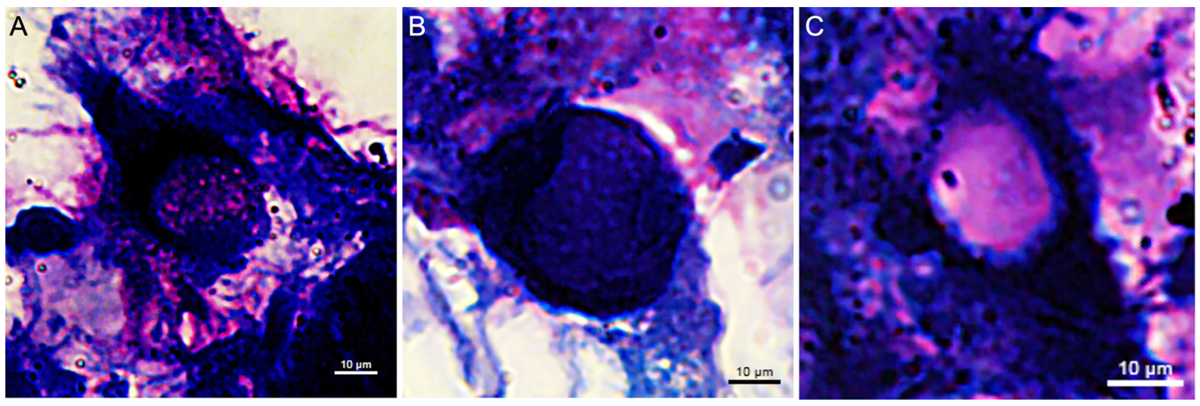
Based on the May-Grünwald Giemsa staining, two hemocyte populations, granular hemocytes (hg) and agranular hyalinocytes (ha), were distinguished by size and the presence of granules (Figure 2).
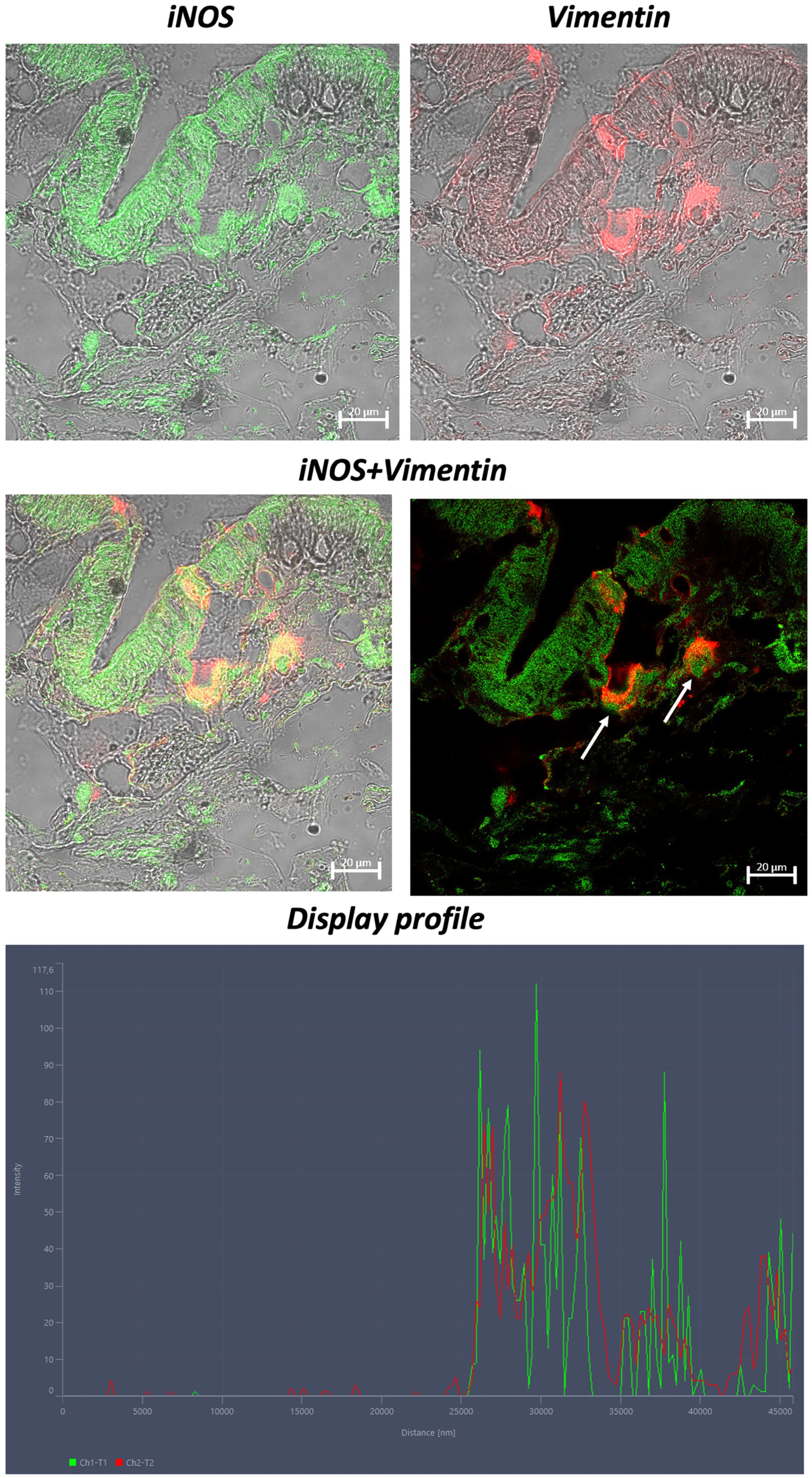
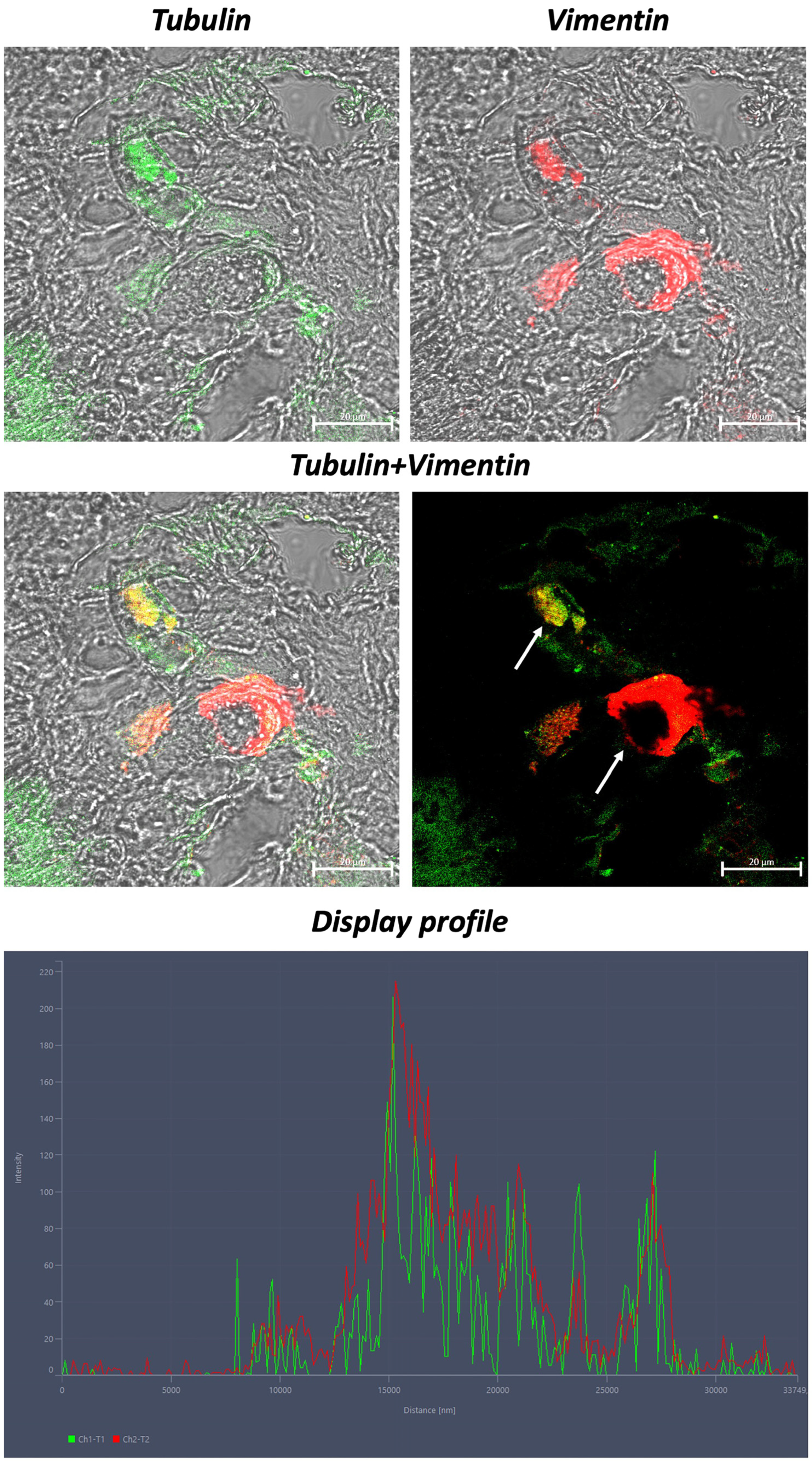
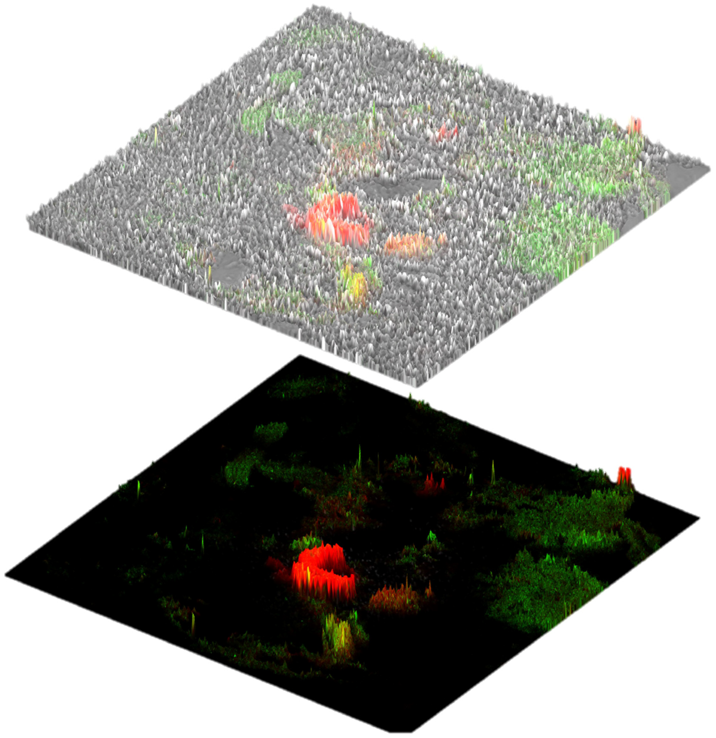
Immunohistochemically, our results show colocalization of TLR2 and iNOS (Figure 3), vimentin and iNOS (Figure 4), and vimentin and α-tubulin (Figure 5) in hemocytes. Not all hemocytes show co-localization of immunoreactivity signal. A high cross-reactivity was found, as shown by the “display profile” function of the confocal microscope (Figure 3, Figure 4 and Figure 5). Zen was also used to recreate the 2.5D graphical reconstruction. This program can directly translate the fluorescence intensity of each position into the height of the histogram and the scale bar color difference. This was performed to further confirm the immunoreactivity of hemocytes to vimentin and α-tubulin (Figure 6).
Quantitative analysis revealed a similar number of hemocytes immunoreactive to TLR2, iNOS, α-tubulin, and vimentin (Table 2).

4. Discussion

A. depilans, commonly named sea hare, is a gastropod mollusk in the Aplysiidae family. Its integument covers the entire soft tissue surface, including the head, foot, body, mantle, and external visceral surfaces [23]. The roles of the integument are to provide a protective barrier and facilitate respiration, locomotion, nutrient absorption, osmoregulation, the secretion of substances, and reproduction [24]. Mucus, defense materials, and shell components are typical secretions. Our histologic results showed that the epidermis of the sea hare is composed of a simple columnar or cuboidal epithelium. There were individual sensory cells, support cells, and mucous cells. The epidermis is covered with a thin layer of mucus supported by subepidermal mucous cells. According to previous investigations, vascular channels are distributed, and muscle fibers and collagen bundles are randomly and orthogonally arranged inside the dermis [25,26]. No true dermis exists, as in vertebrates, but a loose connective tissue representing the subcutis [27]. Several studies have identified hemocytes by histochemical techniques using May-Grünwald Giemsa, distinguishing different types of hemocytes based on size and the presence of granules [28,29,30,31,32,33,34]. We previously identified two populations of hemocytes in A. depilans, distinguished into granular and agranular (also called hyalinocytes), based on size and granulations, stained pink and blue [35]. Following these studies, we identified two types of hemocytes, larger granular, with granules in blue, as reported by Yu et al. (2016) [28], and smaller hyalinocytes in pink.
Aplysia species have been used extensively in toxicology, ecology, and neuroscience because of their bioactive molecules and chemical defenses. In neuroscience, these mollusks have been an important model organism for studying the functions of neurons due to the ease of identifying their nerve cells based on their location, size, and electrophysiological properties [36,37]. The nervous system of gastropods responds to body injury by releasing trophic factors that can promote non-neural tissue repair and cause changes in the nervous system. Despite their wide presence in ecosystems and their relevance as model animals, their internal defense system has only been studied to a limited extent. Our goal was to evaluate the role of hemocytes in neuronal regeneration in this gastropod.
Consistent with previous studies [35], hemocytes show a co-localization of immunoreactivity signals for TLR2 and iNOS. In previous studies, we demonstrated that the presence of different molecules may be related to a different cellular function [35,38]. In this study, not all hemocytes colocalized for the antibodies tested, suggesting a diversification between phagocytic hemocytes that bind TLR2 and cytotoxic hemocytes presenting only iNOS.
In the CNS, nitric oxide (NO) has been implicated both as a mediator of neurotoxicity and as a neuromodulator. The presence of both constitutive NO synthase (cNOS) and inducible NO synthase (iNOS) in neurons suggests that NO has several functions in the brain and supports the possibility that iNOS plays a role in neuronal damage and inflammation as a result of brain microglia activation and cytokine production [39]. Franchini et al. (1995) identified an immunoreactive nitric oxide synthetase (NOS)-like protein in Viviparus ater (De Cristofori & Jan, 1832) hemocytes [40]. Zahoor et al. showed increased NO levels in B. glabrata hemocytes affected by the parasite Schistosoma mansoni (Sambon, 1907) [41]. Hemocytes from resistant snails had a significantly greater increase in NO production 5 h after contact with the parasite [42]. NO is involved in numerous cell signaling events and, depending on the cellular environment, can promote cell survival or cell death. NO has been implicated in many other biological functions, including development and neurotransmission, immune response, feeding behavior, chemosensory activation, olfaction, and stress response. Also, iNOS is present in epithelial cells, where it is involved in tissue repair, as reported by previous studies [43,44,45,46]. Our investigation on confocal microscopy reveals that hemocytes of A. depilans are reactive to iNOS, indicating the neuro-immunomodulator role it plays in the internal defense mechanism of this gastropod.
Toll-like receptors (TLRs) have been recognized as key players in the innate immune response, implicated in recognizing a wide range of bacterial, fungal, and viral molecular patterns [47]. In many invertebrates, TLRs have been linked to pathogen detection and the subsequent production of immune effectors by activating nuclear factor k-activated B cell (NF-kB) signaling [48]. Genomic studies have also revealed that some invertebrates possess a vast repertoire of TLRs, as in the sea urchin S. purpuratus [49]. However, a similar number of membrane-bound receptors have been found in other deuterostomes and protostomes, including the Pacific oyster C. gigas [50]. In previous studies, we evaluated TLR2 presence in several metazoans: mollusks bivalves Polititapes aureus (Gmelin, 1791), Cerastoderma glaucum (Bruguière, 1789) [51], and Mytilus galloprovincialis (Lamarck, 1819) [52]; annelida Lumbricus terrestris (Linnaeus, 1758) [38]; protochordate tunicate Styela plicata (Lesuer, 1823) [53]; chordates cyclostomes Eptatretus cirrhatus (Forster, 1801) [54]; chondrichthyes Scyliorhinus canicula (Linnaeus, 1758) [55]; chordates osteichthyes Carassius auratus auratus (Linnaeus, 1758) [56], Polypterus senegalus (Cuvier, 1829), Lepisosteus oculatus (Winchell, 1864), Clarias batrachus (Linnaeus, 1758) [57], Periophthalmodon schlosseri (Pallas, 1770) [58], and Danio rerio (Hamilton, 1822) [59]; and in the marine mammal Stenella coeruleoalba (Meyen, 1833) [60]. TLR can be found in hemocytes more than in other tissues, and its transcription is significantly upregulated in bacterial and viral infections. It appears that mollusks maintain a TLR signaling cascade like vertebrates. Single cysteine cluster (scc) and multiple cysteine cluster (mcc) TLRs, two distinct groups of structurally dissimilar coexisting receptors, are present in both bivalves and gastropods [61]. However, it is recognized that TLRs play a critical role in mediating the molluscan immune response [62]. The mccTLRs have been linked to antibacterial and antiviral responses in the abalone Haliotis (Linnaeus, 1758) [63], and a TLR of the snail B. glabrata (which was found to be closely associated with samples resistant to trematode infections S. mansoni) [64].
Tubulin is a dimeric protein that helps to produce microtubules, which are essential intracellular structures that regulate critical activities such as growth and cell division [65], and it is highly conserved in metazoans [66]. Tubulin is a component of neurons in mollusks, as shown by Kaplan et al. (1992) in a study on Doryteuthis pealeii (Lesuer, 1821) [67]. Research by Nejatbakhsh et al. (2011) demonstrated its role in axonal growth and degeneration in L. stagnalis [68]. Tubulin forms microtubules in the form of subunit heterodimers of α- and β-tubulin. Tubulin isotypes are shown to be highly conserved among vertebrates. Interestingly, several cephalopods show these isotypes are comparable to those in vertebrates, both in structure and function, involved in neuronal growth [69,70]. A study by Moccia et al. (2003) identified, in one of the cytoskeletal mRNAs, the Aplysia homolog of α1-tubulin [70]. Moreover, the role of α-tubulin in the coelomocytes of L. terrestris was previously demonstrated [38]. Consistent with the reported data, our results showed hemocytes immunoreactive to α-tubulin, suggesting a probable involvement of these cells in neuronal repair mechanisms.
Vimentin has been identified in the cells and tissues of many organs. It has been described primarily in various cancers and diseases of the immune system in recent decades [71,72]. Several studies have focused mainly on the role of vimentin in immunity [73,74] and cellular functions [75,76], suggesting that this molecule exerts various effects on lesions and diseases of the nervous system. It has been demonstrated that vimentin is involved in the promotion/inhibition of axonal regeneration after spinal cord injury (SCI) in mice [77]. Vimentin is generally found in both gray and white matter in the spinal cord, and its levels are significantly increased after SCI, suggesting that vimentin may be involved in the pathogenesis of traumatic spinal cord injury. Early studies found that silencing vimentin and GFAP (glial fibrillary acidic protein) alleviates the excessive proliferation of reactive astrocytes and promotes the regeneration of supraspinal axons, leading to neural circuit reconstruction and locomotor functional rehabilitation [78,79,80]. In addition, although vimentin is a mainly intracellular cytoskeletal protein involved in the regulation of cellular stiffness, cell motility, and cytoplasmic organization, it can also appear within the extracellular matrix through secretion and on the surface of various cells, often in association with axonal plasticity, inflammation, and bacterial onset [81]. Vimentin released from reactive astrocytes and activated macrophages is considered a novel facilitator of axonal regeneration, and recombinant vimentin exerts neurotrophic effects by promoting axonal extension after SCI [82]. Recently, the novel functions of vimentin in neural stem cells (NSCs) were found. It has been shown that vimentin drives proteasomes (molecular scavengers capable of digesting targeted proteins) to clusters of damaged proteins that need to be removed for cells to function properly. Mice unable to produce vimentin had a reduced ability to create new neurons from stem cells at a young age, suggesting that vimentin is essential for keeping neuronal stem cells spry and productive during aging [83]. In this study, we showed the presence of vimentin in hemocytes; this could indicate a putative role of these cells in neurogenerative interactions. Cells of the blood lineage are associated not only with the functions that are usually attributed to them but also with neuroactive substances that induce other types of cells to differentiate into neural cells. Another possibility is that cells of the blood lineage are the ones that ultimately differentiate into neural cells, highlighting an important functional adaptation [10].
A 2022 in silico immunomic study by Kron showed, through the use of InterProScan and OrthoFinder, that the Aplysia californica (J.G. Cooper, 1863) genome encodes orthologs of all key components of the classical Toll-like receptor (TLR) signaling pathway, including TLR2 [84]. In addition, the study showed that this gastropod also preserves many nucleotide receptors and antiviral receptors that play a crucial role in vertebrate viral defense. In A. californica, 39 genes with Toll/interleukin-1 (TIR) domains and leucine-rich domains (LRRs) characteristic of TLRs were found [84]. Phylogenetic clustering of the TLR sequences of A. californica, Biomphalaria glabrata (Say, 1818), Crassosteraea gigas (Thunberg, 1793), Nematostella vectensis (Stephenson, 1935), Drosophila melanogaster (Meigen, 1830), Strongylocentrotus purpuratus (Stimpson, 1857), and humans revealed lineage-specific trends in the diversification of TLRs [84]. However, a number of free radical-generating enzymes have also been identified, including three dual oxidases (DUOX) and two associated assembly factors (DUOXA), seven nitric oxide synthase (NOS), five NADPH oxidase, and a peroxidase (loc100533347) [84]. A transcriptomics study (2018) demonstrated, via qPCR, that A. californica expresses genes encoding for iNOS [85]; also via qPCR, the expression of several genes in A. californica was evaluated using iNOS primers designed via a primer-BLAST tool (F: CCGCCGCTCTAATACTTA R: TTCATCAAGGAATTATACA) [86]. Bodnarova et al. (2005) demonstrated biochemically that functionally active NOS is expressed in A. californica, in all its isoforms; they also suggested that Aplysia NOS shares common enzymatic features with constitutive mammalian and insect NOS [87]. Perlson et al. (2005), through a proteomics study, showed that the intermediate filaments in Lymnea stagnalis (Linnaeus, 1758) were mostly homologous to the vertebrate vimentin expressed in developing neurons or nerve cells following damage [88].
In conclusion, our study on integument samples, a mechanical–biological barrier in contact with the external environment, where hemocytes may be more reactive, provides deeper insight into the internal defense system of A. depilans. As reported in the literature, hemocytes play a key role in the neuronal regeneration of several mollusks and invertebrates. Coherently, our study evaluated the putative neuro-regenerative role of these cells in A. depilans using vimentin and α-tubulin antibodies. Furthermore, the hemocytes examined confirmed their role in the defense response presenting TLR2 and iNOS. These data may suggest the potential involvement of these cells in the neuronal regeneration of this gastropod, supporting a morpho-functional adaptation of these cells. Although the antibodies tested in A. depilans have been validated, further studies are necessary to delve deeper into the data obtained to advance the understanding of the mechanisms of defense, repair, and neuronal regeneration of mollusks.

Author Contributions

Conceptualization, A.A.; formal analysis, A.A. and A.F.; investigation, A.A., A.F., L.M., S.M., A.M., and M.A.; data curation, A.A., A.F., L.M., S.M., A.M., and M.A.; writing—original draft preparation, A.A.; writing—review and editing, A.A. and M.A.; visualization, A.F.; supervision A.A. and M.A. All authors have read and agreed to the published version of the manuscript.

Funding

This research received no external funding.

Institutional Review Board Statement

Ethical review and approval were waived for this study because no experimental procedures were applied to samples. Moreover, the use of gastropod mollusks is not subjected to ethical restrictions.

Informed Consent Statement

Not applicable.

Data Availability Statement

Data are contained within the article.

Conflicts of Interest

The authors declare no conflicts of interest.

References

  1. Alesci, A.; Pergolizzi, S.; Lo Cascio, P.; Fumia, A.; Lauriano, E.R. Neuronal Regeneration: Vertebrates Comparative Overview and New Perspectives for Neurodegenerative Diseases. Acta Zool. 2022, 103, 129–140. [Google Scholar] [CrossRef]
  2. Brockes, J.P.; Kumar, A. Comparative Aspects of Animal Regeneration. Annu. Rev. Cell Dev. Biol. 2008, 24, 525–549. [Google Scholar] [CrossRef] [PubMed]
  3. Sinigaglia, C.; Averof, M. The Multifaceted Role of Nerves in Animal Regeneration. Curr. Opin. Genet. Dev. 2019, 57, 98–105. [Google Scholar] [CrossRef]
  4. Blundon, J.A.; Sheller, R.A.; Moehlenbruck, J.W.; Bittner, G.D. Effect of Temperature on Long-Term Survival of Anucleate Giant Axons in Crayfish and Goldfish. J. Comp. Neurol. 1990, 297, 377–391. [Google Scholar] [CrossRef]
  5. Krause, T.L.; Bittner, G.D. Rapid Morphological Fusion of Severed Myelinated Axons by Polyethylene Glycol. Proc. Natl. Acad. Sci. USA 1990, 87, 1471–1475. [Google Scholar] [CrossRef] [PubMed]
  6. Mokhtar, D.M.; Zaccone, G.; Alesci, A.; Kuciel, M.; Hussein, M.T.; Sayed, R.K.A. Main Components of Fish Immunity: An Overview of the Fish Immune System. Fishes 2023, 8, 93. [Google Scholar] [CrossRef]
  7. Beschin, A.; Bilej, M.; Torreele, E.; De Baetselier, P. On the Existence of Cytokines in Invertebrates. Cell. Mol. Life Sci. 2001, 58, 801–814. [Google Scholar] [CrossRef]
  8. Chiaramonte, M.; Russo, R. The Echinoderm Innate Humoral Immune Response. Ital. J. Zool. 2015, 82, 300–308. [Google Scholar] [CrossRef]
  9. Schmidt, M. The Olfactory Pathway of Decapod Crustaceans—An Invertebrate Model for Life-Long Neurogenesis. Chem. Senses 2007, 32, 365–384. [Google Scholar] [CrossRef]
  10. da Silva, P.C.; de Abreu, I.S.; Cavalcante, L.A.; De Barros, C.M.; Allodi, S. Role of Hemocytes in Invertebrate Adult Neurogenesis and Brain Repair. Invertebr. Surviv. J. 2015, 12, 142–154. [Google Scholar]
  11. De Abreu, I.S.; Wajsenzon, I.J.R.; Dias, J.C.; Allodi, S.; Monteiro-de-Barros, C. Central Nervous System Regeneration in Ascidians: Cell Migration and Differentiation. Cell Tissue Res. 2022, 390, 335–354. [Google Scholar] [CrossRef] [PubMed]
  12. Sardar, S.; Saha, N.; Ghosh, A. Microglia, the Sentinel of Brain in the Evolution of Nervous System from Invertebrate to Vertebrate: A Short Review. J. Sci. Res. 2022, 14, 685. [Google Scholar] [CrossRef]
  13. Benton, J.L.; Kery, R.; Li, J.; Noonin, C.; Söderhäll, I.; Beltz, B.S. Cells from the Immune System Generate Adult-Born Neurons in Crayfish. Dev. Cell 2014, 30, 322–333. [Google Scholar] [CrossRef] [PubMed]
  14. Baylor, D.A.; Nicholls, J.G. Patterns of Regeneration between Individual Nerve Cells in the Central Nervous System of the Leech. Nature 1971, 232, 268–270. [Google Scholar] [CrossRef] [PubMed]
  15. Jansen, J.K.S.; Nicholls, J.G. Regeneration and Changes in Synaptic Connections between Individual Nerve Cells in the Central Nervous System of the Leech. Proc. Natl. Acad. Sci. USA 1972, 69, 636–639. [Google Scholar] [CrossRef] [PubMed]
  16. Elliott, E.J.; Muller, K.J. Sprouting and Regeneration of Sensory Axons after Destruction of Ensheathing Glial Cells in the Leech Central Nervous System. J. Neurosci. 1983, 3, 1994–2006. [Google Scholar] [CrossRef]
  17. Boidin-Wichlacz, C.; Vergote, D.; Slomianny, C.; Jouy, N.; Salzet, M.; Tasiemski, A. Morphological and Functional Characterization of Leech Circulating Blood Cells: Role in Immunity and Neural Repair. Cell. Mol. Life Sci. 2012, 69, 1717–1731. [Google Scholar] [CrossRef]
  18. Chaves da Silva, P.G.; Corrêa, C.L.; de Carvalho, S.L.; Allodi, S. The Crustacean Central Nervous System in Focus: Subacute Neurodegeneration Induces a Specific Innate Immune Response. PLoS ONE 2013, 8, e80896. [Google Scholar] [CrossRef]
  19. Chaves-da-Silva, P.G.; De Barros, C.M.; Lima, F.R.S.; Biancalana, A.; Martinez, A.M.B.; Allodi, S. Identity of the Cells Recruited to a Lesion in the Central Nervous System of a Decapod Crustacean. Cell Tissue Res. 2010, 342, 179–189. [Google Scholar] [CrossRef]
  20. Shapiro, H.M.; Mandy, F. Cytometry in Malaria: Moving beyond Giemsa. Cytometry 2007, 71 Pt A, 643–645. [Google Scholar] [CrossRef]
  21. Woods, G.L.; Walker, D.H. Detection of Infection or Infectious Agents by Use of Cytologic and Histologic Stains. Clin. Microbiol. Rev. 1996, 9, 382–404. [Google Scholar] [CrossRef]
  22. Damsgaard, T.E.; Olesen, A.B.; Sørensen, F.B.; Thestrup-Pedersen, K.; Schiøtz, P.O. Mast Cells and Atopic Dermatitis. Stereological Quantification of Mast Cells in Atopic Dermatitis and Normal Human Skin. Arch. Dermatol. Res. 1997, 289, 256–260. [Google Scholar] [CrossRef]
  23. Alesci, A.; Pergolizzi, S.; Mokhtar, D.M.; Fumia, A.; Aragona, M.; Lombardo, G.P.; Messina, E.; D’Angelo, R.; Cascio, P.L.; Sayed, R.K. Morpho-Structural Adaptations of the Integument in Different Aquatic Organisms. Acta Histochem. 2023, 125, 152031. [Google Scholar] [CrossRef] [PubMed]
  24. LaDouceur, E.E.B.; Wynne, J.; Garner, M.M.; Nyaoke, A.; Keel, M.K. Lesions of Copper Toxicosis in Captive Marine Invertebrates with Comparisons to Normal Histology. Vet. Pathol. 2016, 53, 648–658. [Google Scholar] [CrossRef] [PubMed]
  25. Anderson, E.T.; Davis, A.S.; Law, J.M.; Lewbart, G.A.; Christian, L.S.; Harms, C.A. Gross and Histologic Evaluation of 5 Suture Materials in the Skin and Subcutaneous Tissue of the California Sea Hare (Aplysia Californica). J. Am. Assoc. Lab. Anim. Sci. 2010, 49, 64–68. [Google Scholar]
  26. Ponder, W.F.; Lindberg, D.R.; Ponder, J.M. Biology and Evolution of the Mollusca; CRC Press: Boca Raton, FL, USA, 2019; Volume 1. [Google Scholar]
  27. Cunha, C.M.; Simone, L.R.L. Morphological Re-Description of Aplysia Depilans (Gastropoda: Anaspidea): New Insights into the Anatomy of the Anaspideans. J. Mar. Biol. Assoc. UK 2019, 99, 595–610. [Google Scholar] [CrossRef]
  28. Yu, Y.; Cao, Y.; Xia, Y.; Liu, F. Wright–Giemsa Staining to Observe Phagocytes in Locusta Migratoria Infected with Metarhizium Acridum. J. Invertebr. Pathol. 2016, 139, 19–24. [Google Scholar] [CrossRef] [PubMed]
  29. Zhang, Z.; Shao, M.; Hokang, K. Classification of Haematopoietic Cells and Haemocytes in Chinese Prawn Fenneropenaeus Chinensis. Fish Shellfish Immunol. 2006, 21, 159–169. [Google Scholar] [CrossRef]
  30. Battison, A.; Cawthorn, R.; Horney, B. Classification of Homarus Americanus Hemocytes and the Use of Differential Hemocyte Counts in Lobsters Infected with Aerococcus Viridans Var. Homari (Gaffkemia). J. Invertebr. Pathol. 2003, 84, 177–197. [Google Scholar] [CrossRef]
  31. Cima, F.; Matozzo, V.; Marin, M.G.; Ballarin, L. Haemocytes of the Clam Tapes Philippinarum (Adams & Reeve, 1850): Morphofunctional Characterisation. Fish Shellfish Immunol. 2000, 10, 677–693. [Google Scholar] [CrossRef]
  32. Jeyachandran, S.; Park, K.; Kwak, I.; Baskaralingam, V. Morphological and Functional Characterization of Circulating Hemocytes Using Microscopy Techniques. Microsc. Res. Tech. 2020, 83, 736–743. [Google Scholar] [CrossRef] [PubMed]
  33. Vommaro, M.L.; Kurtz, J.; Giglio, A. Morphological Characterisation of Haemocytes in the Mealworm Beetle Tenebrio Molitor (Coleoptera, Tenebrionidae). Insects 2021, 12, 423. [Google Scholar] [CrossRef] [PubMed]
  34. Impellitteri, F.; Curpăn, A.-S.; Plăvan, G.; Ciobica, A.; Faggio, C. Hemocytes: A Useful Tool for Assessing the Toxicity of Microplastics, Heavy Metals, and Pesticides on Aquatic Invertebrates. Int. J. Environ. Res. Public Health 2022, 19, 16830. [Google Scholar] [CrossRef] [PubMed]
  35. Alesci, A.; Fumia, A.; Albano, M.; Messina, E.; D’Angelo, R.; Mangano, A.; Miller, A.; Spanò, N.; Savoca, S.; Capillo, G. Investigating the Internal System of Defense of Gastropoda Aplysia Depilans (Gmelin, 1791): Focus on Hemocytes. Fish Shellfish Immunol. 2023, 137, 108791. [Google Scholar] [CrossRef]
  36. Frazier, W.T.; Kandel, E.R.; Kupfermann, I.; Waziri, R.; Coggeshall, R.E. Morphological and Functional Properties of Identified Neurons in the Abdominal Ganglion of Aplysia Californica. J. Neurophysiol. 1967, 30, 1288–1351. [Google Scholar] [CrossRef]
  37. Kriegstein, A.R. Development of the Nervous System of Aplysia Californica. Proc. Natl. Acad. Sci. USA 1977, 74, 375–378. [Google Scholar] [CrossRef]
  38. Alesci, A.; Capillo, G.; Fumia, A.; Albano, M.; Messina, E.; Spanò, N.; Pergolizzi, S.; Lauriano, E.R. Coelomocytes of the Oligochaeta Earthworm Lumbricus Terrestris (Linnaeus, 1758) as Evolutionary Key of Defense: A Morphological Study. Zool. Lett. 2023, 9, 5. [Google Scholar] [CrossRef]
  39. Minc-Golomb, D.; Tsarfaty, I.; Schwartz, J.P. Expression of Inducible Nitric Oxide Synthase by Neurones Following Exposure to Endotoxin and Cytokine. Br. J. Pharmacol. 1994, 112, 720–722. [Google Scholar] [CrossRef]
  40. Franchini, A.; Conte, A.; Ottaviani, E. Nitric Oxide: An Ancestral Immunocyte Effector Molecule. Adv. Neuroimmunol. 1995, 5, 463–478. [Google Scholar] [CrossRef]
  41. Zahoor, Z.; Davies, A.J.; Kirk, R.S.; Rollinson, D.; Walker, A.J. Nitric Oxide Production by Biomphalaria Glabrata Haemocytes: Effects of Schistosoma Mansoni ESPs and Regulation through the Extracellular Signal-Regulated Kinase Pathway. Parasites Vectors 2009, 2, 18. [Google Scholar] [CrossRef]
  42. Wright, B.; Lacchini, A.H.; Davies, A.J.; Walker, A.J. Regulation of Nitric Oxide Production in Snail (Lymnaea Stagnalis) Defence Cells: A Role for PKC and ERK Signalling Pathways. Biol. Cell 2006, 98, 265–278. [Google Scholar] [CrossRef] [PubMed]
  43. Frank, S.; Kämpfer, H.; Wetzler, C.; Pfeilschifter, J. Nitric Oxide Drives Skin Repair: Novel Functions of an Established Mediator. Kidney Int. 2002, 61, 882–888. [Google Scholar] [CrossRef] [PubMed]
  44. Stallmeyer, B.; Kämpfer, H.; Kolb, N.; Pfeilschifter, J.; Frank, S. The Function of Nitric Oxide in Wound Repair: Inhibition of Inducible Nitric Oxide-Synthase Severely Impairs Wound Reepithelialization. J. Investig. Dermatol. 1999, 113, 1090–1098. [Google Scholar] [CrossRef] [PubMed]
  45. Cals-Grierson, M.-M.; Ormerod, A.D. Nitric Oxide Function in the Skin. Nitric Oxide 2004, 10, 179–193. [Google Scholar] [CrossRef]
  46. Mansfield, K.; Naik, S. Unraveling Immune-Epithelial Interactions in Skin Homeostasis and Injury. Yale J. Biol. Med. 2020, 93, 133–143. [Google Scholar] [PubMed]
  47. Alesci, A.; Lauriano, E.R.; Fumia, A.; Irrera, N.; Mastrantonio, E.; Vaccaro, M.; Gangemi, S.; Santini, A.; Cicero, N.; Pergolizzi, S. Relationship between Immune Cells, Depression, Stress, and Psoriasis: Could the Use of Natural Products Be Helpful? Molecules 2022, 27, 1953. [Google Scholar] [CrossRef]
  48. Zheng, L.; Zhang, L.; Lin, H.; McIntosh, M.T.; Malacrida, A.R. Toll-like Receptors in Invertebrate Innate Immunity. Invertebr. Surviv. J. 2005, 2, 105–113. [Google Scholar]
  49. Chou, S.-T.; Lin, T.-M.; Yang, H.-Y.; Fugmann, S.D. Functional Characterization of the MyD88 Homologs in Strongylocentrotus Purpuratus. Dev. Comp. Immunol. 2023, 139, 104580. [Google Scholar] [CrossRef]
  50. Zhang, L.; Li, L.; Zhang, G. A Crassostrea Gigas Toll-like Receptor and Comparative Analysis of TLR Pathway in Invertebrates. Fish Shellfish Immunol. 2011, 30, 653–660. [Google Scholar] [CrossRef]
  51. Alesci, A.; Albano, M.; Fumia, A.; Messina, E.; Miller, A.; Di Fresco, D.; de Oliveira Fernandes, J.M.; Spanò, N.; Savoca, S.; Capillo, G. Shell Formation in Two Species of Bivalves: The Role of Mantle Cells and Haemocytes. Zool. J. Linn. Soc. 2023, zlad099. [Google Scholar] [CrossRef]
  52. Alesci, A.; Di Paola, D.; Fumia, A.; Marino, S.; D’Iglio, C.; Famulari, S.; Albano, M.; Spanò, N.; Lauriano, E.R. Internal Defense System of Mytilus Galloprovincialis (Lamarck, 1819): Ecological Role of Hemocytes as Biomarkers for Thiacloprid and Benzo[a]Pyrene Pollution. Toxics 2023, 11, 731. [Google Scholar] [CrossRef] [PubMed]
  53. Alesci, A.; Pergolizzi, S.; Lo Cascio, P.; Capillo, G.; Lauriano, E.R. Localization of Vasoactive Intestinal Peptide and Toll-like Receptor 2 Immunoreactive Cells in Endostyle of Urochordate Styela Plicata (Lesueur, 1823). Microsc. Res. Tech. 2022, 85, 2651–2658. [Google Scholar] [CrossRef]
  54. Alesci, A.; Pergolizzi, S.; Savoca, S.; Fumia, A.; Mangano, A.; Albano, M.; Messina, E.; Aragona, M.; Lo Cascio, P.; Capillo, G. Detecting Intestinal Goblet Cells of the Broadgilled Hagfish Eptatretus Cirrhatus (Forster, 1801): A Confocal Microscopy Evaluation. Biology 2022, 11, 1366. [Google Scholar] [CrossRef] [PubMed]
  55. Lauriano, E.R.; Pergolizzi, S.; Aragona, M.; Montalbano, G.; Guerrera, M.C.; Crupi, R.; Faggio, C.; Capillo, G. Intestinal Immunity of Dogfish Scyliorhinus Canicula Spiral Valve: A Histochemical, Immunohistochemical and Confocal Study. Fish Shellfish Immunol. 2019, 87, 490–498. [Google Scholar] [CrossRef]
  56. Alesci, A.; Pergolizzi, S.; Fumia, A.; Calabrò, C.; Lo Cascio, P.; Lauriano, E.R. Mast Cells in Goldfish (Carassius Auratus) Gut: Immunohistochemical Characterization. Acta Zool. 2023, 104, 366–379. [Google Scholar] [CrossRef]
  57. Alesci, A.; Capillo, G.; Fumia, A.; Messina, E.; Albano, M.; Aragona, M.; Lo Cascio, P.; Spanò, N.; Pergolizzi, S.; Lauriano, E.R. Confocal Characterization of Intestinal Dendritic Cells from Myxines to Teleosts. Biology 2022, 11, 1045. [Google Scholar] [CrossRef] [PubMed]
  58. Alesci, A.; Capillo, G.; Mokhtar, D.M.; Fumia, A.; D’Angelo, R.; Lo Cascio, P.; Albano, M.; Guerrera, M.C.; Sayed, R.K.; Spanò, N. Expression of Antimicrobic Peptide Piscidin1 in Gills Mast Cells of Giant Mudskipper Periophthalmodon Schlosseri (Pallas, 1770). Int. J. Mol. Sci. 2022, 23, 13707. [Google Scholar] [CrossRef] [PubMed]
  59. Alesci, A.; Albano, M.; Savoca, S.; Mokhtar, D.M.; Fumia, A.; Aragona, M.; Lo Cascio, P.; Hussein, M.M.; Capillo, G.; Pergolizzi, S. Confocal Identification of Immune Molecules in Skin Club Cells of Zebrafish (Danio Rerio, Hamilton 1882) and Their Possible Role in Immunity. Biology 2022, 11, 1653. [Google Scholar] [CrossRef]
  60. Lauriano, E.R.; Silvestri, G.; Kuciel, M.; Żuwala, K.; Zaccone, D.; Palombieri, D.; Alesci, A.; Pergolizzi, S. Immunohistochemical Localization of Toll-like Receptor 2 in Skin Langerhans’ Cells of Striped Dolphin (Stenella Coeruleoalba). Tissue Cell 2014, 46, 113–121. [Google Scholar] [CrossRef]
  61. Gerdol, M.; Venier, P.; Pallavicini, A. The Genome of the Pacific Oyster Crassostrea Gigas Brings New Insights on the Massive Expansion of the C1q Gene Family in Bivalvia. Dev. Comp. Immunol. 2015, 49, 59–71. [Google Scholar] [CrossRef]
  62. Ren, Q.; Lan, J.-F.; Zhong, X.; Song, X.-J.; Ma, F.; Hui, K.-M.; Wang, W.; Yu, X.-Q.; Wang, J.-X. A Novel Toll like Receptor with Two TIR Domains (HcToll-2) Is Involved in Regulation of Antimicrobial Peptide Gene Expression of Hyriopsis Cumingii. Dev. Comp. Immunol. 2014, 45, 198–208. [Google Scholar] [CrossRef] [PubMed]
  63. Elvitigala, D.A.S.; Premachandra, H.K.A.; Whang, I.; Nam, B.-H.; Lee, J. Molecular Insights of the First Gastropod TLR Counterpart from Disk Abalone (Haliotis Discus Discus), Revealing Its Transcriptional Modulation under Pathogenic Stress. Fish Shellfish Immunol. 2013, 35, 334–342. [Google Scholar] [CrossRef]
  64. Pila, E.A.; Tarrabain, M.; Kabore, A.L.; Hanington, P.C. A Novel Toll-like Receptor (TLR) Influences Compatibility between the Gastropod Biomphalaria Glabrata, and the Digenean Trematode Schistosoma Mansoni. PLoS Pathog. 2016, 12, e1005513. [Google Scholar] [CrossRef] [PubMed]
  65. Breviario, D.; Gianì, S.; Morello, L. Multiple Tubulins: Evolutionary Aspects and Biological Implications. Plant J. 2013, 75, 202–218. [Google Scholar] [CrossRef]
  66. Breuss, M.W.; Leca, I.; Gstrein, T.; Hansen, A.H.; Keays, D.A. Tubulins and Brain Development—The Origins of Functional Specification. Mol. Cell. Neurosci. 2017, 84, 58–67. [Google Scholar] [CrossRef] [PubMed]
  67. Kaplan, B.B.; Gioio, A.E.; Capano, C.P.; Crispino, M.; Giuditta, A. β-Actin and β-Tubulin Are Components of a Heterogeneous mRNA Population Present in the Squid Giant Axon. Mol. Cell. Neurosci. 1992, 3, 133–144. [Google Scholar] [CrossRef]
  68. Nejatbakhsh, N.; Guo, C.-H.; Lu, T.Z.; Pei, L.; Smit, A.B.; Sun, H.-S.; Van Kesteren, R.E.; Feng, Z.-P. Caltubin, a Novel Molluscan Tubulin-Interacting Protein, Promotes Axonal Growth and Attenuates Axonal Degeneration of Rodent Neurons. J. Neurosci. 2011, 31, 15231–15244. [Google Scholar] [CrossRef]
  69. Ludueña, R.F. Possible Roles of Specific Amino Acids in β-Tubulin Isotypes in the Growth and Maintenance of Neurons: Novel Insights From Cephalopod Mollusks. Front. Mol. Neurosci. 2022, 15, 838393. [Google Scholar] [CrossRef]
  70. Moccia, R.; Chen, D.; Lyles, V.; Kapuya, E.; Yaping, E.; Kalachikov, S.; Spahn, C.M.T.; Frank, J.; Kandel, E.R.; Barad, M.; et al. An Unbiased cDNA Library Prepared from Isolated Aplysia Sensory Neuron Processes Is Enriched for Cytoskeletal and Translational mRNAs. J. Neurosci. 2003, 23, 9409–9417. [Google Scholar] [CrossRef]
  71. Kidd, M.E.; Shumaker, D.K.; Ridge, K.M. The Role of Vimentin Intermediate Filaments in the Progression of Lung Cancer. Am. J. Respir. Cell Mol. Biol. 2014, 50, 1–6. [Google Scholar] [CrossRef]
  72. Chen, Z.; Fang, Z.; Ma, J. Regulatory Mechanisms and Clinical Significance of Vimentin in Breast Cancer. Biomed. Pharmacother. 2021, 133, 111068. [Google Scholar] [CrossRef] [PubMed]
  73. Kuna, A.T. Mutated Citrullinated Vimentin Antibodies in Rheumatoid Arthritis. Clin. Chim. Acta 2012, 413, 66–73. [Google Scholar] [CrossRef] [PubMed]
  74. Musaelyan, A.; Lapin, S.; Nazarov, V.; Tkachenko, O.; Gilburd, B.; Mazing, A.; Mikhailova, L.; Shoenfeld, Y. Vimentin as Antigenic Target in Autoimmunity: A Comprehensive Review. Autoimmun. Rev. 2018, 17, 926–934. [Google Scholar] [CrossRef]
  75. Ivaska, J.; Pallari, H.-M.; Nevo, J.; Eriksson, J.E. Novel Functions of Vimentin in Cell Adhesion, Migration, and Signaling. Exp. Cell Res. 2007, 313, 2050–2062. [Google Scholar] [CrossRef]
  76. Patteson, A.E.; Vahabikashi, A.; Goldman, R.D.; Janmey, P.A. Mechanical and Non-Mechanical Functions of Filamentous and Non-Filamentous Vimentin. Bioessays 2020, 42, 2000078. [Google Scholar] [CrossRef] [PubMed]
  77. Ridge, K.M.; Eriksson, J.E.; Pekny, M.; Goldman, R.D. Roles of Vimentin in Health and Disease. Genes Dev. 2022, 36, 391–407. [Google Scholar] [CrossRef]
  78. Menet, V.; Prieto, M.; Privat, A.; Ribotta, M.G. y Axonal Plasticity and Functional Recovery after Spinal Cord Injury in Mice Deficient in Both Glial Fibrillary Acidic Protein and Vimentin Genes. Proc. Natl. Acad. Sci. USA 2003, 100, 8999–9004. [Google Scholar] [CrossRef] [PubMed]
  79. Toyooka, T.; Nawashiro, H.; Shinomiya, N.; Shima, K. Down-Regulation of Glial Fibrillary Acidic Protein and Vimentin by RNA Interference Improves Acute Urinary Dysfunction Associated with Spinal Cord Injury in Rats. J. Neurotrauma 2011, 28, 607–618. [Google Scholar] [CrossRef]
  80. Desclaux, M.; Perrin, F.E.; Do-Thi, A.; Prieto-Cappellini, M.; Gimenez, Y.; Ribotta, M.; Mallet, J.; Privat, A. Lentiviral-Mediated Silencing of Glial Fibrillary Acidic Protein and Vimentin Promotes Anatomical Plasticity and Functional Recovery after Spinal Cord Injury: Glial Scar Suppression in Mouse SCI. J. Neurosci. Res. 2015, 93, 43–55. [Google Scholar] [CrossRef]
  81. Ludin, B.; Matus, A. The Neuronal Cytoskeleton and Its Role in Axonal and Dendritic Plasticity. Hippocampus 1993, 3, 61–71. [Google Scholar] [CrossRef]
  82. Shigyo, M.; Tohda, C. Extracellular Vimentin Is a Novel Axonal Growth Facilitator for Functional Recovery in Spinal Cord-Injured Mice. Sci. Rep. 2016, 6, 28293. [Google Scholar] [CrossRef] [PubMed]
  83. Morrow, C.S.; Porter, T.J.; Xu, N.; Arndt, Z.P.; Ako-Asare, K.; Heo, H.J.; Thompson, E.A.; Moore, D.L. Vimentin Coordinates Protein Turnover at the Aggresome during Neural Stem Cell Quiescence Exit. Cell Stem Cell 2020, 26, 558–568. [Google Scholar] [CrossRef] [PubMed]
  84. Kron, N.S. In Search of the Aplysia Immunome: An In Silico Study. BMC Genom. 2022, 23, 543. [Google Scholar] [CrossRef] [PubMed]
  85. Greer, J.B.; Schmale, M.C.; Fieber, L.A. Whole-Transcriptome Changes in Gene Expression Accompany Aging of Sensory Neurons in Aplysia Californica. BMC Genom. 2018, 19, 529. [Google Scholar] [CrossRef] [PubMed]
  86. Pereira, R.B.; Pereira, D.M.; Jiménez, C.; Rodríguez, J.; Nieto, R.M.; Videira, R.A.; Silva, O.; Andrade, P.B.; Valentão, P. Anti-Inflammatory Effects of 5α,8α-Epidioxycholest-6-En-3β-Ol, a Steroidal Endoperoxide Isolated from Aplysia Depilans, Based on Bioguided Fractionation and NMR Analysis. Mar. Drugs 2019, 17, 330. [Google Scholar] [CrossRef]
  87. Bodnárová, M.; Martásek, P.; Moroz, L.L. Calcium/Calmodulin-Dependent Nitric Oxide Synthase Activity in the CNS of Aplysia Californica: Biochemical Characterization and Link to cGMP Pathways. J. Inorg. Biochem. 2005, 99, 922–928. [Google Scholar] [CrossRef]
  88. Perlson, E.; Hanz, S.; Ben-Yaakov, K.; Segal-Ruder, Y.; Seger, R.; Fainzilber, M. Vimentin-Dependent Spatial Translocation of an Activated MAP Kinase in Injured Nerve. Neuron 2005, 45, 715–726. [Google Scholar] [CrossRef]
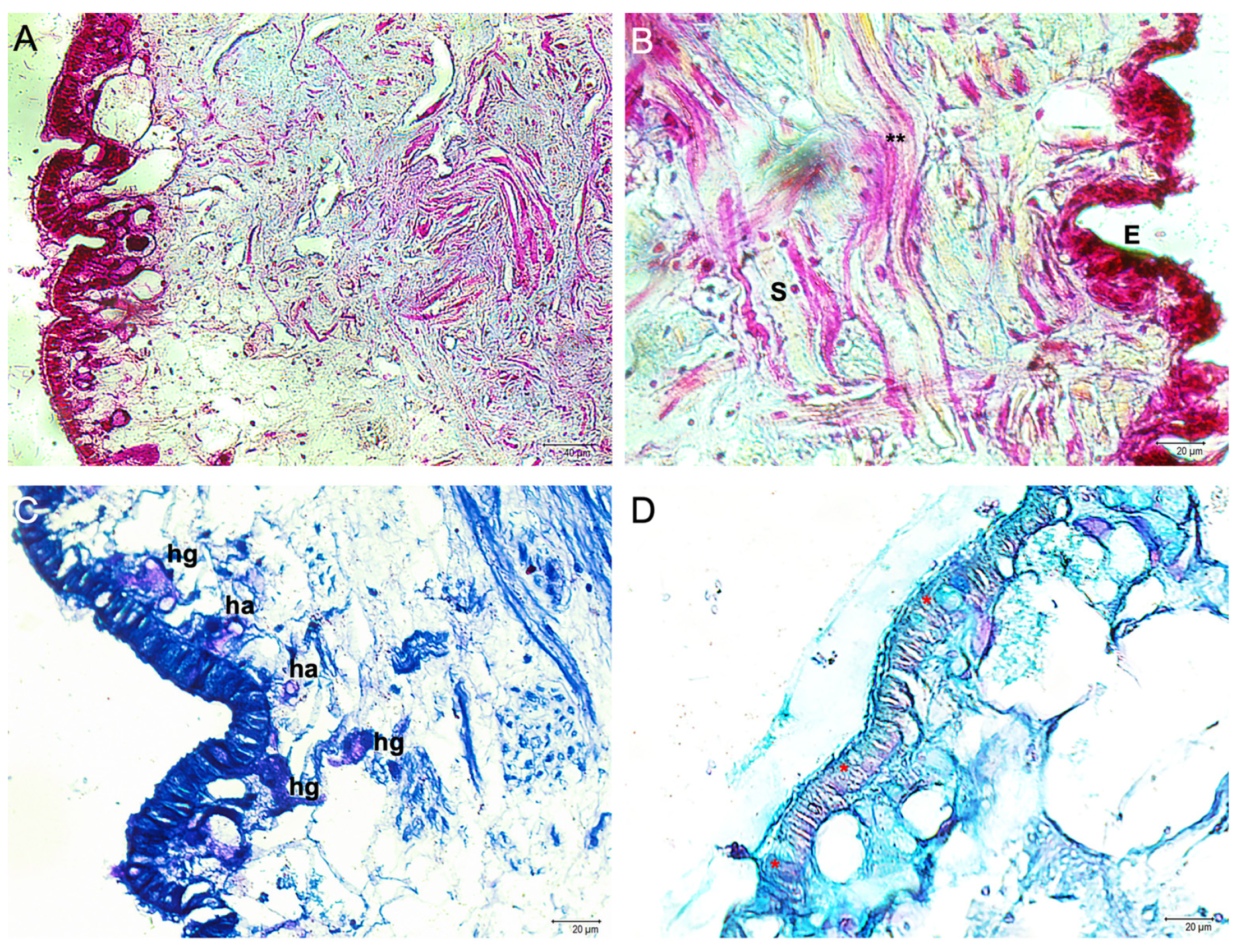
Figure 1. Integument sections of A. depilans. (A) Mallory trichrome, 20×, scale bar 40 μm. (B) Mallory trichrome, 40×, scale bar 20 μm. A simple, monolayered epithelium (E) with columnar epithelial cells is evident. Bundles of collagen and muscle fibers (**) are present in the underlying connective tissue (subcutis, S). (C) May-Grünwald Giemsa, 40×, scale bar 20 μm. Several granular (hg) and agranular hemocytes (ha), also called hyalinocytes, are evident near the epithelium and in the subcutis. (D) Alcian blue/PAS, 40×, scale bar 20 μm. In the columnar epithelium acid-secreting mucous cells (*) are present.
Fishes 09 00032 g001
Figure 2. Different types of hemocytes in the integument of A. depilans, May-Grünwald Giemsa, 100×, scale bar 10 μm. (A,B) Hemocyte with blue granules (mean cell length: 18.34 ± 1.91). (C) Hemocyte without granules (hyalinocyte) (mean cell length: 10.15 ± 0.41).
Fishes 09 00032 g002
Figure 3. Immunofluorescence on integument sections of A. depilans. Hemocytes immunoreactive for iNOS (green) and TLR2 (red) are evident (arrows). Some hemocytes bind only iNOS antibody. Yellow color shows the overlapping (presence) of both antibodies, indicating the colocalization of the iNOS (green) and TLR2 (red).
Fishes 09 00032 g003
Figure 4. Immunofluorescence on integument section of A. depilans. Hemocytes immunoreactive for iNOS (green) and vimentin (red) are evident (arrows). Some hemocytes bind only iNOS. Yellow color shows the overlapping (presence) of both antibodies indicating the colocalization of the iNOS (green) and vimentin (red).
Fishes 09 00032 g004
Figure 5. Immunofluorescence on integument section of A. depilans. Hemocytes (arrows) are immunoreactive for α-tubulin (green) and vimentin (red). Yellow color shows the overlapping (presence) of both antibodies indicating the colocalization of the tubulin (green) and vimentin (red).
Fishes 09 00032 g005
Figure 6. Graphical representation by “2.5D” confocal microscope function of hemocytes immunoreactive to vimentin and α-tubulin.
Fishes 09 00032 g006
Table 1. Data on the antibodies used in this research.
Primary AntibodySupplierCatalogue NumberSourceDilution
Anti-iNOSSanta Cruz Biotechnology, Inc., Dallas, TX, USAsc-7271Mouse1:200
Anti-TLR2Active Motif, La Hulpe, Belgium40981Rabbit1:100
Anti-α-TubulinSigma-Aldrich, Saint Louis, MO, USAT6793Mouse1:200
Anti-VimentinSigma-Aldrich, Saint Louis, MO, USASAB1305445Rabbit1:150
Secondary antibodySupplierCatalogue numberSourceDilution
Alexa Fluor 488 Donkey anti-Mouse IgG FITC conjugatedMolecular Probes, Invitrogen, Waltham, Massachusetts, USAA-21202Donkey1:300
Alexa Fluor 594 Donkey anti-Rabbit IgG TRITC conjugatedMolecular Probes, Invitrogen, Waltham, Massachusetts, USAA-21207Donkey1:300
Table 2. Quantitative analysis data (n = 3) *.
No. of Hemocytes per Slide
iNOS+347.83 ± 47.92
TLR2+387.32 ± 40.87
iNOS+TLR2+227.36 ± 25.36
Vimentin+325.28 ± 37.47
iNOS+Vimentin+233.47 ± 28.28
α-Tubulin+352.67 ± 38.11
α-Tubulin+Vimentin+216.30 ± 16.58
* Number of specimens utilized.
Disclaimer/Publisher’s Note: The statements, opinions and data contained in all publications are solely those of the individual author(s) and contributor(s) and not of MDPI and/or the editor(s). MDPI and/or the editor(s) disclaim responsibility for any injury to people or property resulting from any ideas, methods, instructions or products referred to in the content.