Article

Lysozyme Activity in the Hemolymph of Octopus vulgaris (Cuvier, 1797) Following Challenge with Gram-Negative Bacteria: Insights into Temperature-Driven Innate Immune Response

by
Daniella-Mari White
*,
Eleni Anastasiadou
,
Michail-Aggelos Valsamidis
and
Vasileios Bakopoulos
*
Department of Marine Sciences, School of the Environment, University of the Aegean, University Hill, 81100 Mytilene, Lesvos, Greece
*
Authors to whom correspondence should be addressed.
Fishes 2025, 10(9), 428; https://doi.org/10.3390/fishes10090428
Submission received: 22 July 2025 / Revised: 18 August 2025 / Accepted: 25 August 2025 / Published: 1 September 2025
(This article belongs to the Special Issue Advances in Pathology of Aquatic Animals)

Abstract

As aquaculture expands globally, understanding immune responses in non-traditional farmed species like Octopus vulgaris under varying environmental conditions is increasingly important. This study investigated lysozyme activity, a key innate immune marker, in cell-free hemolymph of O. vulgaris following experimental challenge with four Gram-negative fish pathogens (Photobacterium damselae subsp. piscicida, P. damselae subsp. damselae, Vibrio alginolyticus, and V. anguillarum O1) at two temperatures (21 ± 0.5 °C and 24 ± 0.5 °C). These pathogens were selected because octopus farming frequently occurs near fish aquaculture facilities, raising the potential for pathogen crossover. A total of 216 wild octopuses were injected intramuscularly or intravenously and sampled on days 0, 3, and 7 post-challenge. Lysozyme activity varied by pathogen, injection route, sampling time, and temperature. A significant time- and temperature-dependent increase was observed, especially in IM-challenged groups exposed to Photobacterium species. Elevated temperatures supported a more prolonged immune response. These results highlight lysozyme as a responsive biomarker of innate immunity in O. vulgaris and emphasize the role of environmental factors in immune modulation. This work provides a foundation for disease monitoring and health management in cephalopod aquaculture. Future research should examine long-term lysozyme dynamics, broader pathogen exposure, molecular mechanisms, and additional environmental stressors such as salinity and pollution.
Key Contribution: Lysozyme activity is a sensitive biomarker of immune response in Octopus vulgaris. Temperature and pathogen type significantly affect immune modulation in octopus.

Graphical Abstract

1. Introduction

Aquaculture is the fastest-growing sector of primary production worldwide. However, disease outbreaks remain a major constraint, posing significant risks to its expansion and negatively impacting the economy and socio-economic development in regions dependent on aquaculture and fisheries [1].
The common octopus, Octopus vulgaris Cuvier, 1797, is among the most commercially valuable cephalopods in terms of landings and market price [2]. In southern Europe, where octopus is a staple in traditional diets [3], production reached approximately 19 thousand metric tons in 2023 [4]. Over the past three decades, demand for O. vulgaris has increased dramatically [5], outpacing the availability of wild stocks and driving interest in sustainable farming practices. Consequently, research on the species’ physiology, health, and immune function has become increasingly relevant. Despite this, knowledge of cephalopod immune mechanisms remains limited [6].
Unlike vertebrates, cephalopods lack an adaptive immune system and immunological memory, relying solely on innate immune responses. This renders traditional disease prevention strategies, such as vaccination, ineffective [6]. Their immune defense is primarily mediated by hemocytes, which, as complex and multifunctional biological entities, play critical roles in phagocytosis, encapsulation, inflammation, and wound healing [7].
In addition to cellular mechanisms, humoral factors play a central role in the innate immune response. Among these, lysozyme is a key and evolutionarily conserved antimicrobial enzyme, capable of hydrolyzing the β-(1,4)-glycosidic bonds in bacterial peptidoglycan, leading to cell lysis. While historically associated with defense against Gram-positive bacteria, lysozyme can also act against Gram-negative species, particularly in synergy with other immune factors [8,9,10]. In cephalopods, lysozyme activity has been detected in cell-free hemolymph, establishing it as a useful biomarker of immune function [9,11].
Because hemolymph circulates systemically, measured lysozyme activity primarily reflects overall immune function, though localized responses at infection sites may also contribute [12,13]. This dual origin highlights the complexity of innate immune responses in cephalopods and other invertebrates.
In addition to O. vulgaris, lysozyme activity has been documented in other invertebrates, such as crustaceans (Litopenaeus vannamei) [14], mollusks (Octopus ocellatus) [13], and insects (Drosophila melanogaster) [15], as well as in vertebrates like fish [16,17,18]. It is a highly conserved component of innate immunity across diverse taxa, including many invertebrates such as mollusks, crustaceans, and insects [10], highlighting its evolutionary importance in species lacking adaptive immunity [13].
The intensification of aquaculture raises concerns about pathogen transmission between farmed species [19]. In particular, teleost fish farming in adjacent facilities may pose a threat to octopus mariculture via waterborne pathogen transfer [9,19,20]. Successful industrial farming of octopus requires understanding infectious diseases transmitted from wild and cultured populations [21,22]. Given the rising interest in commercial-scale O. vulgaris farming, understanding the species’ immune response to potential pathogens is essential for sustainable aquaculture development [21,22]. Since octopuses are often cultured near fish farms, bacterial pathogens affecting fish may also threaten octopus health [20]. The genera Photobacterium and Vibrio have been reported in the literature as being involved in infections of cephalopods [22]. Mortality in octopuses has been recorded following infections with Vibrio carchariae and Vibrio lentus. In addition, Vibrio alginolyticus was isolated from skin lesions in cultured Octopus vulgaris [23].
Temperature is a key environmental factor shaping immune function in mollusks, including cephalopods. In Octopus maya, individuals from warmer habitats (>27 °C) showed reduced hemocyte counts and hemocyanin concentrations but elevated phenoloxidase activity, while plasma lysozyme remained low—indicative of effector-specific thermal sensitivities [24,25]. Experimental infections in O. vulgaris demonstrated that phagocytosis rates vary with both temperature and pathogen species, revealing that modest thermal changes within culture ranges can significantly modulate cellular responses in a pathogen-specific manner [26,27]. Similar patterns occur in bivalves: in Ruditapes philippinarum, challenge with Vibrio tapetis at different temperatures (8–21 °C) significantly impacted disease progression, hemocyte counts, and lysozyme activity, with improved immune performance at higher temperatures (21 °C) associated with lower disease prevalence [28]. In Mytilus edulis, elevated temperature enhanced antibacterial activity of cell-free hemolymph, but lysozyme responses remained stable across 5–20 °C, highlighting that not all humoral components share the same thermal sensitivity [29]. These findings suggest that warming can differentially modulate humoral and cellular arms of immunity, with effects depending on the effector measured, the assay matrix, and the pathogen involved, factors directly relevant to interpreting lysozyme activity in O. vulgaris under Gram-negative challenge.
In this context, lysozyme activity offers a reliable biomarker for assessing the innate immune status of O. vulgaris under pathogenic challenge. This study evaluates lysozyme response in cell-free hemolymph following exposure to Gram-negative fish pathogens at different temperatures, providing insights into the temperature-dependent dynamics of octopus immunity and informing future strategies for health management in cephalopod aquaculture.

2. Materials and Methods

2.1. Subjects

Octopuses (O. vulgaris) of both sexes (n = 216) were collected alive by a fisherman using pots during the legally permitted fishing periods, in accordance with national fisheries legislation. Specimens were obtained from the southeastern waters of the Mytilene Strait, Lesvos Island, Greece, during the periods indicated in Table 1 [9,30,31].
Following collection, specimens were transferred to the wet laboratory facilities within 10 min in separate containers with continuous aeration to ensure adequate oxygenation throughout transport. The wet laboratory is part of the Laboratory of Ichthyology, Aquaculture and Aquatic Animal Health (ICHTHYAI) (Government Gazette Issue 1255/28-4-2016) [9,30,31].
ICHTHYAI holds all necessary permits for the production (EL83 BIObr01), supply (EL83 BIOsup01), and experimentation (E83 BioExp01) on aquatic organisms, in accordance with Presidential Decree 56/2013 and Directive 2010/63/EU (Decision No. 4053/14-3-2017 issued by the competent Regional Veterinary Authority). Additionally, specific permits were granted by the Directorate of Rural Affairs of North Aegean, Department of Agricultural Farms and Fishery, Decentralized Administration of Aegean (permits 29623/19-5-2017, 51948/5-9-2018, and 20564/11-4-2019) to cover the needs of this experimental study.
A total of 34 females and 182 males were used in the study, with weights ranging from 0.450 to 1.115 kg (mean ± SD: 0.725 ± 0.137 kg). Due to the wild origin of the specimens, no separation based on sex or weight was performed. Therefore, the experimental groups were formed randomly, based on availability at the time of collection. The experimental challenge protocol was approved by Decision No. 5379/4-4-2017 of the competent Regional Veterinary Authority [9,30,31,32].

2.2. Aquaria Systems

All specimens were maintained in a recirculating, UV-treated seawater tank system at ICHTHYAI and acclimatized for 10 days [9,30,31,32]. They were fed once daily with defrosted or fresh crabs and teleost fish at a rate of 5% of their biomass [8,33,34,35]. Water temperature was controlled at either 21 ± 0.5 °C or 24 ± 0.5 °C, depending on experimental requirements. Aeration was provided by air pumps, and salinity was maintained between 36–39‰. Ammonia, nitrite, and nitrate levels were monitored daily [9,30,31,32]. The selected temperature points (21 ± 0.5 °C and 24 ± 0.5 °C) are based on current sea temperature averages in the Mytilene Strait region [36] and projected increases due to climate change, with estimates of a rise between 1.8 °C and 3.5 °C by 2100 in the Eastern Mediterranean [37]. This design allows the simulation of both present and near-future conditions. Moreover, both temperatures fall within the thermal tolerance range of octopods (13–28 °C) and are close to their optimum for metabolic efficiency (~20 °C) [38,39], ensuring ecological relevance and biological validity of the experimental setup. The animals were maintained under strictly controlled conditions, using sterilized seawater and equipment to minimize external contamination and environmental stressors.

2.3. Bacteria, Media, and Culture Conditions

For experimental purposes, four fish pathogenic bacterial species were used: Photobacterium damselae subsp. piscicida (Phdp hereafter), Photobacterium damselae subsp. damselae (Phdd hereafter), Vibrio alginolyticus (VA hereafter), and Vibrio anguillarum O1 (VAO1, hereafter). Pathogen isolates were stored at −85 °C using the Microbead Cryoprotect® system (Protect Microorganism Preservation System; Technical Service Consultants Ltd., Microbiology House, Fir Street, Heywood, Lancashire OL10 1NW, UK). To revive the stored bacteria, a microbead was added to culture medium [40] composed of Brain Heart Infusion (BHI) agar (HiMedia Laboratories GmbH, Modautal, Germany) supplemented with 2% NaCl and incubated for at least 48 h at room temperature with mild shaking. Then, a colony of each pathogen was inoculated into BHI broth supplemented with 2% NaCl (Sigma-Aldrich, St. Louis, MO, USA) and incubated for 48 h at either 21 ± 0.5 °C or 24 ± 0.5 °C with mild shaking. The cultures were then centrifuged at 1750× g for 45 min at 4 °C (Thermo Fisher Scientific; Heraeus Megafuge 16R; Centrifuge Thermo Electron LED, GmbH, Zweigniederlassung Osterode Am Kalkberg, 37520 Osterode Am Harz Germany) to pellet the bacterial cells. The supernatants were discarded, and bacterial cell suspensions were prepared in sterile 2% NaCl at an optical density (OD) of ~1 at 605 nm (Hitachi, U-2900 Spectrometer, Hitachi High-Tech Science Corporation, Minato-ku, Tokyo 105-8717, Japan), corresponding to approximately 109 bacterial cells/mL, as previously determined by plate count assay [9].

2.4. Challenge/Injections of Octopods

The octopods were challenged either intramuscularly (IM, hereafter) at the second right arm or intravenously (IV, hereafter) at the right branchial heart ventricle with injection volume 100 μL; of a bacterial suspension containing 109 cells/mL (Phdp, Phdd, VA, or VAO1), or with 100 μL of sterile 2% NaCl as negative controls via the same routes [41]. Pathogen injection challenge was chosen as the infection method to ensure accurate dosing and to simulate either an injury scenario (IM infection) or systemic pathogen presence (IV infection) [9,30,31,32]. For clarity, the following abbreviations were used: I for challenged specimens; C for control specimens; M for IM injected; and V for IV injected. The days post challenge/injection were denoted as D0, D3, and D7. Three different octopods were used for each challenge/injection route and sampling point, totaling 27 octopods per temperature and bacterial treatment. Specimens were monitored daily for behavioral changes, skin lesions, food intake, and mortality [9]. D0 group samples were collected from untreated, healthy octopuses prior to any injection, serving as true baseline controls. Different individuals were sampled at each time point (D0, D3, D7) to avoid handling stress and ensure independent measurements. Hemolymph was collected from three animals at D0, and from 12 per group at D3 and D7. Only visibly healthy specimens were included, following established protocols to minimize individual variation and experimental bias [9,30,31,32].

2.5. Hemolymph Sampling

Hemolymph was collected from specimens using syringes pre-filled with Alsever’s solution (0.50 g/L (2.6 mM) Citric Acid (Alfa Aesar, Ward Hill, MA, USA), 4.2 g/L (0.72 M) Sodium Chloride (Penta, Chrudim, Czech Republic), 7.98 g/L (27 mM) Tri-Sodium Citrate 2aq (Chem-Lab, Zedelgem, Belgium), 20.5 g/L (113.8 mM) D-glucose (Merck, Darmstadt, Germany), pH: 6.5–6.9. as an anticoagulant on days 0, 3, and 7 post-challenge. Following collection, the hemolymph was transferred to sterile glass vials and stored at 4 °C. Cell-free hemolymph was then obtained by centrifugation at 750× g for 3 min at 4 °C (Thermo Scientific™ Pico™ 21 Microcentrifuge; Cat. No. 75002477; Thermo Fisher Scientific Inc., Waltham, Massachusetts, USA), aliquoted, and stored at −85 °C until analysis [9,30,31,32].
Octopuses were anesthetized by immersion in 5% magnesium chloride (Chem-Lab, Zedelgem, Belgium) seawater solution (pH 7.5–8) for 9–12 min under aeration before handling and hemolymph sampling, which were both performed according to previously published protocols [9,30,31,32]. Non-lethal hemolymph sampling was performed on all animals except those sampled on day 7, which were euthanized for tissue collection [9]; surviving octopuses were monitored for 15 days and then released back into their natural habitat in accordance with local regulations and animal welfare guidelines [9,31].
Individual hemolymph samples were collected separately from each octopus specimen to capture biological variability among wild-caught animals. Three biological replicates per treatment and time point were analyzed. Sampling began after a stabilization period of more than ten days to ensure animal acclimation and minimize temporal bias. This approach allowed us to preserve individual immune response differences, avoiding the masking effects of sample pooling, and provided a robust basis for statistical analysis.

2.6. Lysozyme Activity (Test)

Lysozyme activity was quantified by assessing the enzyme’s capacity to hydrolyze the bacterial cell wall of Micrococcus lysodeikticus following the protocol of Ellis [42] with minor modifications. Briefly, in a 96-well microtiter plate, 100 μL aliquots of hemolymph, subjected to three twofold serial dilutions in phosphate buffer (0.05 M, pH 6.2), (PBS was prepared using NaH2PO4·2H2O (Sigma-Aldrich, St. Louis, MO, USA), Na2HPO4·2H2O (Merck, Darmstadt, Germany), and NaCl (Sigma-Aldrich, St. Louis, MO, USA), were combined with an equal volume (100 μL) of a 0.4 mg/mL suspension of M. lysodeikticus (Sigma-Aldrich, St. Louis, MO, USA) in the same buffer. The reaction mixtures were incubated at 30 °C, and the optical density at 450 nm (OD450) was measured immediately (time 0) and after 5 min of incubation. Hen egg white lysozyme (HEW) (HEW; Sigma-Aldrich, St. Louis, MO, USA) served as a positive control, prepared in serial dilutions starting at 1.6 mg/mL in phosphate-buffered saline (PBS), while phosphate buffer alone was used as a negative control in place of hemolymph.
Lysozyme activity was expressed in units per milliliter (Units/mL), defined as the amount of enzyme; one unit of enzyme causes a decrease in absorbance of 0.001 per minute at 450 nm [43]. The process can be represented as [44]:
M. lysodeikticus cells → (Lysozyme) → M. lysodeikticus cells (lysed)
The following equation was applied for activity calculation [43]:
Lysozyme activity (units/mL) = (ΔA450 × df × 0.1)/0.001
where
  • ΔA450 is the change in absorbance between 0 and 5 min;
  • df represents the dilution factor;
  • (mL) corresponds to the volume of the M. lysodeikticus (0.4 mg/mL) suspension in the reaction mixture;
  • Corresponds to the absorbance decrease defined as one Unit of lysozyme activity (one unit of lysozyme causes a decrease in absorbance ΔA450 of 0.001/min).
This method provides a reliable quantitative measure of lysozyme enzymatic activity in hemolymph [42].

2.7. Statistical Analysis

All measurements were performed in triplicate. Data were assessed for normality using the Kolmogorov–Smirnov test and Shapiro–Wilk tests, as the latter is particularly suitable for small sample sizes [45]. The results of both tests were consistent, confirming the robustness of the normality assessment. Data analysis and graphing were conducted in IBM SPSS Statistics v20. Comparisons were made across the following:
(i)
Temperatures for the same bacterium, day, and injection route;
(ii)
Time points for the same bacterium, route, and temperature;
(iii)
Challenged vs. control (non-challenged) specimens at the same route, day, and temperature.
Results are reported as mean ± standard deviation (text), and as mean ± standard error (graphic). A p-value ≤ 0.05 was considered statistically significant.

3. Results

No mortality was recorded during the experimental challenges with any of the tested pathogens (Phdp, Phdd, VA, or VAO1). However, certain behavioral alterations were observed, including reduced appetite, lethargy, and hyperpnea in some individuals. Approximately 25% of octopods intramuscularly (IM) challenged with Vibrio species exhibited transient discoloration, characterized by a pale grey tone, at the injection site, lasting for 1 to 2 h post-injection. Moreover, self-amputation of the injected arm was observed in 8.3% of IM-injected individuals following Vibrio exposure [9,30,31,32].

3.1. Lysozyme Activity (Results)

3.1.1. Challenge with Phdp

Cell-free hemolymph lysozyme activity was detected in all samples and is illustrated in Figure 1a,b at 21 ± 0.5 °C και 24 ± 0.5 °C, respectively. Baseline lysozyme levels in D0 controls were calculated at 43.99 ± 12.42 and 79.33 ± 16.61 Units/mL at 21 ± 0.5 °C and 24 ± 0.5 °C, respectively.
At 21 ± 0.5 °C (Figure 1a), lysozyme activity in the CIM control group showed a significant decrease over time, whereas activity levels in the CIV group remained stable between D3 and D7. Conversely, at 24 ± 0.5 °C (Figure 1b), lysozyme activity remained constant in the CIM group, while a significant decline was observed in the CIV group between D3 and D7 (Supplementary Table S1a).
In the challenged groups at 21 ± 0.5 °C, lysozyme activity decreased significantly over time in the IM group, whereas it remained relatively stable in the IV group. Importantly, both challenged groups exhibited significantly higher lysozyme activity compared to their respective control groups (Supplementary Table S1b).
At 24 ± 0.5 °C (Figure 1b), lysozyme activity in the IM group showed a slight decreasing trend from D3 to D7, though not statistically significant. In contrast, the IV group demonstrated a significant reduction over the same period.
A significant shift in lysozyme activity was observed between D0 and D3 in the control groups at 21 ± 0.5 °C, and in the IV control and challenged groups at 24 ± 0.5 °C, with notably lower activity levels at D3. In contrast, lysozyme activity remained stable in the challenged groups at 21 ± 0.5 °C and in the IM control and challenged groups at 24 ± 0.5 °C.
Finally, a comparison of challenged specimens between temperatures on D7 indicated that temperature influenced lysozyme activity, depending on the route of challenge (Supplementary Table S1d).

3.1.2. Challenge with Phdd

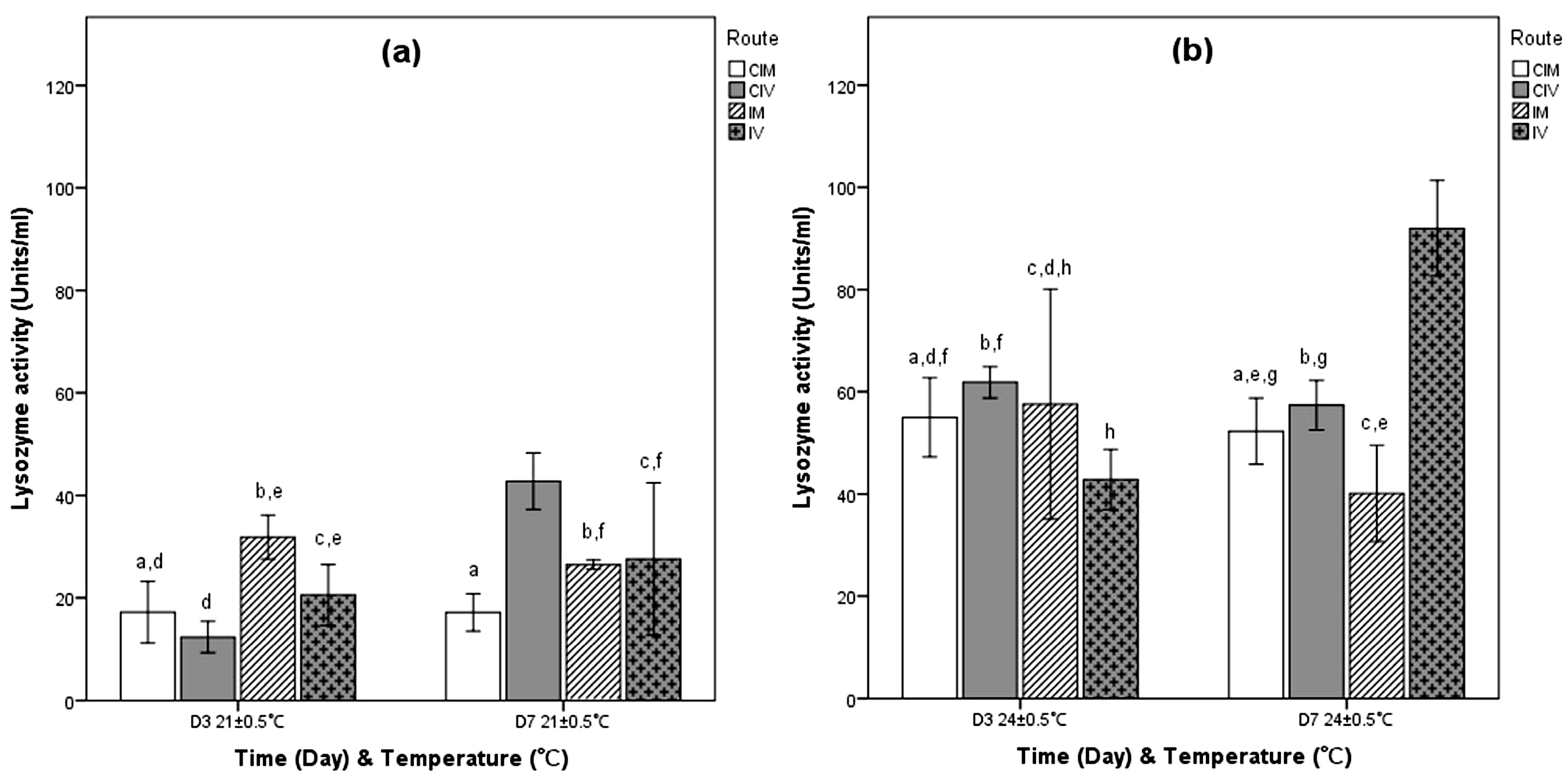
Lysozyme activity in hemolymph was detected in all samples and is illustrated in Figure 2a,b, respectively. Baseline lysozyme levels in D0 controls were calculated at 115.63 ± 10.67 and 60.46 ± 26.24 Units/mL at 21 ± 0.5 °C and 24 ± 0.5 °C, respectively.
As shown in Figure 2a, at 21 ± 0.5 °C, lysozyme activity in the CIM control group increased significantly between time points (Supplementary Table S2a). In contrast, at 24 ± 0.5 °C, activity showed a significant decrease. Interestingly, in the CIV group, lysozyme activity remained stable from D3 to D7 at both temperatures (21 ± 0.5 °C and 24 ± 0.5 °C).
In the IM-challenged group at 21 ± 0.5 °C, lysozyme activity decreased significantly over time, whereas in the IV-challenged group, it increased significantly. At 24 ± 0.5 °C, lysozyme activity levels in the IM- and IV-challenged groups remained stable between sampling points.
A notable shift in lysozyme activity was observed between the D0 and D3 samplings for both control and challenged specimens at 21 ± 0.5 °C, with lower activity levels recorded, except for the D3 IM group, where no significant decrease was detected. Similarly, at 24 ± 0.5 °C, lower activity levels were also recorded in the D3 groups, with statistically significant differences observed only in the challenged groups.
Comparing challenged specimens between temperatures revealed that, at D3, lower temperature had a positive effect on the immune response of the IM-challenged group, as indicated by lysozyme activity levels in the hemolymph (Supplementary Table S2d).

3.1.3. Challenge with VA

Lysozyme activity in hemolymph was detected in all samples and is illustrated in Figure 3a,b, respectively. Baseline lysozyme levels in D0 controls were calculated at 40.95 ± 6.00 and 50.47 ± 8.91 Units/mL at 21 ± 0.5 °C and 24 ± 0.5 °C, respectively.
As shown in Figure 3a, at 21 ± 0.5 °C, lysozyme activity in the CIM group remained stable across sampling points. In contrast, the CIV group showed a significant increase from D3 to D7 (Supplementary Table S3a). At 24 ± 0.5 °C (Figure 3b), lysozyme activity levels in the control CIM and CIV groups remained unchanged between sampling points.
Lysozyme activity in both challenged groups at 21 ± 0.5 °C remained relatively stable across time points. However, the effect of temperature was notable, as all groups exhibited high lysozyme activity. At 24 ± 0.5 °C, lysozyme levels in the IM-challenged group remained unchanged between sampling points, whereas a significant decrease in activity was observed in the IV control group (Supplementary Table S3a).
A comparison of lysozyme activity levels between D0 and D3 for both control and challenged specimens at 21 ± 0.5 °C revealed a notable decrease in D3 specimens, except for the D3 IM group. Conversely, at 24 ± 0.5 °C, all groups showed an increase in activity, except for the D3 IV group, which did not follow this trend.
Consistent with these findings, a comparison between challenged specimens at both temperatures indicated that lower temperature negatively affected the lysozyme activity response in all challenged specimens (Figure 3a,b, and Supplementary Table S3d).

3.1.4. Challenge with VAO1

Lysozyme activity in hemolymph was detected in all samples and is illustrated in Figure 4a,b, respectively. Baseline lysozyme levels in D0 controls were calculated at 101.35 ± 10.95 and 74.00 ± 21.83 Units/mL at 21 ± 0.5 °C and 24 ± 0.5 °C, respectively.
Lysozyme activity at 21 ± 0.5 °C (Figure 4a) decreased significantly in the CIM groups but remained unchanged in the CIV groups between time points D3 and D7. At 24 ± 0.5 °C (Figure 4b), lysozyme activity in the CIM group remained relatively stable between sampling points, whereas in the CIV group, a significant increase was observed over time (Supplementary Table S4a).
As shown in Figure 4a, lysozyme activity in both challenged groups at 21 ± 0.5 °C increased significantly between sampling points. In contrast, at 24 ± 0.5 °C, the elevated temperature resulted in consistently high levels of lysozyme activity at both sampling points.
Comparison of lysozyme activity levels between D0 and D3 in both control and challenged specimens at 21 ± 0.5 °C revealed notably lower activity levels in all D3 specimens. The opposite trend was observed at 24 ± 0.5 °C in all groups except for the D3 CIV group.
A comparison of challenged specimens at different temperatures showed that higher temperatures positively influenced the immune response of the IM-challenged group on D3. Specifically, lysozyme activity in this group was significantly higher at 24 ± 0.5 °C compared to the IM-challenged group at 21 ± 0.5 °C (Supplementary Table S4d). The opposite pattern was observed in the IV group on D7, as extremely high lysozyme activity was recorded at the lower temperature.

4. Discussion

According to our findings, the levels of lysozyme activity detected in the cell-free hemolymph of O. vulgaris were lower than those reported in challenged marine teleosts (at levels of 100–1000 Units/mL in Nile tilapia, Oreochromis niloticus and 600–900 Units/mL in striped bass Morone chrysops × Morone saxatilis) [18,46], yet higher than those found in other fish species (29.40 Units/mL in red seabream, Pagrus major, and 76.3 Units/mL in Japanese eel, Anguilla japonica) [47,48]. Nevertheless, our results align with a recent study that demonstrated significant time- and temperature-dependent variation in both control and challenged groups [18]. Additional variability in lysozyme activity was observed in relation to the pathogen species and the route of administration.
As noted by Saurabh & Sahoo [49], hemolymph lysozyme levels may fluctuate due to several intrinsic and extrinsic factors, including sex, reproductive status, seasonal variation, and stress. In the present study, although direct measurements of all such factors were not performed, appropriate controls were implemented to minimize their potential impact on the experimental results. The sex of each individual was recorded during sampling, as documented in our previous studies [9,30,31,32]; however, sex-specific comparisons were not statistically tested in this study. This was primarily due to the use of wild-caught specimens, which made it logistically challenging to obtain a balanced number of males and females. As a result, the unbalanced sex distribution limited the feasibility of conducting meaningful statistical analyses based on sex.
Nonetheless, we acknowledge that sex may influence immune and physiological responses and recognize it as an important factor for future research. We therefore recommend that future studies utilize aquaculture-reared individuals, where sex ratios can be controlled, to enable a more comprehensive assessment of sex-related differences [9,30,31,32].
Similarly, a study of Locatello et al. [12] reported variation in lysozyme activity in the hemolymph of both control and lipopolysaccharide (LPS)-challenged O. vulgaris individuals, reinforcing the complexity of interpreting this biomarker. A comparable trend was identified in our data, whereby baseline lysozyme levels at D0 were typically lower than those at D3 and D7 in both challenged and control animals, indicating a time-and temperature-dependent immune modulation. This increase over time suggests that the octopus’s innate immune system likely utilizes lysozyme to mitigate microbial threats or to respond to tissue injury caused by injection [50].
Notably, in our study, IM-challenged animals exposed to Photobacterium damselae subsp. piscicida (Phdp) and Ph. damselae subsp. damselae (Phdd) exhibited elevated lysozyme activity on D3 at 21 ± 0.5 °C, compared to other groups. Furthermore, at 24 ± 0.5 °C, IM-challenged animals maintained relatively stable and elevated lysozyme activity throughout the experiment, whereas other groups under the same conditions displayed a similar trend but with a lower magnitude (~50%) across all eight experimental combinations.
Previous studies have yielded mixed results. For example, lysozyme levels increased following bacterial challenge in the hard clam Mercenaria mercenaria [51], whereas Locatello et al. [12] reported decreased serum lysozyme activity in O. vulgaris following LPS challenge compared to controls. Malham et al. [52] observed no temporal changes in tissue lysozyme levels in control or Vibrio anguillarum-challenged individuals, contrary to our findings. In contrast, Grimaldi et al. [53] demonstrated a transient spike in lysozyme activity within 4 h of O. vulgaris exposure to titanium dioxide nanoparticles, with levels normalizing by 24 h, suggesting a short-lived response. Lysozyme remains one of the most fundamental and well-studied components of the humoral immune response, due to its broad antimicrobial activity and evolutionary conservation [54].
The persistent lysozyme response observed in O. vulgaris could indicate mechanisms of immune priming or tolerance, reflecting a complex regulation of innate immunity. These findings not only deepen our understanding of the immune response dynamics in O. vulgaris but also have important implications for cephalopod aquaculture practice.
Although lysozyme is classically known for its lytic activity against Gram-positive bacteria, it plays a broader role in invertebrate immunity, including signaling functions and responses to stress or tissue injury. Studies have demonstrated lysozyme activation in cephalopods and other mollusks following exposure to Gram-negative pathogens, indicating additional immunological roles beyond bacterial lysis [6,9,11,12,13,16,17,18]. In the present study, lysozyme was selected as a general biomarker of humoral immune response, due to its evolutionary conservation and functional versatility. However, future studies should include complementary immune markers, such as antimicrobial peptides or immune-related gene expression, to provide a more comprehensive assessment of the host–pathogen interaction in cephalopods.
Elevated lysozyme levels may serve as an early biomarker of immune activation and general health status in cultured cephalopods. Routine monitoring of lysozyme activity in hemolymph could help detect subclinical infections or stress before clinical signs emerge, allowing timely interventions that improve disease prevention, reduce mortality, and enhance welfare and production efficiency. Future studies should consider the use of aquaculture-sourced animals, where sex and size can be standardized, an approach still under development for this species, especially in Europe. In addition, to further validate the infection model, additional trials using immersion-based infection methods over longer exposure periods are required.
Concerning the statistical analysis, the normality of the data was evaluated using both Kolmogorov–Smirnov and Shapiro–Wilk tests, which yielded consistent results despite the small sample size (n = 3 per group). This agreement supports the robustness of the statistical approach applied in the present study. Given the inherent limitations of experiments with wild-caught cephalopods, the application of more complex models such as repeated measures ANOVA would not increase reliability under these constraints and may instead yield spurious outcomes.

5. Conclusions

  • This study highlights the significance of lysozyme activity as a potential biomarker of innate immune function in O. vulgaris under pathogenic challenge and varying environmental temperatures.
  • Lysozyme activity in cell-free hemolymph varied significantly depending on the following:
    Pathogen type;
    Route of challenge;
    Time point post-injection;
    Environmental temperature.
  • Specimens challenged intramuscularly, especially with Photobacterium damselae subsp. piscicida and damselae, induce stronger lysozyme responses, especially at 21 ± 0.5 °C on day 3 post-injection.
    The observed peak activity suggests a temperature- and time-dependent activation pattern of O. vulgaris immunity.
  • Notably, lysozyme activity in cephalopods appears to persist longer than expected and previously assumed, possibly indicating a more prolonged role in immune defense.
    This sustained enzymatic response might be central to bacterial neutralization in cephalopods.
  • Lysozyme serves as a non-specific yet informative immune indicator of immune status, although environmental land biological variability must be taken into account.
  • Future research should focus on the following:
    Long-term patterns of lysozyme activity beyond early-phase responses;
    A wider array of pathogens and challenge methods;
    Molecular pathways regulating lysozyme expression and interaction with other immune components;
    Additional environmental factors, such as salinity, pollution, and ocean acidification.
  • Overall, applying lysozyme activity as a biomarker for health monitoring in octopus aquaculture could contribute meaningfully to developing sustainable and resilient farming practices.

Supplementary Materials

The following supporting information can be downloaded at: https://www.mdpi.com/article/10.3390/fishes10090428/s1.

Author Contributions

D.-M.W.: Conceptualization, experimental design, sampling, laboratory work, lysozyme analysis, data collection and analysis, manuscript writing and editing; E.A.: Lysozyme analysis; M.-A.V.: Sampling; V.B.: Conceptualization, experimental design, supervision, manuscript review and editing. All authors have read and agreed to the published version of the manuscript.

Funding

This research is co-financed by Greece and the European Union (European Social Fund-ESF) through the Operational Programme «Human Resources Development, Education and Lifelong Learning 2014–2020» in the context of the project “Effect of climate change on the infectiousness of pathogenic microorganisms and the physiology of sensitive host aquatic organisms” (MIS 5004276).

Institutional Review Board Statement

The experimental protocol was approved by Decision No. 5379/04-04-2017 issued by the competent Regional Veterinary Authority. The wet laboratory is part of the Laboratory of Ichthyology, Aquaculture and Aquatic Animal Health (ICHTHYAI) (Government Issue 1255/28-4-2016). ICHTHYAI holds all necessary permits for the production (EL83 BIObr01), supply (EL83 BIOsup01), and experimentation (E83 BioExp01) on aquatic organisms, in accordance with Presidential Decree 56/2013 and Directive 2010/63/EU (Decision No. 4053/14-3-2017 issued by the competent Regional Veterinary Authority). Specific permits were granted by the Directorate of Rural Affairs of North Aegean, Department of Agricultural Farms and Fishery, Decentralized Administration of Aegean (permits 29623/19-5-2017, 51948/5-9-2018, and 20564/11-4-2019) to cover the needs of this experimental study.

Informed Consent Statement

Not applicable.

Data Availability Statement

Data are contained within the article and supplementary materials.

Acknowledgments

The authors would like to thank Kantham Papanna, Ogut Hamdi, and Pantelis Katharios for their kind donations of bacterial strains according to White and White et al. [9,31,32].

Conflicts of Interest

The authors declare no conflict of interest.

References

  1. Garlock, T.M.; Asche, F.; Anderson, J.L.; Eggert, H.; Anderson, T.M.; Che, B.; Chávez, C.A.; Chu, J.; Chukwuone, N.; Dey, M.M.; et al. Environmental, economic, and social sustainability in aquaculture: The aquaculture performance indicators. Nat. Commun. 2024, 15, 527, Erratum in Nat. Commun. 2024, 15, 5965. [Google Scholar] [CrossRef]
  2. Vaz-Pires, P.; Seixas, P.; Barbosa, A. Aquaculture potential of the common octopus (Octopus vulgaris Cuvier, 1797): A review. Aquaculture 2004, 238, 221–238. [Google Scholar] [CrossRef]
  3. Almeida, D.; Domínguez-Pérez, D.; Matos, A.; Agüero-Chapin, G.; Osório, H.; Vasconcelos, V.; Campos, A.; Antunes, A. Putative antimicrobial peptides of the posterior salivary glands from the cephalopod Octopus vulgaris revealed by exploring a composite protein database. Antibiotics 2020, 9, 757. [Google Scholar] [CrossRef] [PubMed]
  4. EUWFOP. European Union World Fisheries Overview Platform; EUWFOP: Brussels, Belgium, 2023; Available online: https://www.reportlinker.com/dataset/15d0ef2740e108bb3c5bb5a19cc3e1bdad910695 (accessed on 16 July 2025).
  5. Sauer, W.H.H.; Gleadall, I.G.; Downey-Breedt, N.; Doubleday, Z.; Gillespie, G.; Haimovici, M.; Ibáñez, C.M.; Katugin, O.N.; Leporati, S.; Lipinski, M.R.; et al. World octopus fisheries. Rev. Fish. Sci. Aquac. 2019, 29, 279–429. [Google Scholar] [CrossRef]
  6. Gestal, C.; Castellanos-Martínez, S. Understanding the cephalopod immune system based on functional and molecular evidence. Fish Shellfish Immunol. 2015, 46, 120–130. [Google Scholar] [CrossRef]
  7. Sacchi, S.; Malagoli, D.; Franchi, N. The invertebrate immunocyte: A complex and versatile model for immunological, developmental, and environmental research. Cells 2024, 13, 2106. [Google Scholar] [CrossRef]
  8. Rodríguez-Domínguez, H.; Soto-Búa, M.; Iglesias-Blanco, R.; Crespo-González, C.; Arias-Fernández, C.; García-Estévez, J. Preliminary study on the phagocytic ability of Octopus vulgaris Cuvier, 1797 (Mollusca: Cephalopoda) haemocytes in vitro. Aquaculture 2006, 254, 563–570. [Google Scholar] [CrossRef]
  9. White, D.M. Studies on Pathology and Immunology of the Common Octopus (Octopus vulgaris Cuvier, 1797). Ph.D. Thesis, University of the Aegean, Mytilini, Greece, 2023. Available online: https://www.didaktorika.gr/eadd/handle/10442/54760 (accessed on 4 August 2025).
  10. Van Herreweghe, J.M.; Michiels, C.W. Invertebrate lysozymes: Diversity and distribution, molecular mechanism and in vivo function. J. Biosci. 2012, 37, 327–348. [Google Scholar] [CrossRef]
  11. Matozzo, V.; Chinellato, A.; Munari, M.; Finos, L.; Bressan, M.; Marin, M.G. First evidence of immunomodulation in bivalves under seawater acidification and increased temperature. PLoS ONE 2012, 7, e33820. [Google Scholar] [CrossRef]
  12. Locatello, L.; Fiorito, G.; Finos, L.; Rasotto, M.B. Behavioural and immunological responses to an immune challenge in Octopus vulgaris. Physiol. Behav. 2013, 122, 93–99. [Google Scholar] [CrossRef] [PubMed]
  13. Li, H.; Wei, X.; Yang, J.; Zhang, R.; Zhang, Q.; Yang, J. The bacteriolytic mechanism of an invertebrate-type lysozyme from mollusk Octopus ocellatus. Fish Shellfish Immunol. 2019, 93, 232–239. [Google Scholar] [CrossRef]
  14. Hu, F.; Wang, Y.; Hu, J.; Bao, Z.; Wang, M. A novel c-type lysozyme from Litopenaeus vannamei exhibits potent antimicrobial activity. Fish Shellfish Immunol. 2022, 131, 108485. [Google Scholar] [CrossRef]
  15. Marra, A.; Hanson, M.A.; Kondo, S.; Erkosar, B.; Lemaitre, B. Drosophila antimicrobial peptides and lysozymes regulate gut microbiota composition and abundance. mBio 2021, 12, e0082421. [Google Scholar] [CrossRef]
  16. Montoya, L.N.F.; Martins, T.P.; Gimbo, R.Y.; Zanuzzo, F.S.; Urbinati, E.C. β-Glucan-induced cortisol levels improve the early immune response in matrinxã (Brycon amazonicus). Fish Shellfish Immunol. 2017, 60, 197–204. [Google Scholar] [CrossRef]
  17. Amphan, S.; Unajak, S.; Printrakoon, C.; Areechon, N. Feeding-regimen of β-glucan to enhance innate immunity and disease resistance of Nile tilapia, Oreochromis niloticus Linn., against Aeromonas hydrophila and Flavobacterium columnare. Fish Shellfish Immunol. 2019, 87, 120–128. [Google Scholar] [CrossRef] [PubMed]
  18. Biller, J.; Polycarpo, G.; Moromizato, B.S.; Sidekerskis, A.; Silva, T.; Reis, I.; Fierro-Castro, C. Lysozyme activity as an indicator of innate immunity of tilapia (Oreochromis niloticus) when challenged with LPS and Streptococcus agalactiae. Rev. Bras. Zootec. 2021, 50, e20210053. [Google Scholar] [CrossRef]
  19. FAO. Expanding Sustainable Aquaculture Production; FAO: Rome, Italy, 2025; Available online: https://openknowledge.fao.org/server/api/core/bitstreams/9df19f53-b931-4d04-acd3-58a71c6b1a5b/content/sofia/2022/expanding-sustainable-aquaculture-production.html (accessed on 15 July 2025).
  20. Hanlon, R.T.; Forsythe, J.W.; Cooper, K.M.; Dinuzzo, A.R.; Folse, D.S.; Kelly, M.T. Fatal penetrating skin ulcers in laboratory-reared octopuses. J. Invertebr. Pathol. 1984, 44, 67–83. [Google Scholar] [CrossRef] [PubMed]
  21. Rigos, G.; Katharios, P. Pathological obstacles of newly-introduced fish species in Mediterranean mariculture: A review. Rev. Fish Biol. Fish. 2010, 20, 47–70. [Google Scholar] [CrossRef]
  22. Gestal, G.; Pascual, S.; Guerra, A.; Fiorito, G.; Vieites, J.M. Handbook of Pathogens and Diseases in Cephalopods; Springer Open: Berlin/Heidelberg, Germany, 2019; pp. 1–234. [Google Scholar] [CrossRef]
  23. Farto, R.; Fichi, G.; Gestal, C.; Pascual, S.; Nieto, T.P. Bacteria-Affecting Cephalopods. In Handbook of Pathogens and Diseases in Cephalopods; Gestal, C., Pascual, S., Guerra, Á., Fiorito, G., Vieites, J., Eds.; Springer: Berlin/Heidelberg, Germany, 2019; pp. 127–142. [Google Scholar] [CrossRef]
  24. Pascual, C.; Angeles-González, L.E.; Rodríguez-Flores, C.; Rosas, C. Sea surface temperature modulates physiological and immunological condition of Octopus maya. Front. Physiol. 2019, 10, 739. [Google Scholar] [CrossRef]
  25. Ángeles-González, L.E.; Pascual, C.; Mascaró, M.; Sánchez, A.; Gallardo, P.; Rodríguez-Flores, C.; Rosas, C. Linking inferred laboratory-derived temperature stress to immunocompetence in the Mayan octopus (Octopus maya). Front. Mar. Sci. 2025, 12, 1524296. [Google Scholar] [CrossRef]
  26. Castillo, M.G.; Goodson, M.S.; McFall-Ngai, M.J. The immune response of cephalopods from head to foot. Fish Shellfish Immunol. 2015, 46, 145–160. [Google Scholar] [CrossRef]
  27. Day, R.J.; Doubleday, Z.A.; Semmens, J.M. Exercise stress affects immune and metabolic physiology in the pale octopus (Octopus pallidus). Fish Shellfish Immunol. 2024, 145, 108896. [Google Scholar] [CrossRef]
  28. Paillard, C.; Allam, B.; Oubella, R. Effect of temperature on defense parameters in Manila clam Ruditapes philippinarum challenged with Vibrio tapetis. Dis. Aquat. Org. 2004, 59, 249–262. [Google Scholar] [CrossRef]
  29. Papadopoulos, M.; Bort, J.; Theodorou, J.A.; Katsiadaki, I. Molecular immune responses of Ruditapes decussatus to Vibrio splendidus: Effects of temperature on lysozyme gene expression. Fishes 2025, 10, 72. [Google Scholar] [CrossRef]
  30. White, D.M.; Valsamidis, M.A.; Bakopoulos, V. In vitro hemocyte phagocytosis activation after experimental infection of common octopus, Octopus vulgaris (Cuvier, 1797) with Photobacterium damselae subsp. piscicida or Vibrio alginolyticus at different temperatures and infection routes. J. Invertebr. Pathol. 2022, 191, 107754. [Google Scholar] [CrossRef]
  31. White, D.M.; Valsamidis, M.A.; Kokkoris, G.D.; Bakopoulos, V. The effect of temperature and challenge route on in vitro hemocyte phagocytosis activation after experimental challenge of common octopus, Octopus vulgaris (Cuvier, 1797) with either Photobacterium damselae subsp. damselae or Vibrio anguillarum O1. Microb. Pathog. 2023, 174, 105955. [Google Scholar] [CrossRef]
  32. White, D.M.; Valsamidis, M.A.; Bakopoulos, V. Hemolymph activities after experimental infection of common octopus, Octopus vulgaris (Cuvier, 1797) with Photobacterium damselae subsp. piscicida. In Proceedings of the “HydroMediT” 4th International Congresses on Applied Ichthyology, Oceanography, and Aquatic Environment, Mytilene, Lesvos, Greece, 30 May–2 June 2021. [Google Scholar]
  33. Miliou, H.; Fintikaki, M.; Tzitzinakis, M.; Kountouris, T.; Verriopoulos, G. Fatty acid composition of the common octopus, Octopus vulgaris, in relation to rearing temperature and body weight. Aquaculture 2006, 256, 311–322. [Google Scholar] [CrossRef]
  34. Prato, E.; Portacci, G.; Biandolino, F. Effect of diet on growth performance, feed efficiency and nutritional composition of Octopus vulgaris. Aquaculture 2010, 309, 203–211. [Google Scholar] [CrossRef]
  35. Petza, D.; Katsanevakis, S.; Lykouri, N.; Spiliotis, V.; Verriopoulos, G. Investigation of the potential effect of diet, body mass and maturity on growth and feed performance of common Octopus vulgaris: An information theory approach. Aquac. Nutr. 2011, 17, e348–e361. [Google Scholar] [CrossRef]
  36. Weather Stars. 2022. Available online: https://seatemperature.info/mytilini-water-temperature.html#google_vignette (accessed on 3 June 2022).
  37. UN. UN Environment Programme. Mediterranean Action Plan-Barcelona Convection; UN: New York, NY, USA, 2022; Available online: https://www.unep.org/unepmap/resources/factsheets/climate-change. (accessed on 3 June 2022).
  38. Katsanevakis, S.; Protopapas, N.; Miliou, H.; Verriopoulos, G. Effect of temperature on specific dynamic action in the common octopus, Octopus vulgaris (Cephalopoda). Mar Biol. 2005, 146, 733–738. [Google Scholar] [CrossRef]
  39. Noyola, J.; Caamal-Monsreal, C.; Díaz, F.; Re, D.; Sánchez, A.; Rosas, C. Thermopreference, tolerance and metabolic rate of early stages juvenile Octopus maya acclimated to different temperatures. J. Therm. Biol. 2013, 38, 14–19. [Google Scholar] [CrossRef]
  40. Hanif, A.; Bakopoulos, V.; Dimitriadis, G.J. Maternal transfer of humoral specific and non-specific immune parameters to sea bream (Sparus aurata) larvae. Fish Shellfish Immunol. 2004, 17, 411–435. [Google Scholar] [CrossRef]
  41. Bakopoulos, V.; White, D.; Valsamidis, M.A.; Vasilaki, F. Experimental infection of Octopus vulgaris (Cuvier, 1797) with Photobacterium damsela subsp. piscicida. Immunohistochemical tracking of antigen and tissue responses. J. Invertebr. Pathol. 2017, 144, 24–31. [Google Scholar] [CrossRef]
  42. Ellis, A.E. Lysozyme assays. In Techniques in Fish Immunology; Stolen, J.S., Fletcher, T.C., Anderson, D.P., Roberson, B.S., van Muiswinkel, W.B., Eds.; SOS Publications: Cambridge, UK, 1990; pp. 101–103. Available online: https://scholar.google.com/scholar_lookup?title=Lysozyme%20assays&author=A.E.%20Ellis&publication_year=1990&pages=101-103 (accessed on 28 August 2025).
  43. Sigma-Aldrich. Enzymatic Assay of Lysozyme; Sigma-Aldrich: Shanghai, China, 2025; Available online: https://www.sigmaaldrich.com/GR/en/technical-documents/protocol/protein-biology/enzyme-activity-assays/enzymatic-assay-of-lysozyme (accessed on 14 July 2025).
  44. Cerón, A.; Costa, S.; Imbernon, R.; Queiroz, R.; Castro, J.; Ferraz, H.; Oliveira, R.; Costa, S. Study of stability, kinetic parameters and release of lysozyme immobilized on chitosan microspheres by crosslinking and covalent attachment for cotton fabric functionalization. Process Biochem. 2023, 128, 116–125. [Google Scholar] [CrossRef]
  45. Norusis, M.J. SPSS, version 12.0 Guide to Data Analysis; Prentice Hall PTR: Centennial, CO, USA, 2004.
  46. Sealey, W.M.; Gatlin, D.M. Dietary vitamin C and vitamin E interact to influence growth and tissue composition of juvenile hybrid striped bass (Morone chrysops X M. saxatilis) head–kidney cells. J. Nutr. 2002, 132, 748–755. [Google Scholar] [CrossRef]
  47. Ji, S.C.; Takaoka, O.; Jeong, G.S.; Lee, S.W.; Ishimaru, K.; Seoka, M.; Takii, K. Dietary medicinal herbs improve growth and some non-specific immunity of red sea bream Pagrus major. Fish. Sci. 2007, 73, 63–69. [Google Scholar] [CrossRef]
  48. Ren, T.; Koshio, S.; Ishikawa, M.; Yokoyama, S.; Micheal, F.R.; Uyan, O.; Tung, H.T. Influence of dietary vitamin C and bovine lactoferrin on blood chemistry and non-specific immune responses of Japanese eel, Anguilla japonica. Aquaculture 2007, 267, 31–37. [Google Scholar] [CrossRef]
  49. Saurabh, S.; Sahoo, P.K. Lysozyme: An important defence molecule of fish innate immune system. Aquac. Res. 2008, 39, 223–239. [Google Scholar] [CrossRef]
  50. Le Pabic, C.; Safi, G.; Serpentini, A.; Lebel, J.M.; Robin, J.P.; Koueta, N. Prophenoloxidase system, lysozyme and protease inhibitor distribution in the common cuttlefish Sepia officinalis. Comp. Biochem. Physiol. B 2014, 172–173, 96–104. [Google Scholar] [CrossRef] [PubMed][Green Version]
  51. Cheng, T.C. The role of lysosomes in molluscan inflammation. Am. Zool. 1983, 23, 129–144. [Google Scholar] [CrossRef]
  52. Malham, S.K.; Runham, N.W. A brief review of the immunobiology of Eledone cirrhosa. S. Afr. J. Mar. Sci. 1998, 20, 385–391. [Google Scholar] [CrossRef]
  53. Grimaldi, A.M.; Belcari, P.; Pagano, E.; Cacialli, F.; Locatello, L. Immune responses of Octopus vulgaris (Mollusca: Cephalopoda) exposed to titanium dioxide nanoparticles. J. Exp. Mar. Biol. Ecol. 2013, 447, 123–127. [Google Scholar] [CrossRef]
  54. Mukherjee, K.; Moroz, L.L. Evolution of g-type lysozymes in metazoa: Insights into immunity and digestive adaptations. Front. Cell Dev. Biol. 2024, 12, 1487920. [Google Scholar] [CrossRef] [PubMed]
Figure 1. Hemolymph lysozyme activity of O. vulgaris after IM or IV injection with either Phdp or NaCl. Results are expressed as the Units/mL of hemolymph from 3 octopus per group: (a) at 21 ± 0.5 °C and (b) at 24 ± 0.5 °C. D: day; CIM: controls injected IM; CIV: controls injected IV; IM: challenged IM; IV: challenged IV. Mean values (±SE); same letter indicates no significance (p > 0.05): identical letters are shared only by groups that do not differ significantly (p > 0.05). Groups without letters are significantly different from all others. One unit of lysozyme activity was defined as a reduction in absorbance of 0.001/min.
Fishes 10 00428 g001
Figure 2. Hemolymph lysozyme activity of O. vulgaris after IM or IV injection with either Phdd or NaCl. Results are expressed as the Units/mL of hemolymph from 3 octopus per group: (a) at 21 ± 0.5 °C and (b) at 24 ± 0.5 °C. D: day; CIM: controls injected IM; CIV: controls injected IV; IM: challenged IM; IV: challenged IV. Mean values (±SE); same letter indicates no significance (p > 0.05): identical letters are shared only by groups that do not differ significantly (p > 0.05). Groups without letters are significantly different from all others. One unit of lysozyme activity was defined as a reduction in absorbance of 0.001/min.
Fishes 10 00428 g002
Figure 3. Hemolymph lysozyme activity of O. vulgaris after IM or IV injection with either VA or NaCl. Results are expressed as the Units/mL of hemolymph from 3 octopus per group: (a) at 21 ± 0.5 °C and (b) at 24 ± 0.5 °C. D: day; CIM: controls injected IM; CIV: controls injected IV; IM: challenged IM; IV: challenged IV. Mean values (±SE); same letter indicates no significance (p > 0.05): identical letters are shared only by groups that do not differ significantly (p > 0.05). Groups without letters are significantly different from all others. One unit of lysozyme activity was defined as a reduction in absorbance of 0.001/min.
Fishes 10 00428 g003
Figure 4. Hemolymph lysozyme activity of O. vulgaris after IM or IV injection with either VAO1 or NaCl. Results are expressed as the Units/mL of hemolymph from 3 octopuses per group: (a) at 21 ± 0.5 °C and (b) at 24 ± 0.5 °C. D: day; CIM: controls injected IM; CIV: controls injected IV; IM: challenged IM; IV: challenged IV. Mean values (±SE); same letter indicates no significance (p > 0.05): identical letters are shared only by groups that do not differ significantly (p > 0.05). Groups without letters are significantly different from all others. One unit of lysozyme activity was defined as a reduction in absorbance of 0.001/min.
Fishes 10 00428 g004
Table 1. Sampling periods of O. vulgaris specimens [9,30,31].
Bacterial PathogenSampling Period
Photobacterium damselae subsp. Piscicida
21 ± 0.5 °C or 24 ± 0.5 °C
October 2017 (n = 27; 21 ± 0.5 °C)
November 2017 (n = 27; 24 ± 0.5 °C)
Photobacterium damselae subsp. Damselae
21 ± 0.5 °C or 24 ± 0.5 °C
April 2019 (n = 27; 21 ± 0.5 °C)
May 2019 (n = 27; 24 ± 0.5 °C)
Vibrio alginolyticus
21 ± 0.5 °C or 24 ± 0.5 °C
February 2018 (n = 27; 21 ± 0.5 °C)
March 2018 (n = 27; 24 ± 0.5 °C)
Vibrio anquillarum O1
21 ± 0.5 °C or 24 ± 0.5 °C
October 2019 (n = 27; 21 ± 0.5 °C)
November 2019 (n = 27; 24 ± 0.5 °C)
Disclaimer/Publisher’s Note: The statements, opinions and data contained in all publications are solely those of the individual author(s) and contributor(s) and not of MDPI and/or the editor(s). MDPI and/or the editor(s) disclaim responsibility for any injury to people or property resulting from any ideas, methods, instructions or products referred to in the content.