Article

Bioactive Substance Derived from Mealworm Larvae (Tenebrio molitor) Potentially Induces Immune Performance of Zebrafish (Danio rerio)

by
Ibnu Bangkit Bioshina Suryadi
1,2,
Muhammad Fariz Zahir Ali
3,4,
Haruki Nishiguchi
4,
Saita Akanuma
1,
Chiemi Miura
4 and
Takeshi Miura
4,*
1
The United Graduate School of Agricultural Sciences, Ehime University, 3-5-7 Tarumi, Matsuyama 790-8566, Japan
2
Department of Fisheries, Faculty of Fisheries and Marine Sciences, Universitas Padjadjaran, Jalan Ir. Soekarno KM 21, Sumedang 45363, Indonesia
3
Research Center for Marine and Land Bio Industry, National Research and Innovation Agency, Jl. Raya Senggigi, Kodek Bay, Pemenang 83352, Indonesia
4
Graduate School of Agriculture, Ehime University, 3-5-7 Tarumi, Matsuyama 790-8566, Japan
*
Author to whom correspondence should be addressed.
Fishes 2025, 10(6), 285; https://doi.org/10.3390/fishes10060285
Submission received: 30 April 2025 / Revised: 29 May 2025 / Accepted: 4 June 2025 / Published: 10 June 2025
(This article belongs to the Section Sustainable Aquaculture)

Abstract

Mealworm (Tenebrio molitor) larvae meal is a notable alternative protein source that is widely used in the aquaculture industry. Recently, it has also gained recognition for enhancing the immune performance of various aquaculture species. However, studies identifying the specific bioactive substances responsible for the immunomodulatory effects of mealworm larvae meal remain limited. In this study, we isolated and purified a bioactive substance from mealworm and incorporated it into zebrafish (Danio rerio) diets at concentrations of 0, 10, and 100 µg/g; the zebrafish were fed this diet for 14 days. To assess the transcriptional changes induced by the bioactive substance, we performed RNA sequencing and qRT-PCR analysis on intestinal and liver tissues. Subsequently, zebrafish were challenged with Edwardsiella tarda via immersion in order to evaluate the protective effects of the bioactive substance. The results demonstrated that a dietary inclusion of 100 µg/g of the bioactive substance optimized the immune performance of zebrafish. Additionally, challenge tests revealed that the dietary inclusion of the bioactive substance from mealworms positively influenced pathogen resistance, although these effects were not consistently significant.
Key Contribution: This paper discusses the identification and purification of a possible novel bioactive substance from mealworm (Tenebrio molitor) larvae that demonstrates potential as an immunomodulator in aquaculture. This substance was shown to enhance nitric oxide production in RAW264.7 macrophage cells in vitro and modulate cytokine expression in zebrafish (Danio rerio), ultimately increasing their lifespan when challenged with Edwardsiella tarda.

1. Introduction

Various methods have been used in aquaculture to prevent disease outbreaks. One approach that has sparked interest is the use of insect-based products as immunostimulants due to the fact that insects produce several substances that can improve animal health [1,2,3,4]. Compared with conventional animal protein sources, insects have gained attention in the animal feed industry due to their ability to thrive in challenging environments that are often contaminated with a wide range of microorganisms [5]. Cultured insects can synthesize numerous native bioactive peptides with antibacterial, antifungal, and antiviral properties, which is as a result of being raised in these types of environments [2,3,6].
Among the various types of insect feed used in aquaculture, mealworm (Tenebrio molitor) larvae are one of the most widely used [7], and their extensive application has resulted in the development and establishment of large-scale commercial rearing systems [8]. In addition to being a source of protein in aquafeed, mealworms also serve as immune enhancers; this property has been reported in various aquaculture fish [9] including in yellow catfish [10], mandarin fish [11], vannamei [12,13], and rainbow trout [14,15].
Our previous research demonstrated that insect-derived bioactive substances—such as dipterose-BC, dipterose-BSF, silkrose-AY, and silkrose-BM—can enhance innate immune responses [6,16,17,18]. These immunostimulatory effects are likely mediated through pathogen-associated molecular pattern (PAMP)-like activity [19,20], as these compounds may be recognized by pattern recognition receptors (PRRs) on immune cells [21], thereby initiating innate immune activation.
In the present study, we extracted and purified bioactive substances from mealworms, followed by an assessment of the immunomodulatory potential of these substances. This capability was first explored by evaluating the nitric oxide (NO) activity in RAW264.7 macrophage cells, along with analyses of their protein, organic acid, and sugar content. We then explored the immunomodulatory effects of these bioactive substances during a 14-day dietary treatment in zebrafish (Danio rerio), followed by transcriptomic analysis. Following the feeding trial, zebrafish were challenged with Edwardsiella tarda and were monitored for 7 days. In this study, we aimed to evaluate the feasibility of using mealworm-derived bioactive substances as immunomodulatory agents in aquaculture.

2. Materials and Methods

2.1. In Vitro Analysis

2.1.1. Extraction and Isolation of Bioactive Substances from Mealworm Larvae

In these experiments, mealworms from stock that had been continuously bred since 2015 at the Fish Reproductive Physiology Laboratory, Ehime University, Japan, were used. The extraction of bioactive substances from mealworm larvae was carried out as follows. The mealworm meal was diluted with 10 volumes of ultrapure water and mixed gently for 24 h at 4 °C. The mixture was then centrifuged at 20,000× g for 1 h at room temperature, and the supernatant was concentrated to approximately one-tenth of its original volume via evaporation in a water bath at 50 °C.

2.1.2. Purification of Bioactive Substances from Mealworm Larvae Extract

Bioactive substances from mealworm larvae were purified using modifications of the methods used in previous studies [6,16,17,18]. To precipitate the bioactive substances, the mealworm larvae extract was mixed with four volumes of 99.5% (v/v) ethanol and gently stirred overnight at 4 °C. Centrifugation (20,000× g for 15 min) was then used to isolate the bioactive substances, which were washed three times with 75% ethanol and dried in a draft chamber. To separate any remaining low-molecular-weight compounds, the precipitates were dissolved in 20 mM Tris-HCl (tris (hydroxymethyl) aminomethane hydrochloride; pH 8.0), shaken overnight at 4 °C, and centrifuged at 20,000× g for 15 min. The crude bioactive substances were obtained from the collected precipitate after centrifugation.
The first step in the purification of bioactive substances was accomplished using anion-exchange chromatography (AEC) on a HiPrep Q XL 16/10 column (GE Healthcare, Chicago, IL, USA). The crude bioactive substances were applied to a pre-equilibrated column, which had been prepared with 20 mM Tris-HCl (pH 8.0). Elution steps were conducted using two solutions—20 mM Tris-HCl (pH 8.0) and 20 mM Tris-HCl with 1 M NaCl (sodium chloride; pH 8.0). A stepwise gradient of NaCl at concentrations of 20%, 40%, and 100% was used at a flow rate of 2.0 mL/min to create the fractions. The eluted fractions were collected automatically. They then underwent protein evaluation using UV-Vis spectrophotometer (Bio-Rad, Hercules, CA, USA) at 280 nm [16], were evaluated for NO production in RAW264.7 macrophage cells, and their sugar content was measured using the phenol–sulfuric acid (PSA) method, with glucose as the standard [22]. Fractions exhibiting NO activity and sugar content were pooled and dialyzed (3500 MWCO; Thermo Fisher Scientific, Libertyville, IL, USA) in 20 mM Tris-HCl (pH 8.0) to be used in the next step, following our previous foundings [16,17,18].
The positive fractions were injected into an ENrich SEC (size-exclusion chromatography) 650 gel-filtration chromatography column (Bio-Rad, Hercules, CA, USA), which had been pre-equilibrated with 20 mM Tris-HCl and 200 mM NaCl (pH 8.0). Elution was performed at a flow rate of 2.5 mL/min using the same solution. The eluates were collected automatically, and the protein was evaluated using protein evaluation using UV-Vis spectrophotometer (Bio-Rad, CA, USA); total sugar content was evaluated using the PSA method. Each eluted fraction was also tested for NO activity in RAW264.7 cells to assess its immunomodulatory capacity as explained above. This approach was selected because NO is a well-established indicator of immune activation in macrophages and provides a reliable in vitro proxy for evaluating immunostimulatory potential prior to in vivo application. Fractions exhibiting the greatest NO activity (regardless of sugar activity) were pooled and precipitated overnight at 4 °C by adding four volumes of 99.5% (v/v) ethanol. The resulting precipitate was centrifuged (20,000× g for 15 min) and lyophilized to obtain the bioactive substance.

2.1.3. RAW264.7 Cell Culture

Cells of the RAW264.7 cell line, derived from mouse macrophages, were obtained from the Cell Bank at the RIKEN BioResource Center (Tsukuba, Japan). The cells were cultured in minimal essential medium [MEM] supplemented with 10% fetal bovine serum, 0.1 mM non-essential amino acids, 100 U/mL penicillin, and 100 µg/mL streptomycin (Life Technologies, Waltham, MA, USA). The cells were maintained at 37 °C in a humidified atmosphere containing 5% CO2 (carbon dioxide).

2.1.4. NO Activity Assay

NO levels in the macrophage culture medium were measured using a Griess Reagent System Kit (Promega, Madison, WI, USA), according to the manufacturer’s instructions. Cells were seeded at a density of 1 × 10⁵ cells per well in 96-well culture plates. A 20 µL aliquot from each fraction (obtained from AEC and SEC) was diluted in supplemented MEM, as previously described. Lipopolysaccharide (LPS, 100 ng/mL) was used as a positive control. Samples were incubated with the cells for 24 h at 37 °C. Nitrite concentrations in the culture medium were quantified using a sodium nitrite (NaNO2) standard curve.

2.1.5. Determination of the Molecular Weight of the Mealworm-Derived Bioactive Substance

The molecular weight of the bioactive substance from mealworms was determined using gel-filtration chromatography in combination with high-performance liquid chromatography (HPLC) (Hitachi, Chiyoda, Japan), following the methods outlined in previous studies [6,16,17,18]. Briefly, 1 mg/mL of the pure bioactive substance was dissolved in 0.2 M phosphate buffer (PB) and filtered through a 0.22 µm membrane. The sample solution was then applied to a Shodex SB-803 HQ size-exclusion chromatography column (Resonac, Minato, Japan) and eluted with 0.2 M PB (phosphate buffer; pH 7.5) at a flow rate of 0.5 mL/min. Detection was carried out using a refractive index detector, with the column temperature being maintained at 35 °C. Pullulans of various molecular weights (P-5, P-10, P-20, P-50, P-100, P-200, P-800, and P-2500) were used as molecular weight standards, and their retention times were plotted against the logarithms of their molecular weights in order to generate a standard curve. Since the bioactive substance was barely detected by RI (refractive index), fractions were collected and subjected to NO assay to confirm biological activity; these steps are in accordance with Ali et al. (2019) [16]. The molecular weight of the bioactive substance was then calculated using the calibration equation derived from the pullulan standard curve.

2.2. In Vivo Analysis

2.2.1. Experimental Zebrafish

The zebrafish (Danio rerio) used in this study were individuals that had been bred consecutively since 2018 at the Fish Reproductive Physiology Laboratory, Ehime University. The mean weight of the zebrafish was 0.8 ± 0.1 g, and the mean fork length was 4.26 ± 0.4 cm. The maintenance conditions were as follows: the water temperature was maintained at 27 ± 1 °C, with continuous 24-h aeration and filtration. Fish were fed to satiation twice daily using a control diet (Table S1) at 09:00 and 17:00; they were kept on a 14-h light/10-h dark cycle. Water quality was preserved by changing 30% of the water twice per week. All experiments were conducted in accordance with protocols approved by the Institutional Animal Care and Use Committee (IACUC) of Ehime University (Permit Number: 08K2-1). To minimize suffering during surgical procedures, ethylene glycol monophenyl ether at a concentration of 0.2% was used for euthanasia.

2.2.2. Preparation of the Mealworm-Derived Bioactive Substance Diet

The dry ingredients were manually mixed, followed by the addition of soybean oil, water (Table S1), and the freeze-dried bioactive substance derived from mealworm larvae at final concentrations of 0, 10, and 100 µg/g. The mixture was pelletized using an ABV-120 L cylindrical granulator (Akira Kiko, Fukui, Japan), before being air-dried for 1–2 days at 60 °C and sieved to a size of 2–3 mm. As the quantity of bioactive substance added was negligible, its nutritional impact was considered insignificant.

2.2.3. Evaluation of Zebrafish Cytokines Using qRT-PCR to Confirm mRNA (Messenger Ribonucleic Acid) Sequencing After Dietary Treatment with the Mealworm-Derived Bioactive Substance

A total of 360 zebrafish were used in this study. They were randomly distributed into six aquariums (45 × 25 × 25 cm), with 20 fish per aquarium. Each treatment was applied to two aquariums (i.e., in duplicate) and repeated three times. The water temperature was maintained at 27 °C, with continuous aeration and filtration. A 14-h light/10-h dark cycle was used, and 30% of the water was replaced twice weekly. After 14 days of feeding, five fish from each aquarium were euthanized and dissected to collect liver and intestine samples for RNA extraction. The cytokines investigated included tumor necrosis factor (TNF)-α; interleukin (IL)-1β, IL-6, and IL-8 (pro-inflammatory cytokines); and transforming growth factor (TGF)-β and IL-10 (anti-inflammatory cytokines). RNA isolation was performed following the ISOGEN II (Nippon Gene, Tokyo, Japan) reagent protocol. To verify RNA concentration and quality, the A280/A260 ratios of the extracted RNA were measured using a P330 NanoPhotometer (Munich, Germany).
Quantitative reverse transcription–polymerase chain reaction (qRT-PCR) was performed to analyze six genes using the PowerTrack SYBR Green Master Mix (Thermo Fisher Scientific, Vilnius, Lithuania) protocols, with confirmation assays conducted (n = 5 per group). First-strand complementary deoxyribonucleic acid (cDNA) was synthesized from 500 ng of total RNA using a High-Capacity cDNA Reverse Transcript Kit (Thermo Fisher, Vilnius, Lithuania). The thermocycling process was carried out in 96-well Multiplate PCR Plates (Bio-Rad Laboratories, Tokyo, Japan) using a qRT-PCR detection system (Bio-Rad Laboratories) under the following conditions: 30 s at 95 °C, followed by 40 cycles of 5 s at 95 °C, and 5 s at 55 °C. Melting curves were analyzed to confirm that each gene produced a single product after amplification. The comparative threshold (CT) cycle method [23] was used to measure relative gene expression, with elongation factor 1-alpha (EF1A) serving as an internal reference. EF1A was selected as the zebrafish housekeeping gene because it remains relatively stable under various conditions, including during development, in different tissue types, as well as following chemical treatments [24]. The primers used for qRT-PCR are listed in Table S2.
RNA sequencing (RNA-seq) analysis was performed on four fish per treatment (n = 4), comparing the control group with the 100 μg/g mealworm bioactive substance group. Total RNA was adjusted to a minimum concentration of 50 ng in a final volume of 20 μL. Genomic DNA was completely digested using a TURBO DNA-free™ Kit (Invitrogen, Waltham, MA, USA). The total RNA concentration was measured with a Qubit™ RNA HS Assay Kit (Thermo Fisher Scientific, Waltham, MA, USA), while RNA quality was assessed using an RNA R1 Cartridge (BiOptic, New Taipei, Taiwan) on a Qsep100 DNA Fragment Analyzer. Library preparation was conducted using a KAPA Stranded mRNA-Seq Kit (KAPA Biosystems, Wilmington, MA, USA), following the manufacturer’s protocol. The concentration of the final library DNA solution was determined using Qubit™ and dsDNA HS Assay Kits (Thermo Fisher Scientific, Waltham, MA, USA), and its quality was verified with a fragment analyzer and a dsDNA 915 Reagent Kit (Advanced Analytical Technologies, Ames, IA, USA).
DNA nanoballs (DNBs) were synthesized using a DNBSEQ-G400RS High-throughput Sequencing Set (MGI Tech Co., Ltd., Shenzhen, China) after library DNA was cyclized with an MGIEasy Circularization Kit (MGI Tech Co., Ltd.). Sequencing was performed on a NextSeq 500 system (Illumina, San Diego, CA, USA) at a depth of 2 × 76 bp. Adapter sequences were removed using Cutadapt (ver. 1.9.1) [25], while reads with a quality score of less than 20 or paired reads shorter than 30 bases were filtered out using Sickle (ver. 1.33). The filtered reads were mapped to the Danio rerio reference genome (GRCz11-GCA_000002035.4) using STAR (ver. 2.7.11b), generating BAM-formatted files. The BAM files were indexed with SAMtools (ver. 1.19.2) [26], and gene-level read counts were obtained using FeatureCounts (ver. 3.18) [27]. Differentially expressed genes (DEGs) were identified using the TMM-edgeR-TMM pipeline after normalization with the DEGES algorithm in TCC (ver. 1.38) [28]. Genes were considered differentially expressed if they had a log2 fold-change of ≥1 or ≤−1, with a false discovery rate (FDR) of <0.05. Volcano plots and heatmaps were generated using ggplot2 (ver. 3.4.3) [29] and metaseqR2 (ver. 1.10) [30]. Gene ontology (GO) enrichment and pathway enrichment analysis were conducted using ClusterProfiler (ver. 3.18.1) [31], incorporating the Kyoto Encyclopedia of Genes and Genomes (KEGG) database (https://www.kegg.jp/; accessed on 13 January 2025).

2.2.4. Zebrafish Challenge Test Using Edwardsiella tarda

Edwardsiella tarda was cultured in 500 mL of LB (lysogeny broth) medium for 17–18 h at 28 °C, with 200 µL of inoculant per 200 mL in a shaker incubator (170 rpm). After reaching the desired density, E. tarda was washed twice with PBS (6000 rpm at 4 °C for 15 min). The challenge test was conducted after 14 days of feeding treatment in order to analyze the immunomodulatory response to the mealworm-derived bioactive substance in the immune system of the fish. The fish were maintained under the same conditions as described in the previous section. Ten fish were randomly selected from each aquarium and were immersed for 8 h in water containing E. tarda at an optical density of 0.5 at 660 nm (1 × 10⁷ colony forming unit (CFU)/mL), using an 8:2 ratio of water-to-E. tarda solution in PBS (final concentration 2 × 10⁶ CFU/mL). Then, the challenged fish were randomly distributed into aquariums measuring 45 × 25 × 25 cm at a density of 10 fish per aquarium, with three replicates per dietary treatment. The experimental feeding treatment continued during the challenge test, and the survival rate of the zebrafish was observed for 7 days.

2.2.5. Statistical Analysis

The standard error of the mean (SEM) was used to present all data. Statistical analyses were performed using EZR version 1.64 (https://www.jichi.ac.jp/saitama-sct/SaitamaHP.files/statmed.html; accessed from 14 to 31 December 2024). mRNA expression was analyzed using non-parametric statistical methods due to the non-normal distribution of the variables. The Kruskal–Wallis test was used to determine if there were statistically significant differences among the groups. When significant differences were detected, pairwise comparisons were conducted using the Mann–Whitney U test. To account for multiple comparisons and control the family-wise error rate, the p-values obtained from the Mann–Whitney U tests were adjusted using Holm’s method. Survival rates were examined using the Kaplan–Meier method and were subjected to a log-rank test followed by Holm’s post hoc test. Results were considered significant at p < 0.05.

3. Results

3.1. In Vitro Analysis

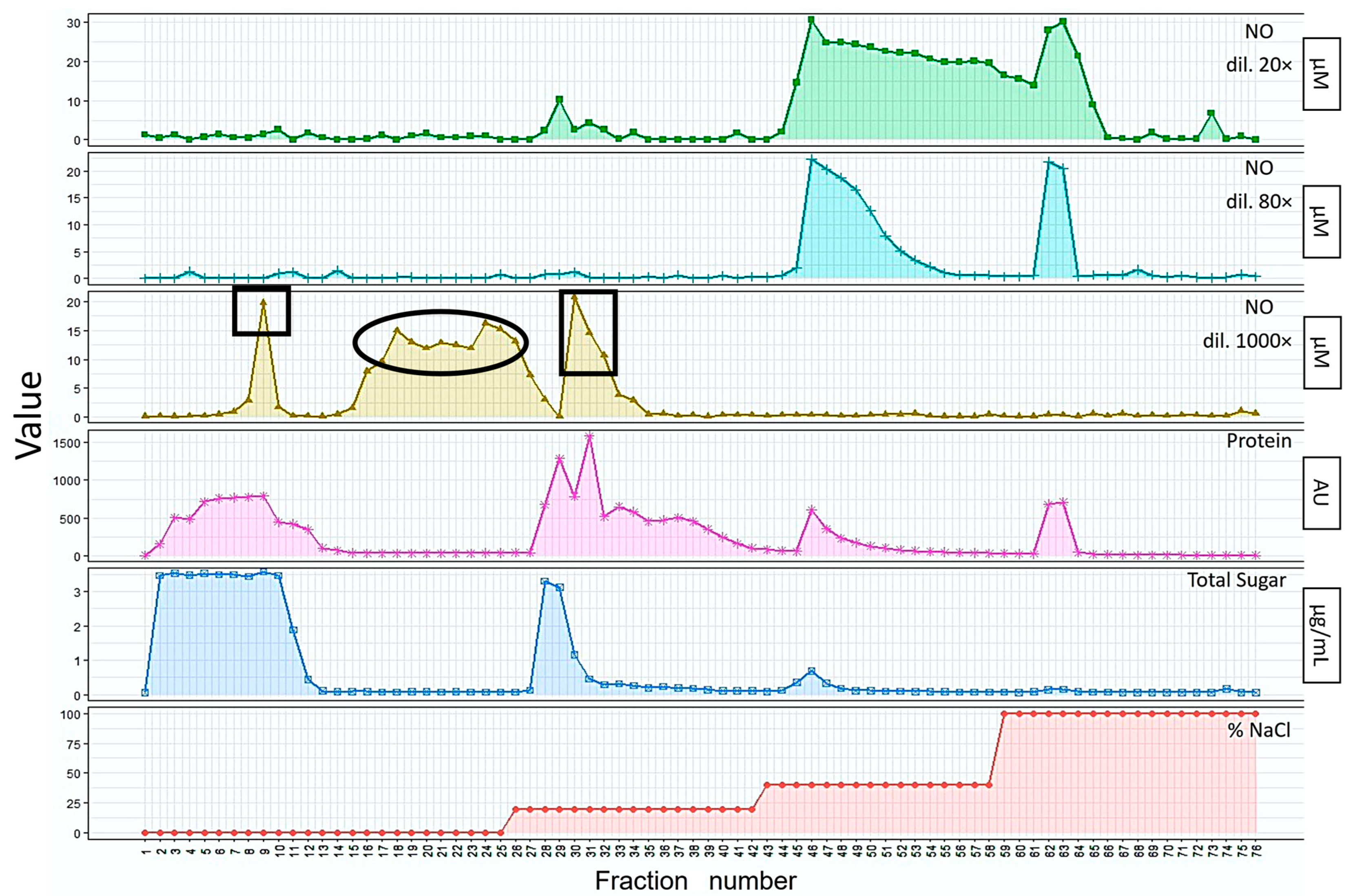
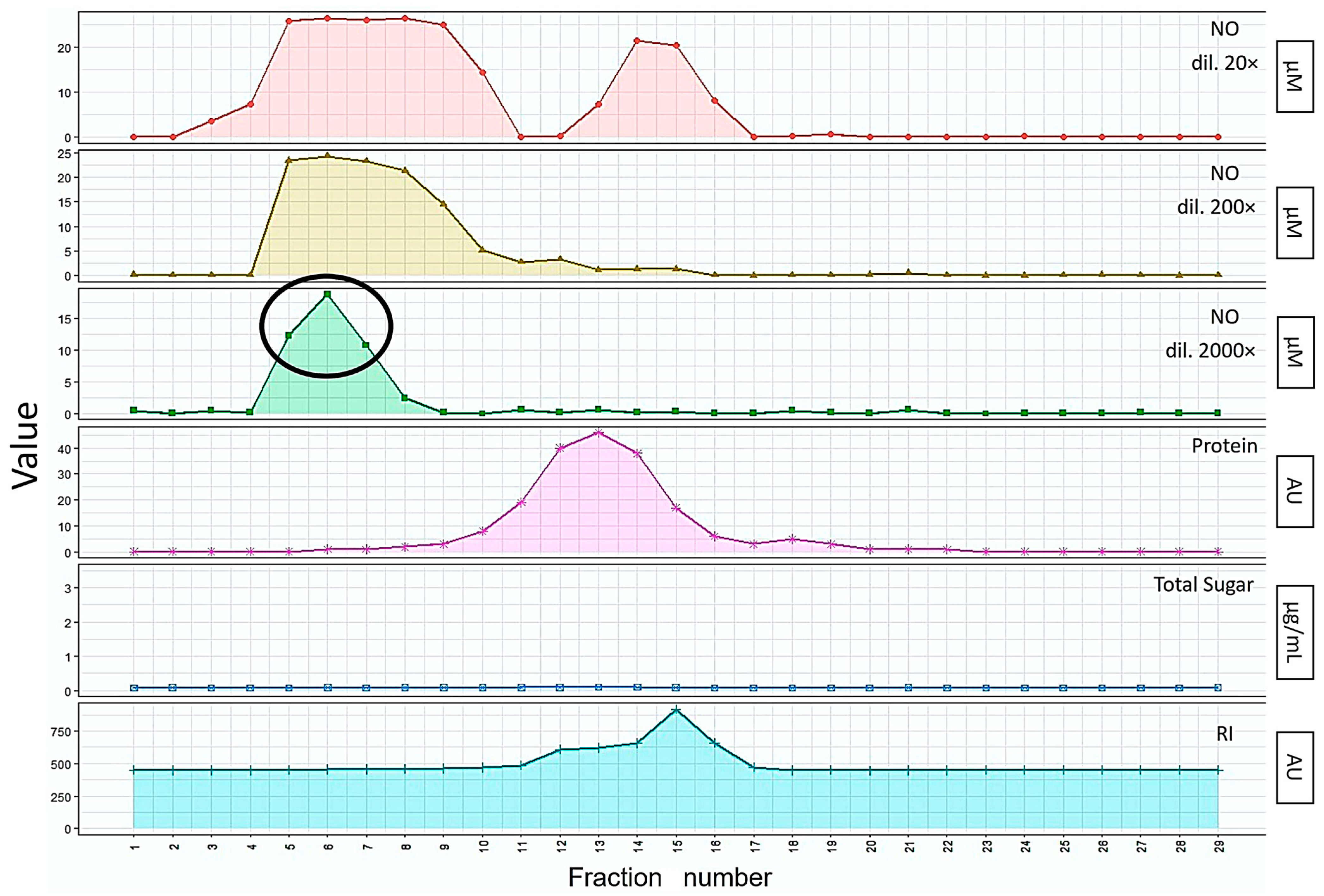
3.1.1. Extraction and Isolation of the Bioactive Substances from Mealworm Larvae

The crude bioactive substances were extracted from mealworm larvae through sequential processing steps, including water extraction, ethanol precipitation, and centrifugation. The immunomodulatory bioactive fraction was isolated by evaluating NO production in RAW264.7 cells. NO-positive fractions were then collected and further separated using AEC on a HiPrep Q XL 16/10 column (GE Healthcare, Chicago, IL, USA) with a linear NaCl gradient from 20% to 100%. Fractions exhibiting high levels of NO activity were selected and further purified via gel-filtration chromatography on a fast protein liquid chromatography system using an ENrich SEC 650 column (Bio-Rad, CA, USA), with 20 mM Tris-HCl containing 200 mM NaCl (pH 8.0) as the elution buffer (Figure 1). This method was adapted from our previous work on insect-derived bioactive substances [6,18,19,20].
From Figure 1, it can be observed that the immunomodulatory peak of the mealworm-derived bioactive substances could be clearly recorded at a 1000-fold dilution. At 20- and 80-fold dilutions, the NO activity was so high that it caused cell death in the RAW cells, rendering them unreadable by the spectrophotometer. Three groups of fractions displayed the greatest NO activity—the first was fraction 9, the second spanned fractions 18 to 26, and the third included fractions 30 to 32. As protein and sugar were detected in the first and third groups (black squares in Figure 1), we presumed these might be glycoproteins or proteoglycans. Based on our previous studies, the insect-derived bioactive substances typically do not contain proteins. Therefore, for further analysis the second group was selected.
Following further purification using size-exclusion chromatography, the results showed that fractions 5 to 7 had the greatest NO activity at up to 2000-fold dilution, with no detectable sugar, protein, or organic acid content (as determined according to the refractive index) (Figure 2). This finding differs from those of our previous study, in which the bioactive substances contained sugars (polysaccharides). Therefore, we presume that this may be a novel bioactive substance derived from mealworms.

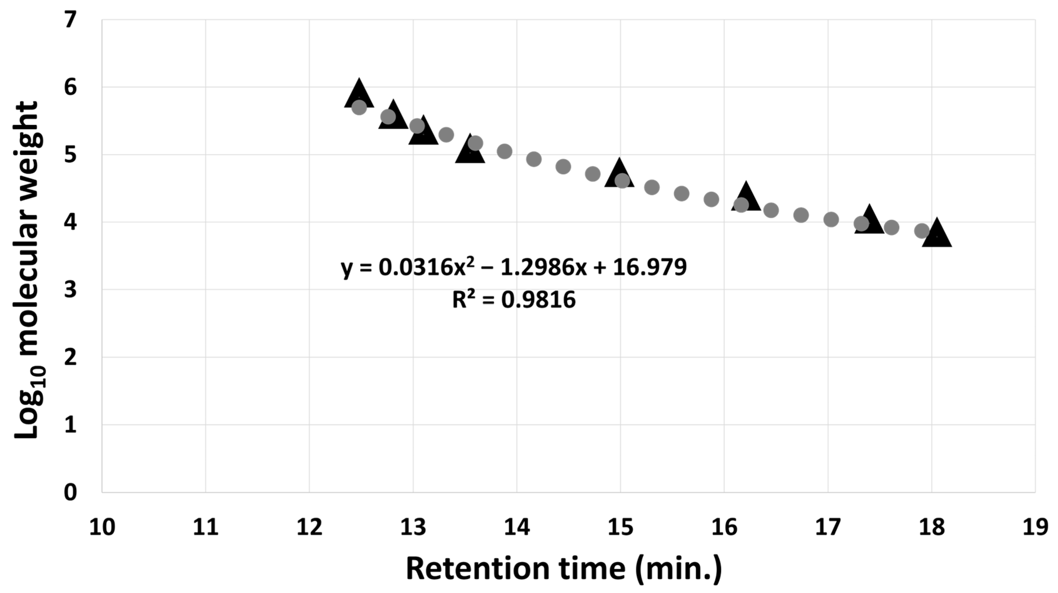
3.1.2. Molecular Weight Determination of Mealworm Bioactive Substance

The molecular weight of the mealworm-derived bioactive substance was determined using HPLC on a Shodex SB-803 HQ column (Resonac, Minato, Japan). The retention times were 12.79 and 12.81 min (in duplicate), corresponding to an average molecular weight of 342.21 kDa. This value was calculated based on the regression equation derived from a standard curve generated using pullulan P-series standards (P-5, P-10, P-20, P-50, P-100, P-200, P-400, and P-800) (Figure 3).

3.2. In Vivo Analysis

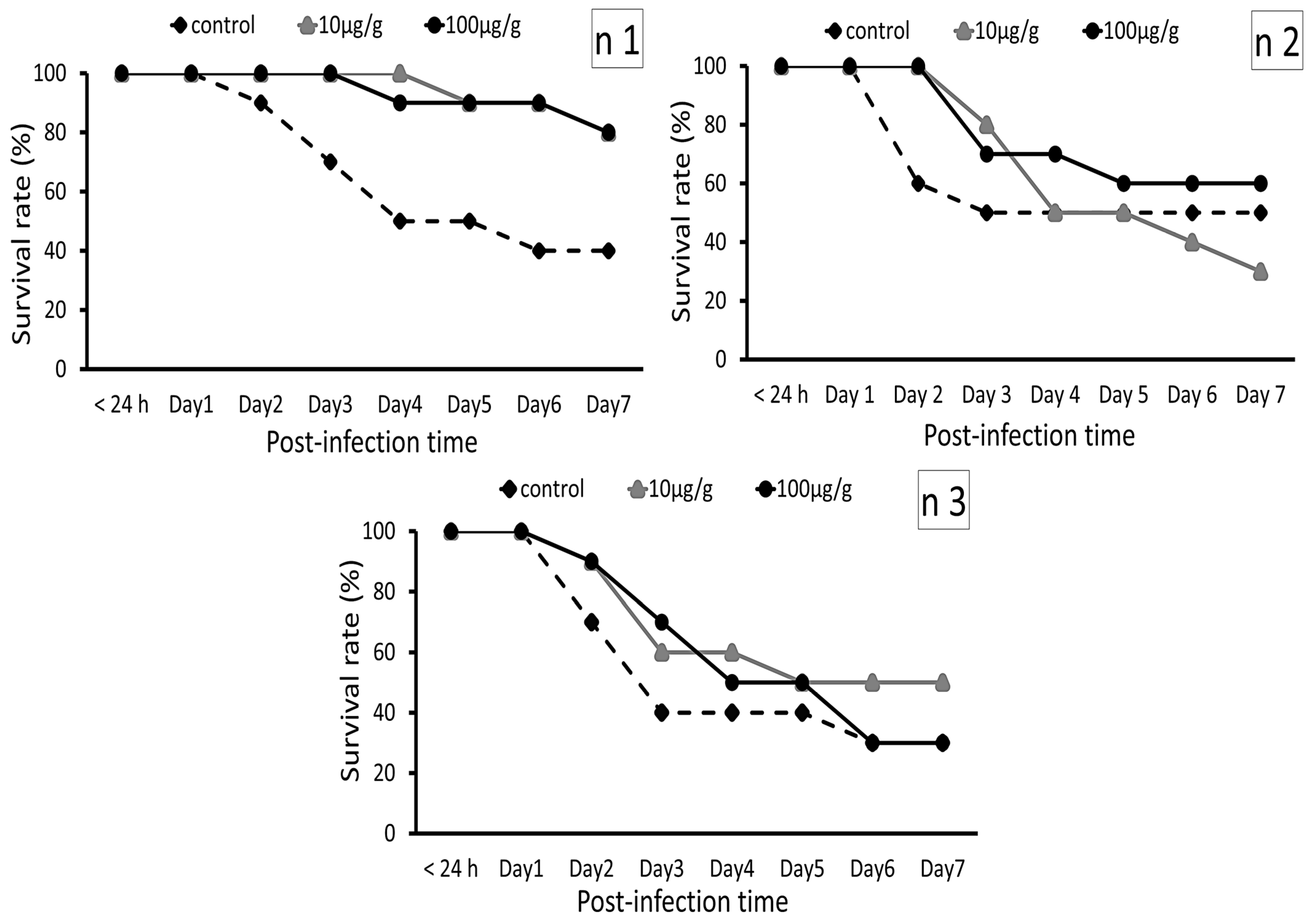
3.2.1. Zebrafish Post-Challenge Evaluation

A challenge test was conducted to assess immunomodulatory responses after zebrafish had been fed on a diet containing mealworm-derived bioactive substance for 14 days (Figure 4). The zebrafish were challenged by E. tarda using the immersion method and were observed for 7 days. The results indicated that the control group experienced its first mortality on day 2 post-challenge, which is earlier than that of the treatment groups. Although no significant differences were observed between the control and treatment groups, the results suggest that the time to mortality was delayed in all treatment groups.

3.2.2. RNA Sequencing Analysis of Zebrafish After Receiving Dietary Mealworm-Derived Bioactive Substance Treatment

Following the challenge test, 100 μg/g of the mealworm-derived bioactive substance was identified to be the optimal concentration for enhancing immunity as a feed supplement (Figure 4). Therefore, we present the challenge test results first, followed by the gene expression and RNA sequencing (RNA-seq) data. To investigate transcriptomic alterations, RNA-seq analysis was performed on the liver and intestine samples from zebrafish that were fed the mealworm-derived bioactive substance (100 μg/g) for 14 days; these were compared to the control group. The proportion of clean reads obtained from RNA-seq ranged from 96.27% to 98.43%, while raw reads ranged from 42,561,946 to 102,178,578 (Table S3).
On average, a successful mapping rate of 90.82% was achieved when clean reads were aligned to the reference genome assembly GRCz11 (GCF_000002035.6) using STAR (version 2.7.11) (Table S4). To visualize the pattern of DEGs, heatmaps based on normalized counts were generated for liver (Figure S1) and intestinal (Figure S2) samples. These data indicated no significant differences between the control and 100 μg/g groups.

3.2.3. Differentially Expressed Genes in Response to Dietary Mealworm-Derived Bioactive Substance

A threshold of log2 fold-change > 1 with FDR < 0.05 was applied to identify DEGs. In the zebrafish intestine samples, DEG analysis using the TCC package (version 1.38) identified 123 downregulated genes and one upregulated gene in the group fed 100 μg/g of mealworm-derived bioactive substance compared with the control group (fed the basal diet). In the zebrafish liver samples, the same analysis revealed one upregulated gene and two downregulated genes (Figure S4A,B).

3.2.4. Differentially Expressed Genes and Functional Enrichment Obtained via Gene Ontology Analysis

After zebrafish were fed the mealworm-derived bioactive substance for 14 days, the molecular pathways in their liver and intestines were examined using GO enrichment analysis. An over-representation analysis (ORA) was then conducted using ClusterProfiler software (version 3.18.1) to compare the DEG results between the control group and the 100 μg/g of dietary mealworm-derived bioactive substance group. Following the dietary mealworm-derived bioactive substance treatment, ORA revealed that there were significant transcriptional changes, particularly in the intestine (p < 0.05). The GO terms associated with these DEGs suggest alterations in various molecular functions (MFs) (Figure S3A,B), cellular components (CCs) (Figure S5), and biological processes (BPs) (Figure S6).
Zebrafish also exhibited significant transcriptional changes following the dietary mealworm-derived bioactive substance treatment. These changes included the downregulation of genes associated with protease inhibition and extracellular matrix components, as well as processes such as coagulation and wound healing. Additionally, the upregulation of genes was involved in the positive regulation of type I interferon production.

3.2.5. Determination of Differentially Expressed Genes Associated with Immune Responses

Immune-related DEGs were identified and classified using data from the literature, the NCBI database, GO annotation, and the KEGG pathway database. Transcriptomic analysis revealed the significant modulation of immune-related genes in zebrafish liver and intestine samples following the 14-day dietary administration of the mealworm-derived bioactive substance. DEGs with a |log2 fold-change| ≥ 1 and an FDR < 0.05 were identified. In the liver, a notable set of immune genes exhibited altered expression patterns. Similarly, in the intestine, the mealworm-derived bioactive substance led to the downregulation of several immune-related genes. The specific genes and their expression ratios are detailed in Table S5.

3.2.6. KEGG Pathway Analysis of Differentially Expressed Genes

A signaling pathway evaluation in the zebrafish intestine and liver samples was conducted using KEGG pathway analysis in ClusterProfiler software. DEGs between the control and dietary mealworm-derived bioactive substance groups were mapped to the KEGG pathway database (https://www.kegg.jp/; accessed on 20 January 2025) and were analyzed for statistical significance (p < 0.05). In the intestine, two signaling pathways were significantly enriched—phenylalanine metabolism (dre00360) and ferroptosis (dre04216). However, no significantly enriched pathways were detected in the liver (Table S6).

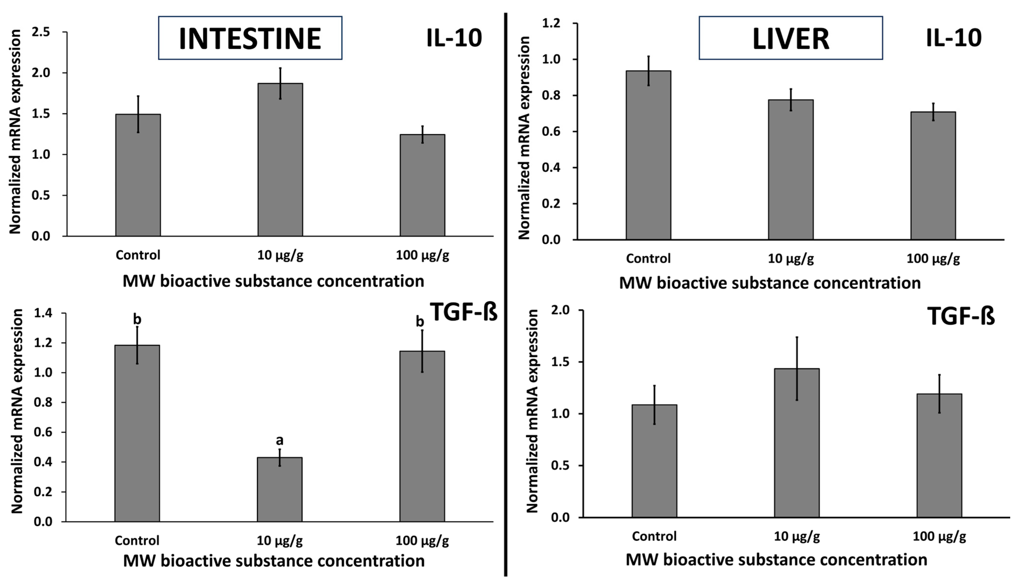
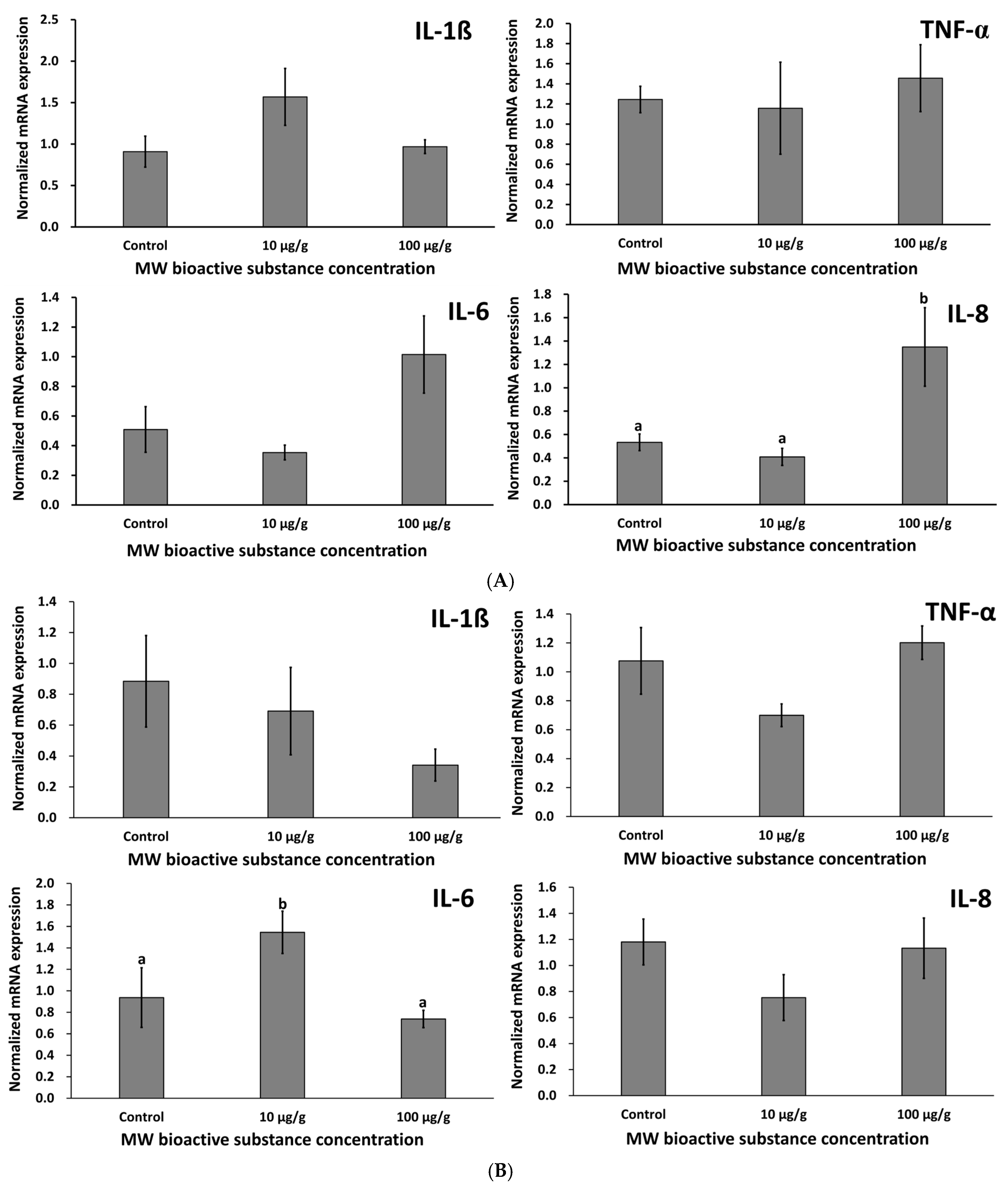
3.2.7. Evaluation of Zebrafish Cytokines

The zebrafish were fed on the mealworm-derived bioactive substance for 14 days at the following doses: 0, 10, and 100 µg/g. Subsequently, five fish from each treatment group were euthanized and dissected to collect liver and intestine samples for RNA extraction. The cytokines analyzed included TGF-β and IL-10 (anti-inflammatory cytokines) (Figure 5), as well as TNF-α, IL-1β, IL-6, and IL-8 (pro-inflammatory cytokines) (Figure 6).
The expression levels of IL-10 in both the zebrafish intestine and liver samples were slightly decreased by the diet containing 100 µg/g mealworm-derived bioactive substance. The highest IL-10 expression in the intestine was observed at 10 µg/g, while in the liver, IL-10 expression was in a dose-dependent manner. We observed that a moderate dose (10 µg/g) significantly suppressed TGF-β expression in the intestine. However, the same dose appeared to increase TGF-β expression in the liver, although no significant differences were observed between the control and treatment groups (p > 0.05).
Although most pro-inflammatory responses showed no significant differences, they tended to increase in the liver following 100 µg/g of the mealworm-derived bioactive substance treatment compared with the control group, except for IL-1β (Figure 6A,B). In the intestine, the 10 µg/g treatment generally resulted in lower mRNA expression levels compared to the control group, with the exception of IL-6. Meanwhile, the 100 µg/g treatment showed expression levels nearly identical to the control group.

4. Discussion

In the present study, a substance with immunomodulatory potential was isolated from mealworms. We used the NO assay to determine the bioactive substances, since it is a well-established indicator of immune activation in macrophages stimulated by natural-derived products [32,33]. However, this substance was not a polysaccharide, unlike the previously reported silkrose in silkworms [21,34] and dipterose in BSF [35]. Additionally, the absence of absorption at 280 nm suggested that it was not a protein.
Various insect species are known to contain multiple bioactive substances with beneficial characteristics [36,37,38]. These substances exhibit diverse pharmacological traits, including anti-tumor, anti-cancer, antiviral, and antimicrobial activities, as well as the ability to enhance resistance to various diseases in humans, animals, and fish [34,39,40,41]. Our study determined that the molecular weight of the mealworm-derived bioactive substance was 342.21 kDa. Previous studies involving bioactive substances derived from insects have reported molecular weights of 147 kDa for dipterose-BSF [16], 315 kDa for silkrose-AY [6], 1010 kDa for dipterose-BC [6], and 1150 kDa for silkrose-BM [17]. As mentioned previously, this mealworm-derived substance contained no sugar, protein, or organic acid. Potential classifications for the mealworm-derived bioactive substance include polysaccharide derivatives [42], polyphenols [43], or even a novel biopolymer [44]. This hypothesis is supported by evidence that insects can synthesize unique biopolymers as components of their cuticle [45], secretions [46], or defensive mechanisms [47]. Despite these findings, the exact identity of this mealworm-derived substance remains unknown. A further chemical and structural analysis of this substance is required.
We conducted an E. tarda challenge test to assess the immunomodulatory potential of the mealworm-derived bioactive substance in zebrafish. The most effective dose for reducing mortality was 100 µg/g (with an average survival rate of 56.67%). However, no significant differences in survival rates were observed between the control and treatment groups. These findings are consistent with an earlier report stating that dipterose-BSF, which is a bioactive substance derived from the black soldier fly, enhanced immunity at the transcriptomic level but did not significantly protect zebrafish when challenged with E. tarda [35]. In contrast, silkrose-BM, which is a bioactive substance derived from silkworm, has been shown to confer strong immune protection in penaeid shrimp [18] and yellowtail [32].
There are two possible explanations for why the mealworm-derived bioactive substance did not significantly alter survival rates in zebrafish. The first is that the mealworm-derived bioactive substance may be poorly compatible with zebrafish, as the effectiveness of these types of substances can vary according to fish species, inclusion levels, and overall diet formulation [48,49]. The second possibility is that the mealworm-derived bioactive substance may require synergistic interactions with other compounds to enhance fish immunity. For instance, Su et al. (2017) demonstrated that replacing 27% of fish meal with mealworm meal improved the survival rate of yellow catfish challenged with E. ictalurid, achieving a survival rate of up to 87.2% [10]. Similarly, Sharifinia et al. (2023) reported that 100% mealworm meal replacement did not impair the growth and enhanced the immune response of Litopenaeus vannamei, with survival rates comparable to the control group (approximately 80%) after 60 days of dietary treatment [50].
RNA-seq analysis indicated that there was a downregulation of genes associated with (a) complement and coagulation systems, (b) chemokine expression, and (c) superoxide dismutase 3 (SOD3) in zebrafish intestines following dietary supplementation with the mealworm-derived bioactive substance (Table S5). We presume that the observed immunosuppression in zebrafish may reflect a transition to the immune resolution phase, through which the organism seeks to restore homeostasis and prevent excessive or chronic inflammation. In contrast, the stimulation of NO production in mouse macrophages reflects early-stage immune activation, which is expected during in vitro screening of bioactive compounds. These differences likely reflect tissue- and species-specific immune regulation mechanisms, as well as differences between in vivo and in vitro responses.
Regarding (a), during the acute phase of an immune response, complement and coagulation activities are upregulated to counter pathogens and initiate healing [51,52]. As the immune response resolves, it is crucial that these systems are downregulated to prevent chronic inflammation and coagulation disorders. Following an initial immune challenge, genes involved in the complement and coagulation cascades are downregulated to facilitate the resolution of inflammation and restore tissue homeostasis [53]. In terms of (b), chemokines are pivotal in recruiting leukocytes to sites of infection or injury [54,55]. The observed downregulation of chemokine genes suggests a reduction in leukocyte recruitment to the intestine, which may indicate a shift toward resolving inflammation. In Atlantic salmon (Salmo salar), the differential expression of chemokines has been linked to variations in immune cell recruitment during infection, highlighting the importance of chemokine regulation in immune responses [56]. Finally, regarding (c), SOD3 plays a critical role in detoxifying reactive oxygen species in the extracellular environment, protecting tissues from oxidative damage during inflammatory responses [57,58]. The downregulation of SOD3 may reflect a decreased oxidative stress burden as the immune response resolves [59]. In rainbow trout (Oncorhynchus mykiss), a significant decrease in SOD activity was observed post-infection, correlating with the resolution phase of the immune response [60].
Collectively, these genes play critical roles in multiple aspects of immune function, including innate defense (e.g., C3), inflammation control (e.g., serpins and acute-phase proteins such as C-reactive protein and haptoglobin), and leukocyte trafficking (e.g., chemokines and their receptors) [51,61,62,63,64]. The coordinated downregulation of these genes likely reflects a systemic shift toward the restoration of intestinal immune balance in zebrafish.
The KEGG pathway schematic for phenylalanine metabolism (dre00360) illustrates that phenylalanine serves as a precursor for tyrosine, which is essential for the synthesis of catecholamines and melanin, both of which are involved in immune signaling [65] (Figure S7). In fish, amino acid metabolism plays a crucial role in immune regulation, and phenylalanine-derived metabolites have been shown to modulate oxidative stress, which is a key factor in maintaining immune homeostasis [66]. The activation of ferroptosis suggests a complex interaction between cell death, iron metabolism, and immune responses [67], potentially indicating alterations in gut immunity following mealworm-derived bioactive substance treatment (Figure S8).
In fish, the proper functioning of the intestine and liver requires a balance of pro-inflammatory and anti-inflammatory cytokines [68]. TGF-β and IL-10 are well known for their anti-inflammatory effects [69]. In our study, although neither cytokine showed significant differences between the control and treatment groups, IL-10 expression tended to be suppressed, while TGF-β expression tended to increase in both the intestine and liver following 100 µg/g dietary treatment with the mealworm-derived bioactive substance. This suggests an attempt to restore balance during inflammation. This finding aligns with a previous report that TGF-β immunomodulatory effects are predominantly suppressive [70]. Furthermore, the increased expression of TGF-β has been observed in fish immune system-associated tissues [71].
The findings of this study regarding pro-inflammatory gene expression, particularly IL-1β (Figure 6), are in accordance with a study in which black soldier fly meal (BSFM) was used as a replacement in Nile tilapia feed [72]. In that study, it was found that in the intestine, IL-1β expression was downregulated when the diet was replaced with more than 50% BSFM, while other inflammatory cytokines, such as IL-8, IL-10, and TNF-α, were unaffected. These findings strongly suggest that homeostasis is maintained when fish are introduced to a novel diet.
IL-8 and TNF-α are produced by macrophages and are known to stimulate an inflammatory response [68,73]. They are also widely recognized as pro-inflammatory cytokines [74]. IL-6 can function as both a pro- and anti-inflammatory cytokine [75] and is upregulated in response to PAMPs such as peptidoglycan [76], LPS [77], and bacterial DNA [78]. In this study, the mealworm-derived bioactive substance, up to a dose of 100 µg/g, did not induce overexpression of pro-inflammatory cytokines in the intestine (Figure 6B). This lack of overexpression is an indicator of gut integrity, as excessive inflammation can damage the intestinal mucosal barrier [79]. Interestingly, IL-8 expression was significantly upregulated in the liver (Figure 6A). IL-8 is a chemokine that is activated through pattern recognition receptors (PRRs); it plays an essential role in host defense [80], especially in response to viral [81], bacterial [82], and parasitic infections [83]. The upregulation of IL-8 in the liver suggests that the 100 µg/g dose of the bioactive substance may function as an immunomodulator, stimulating localized immune readiness without eliciting harmful systemic inflammation.
Our findings demonstrate that the mealworm-derived bioactive substance can regulate inflammatory cytokine expression in fish. Despite the lack of a significant expression of pro-inflammatory cytokines in the liver, an increased trend in their expression at the 100 µg/g dose was observed. The liver is widely recognized to be an “immunity hub” because it can activate inflammatory responses through the chemokine-mediated recruitment of monocytes, neutrophils, natural killer cells, and natural killer T cells [84]. It also balances anti-inflammatory and pro-inflammatory T cell activity [85] and processes immune molecules, antigens, microbial products, and lymphocytes via enterohepatic circulation [85,86]. Additionally, gut immunity can be modulated by bile, which contains immune system molecules such as cytokines, chemokines, and antibodies [87]. These findings suggest that the mealworm-derived bioactive substance may have systemic immunomodulatory effects, contributing to balanced inflammatory responses in fish.

5. Conclusions

Transcriptome analysis revealed that dietary supplementation with the mealworm-derived bioactive substance at a concentration of 100 µg/g modulated immune responses in zebrafish, potentially enhancing resistance to bacterial infections. Although the survival rate showed no significant difference in the treatment groups compared with the control group, the delayed mortality observed in the 100 µg/g treatment group suggests a protective effect compared with the control. These findings highlight the potential of insect-derived bioactive substances as a promising strategy for disease management in aquaculture. Given the possibility of it being a novel bioactive substance, further characterization of the mealworm-derived bioactive substance is warranted to elucidate its immunomodulatory mechanisms and optimize its application in aquafeeds. In addition, longer immunostimulation periods could overwhelm and suppress the immune system, and further study with low dose for longer periods, more practical in fish farm settings, should be studied in the future.

Supplementary Materials

The following supporting information can be downloaded at https://www.mdpi.com/article/10.3390/fishes10060285/s1. Figure S1. Heatmap of differentially expressed genes (DEGs) in the zebrafish liver after 14 days of dietary supplementation with the mealworm bioactive substance. Samples (n = 4) are represented in columns, while DEGs are displayed in rows. Sample groupings are labeled as control (C) and 100 µg/g treatment (100), with individual sample numbers indicated. Figure S2. Heatmap of differentially expressed genes (DEGs) in the zebrafish intestine after 14 days of dietary supplementation with the mealworm bioactive substance. Samples (n = 4) are represented in columns, while DEGs are displayed in rows. Sample groupings are labeled as control (C) and 100 µg/g treatment (100), with individual sample numbers indicated. Figure S3. Gene Ontology (GO) analysis of molecular function (MF) terms in the zebrafish intestine based on differentially expressed genes (DEGs) identified in the comparison between the mealworm bioactive substance group and the control group. (a) Upregulated DEGs; (b) downregulated DEGs. Figure S4. Transcriptional profiling analysis of the intestine (a) and liver (b) of zebrafish from the control group compared to those fed a diet containing 100 μg/g of mealworm bioactive substance. DEGs were identified using the TCC package in R software (version 1.38). Figure S5. Gene Ontology (GO) analysis of cellular components (CC) terms in the zebrafish intestine based on differentially expressed genes (DEGs) identified in the comparison between the mealworm bioactive substance group and the control group. Figure S6. Gene Ontology (GO) analysis of biological process (BP) terms in the zebrafish intestine based on differentially expressed genes (DEGs) identified in the comparison between the mealworm bioactive substance group and the control group. (a) Upregulated DEGs; (b) Downregulated DEGs. Figure S7. Differentially expressed genes (DEGs) related to protein processing in phenylalanine metabolism (dre00360), identified through the comparison between the mealworm bioactive substance inclusion group and the control (basal diet) group in the zebrafish intestine. Downregulated genes are shown in green, while upregulated genes are shown in red. Figure S8. Differentially expressed genes (DEGs) related to protein processing in ferroptosis (dre04216), identified through the comparison between the mealworm bioactive substance inclusion group and the control (basal diet) group in the zebrafish intestine. Downregulated genes are shown in green, while upregulated genes are shown in red. Table S1. Feed Formulation of Zebrafish. Table S2. Primers used in qRT-PCR. Table S3. Summary of RNA-seq results, including raw reads, clean reads, and Q20 and Q30 quality scores (n = 4). Table S4. Summary of RNA-seq clean reads and mapping results (n = 4). Table S5. Lists of immune-related differentially expressed genes (|log2 fold-change| ≥ 1 and FDR < 0.05) and their expression ratios in the liver and intestine of zebrafish after 14 days fed by mealworm bioactive substance. Table S6. KEGG Pathway Enrichment Analysis (p < 0.05) of DEGs Between Control and 100 µg/g Mealworm Bioactive Substance Group Using ClusterProfiler (ver. 3.18.1).

Author Contributions

Conceptualization: C.M., T.M. and M.F.Z.A.; Methodology: I.B.B.S., M.F.Z.A., S.A. and H.N.; Software: M.F.Z.A. and S.A.; Validation: C.M. and T.M.; Formal analysis: I.B.B.S. and M.F.Z.A.; Investigation: I.B.B.S., M.F.Z.A., S.A. and H.N.; Resources: C.M. and T.M.; Data curation: I.B.B.S., M.F.Z.A., S.A. and H.N.; Writing—original draft preparation: I.B.B.S.; Writing—review and editing: I.B.B.S., M.F.Z.A. and T.M.; Supervision: T.M.; Project administration: C.M.; Funding acquisition: T.M. All authors have read and agreed to the published version of the manuscript.

Funding

This work was supported by the JSPS Kakenhi (25K02097) (https://www.jsps.go.jp/english/e-grants/; accessed on 1 April 2024) and TAIYO OIL CO., LTD (Z21-100094) (https://www.taiyooil.net/english/; accessed on 1 June 2023).

Institutional Review Board Statement

All experiments were conducted in accordance with protocols approved by the Institutional Animal Care and Use Committee (IACUC) of Ehime University (Permit Number: 08K2-1).

Informed Consent Statement

Not applicable.

Data Availability Statement

Supporting data for this research are available from the corresponding author upon reasonable request.

Acknowledgments

We would like to express our sincere gratitude to Yuki Otsu for their invaluable guidance in using R software and for their dedicated assistance in maintaining the fish when we were unable to be on campus. Their support and commitment were instrumental in ensuring the smooth progress of this research.

Conflicts of Interest

The authors declare no conflicts of interest.

References

  1. Ratcliffe, N.; Azambuja, P.; Mello, C.B. Recent advances in developing insect natural products as potential modern day medicines. Evid. Based Complement. Altern. Med. 2014, 2014, 904958. [Google Scholar] [CrossRef] [PubMed]
  2. Park, S.I.; Kim, J.W.; Yoe, S.M. Purification and characterization of a novel antibacterial peptide from black soldier fly (Hermetia illucens) larvae. Dev. Comp. Immunol. 2015, 52, 98–106. [Google Scholar] [CrossRef]
  3. Elhag, O.; Zhou, D.; Song, Q.; Soomro, A.A.; Cai, M.; Zheng, L.; Yu, Z.; Zhang, J. Screening, expression, purification and functional characterization of novel antimicrobial peptide genes from Hermetia illucens (L.). PLoS ONE 2017, 12, e0169582. [Google Scholar] [CrossRef]
  4. Gasco, L.; Finke, M.; van Huis, A. Can diets containing insects promote animal health? J. Insects Food Feed 2018, 4, 1–4. [Google Scholar] [CrossRef]
  5. Katayama, N.; Ishikawa, Y.; Takaoki, M.; Yamashita, M.; Nakayama, S.; Kiguchi, K.; Kok, R.; Wada, H.; Mitsuhashi, J.; Space Agriculture Task Force. Entomophagy: A key to space agriculture. Adv. Space Res. 2008, 41, 701–705. [Google Scholar] [CrossRef]
  6. Ohta, T.; Ido, A.; Kusano, K.; Miura, C.; Miura, T. A novel polysaccharide in insects activates the innate immune system in mouse macrophage RAW264 cells. PLoS ONE 2014, 9, e114823. [Google Scholar] [CrossRef]
  7. Son, Y.J.; Hwang, I.K.; Nho, C.W.; Kim, S.M.; Kim, S.H. Determination of carbohydrate composition in mealworm (Tenebrio molitor L.) larvae and characterization of mealworm chitin and chitosan. Foods 2021, 10, 640. [Google Scholar] [CrossRef]
  8. Liu, C.; Masri, J.; Perez, V.; Maya, C.; Zhao, J. Growth performance and nutrient composition of mealworms (Tenebrio molitor) fed on fresh plant materials-supplemented diets. Foods 2020, 9, 151. [Google Scholar] [CrossRef]
  9. Gasco, I.; Józefiak, A.; Henry, M. Beyond the protein concept: Health aspects of using edible insects on animals. J. Insects Food Feed 2021, 7, 715–741. [Google Scholar] [CrossRef]
  10. Su, J.; Gong, Y.; Cao, S.; Lu, F.; Hand, D.; Liu, H.; Jin, J.; Yang, Y.; Zhu, X.; Xie, S. Effects of dietary Tenebrio molitor meal on the growth performance, immune response and disease resistance of yellow catfish (Pelteobagrus fulvidraco). Fish Shellfish Immunol. 2017, 69, 59–66. [Google Scholar] [CrossRef]
  11. Sankian, Z.; Khosravi, S.; Kim, Y.O.; Lee, S.M. Effects of dietary inclusion of yellow mealworm (Tenebrio molitor) meal on growth performance, feed utilization, body composition, plasma biochemical indices, selected immune parameters and antioxidant enzyme activities of mandarin fish (Siniperca scherzeri) juveniles. Aquaculture 2018, 496, 79–87. [Google Scholar] [CrossRef]
  12. Choi, I.H.; Kim, J.M.; Kim, N.J.; Kim, J.D.; Park, C.; Park, J.H.; Chung, T.H. Replacing fish meal by mealworm (Tenebrio molitor) on the growth performance and immunologic responses of white shrimp (Litopenaeus vannamei). Acta Sci. Anim. Sci. 2018, 40, e35015. [Google Scholar] [CrossRef]
  13. Motte, C.; Rios, A.; Lefebvre, T.; Do, H.; Henry, M.; Jintasataporn, O. Replacing fish meal with defatted insect meal (yellow mealworm Tenebrio molitor) improves the growth and immunity of Pacific white shrimp (Litopenaeus vannamei). Animals 2019, 9, 258. [Google Scholar] [CrossRef] [PubMed]
  14. Henry, M.A.; Gai, F.; Enes, P.; Peréz-Jiménez, A.; Gasco, L. Effect of partial dietary replacement of fishmeal by yellow mealworm (Tenebrio molitor) larvae meal on the innate immune response and intestinal antioxidant enzymes of rainbow trout (Oncorhynchus mykiss). Fish Shellfish Immunol. 2018, 83, 308–313. [Google Scholar] [CrossRef]
  15. Jeong, S.M.; Khosravi, S.; Mauliasari, I.R.; Lee, S.M. Dietary inclusion of mealworm (Tenebrio molitor) meal as an alternative protein source in practical diets for rainbow trout (Oncorhynchus mykiss) fry. Fish. Aquat. Sci. 2020, 23, 12. [Google Scholar] [CrossRef]
  16. Ali, M.F.Z.; Ohta, T.; Ido, A.; Miura, C.; Miura, T. The dipterose of black soldier fly (Hermetia illucens) induces innate immune response through toll-like receptor pathway in mouse macrophage RAW264.7 cells. Biomolecules 2019, 9, 677. [Google Scholar] [CrossRef]
  17. Ohta, T.; Kusano, K.; Ido, A.; Miura, C.; Miura, T. Silkrose: A novel acidic polysaccharide from the silkmoth that can stimulate the innate immune response. Carbohydr. Polym. 2016, 136, 995–1001. [Google Scholar] [CrossRef]
  18. Ali, M.F.Z.; Yasin, I.A.; Ohta, T.; Hashizume, A.; Ido, A.; Takahashi, T.; Miura, C.; Miura, T. The silkrose of Bombyx mori effectively prevent vibriosis in penaeid prawns via the activation of innate immunity. Sci. Rep. 2018, 8, 8836. [Google Scholar] [CrossRef]
  19. Yan, Z.; Hansson, G.K. Innate immunity, macrophage activation, and atherosclerosis. Immunol. Rev. 2007, 219, 187–203. [Google Scholar] [CrossRef]
  20. Li, D.; Wu, M. Pattern recognition receptors in health and diseases. Signal. Transduct. Target. Ther. 2021, 6, 291. [Google Scholar] [CrossRef]
  21. Ali, M.F.Z.; Kameda, K.; Kondo, F.; Iwai, T.; Kurniawan, R.A.; Ohta, T.; Ido, A.; Takahashi, T.; Miura, C.; Miura, T. Effects of dietary silkrose of Antheraea yamamai on gene expression profiling and disease resistance to Edwardsiella tarda in Japanese medaka (Oryzias latipes). Fish Shellfish Immunol. 2021, 114, 207–217. [Google Scholar] [CrossRef] [PubMed]
  22. Dubois, M.; Gilles, K.A.; Hamilton, J.K.; Rebers, P.A.; Smith, F. Colorimetric method for determination of sugars and related substances. Anal. Chem. 1956, 28, 350–356. [Google Scholar] [CrossRef]
  23. Livak, K.J.; Schmittgen, T.D. Analysis of relative gene expression data using real-time quantitative PCR and the 2−ΔΔCT method. Methods 2001, 25, 402–408. [Google Scholar] [CrossRef]
  24. McCurley, A.T.; Callard, G.V. Characterization of housekeeping genes in zebrafish: Male-female differences and effects of tissue type, developmental stage and chemical treatment. BMC Mol. Biol. 2008, 9, 102. [Google Scholar] [CrossRef]
  25. Martin, M. Cutadapt removes adapter sequences from high-throughput sequencing reads. EMBnet J. 2011, 17, 10–12. [Google Scholar] [CrossRef]
  26. Li, H.; Handsaker, B.; Wysoker, A.; Fennell, T.; Ruan, J.; Homer, N.; Marth, G.; Abecasis, G.; Durbin, R.; 1000 Genome Project Data Processing Subgroup. The sequence alignment/map format and SAMtools. Bioinformatics 2009, 25, 2078–2079. [Google Scholar] [CrossRef]
  27. Liao, Y.; Smyth, G.K.; Shi, W. FeatureCounts: An efficient general purpose program for assigning sequence reads to genomic features. Bioinformatics 2014, 30, 923–930. [Google Scholar] [CrossRef]
  28. Sun, J.; Nishiyama, T.; Shimizu, K.; Kadota, K. TCC: An R package for comparing tag count data with robust normalization strategies. BMC Bioinform. 2013, 14, 219. [Google Scholar] [CrossRef]
  29. Wilkinson, L. Ggplot2: Elegant graphic for data analysis by WICKHAM, H. Biometerics 2011, 67, 678–679. [Google Scholar] [CrossRef]
  30. Moulos, P.; Hatzis, P. Systematic integration of RNA-Seq statistical algorithms for accurate detection of differential gene expression patterns. Nucleid Acid Res. 2015, 43, e25. [Google Scholar] [CrossRef]
  31. Yu, G.; Wang, L.G.; Han, Y.; He, Q.Y. ClusterProfiler: An R package for comparing biological themes among gene clusters. Omics A J. Integr. Biol. 2012, 16, 284–287. [Google Scholar] [CrossRef] [PubMed]
  32. Kumar-Roiné, S.; Matsui, M.; Reybier, K.; Darius, H.T.; Chinain, M.; Pauillac, S.; Laurent, D. Ability of certain plant extracts traditionally used to treat ciguatera fish poisoning to inhibit nitric oxide production in RAW 264.7 macrophages. J. Ethnopharmacol. 2009, 123, 369–377. [Google Scholar] [CrossRef] [PubMed]
  33. Zhang, H.P.; Zhang, D.D.; Ke, Y.; Bian, K. The vasodilatory effects of anti-inflammatory herb medications: A comparison study of four botanical extracts. Evid. Based Complement. Altern. Med. 2017, 2017, 1021284. [Google Scholar] [CrossRef]
  34. Miura, T.; Nishikawa, M.; Otsu, Y.; Ali, M.F.Z.; Hashizume, A.; Miura, C. The effects of silkworm-derived polysaccharide (silkrose) on ectoparasitic infestations in yellowtail (Seriola quinqueradiata) and white trevally (Pseudocaranx dentex). Fishes 2022, 7, 14. [Google Scholar] [CrossRef]
  35. Nishiguchi, H.; Suryadi, I.B.B.; Ali, M.F.Z.; Miura, C.; Miura, T. Dietary black soldier fly (Hermetia illucens)—Dipterose-BSF—Enhanced zebrafish innate immunity gene expression and resistance to Edwardsiella tarda infection. Insects 2024, 15, 326. [Google Scholar] [CrossRef]
  36. Maeda, S.; Kawai, T.; Obinata, M.; Fujiwara, H.; Horiuchi, T.; Saeki, Y.; Sato, Y.; Furusawa, M. Production of human α-interferon in silkworm using a baculovirus vector. Nature 1985, 315, 592–594. [Google Scholar] [CrossRef]
  37. Liou, Y.C.; Tocilj, A.; Davies, P.L.; Jia, Z. Mimicry of ice structure by surface hydroxyls and water of a β-helix antifreeze protein. Nature 2000, 406, 322–324. [Google Scholar] [CrossRef]
  38. Hirose, Y.; Ohta, E.; Kawai, Y.; Ohta, S. Dorsamin-A’s, glycerolipids carrying a dehydrophenylalanine ester moiety from the seed-eating larvae of the bruchid beetle Bruchidius dorsalis. J. Nat. Prod. 2013, 76, 554–558. [Google Scholar] [CrossRef]
  39. Akiyama, N.; Hijikata, M.; Kobayashi, A.; Yamori, T.; Tsuruo, T.; Natori, S. Anti-tumor effect of N-beta-alanyl-5-S-glutathionyldihydroxyphenylalanine (5-S-GAD), a novel anti-bacterial substance from an insect. Anticancer Res. 2000, 20, 357–362. [Google Scholar] [PubMed]
  40. Chernysh, S.; Kim, S.I.; Bekker, G.; Pleskach, V.A.; Filatova, N.A.; Anikin, V.B.; Platonov, V.G.; Bulet, P. Antiviral and antitumor peptides from insects. Proc. Natl. Acad. Sci. USA 2002, 99, 12628–12632. [Google Scholar] [CrossRef]
  41. Yang, P.; Abe, S.; Zhao, Y.P.; An, Y.; Suzuki, K. Growth suppression of rat hepatoma cells by a pentapeptide from Antheraea yamamai. J. Insect Biotechnol. Sericology 2004, 73, 7–13. [Google Scholar] [CrossRef]
  42. Olawuyi, I.G.; Heo, E.; Jeong, M.; Kim, J.H.; Park, J.J.; Chae, J.; Gwon, S.; Lee, S.D.; Kim, H.; Ojulari, O.V.; et al. Acidic polysaccharide from the edible insect Protaetia brevitarsis seulensis activates antiviral immunity to suppress norovirus infection. Carbohydr. Polym. 2025, 347, 122587. [Google Scholar] [CrossRef] [PubMed]
  43. da Silva, E.S.; Xiong, J.; de Medeiros, F.G.M.; Grace, M.; Moncada, M.; Lila, M.A.; Hoskin, R.T. Spray dried insect protein-polyphenol particles deliver health-relevant value-added food ingredients. Future Foods 2024, 9, 100315. [Google Scholar] [CrossRef]
  44. Guarneri, A.; Triunfo, M.; Ianniciello, D.; Tedesco, F.; Salvia, R.; Scieuzo, C.; Schmitt, E.; Capece, A.; Falabella, P. Insect-derived chitosan, a biopolymer for the increased shelf life of white and red grapes. Int. J. Biol. Macromol. 2024, 275, 133149. [Google Scholar] [CrossRef]
  45. Wu, N.; Lin, Q.; Shao, F.; Chen, L.; Zhang, H.; Chen, K.; Wu, J.; Wang, G.; Wang, H.; Wang, H.; et al. Insect cuticle-inspired design of sustainably sourced composite bioplastics with enhanced strength, toughness and stretch-strengthening behavior. Carbohydr. Polym. 2024, 333, 121970. [Google Scholar] [CrossRef]
  46. Shirk, B.D.; Heichel, D.L.; Eccles, L.E.; Rodgers, L.I.; Lateef, A.H.; Burke, K.A.; Stoppel, W.L. Modifying naturally occurring, nonmammalian-sourced biopolymers for biomedical applications. ACS Biomater. Sci. Eng. 2024, 10, 5915–5938. [Google Scholar] [CrossRef]
  47. Sugumaran, M.; Evans, J.J. Catecholamine derivatives as novel crosslinkers for the synthesis of versatile biopolymers. J. Funct. Biomater. 2023, 14, 449. [Google Scholar] [CrossRef]
  48. Mente, E. Insects: An alternative for fish and human nutrition. Public Health Toxicol. 2022, 2, A27. [Google Scholar] [CrossRef]
  49. Zlaugotne, B.; Pubule, J.; Blumberga, D. Advantages and disadvantages of using more sustainable ingredients in fish feed. Heliyon 2022, 8, e10527. [Google Scholar] [CrossRef]
  50. Sharifinia, M.; Bahmanbeigloo, Z.A.; Keshavarzifard, M.; Khanjani, M.H.; Daliri, M.; Koochacknejad, E.; Jasour, M.S. Fishmeal replacement by mealworm (Tenebrio molitor) in diet of farmed Pacific white shrimp (Litopenaeus vannamei): Effects on growth performance, serum biochemistry, and immune response. Aquat. Living Resour. 2023, 36, 19. [Google Scholar] [CrossRef]
  51. Lü, A.; Hu, X.; Wang, Y.; Shen, X.; Zhu, A.; Shen, L.; Ming, Q.; Feng, Z. Comparative analysis of the acute response of zebrafish Danio rerio skin to two different bacterial infections. J. Aquat. Anim. Health 2013, 25, 243–251. [Google Scholar] [CrossRef] [PubMed]
  52. Gerwick, L.; Bayne, C.J. The acute phase response and innate immunity of fish. Dev. Comp. Immunol. 2001, 25, 725–743. [Google Scholar] [CrossRef]
  53. Robledo, D.; Taggart, J.B.; Ireland, J.H.; McAndrew, B.J.; Starkey, W.G.; Haley, C.S.; Hamilton, A.; Guy, D.R.; Mota-Velasco, J.C.; Gheyas, A.A.; et al. Gene expression comparison of resistant and susceptible Atlantic salmon fry challenged with infectious pancreatic necrosis virus reveals a marked contrast in immune response. BMC Genom. 2016, 17, 279. [Google Scholar] [CrossRef] [PubMed]
  54. Sommer, F.; Torraca, V.; Meijer, A.H. Chemokine receptors and phagocyte biology in zebrafish. Front. Immunol. 2020, 11, 325. [Google Scholar] [CrossRef]
  55. Barreda, D.R.; Soliman, A.M. The acute inflammatory response of teleost fish. Dev. Comp. Immunol. 2023, 146, 104731. [Google Scholar] [CrossRef]
  56. Tadiso, T.M.; Krasnov, A.; Skugor, S.; Afanasyef, S.; Hordvik, I.; Nilsen, F. Gene expression analyses of immune responses in Atlantic salmon during early stages of infection by salmon louse (Lepeophtheirus salmonis) revealed bi-phasic responses coinciding with the copepod-chalimus transition. BMC Genom. 2011, 12, 141. [Google Scholar] [CrossRef]
  57. Matthiesen, C.L.; Hu, L.; Torslev, A.S.; Poulsen, E.T.; Larsen, U.G.; Kjaer-Sorensen, K.; Thomsen, J.S.; Brüel, A.; Enghild, J.J.; Oxvig, C.; et al. Superoxide dismutase 3 is expressed in bone tissue and required for normal bone homeostasis and mineralization. Free Radic. Biol. Med. 2021, 164, 399–409. [Google Scholar] [CrossRef]
  58. Wang, Y.; Noman, A.; Zhang, C.; Al-Bukhaiti, W.; Abed, S.M. Effect of fish-heavy metals contamination on the generation of reactive oxygen species and its implications on human health: A review. Front. Mar. Sci. 2024, 11, 1500870. [Google Scholar] [CrossRef]
  59. Hong, Y.; Boiti, A.; Vallone, D.; Foulkes, N.S. Reactive oxygen species signaling and oxidative stress: Transcriptional regulation and evolution. Antioxidants 2024, 13, 312. [Google Scholar] [CrossRef]
  60. Fajardo, C.; Santos, P.; Passos, R.; Vaz, M.; Azeredo, R.; Machado, M.; Fernández-Boo, S.; Baptista, T.; Costas, B. Functional and molecular immune response of rainbow trout (Oncorhynchus mykiss) following challenge with Yersinia ruckeri. Int. J. Mol. Sci. 2022, 23, 3096. Available online: https://pubmed.ncbi.nlm.nih.gov/35328519/ (accessed on 30 January 2025). [CrossRef]
  61. Ricklin, D.; Reis, E.S.; Mastellos, D.C.; Gros, P.; Lambris, J.D. Complement component C3—The “Swiss Army Knife” of innate immunity and host defense. Immunol. Rev. 2017, 274, 33–58. [Google Scholar] [CrossRef] [PubMed]
  62. Delvaeye, M.; Conway, E.M. Coagulation and innate immune responses: Can we view them separately? Blood 2009, 114, 2367–2374. [Google Scholar] [CrossRef] [PubMed]
  63. Smith, N.C.; Rise, M.L.; Christian, S.L. A comparison of the innate and adaptive immune systems in cartilaginous fish, ray-finned fish, and lobe-finned fish. Front. Immunol. 2019, 10, 2292. [Google Scholar] [CrossRef]
  64. Charlie-Silva, I.; Klein, A.; Gomes, J.M.M.; Prado, E.J.R.; Moraes, A.C.; Eto, S.F.; Fernandes, D.C.; Fagliari, J.J.; Junior, J.D.C.; Lima, C.; et al. Acute-phase proteins during inflammatory reaction by bacterial infection: Fish-model. Sci. Rep. 2019, 9, 4776. [Google Scholar] [CrossRef]
  65. Zhang, S.; Wang, C.; Liu, S.; Wang, Y.; Lu, S.; Han, S.; Jiang, H.; Liu, H.; Yang, Y. Effect of dietary phenylalanine on growth performance and intestinal health of triploid rainbow trout (Oncorhynchus mykiss) in low fishmeal diets. Front. Nutr. 2023, 10, 1008822. [Google Scholar] [CrossRef]
  66. Yi, C.; Liang, H.; Huang, D.; Yu, H.; Xue, C.; Gu, J.; Chen, X.; Wang, Y.; Ren, M.; Zhang, L. Phenylalanine plays important roles in regulating the capacity of intestinal immunity, antioxidants and apoptosis in largemouth bass (Micropterus salmoides). Animals 2023, 13, 2980. [Google Scholar] [CrossRef]
  67. Gao, F.; Shi, X.; Pei, C.; Zhao, X.; Zhu, L.; Zhang, J.; Li, L.; Li, C.; Kong, X. The role of ferroptosis in fish inflammation. Rev. Aquac. 2022, 15, 318–332. [Google Scholar] [CrossRef]
  68. Wang, J.; Zhang, C.; Zhang, J.; Xie, J.; Yang, L.; Xing, Y.; Li, Z. The effects of quercetin on immunity, antioxidant indices, and disease resistance in zebrafish (Danio rerio). Fish Physiol. Biochem. 2019, 46, 759–770. [Google Scholar] [CrossRef]
  69. Rymuszka, A.; Adaszek, Å. Pro-and anti-inflammatory cytokine expression in carp blood and head kidney leukocytes exposed to cyanotoxin stress—An in vitro study. Fish Shellfish Immunol. 2012, 33, 382–388. [Google Scholar] [CrossRef]
  70. Harms, C.A.; Kennedy-Stoskopf, S.; Horne, W.A.; Fuller, F.J.; Tompkins, W.A.F. Cloning and sequencing hybrid striped bass (Morone saxatilis x M. chrysops) transforming growth factor-ß (TGF-ß), and development of a reverse transcription quantitative competitive polymerase chain reaction (RT-qcPCR) assay to measure TGF-ß mRNA of teleost fish. Fish Shellfish Immunol. 2000, 10, 61–85. [Google Scholar] [CrossRef]
  71. Sayed, R.K.A.; Zaccone, G.; Capillo, G.; Albano, M.; Mokhtar, D.M. Structural and functional aspects of the spleen in molly fish Poecilia sphenops (Valenciennes, 1846): Synergistic interactions of stem cells, neurons, and immune cells. Biology 2022, 11, 779. [Google Scholar] [CrossRef] [PubMed]
  72. de Oliveira, C.G.; Freitas, D.D.A.; Riberio, P.A.P.; Teixeira, R.R.C.; da Silva, R.F.; Gamarano, P.G.; de AraÚjo, R.D.; Prado, V.G.L.; Guilherme, H.D.O.; Paulino, R.P.; et al. Impact of replacing fish meal with black soldier fly (Hermetia illucens) meal on diet acceptability in juvenile nile tilapia: Palatability and nutritional and health considerations for dietary preference. Aquac. Res. 2024, 2024, 3409955. [Google Scholar] [CrossRef]
  73. Warner, S.J.; Libby, P. Human vascular smooth muscle cells. Target for and source of tumor necrosis factor. J. Immunol. 1989, 142, 100–109. [Google Scholar] [CrossRef] [PubMed]
  74. Nguyen, T.T.T.; Nguyen, H.T.; Wang, P.C.; Chen, S.C. Identification and expression analysis of two pro-inflammatory cytokines, TNF-a and IL-8, in cobia (Rachycentron canadum L.) in response to Streptococcus dysgalactiae infection. Fish Shellfish Immunol. 2017, 67, 159–171. [Google Scholar] [CrossRef]
  75. Eggestøl, H.Ø.; Lunde, H.S.; Haugland, G.T. The proinflammatory cytokines TNF-α and IL-6 in lumpfish (Cyclopterus lumpus L.) -identification, molecular characterization, phylogeny and gene expression analyses. Dev. Comp. Immunol. 2020, 105, 103608. [Google Scholar] [CrossRef]
  76. Chen, H.H.; Lin, H.T.; Foung, Y.F.; Lin, J.H.Y. The bioactivity of teleost IL-6: IL-6 protein in orange-spotted grouper (Epinephelus coioides) induces Th2 cell differentiation pathway and antibody production. Dev. Comp. Immunol. 2012, 38, 285–294. [Google Scholar] [CrossRef]
  77. Bird, S.; Zou, J.; Savan, R.; Kono, T.; Sakai, M.; Woo, J.; Secombes, C. Characterisation and expression analysis of an interleukin 6 homologue in the Japanese pufferfish, Fugu rubripes. Dev. Comp. Immunol. 2005, 29, 775–789. [Google Scholar] [CrossRef]
  78. Castellana, B.; Iliev, D.B.; Sepulcre, M.P.; Mackenzie, S.; Goetz, F.W.; Mulero, V.; Planas, J.V. Molecular characterization of interleukin-6 in the gilthead seabream (Sparus aurata). Mol. Immunol. 2008, 45, 3363–3370. [Google Scholar] [CrossRef]
  79. Zhang, H.; Ran, C.; Teame, T.; Ding, Q.; Hoseinifar, H.; Xie, M.; Zhang, Z.; Yang, Y.; Olsen, R.E.; Gatlin III, D.M.; et al. Research progress on gut health of farmers teleost fish: A viewpoint concerning the intestinal mucosal barrier and the impact of its damage. Rev. Fish Biol. Fish. 2020, 30, 569–586. [Google Scholar] [CrossRef]
  80. Kole, S.; Avunje, S.; Jung, S.J. Differential expression profile of innate immune genes in the liver of olive flounder (Paralichthys olivaceus) against viral haemorrhagic septicaemia virus (VHSV) at host susceptible and non-susceptible temperatures. Aquaculture 2019, 503, 51–58. [Google Scholar] [CrossRef]
  81. Tafalla, C.; Coll, J.; Secombes, C.J. Expression of genes related to the early immune response in rainbow trout (Oncorhynchus mykiss) after viral haemorrhagic septicemia virus (VHSV) infection. Dev. Comp. Immunol. 2005, 29, 615–626. [Google Scholar] [CrossRef] [PubMed]
  82. Chen, L.; He, C.; Baoprasertkul, P.; Xu, P.; Li, P.; Serapion, J.; Waldbieser, G.; Wolters, W.; Liu, Z. Analysis of a catfish gene resembling interleukin-8: cDNA cloning, gene structure, and expression after infection with Edwardsiella ictaluri. Dev. Comp. Immunol. 2005, 29, 135–142. [Google Scholar] [CrossRef] [PubMed]
  83. Covello, J.M.; Bird, S.; Morrison, R.N.; Battaglene, S.C.; Secombes, C.J.; Nowak, B.F. Cloning and expression analysis of three striped trumpeter (Latris lineata) pro-inflammatory cytokines, TNF-alpha, IL-1beta and IL-8, in response to infection by the ectoparasitic, Chondracanthus goldsmidi. Fish Shellfish Immunol. 2009, 26, 773–786. [Google Scholar] [CrossRef] [PubMed]
  84. Heymann, F.; Tacke, F. Immunology in the liver—From homeostasis to disease. Nat. Rev. Gastroenterol. Hepatol. 2016, 13, 88–110. [Google Scholar] [CrossRef]
  85. Ma, H.D.; Wang, Y.H.; Chang, C.; Gershwin, M.E.; Lian, Z.X. The intestinal microbiota and microenvironment in liver. Autoimmun. Rev. 2015, 14, 183–191. [Google Scholar] [CrossRef]
  86. Eickmeier, I.; Seidel, D.; Grün, J.R.; Derkow, K.; Lehnardt, S.; Kühl, A.A.; Hamann, A.; Schott, E. Influence of CD8 T cell priming in liver and gut on the enterohepatic circulation. J. Hepatol. 2014, 60, 1143–1150. [Google Scholar] [CrossRef]
  87. Sipka, S.; Bruckner, G. The immunomodulatory role of bile acids. Int. Arch. Allergy Immunol. 2014, 165, 1–8. [Google Scholar] [CrossRef]
Figure 1. Anion-exchange chromatography profile of the mealworm-derived bioactive substances. The main bioactive fractions are indicated by the black oval, while the black squares indicate fractions containing protein. Abbreviations—NO: nitric oxide assay; dil.: dilution; AU: absorbance unit. The NO assay measured absorbance at 540 nm, the protein assay measured absorbance at 280 nm, and the total sugar assay measured absorbance at 490 nm.
Fishes 10 00285 g001
Figure 2. Size-exclusion chromatography profile of the mealworm-derived bioactive substance, with the main bioactive fractions indicated by the black oval. Abbreviations—NO: nitric oxide assay; dil.: dilution; AU: absorbance unit. NO assay absorbance was measured at 540 nm, protein absorbance was measured at 280 nm, total sugar absorbance was measured at 490 nm, and organic acid was measured using refractive index (RI).
Fishes 10 00285 g002
Figure 3. Regression equation formed by several pullulan P-series standards used to determine the molecular weight of the mealworm-derived bioactive substance. The gray dotted line represents the standard calibration curve, while triangles represent the retention times of the pullulans.
Fishes 10 00285 g003
Figure 4. Zebrafish survival rate after 14 days of dietary supplementation with the mealworm-derived bioactive substance, followed by challenge with E. tarda via immersion and observation for 7 days. Survival rates were analyzed using the Kaplan–Meier method, and significance was investigated using log-rank tests followed by Holm’s post hoc test. No significant differences were observed (p > 0.05). n1: first replicate; n2: second replicate; n3: third replicate.
Fishes 10 00285 g004
Figure 5. Anti-inflammatory cytokine expression in the liver and intestine of zebrafish after 14 days of dietary treatment with the mealworm-derived (MW) bioactive substance. Values were normalized using EF1A as the housekeeping gene and were calculated using the 2−ΔΔCT method. Bars represent the SEM of five samples. Statistical differences among groups were analyzed using the Kruskal–Wallis test. Pairwise comparisons were performed with the Mann–Whitney U test, and p-values were adjusted using Holm’s method to control for multiple comparisons (p < 0.05). Different superscript letters represent significantly different values.
Fishes 10 00285 g005
Figure 6. Pro-inflammatory cytokine expression in the liver (A) and intestine (B) of zebrafish after 14 days of dietary treatment with the mealworm-derived (MW) bioactive substance. Values were normalized using EF1A as the housekeeping gene and were calculated using the 2-ΔΔCT method. Bars represent the SEM of five samples. Statistical differences among groups were analyzed using the Kruskal–Wallis test. Pairwise comparisons were performed with the Mann–Whitney U test, and p-values were adjusted using Holm’s method to control for multiple comparisons (p < 0.05). Different superscript letters represent significantly different values.
Fishes 10 00285 g006
Disclaimer/Publisher’s Note: The statements, opinions and data contained in all publications are solely those of the individual author(s) and contributor(s) and not of MDPI and/or the editor(s). MDPI and/or the editor(s) disclaim responsibility for any injury to people or property resulting from any ideas, methods, instructions or products referred to in the content.

