Article

Cyprinid herpesvirus 2 ORF41 Protein Degrades Pyruvate Dehydrogenase (PDH)-E1β to Promote Viral Replication in Gibel Carp Brain (GiCB) Cells

1
Yangtze River Fisheries Research Institute, Chinese Academy of Fishery Sciences, Wuhan 430223, China
2
College of Fisheries, Huazhong Agricultural University, Wuhan 430070, China
*
Author to whom correspondence should be addressed.
Fishes 2025, 10(3), 107; https://doi.org/10.3390/fishes10030107
Submission received: 27 January 2025 / Revised: 25 February 2025 / Accepted: 28 February 2025 / Published: 3 March 2025
(This article belongs to the Special Issue Prevention and Control of Aquatic Animal Diseases)

Abstract

:
Cyprinid herpesvirus 2 (CyHV-2) is a major pathogen posing a serious threat to crucian carp farming and has led to major economic losses in China’s aquaculture industry. This research aimed to explore how the CyHV-2-ORF41 protein influences viral replication. Firstly, we found that ORF41 overexpression in Gibel carp brain (GiCB) cells significantly enhanced CyHV-2 replication. Subsequently, GST pull-down and LC-MS/MS analyses were conducted to identify ORF41’s protein interactions. The results showed that ORF41 might interact with pyruvate dehydrogenase (PDH)-E1β, an enzyme connecting glycolysis to the tricarboxylic acid (TCA) cycle. Furthermore, ORF41 expression decreased the PDH-E1β levels, leading to pyruvate and lactic acid accumulation. Molecular docking and dynamics simulations confirmed a stable interaction between ORF41 and PDH-E1β. This research not only deepens our understanding of CyHV-2’s mechanisms of infection but also suggests potential targets for therapeutic strategies in aquaculture.
Key Contribution: The ORF41 protein of Cyprinid herpesvirus 2 modulates host metabolic pathways to facilitate viral replication through its interaction with PDH-E1β.

1. Introduction

Cyprinid herpesvirus 2 (CyHV-2) is a double-stranded DNA virus classified under the family Alloherpesviridae and the order Herpesvirales [1]. CyHV-2 infects crucian carp and its variant, the Gibel carp (Carassius auratus gibelio), a species crucial to China’s freshwater aquaculture industry [2,3]. CyHV-2 infection in crucian carp can result in mortality rates exceeding 90%, leading to substantial economic losses in aquaculture [4,5]. Discovered in 1992, CyHV-2 has been studied for over 30 years; however, its pathogenesis remains poorly understood [6]. This lack of understanding has greatly impeded the development of effective strategies for CyHV-2 prevention and control.
Viruses are obligate intracellular parasites lacking their own metabolic systems, relying entirely on the host cell’s metabolic machinery to produce progeny viruses. To meet their own energy and material demands, viruses have evolved various strategies to reprogram and exploit their host’s metabolism [7]. Understanding how viruses regulate or hijack host metabolism provides valuable insights into preventing and controlling viral diseases [8]. Central carbon metabolism, such as glycolysis, provides energy and precursors for synthesizing amino acids, nucleotides, and lipids. Most cancerous and proliferating cells convert glucose into lactate, even with sufficient oxygen; this is a process known as aerobic glycolysis or the Warburg effect [9]. Notably, many viruses induce metabolic changes in host cells resembling those in tumor cells, with replication often depending on these alterations [7]. For example, herpes simplex virus 1 (HSV-1), human cytomegalovirus (HCMV), and hepatitis C virus (HCV) significantly inhibit TCA cycle activity while enhancing glycolysis during infection. Porcine deltacoronavirus (PDCoV) enhances glutaminolysis to support the synthesis of non-essential amino acids and pyrimidines for optimal proliferation. Meanwhile, certain invertebrate viruses (e.g., white spot syndrome virus) also trigger aerobic glycolysis via the PI3K-Akt-mTOR pathway. For many viruses, aerobic glycolysis is crucial during the viral genome replication stages. Glycolysis generates ATP more rapidly than the TCA cycle, supplying quick energy for viral replication. Additionally, certain intracellular intermediates, such as α-ketoglutarate and oxaloacetate, are diverted from the TCA cycle and converted into precursors for nucleic acid synthesis. This ensures the abundant synthesis of viral nucleic acids, thereby facilitating the viral replication process [8].
This study found that ORF41 overexpression in Gibel carp brain (GiCB) cells enhanced CyHV-2 proliferation. Moreover, ORF41 reduced pyruvate dehydrogenase (PDH) levels while increasing pyruvate and lactic acid accumulation in GiCB cells. Furthermore, GST pull-down assays suggested a potential interaction between ORF41 and PDH-E1β. This study aimed to investigate how CyHV-2 ORF41 modulates host metabolism to facilitate viral replication. The results of this study reveal a novel role for CyHV-2 ORF41 in regulating host glycolysis, advancing our understanding of CyHV-2 infection.

2. Materials and Methods

2.1. Virus and Cells

Cyprinid herpesvirus 2 (CyHV-2) was maintained and cultured in our laboratory for experimental use. The CyHV-2-sensitive GiCB cell line (CCTCC NO: C2013179) was developed by our laboratory [5]. GiCB cells were cultured in Medium 199 (HyClone, Logan, UT, USA) supplemented with 10% fetal bovine serum (FBS) (Every Green, Beijing, China) and maintained at 25 °C. The GiCB cells were incubated until the formation of a monolayer. Next, the cells were incubated with CyHV-2 (103 TCID50/mL) at 25 °C until a cellular cytopathic effect (CPE) of more than 80% was noted. The infected cell culture containing CyHV-2 was then subjected to three cycles of freeze-thawing. After that, the virus fluid was gathered and divided into cryovials, which were kept at −80 °C.

2.2. Effects of ORF41 on CyHV-2 Replication and Pyruvate and Lactic Acid Metabolism

The ORF41 gene was amplified by PCR using primers flanked with HindIII and XhoI restriction sites (F: 5′-CCCAAGCTTCCACCATGGAA GACACGGACTGTGCCGTTTGC-3′, R: 5′-CCGCTCGAGTTA AGTCCACAGACAGTCAG-3′). The PCR product and pcDNA3.1 vector were digested with HindIII and XhoI (Thermo Fisher Scientific, Waltham, MA, USA), purified via gel extraction, and ligated using T4 DNA ligase (NEB) to generate the recombinant plasmid pcDNA3.1-ORF41 (Huayu Gene, Wuhan, China). The ligation mixture was transformed into DH5α competent cells, and positive clones were selected on LB agar plates containing ampicillin (100 μg/mL). Plasmid DNA from validated clones (confirmed by Sanger sequencing) was transfected into GiCB cells using TransIntro™ EL Transfection Reagent (TransGen Biotech, Beijing, China) at a 1:2 plasmid DNA-to-reagent ratio following a 15 min incubation. After a six-hour incubation period, the medium was replaced with 1 mL of fresh Medium 199, and the cells were maintained at 25 °C to support optimal conditions for subsequent processes. Twenty-four hours following the transfection, the cells were exposed to CyHV-2 to initiate infection and allow for the study of the viral dynamics. Cellular alterations in response to viral infection were closely monitored, with images being captured at 48 h post infection using an inverted microscope (Leica DMi1, Tokyo, Japan) to assess morphological changes indicative of viral activity. To quantify the viral load, the cell samples (n = 5) were collected, and droplet digital PCR (ddPCR) was employed (Bio-Rad, Hercules, CA, USA), utilizing specifically designed primers (CyHV-2-F: TCGGTTGGACTCGGTTTGTG, CyHV-2-R: CTCGGTCTTGATGCGTTTCTTG, and CyHV-2-probe: FAM-CCGCTTCCAGTCTGGGCCACTACC-BHQ1) for accurate and sensitive detection of the CyHV-2 replication levels. The PCR thermal profile consisted of an initial incubation of 2 min at 95 °C followed by 35 cycles of 45 s at 58 °C, 45 s at 72 °C, 30 s at 95 °C, and a final extension of 2 min at 72 °C [10]. This approach allowed for precise measurement of the viral quantities, ensuring the reliability of the data regarding the efficiency of viral infection and replication in the host cells. Pyruvate and lactic acid were extracted from the cell samples (n = 5) and quantified using commercially available assay kits, following the protocols provided by the manufacturer (BC2200, BC2235, Solarbio, Beijing, China). Briefly, cell lysates were centrifuged at 12,000× g for 10 min at 4 °C to collect the supernatants. For the pyruvate detection, 20 μL of supernatant was mixed with 180 μL of reaction reagent (containing pyruvate oxidase and chromogen) and incubated at 37 °C for 30 min. For the lactic acid, 50 μL of supernatant was reacted with 150 μL of lactate dehydrogenase-based reagent at 37 °C for 20 min. Absorbance was measured at 505 nm (pyruvate) or 570 nm (lactic acid) using a microplate reader, and concentrations were calculated against standard curves.

2.3. Western Blot

GiCB cells were harvested and lysed on ice for 30 min in RIPA lysis buffer to ensure efficient protein extraction. Following lysis, the cell lysate was centrifuged at 12,000× g for 15 min at 4 °C to remove cellular debris, and the supernatant containing the soluble proteins was collected. Protein concentration was quantified using the BCA assay to ensure equal protein loading across samples. For electrophoresis, 20 μg of protein from each sample was mixed with 4× loading buffer and denatured by heating at 95 °C for 5 min. The samples were then separated on a 10% SDS-PAGE gel, which was run at 100 V until the dye front reached the gel’s bottom. Proteins were subsequently transferred to a PVDF membrane (8.5 cm × 6 cm) via a wet transfer method, applying a constant current of 100 mA for 1.5 h. To prevent non-specific binding, the membrane was blocked with 5% BSA in PBS-T for 1 h at room temperature. The membrane was then incubated overnight at 4 °C with primary antibodies (anti-PDH-E1β: PA1-24020048, Gene Creat, Wuhan, China) diluted to 1:1000 in blocking buffer. After washing three times for 10 min with PBS-T, the membrane was incubated with a secondary antibody diluted 1:2000 in blocking buffer for 1 h at room temperature. Following an additional three washes with PBS-T, protein bands were visualized using a chemiluminescent substrate (ECL) and imaged with a ChemiDoc MP Imaging System 4.0. This protocol allowed for precise identification and analysis of protein expression and interactions, critical for understanding the cellular responses to CyHV-2 infection.

2.4. GST Pull-Down Assay

Proteins potentially interacting with ORF41 in GiCB cells were investigated through GST pull-down assays [11]. The recombinant ORF41 protein tagged with GST was cloned and expressed using the pEGX-6p-1 plasmid (0.5 mM IPTG at 30 °C for 4 h) (Gene Creat, Wuhan, China) and subsequently purified with glutathione sepharose beads (General Electric Healthcare, Chicago, IL, USA) following the manufacturer’s protocol. GiCB cells were harvested, homogenized, and washed three times with 0.01 M PBS. The cell homogenate was treated with ice-cold lysis buffer for 30 min, and then centrifuged at 20,000× g for 30 min at 4 °C to collect the supernatant. The lysate was then incubated with either GST-ORF41 or rGST for 2 h at 4 °C, followed by centrifugation at 1000× g for 5 min at 4 °C. The resulting supernatant was analyzed via SDS-PAGE (Yeasen, Shanghai, China).

2.5. Protein Identification by Mass Spectrometry

Following the GST pull-down assay, the protein samples were initially subjected to dehydration and then reduced with 10 mM dithiothreitol (DTT) at 56 °C for 30 min to break the disulfide bonds and aid in protein denaturation. Subsequently, the samples were alkylated with 10 mM iodoacetamide (IAA) at room temperature for 20 min to prevent the reformation of disulfide bonds. After a further dehydration step, the proteins were enzymatically digested with sequence-grade Trypsin (Promega Corporation, Madison, WI, USA), yielding peptide fragments suitable for mass spectrometric analysis. For the LC-MS/MS analysis, the peptide samples were analyzed on an UltiMate 3000 RSLCnano system coupled on-line with a Q Exactive HF mass spectrometer (Thermo Fisher Scientific, Waltham, MA, USA) via a Nanospray Flex ion source. Peptides were first loaded onto a C18 trap column (75 µm × 2 cm, 3 µm particle size, 100 Å pore size, Thermo) and then separated on a reversed-phase C18 analytical column packed in-house with ReproSil-Pur C18-AQ resin (75 µm × 25 cm, 1.9 µm particle size, 100 Å pore size). The chromatographic separation was performed using mobile phase A (0.1% formic acid, 3% DMSO, 97% H2O) and mobile phase B (0.1% formic acid, 3% DMSO, 97% acetonitrile) at a flow rate of 300 nL/min. The MS raw data were analyzed with MaxQuant (2.2.0.0) using the Andromeda database search algorithm. The spectra were searched against the Carassius auratus protein sequence database downloaded from UniProt (20230807) using the following parameters: variable modifications were set as Carbamidomethyl (C)-57.021464, Oxidation (M)-15.994915, Acetyl (Protein N-term)-42.010565, Deamidation (NQ)-0.984016; fixed modification was set as Carbamidomethyl (C)-57.021464; Enzyme digestion specificity was set to Trypsin/P with maximum 2 missed cleavages; Peptide mass tolerance in the first search and main search was set to 20 ppm and 4.5 ppm; Fragment match tolerance was set to 20 ppm. Proteins that could not be distinguished based on unique peptides were grouped together. The final identification results were filtered to a 1% false discovery rate (FDR) at both the peptide and protein levels. To verify the effect of ORF41 on PDH-Eβ, the GiCB cells were transfected with increasing amounts of ORF41 plasmids (0, 0.5 μg, 0.8 μg, and 1 μg). A Western blot analysis was performed on the cell lysates to detect changes in the PDH-E1β levels in the presence of ORF41.

2.6. Binding Mode and Interaction Analysis

To investigate the molecular interactions between ORF41 and PDH-E1β, molecular docking studies were conducted using the software suite HADDOCK 2.4. The 3D structures of PDH-E1β and ORF41 were modeled using AlphaFold3. Docking simulations were performed with ORF41 as the ligand and PDH-E1β as the receptor, generating 2000 complex structures during the rigid-body docking phase. According to the ranking results, a follow-up analysis of the best-performing docking results was performed using PyMOL 2.0.7.

2.7. Conformational Stability Analysis

Molecular dynamics (MD) simulations were executed using GROMACS v2022.3 [12,13]. Prior to simulation, the protein structure was prepared using AmberTools22, employing the General Amber Force Field (GAFF) for parameterization. To ensure accurate modeling of the molecular environment, hydrogenation of the molecules was carried out, and electrostatic potentials were computed using the Restricted Electrostatic Potential (RESP) method via Gaussian 16 W. These processed parameters were incorporated into the system’s topology files for subsequent MD simulations. The simulations were conducted under standard conditions of 300 K and 1 bar, utilizing the Amber99sb-ildn force field in conjunction with TIP3P water molecules as the solvent model. To ensure charge neutrality, sodium (Na⁺) counterions were added in appropriate quantities. The system underwent energy minimization using the steepest descent algorithm to optimize the initial structures and alleviate steric clashes. Following minimization, equilibration was performed in two phases: the isothermal–isovolumetric (NVT) ensemble and the isothermal–isobaric (NPT) ensemble. Each was carried out for 100,000 steps with a time constant of 0.1 ps, totaling 100 ps per phase. The production phase of the MD simulation lasted 100 ns with a timestep of 2 fs, resulting in 5,000,000 simulation steps. After the production run, a trajectory analysis was conducted using GROMACS’ integrated tools to extract key structural and dynamic properties, including the root mean square deviation (RMSD), root mean square fluctuation (RMSF), solvent-accessible surface area (SASA), and radius of gyration (Rg). These analyses provided insights into the stability, flexibility, and overall structural integrity of the system, shedding light on critical aspects of the protein’s dynamic behavior and interactions over the simulation time.

2.8. Statistical Analysis

Statistical analyses were conducted using GraphPad Prism 5.0 (GraphPad Software Company, La Jolla, CA, USA). The statistical significance of the data was assessed using Student’s t test, with the significance level set at p < 0.05. The raw MS data were processed using MaxQuant (v2.2.0.0) with the Andromeda search engine.

3. Results

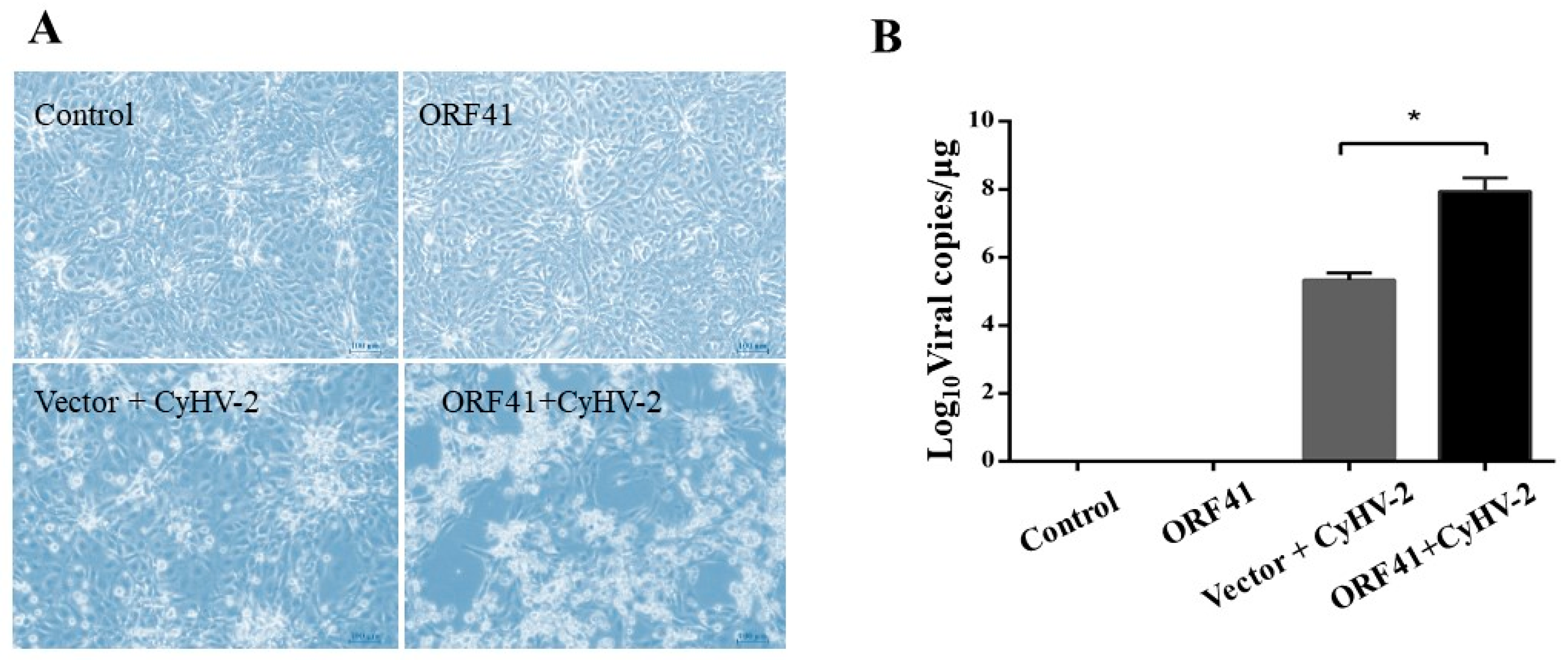
3.1. ORF41 Protein Promotes CyHV-2 Replication

Our morphological observations demonstrated that the expression of ORF41 alone did not induce significant alterations in the GiCB cells. The GiCB cells exhibited a typical cytopathic effect (CPE) after infection with CyHV-2 and displayed disrupted cellular morphology resembling a broken fishing network (Figure 1A). The CPE was notably accentuated in the presence of ORF41 expression (Figure 1A). Furthermore, the quantitative viral load analysis revealed a 48.8% increase in the viral load in the GiCB cells expressing ORF41 (Figure 1B).

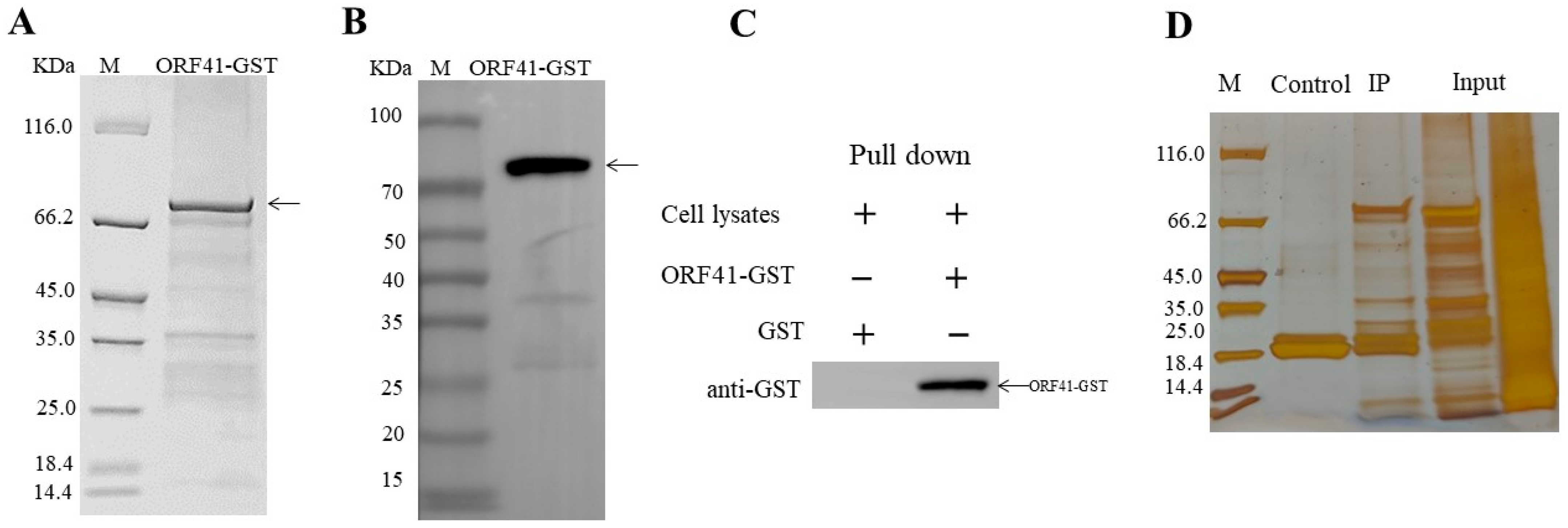
3.2. ORF41 Protein Interacts with PDH-E1β

Recombinant proteins (GST-ORF41) were expressed and purified. The analysis of the purified fusion protein using SDS-PAGE revealed clear bands corresponding to the expected molecular weight of 75.3 kDa (Figure 2A), indicating successful purification. The target protein was further validated through Western blotting, which showed distinct bands at the anticipated position (Figure 2B), confirming its identity. The Western blot results also demonstrated the presence of the target signal in the experimental group (Figure 2C). To explore ORF41’s protein interactions, both GST-glutathione-agarose resin homogenate and GST-ORF41-glutathione-agarose resin homogenate were utilized. The silver staining displayed fewer bands in the control lane (GST-glutathione-agarose resin homogenate), which contrasted with the greater number of bands in the experimental lane (GST-ORF41-glutathione-agarose resin homogenate) (Figure 2D). The information regarding the proteins with high scores was compiled based on an LC-MS/MS analysis and is presented in Table 1. The pyruvate dehydrogenase E1 component subunit beta (PDH-E1β) showed the highest score.

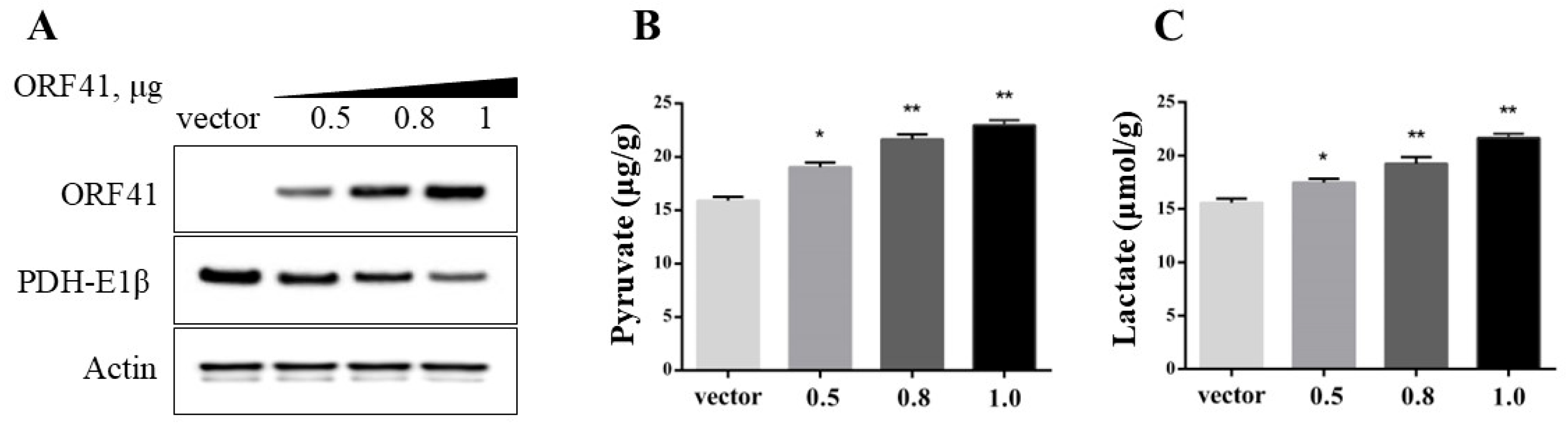
3.3. ORF41 Protein Decreases the Levels of PDH in GiCB Cells

Western blot analysis revealed a concentration-dependent decrease in PDH-E1β protein levels with increasing ORF41 expression (Figure 3A). Concurrently, ORF41 transfection induced a dose-responsive elevation in pyruvate and lactic acid concentrations (Figure 3B).

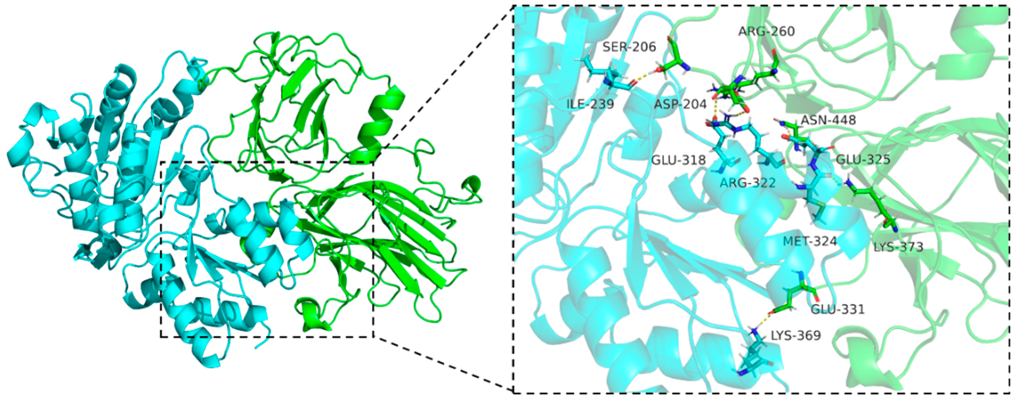
3.4. Molecular Docking

In this docking study, ORF41 was used as the receptor, and PDH-E1β served as the ligand. A three-dimensional binding model was analyzed, with ORF41 depicted in green and PDH-E1β in blue (Figure 4). The key residues are represented as sticks. The hydrogen bonds are represented as yellow dashed lines (Figure 4). The binding energy, represented by a docking score of 2.9 kcal/mol, was calculated. The ORF41 residues ASP-204, SER-206, ARG-260, GLU-331, LYS-373, and ASN-448 of ORF41 can form six hydrogen bonds with PDH-E1β residues ILE-239, GLU-318, ARG-322, MET-324, GLU-325, and ARG-369 on PDH-E1β. These findings confirm the presence of an interaction between ORF41 and PDH-E1β.

3.5. Molecular Dynamics Simulation

Molecular dynamics (MD) simulations were performed to validate the molecular docking results for the ORF41–PDH-E1β complex (Figure 5). The RMSD curves of the complex reached equilibrium after 15 ns, with an average RMSD of 0.4 nm, indicating structural stability during the simulation (Figure 5A). RMSF analysis identified key flexible regions in both ORF41 and PDH-E1β (Figure 5B). Notably, ORF41 exhibited higher flexibility, with residue fluctuations ranging from 0.5 to 0.8 nm, particularly in loop regions and the C-terminal domain. In contrast, most PDH-E1β residues fluctuated within 0.1–0.3 nm, except for the terminal regions (e.g., residues 400–420) where fluctuations reached 0.6–0.8 nm (Figure 5B). Stability analysis confirmed minimal positional deviation of ORF41 when bound to PDH-E1β. The radius of gyration for the complex remained stable, with a mean value of 2.38 nm (Figure 5C). The solvent-accessible surface area (SASA) values stabilized at an average of 295 nm2 (Figure 5D).

4. Discussion

Herpesviral hematopoietic necrosis has had devastating effects on the carp industry [14]. The genome of CyHV-2 contains about 154 open reading frames (ORFs), but the functions of most of its genes are unknown [15]. The ORF41 protein encoded by CyHV-2 is a RING-type (Really Interesting New Gene) E3 ubiquitin ligase. The ubiquitin–proteasome system (UPS) is a central intracellular pathway responsible for protein degradation and functional modification [16]. In this study, we found that ORF41 overexpression in GiCB cells could enhance CyHV-2 proliferation. Moreover, the GST pull-down and LC-MS/MS analyses showed that ORF41 might interact with pyruvate dehydrogenase (PDH)-E1β. Furthermore, we verified that ORF41 could decrease PDH-E1β levels and increase pyruvate and lactic acid levels. Therefore, we suggest that ORF41 promotes viral replication by altering GiCB cell metabolism by interacting with PDH-E1β. These findings offer critical insights into the metabolic strategies of CyHV-2 and their roles in pathogenesis.
Our study demonstrates that CyHV-2 ORF41 enhances viral replication by 48.8% in GiCB cells and intensifies cytopathic effects (CPEs). These results suggest that ORF41 plays an important role in CyHV-2 replication. In recent years, it has been found that many viruses can directly encode ubiquitin ligase to promote their own replication. For example, the ICP0 protein encoded by herpes simplex virus type 1 (HSV-1) is a RING E3 ubiquitin ligase that can facilitate the ubiquitination and degradation of the interferon regulatory factors 3 and 7 (IRF3/7), thereby inhibiting IFN-I production [17]. Therefore, we hypothesized that ORF41 interacts with certain host proteins to promote self-replication. The GST pull-down assay combined with LC-MS/MS showed that ORF41 might interact with PDH-E1β. Meanwhile, molecular docking and dynamics simulations were performed to verify the interaction between ORF41 and PDH-E1β. The molecular docking analysis showed that the ORF41 residues ASP-204, SER-206, ARG-260, GLU-331, LYS-373, and ASN-448 of ORF41 can form six hydrogen bonds with the PDH-E1β residues ILE-239, GLU-318, ARG-322, MET-324, GLU-325, and ARG-369 on PDH-E1β. The stable RMSD, RSMF, Rg, and SASA values indicate that the ORF41–PDH-E1β complex maintains a stable binding mode. These findings provide insight into the structural and dynamic characteristics of ORF41–PDH-E1β interactions.
PDH-E1, present in all known living prokaryotes and eukaryotes, is primarily responsible for catalyzing pyruvate dehydrogenation and acetyl-CoA generation, a process that plays an important role in energy metabolism [18]. Previous studies have reported that the hepatitis B virus (HBV) can inactivate PDH to inhibit the pyruvate-to-acetyl-CoA conversion, resulting in lactate accumulation and mitochondrial toxicity [19]. The Western blot analysis showed that the PDH-Eβ level decreased with the increase in ORF41 concentration. In addition, the transfection of ORF41 in the GiCB cells resulted in a concentration-dependent increase in the pyruvate and lactate levels. Therefore, we hypothesized that CyHV-2 could regulate host glucose metabolism through ORF41 and PDH-Eβ interactions.
Viral replication depends on energy provided by the host’s cell metabolism and biosynthetic precursors. Previous studies have found that many viruses are able to alter their hosts’ metabolism to promote viral replication. For example, HSV-1 and human cytomegalovirus (HCMV) can reprogram the host metabolism, inducing a state in which cells favor glycolysis over oxidative phosphorylation, even in aerobic conditions [20,21]. This reprogramming provides a rapid supply of ATP and intermediates for viral genome replication [22]. In this study, we found that ORF41 can alter the host’s pyruvate metabolism. In our next study, we will further investigate the mechanism by which this process promotes viral replication. Additionally, in vivo studies will be necessary to validate these findings in the context of CyHV-2 infection in crucian carp.

5. Conclusions

In summary, this study’s results have identified ORF41 as a critical modulator of host glycolysis during CyHV-2 infection. By interacting with and inhibiting PDH-E1β, ORF41 redirects the host metabolism to favor glycolysis, creating an optimal environment for viral replication. These findings not only enhance our understanding of CyHV-2 pathogenesis but also open new avenues for developing targeted antiviral strategies. Continued research into the metabolic interplay between CyHV-2 and its host is essential for advancing aquaculture health and mitigating the economic impacts of this devastating pathogen.

Author Contributions

Conceptualization, M.X. and Y.Z.; methodology, M.X. and C.X.; software, Z.H. and Y.M.; validation, M.X., C.X., and N.J.; formal analysis, Z.H. and Y.F.; investigation, M.X. and Z.H.; resources, M.X. and Y.M.; data curation, N.J. and C.X.; writing—original draft preparation, M.X. and C.X.; writing—review and editing, M.X. and Y.F.; visualization, Y.M.; supervision, Y.Z.; project administration, Y.F. and Y.Z.; funding acquisition, Y.F. and Y.Z. All authors have read and agreed to the published version of the manuscript.

Funding

This work was supported by the Nature Science Foundation of Hubei Province (2023AFB407), the Earmarked Fund for CARS (CARS-45), Chongqing Aquatic Science and Technology Innovation Alliance (CQFTIU202504-1), the National Freshwater Aquatic Germplasm Resource Center (FGRC18537), and the Central Public-interest Scientific Institution Basal Research Fund (2023TD46).

Institutional Review Board Statement

Not applicable.

Informed Consent Statement

Not applicable.

Data Availability Statement

The data presented in this study are available on request from the corresponding author.

Acknowledgments

We would like to thank Wuhan Gene Create Technology Co., Ltd. for their support in carrying out this study.

Conflicts of Interest

The authors declare no conflicts of interest.

References

  1. Xu, J.; Zeng, L.; Zhang, H.; Zhou, Y.; Ma, J.; Fan, Y. Cyprinid herpesvirus 2 infection emerged in cultured gibel carp, Carassius auratus gibelio in China. Vet. Microbiol. 2013, 166, 138–144. [Google Scholar] [CrossRef] [PubMed]
  2. Zhu, M.; Dai, Y.P.; Tong, X.Y.; Zhang, Y.X.; Zhou, Y.; Cheng, J.L.; Jiang, Y.T.; Yang, R.L.; Wang, X.Y.; Cao, G.L.; et al. Circ-Udg Derived from Cyprinid Herpesvirus 2 Promotes Viral Replication. Microbiol. Spectr. 2022, 10, e00943-22. [Google Scholar] [CrossRef] [PubMed]
  3. Song, Y.; Zhang, Y.; Xiao, S.; Li, P.; Lu, L.; Wang, H. Akt inhibitors prevent CyHV-2 infection in vitro. Fish Shellfish. Immunol. 2024, 154, 109940. [Google Scholar] [CrossRef] [PubMed]
  4. Wang, L.; He, J.; Liang, L.; Zheng, X.; Jia, P.; Shi, X.; Lan, W.; Xie, J.; Liu, H.; Xu, P. Mass mortality caused by Cyprinid Herpesvirus 2 (CyHV-2) in Prussian carp (Carassius gibelio) in China. Bull. Eur. Assoc. Fish Pathol. 2012, 32, 164–173. [Google Scholar]
  5. Ma, J.; Jiang, N.; LaPatra, S.E.; Jin, L.; Xu, J.; Fan, Y.; Zhou, Y.; Zeng, L. Establishment of a novel and highly permissive cell line for the efficient replication of cyprinid herpesvirus 2 (CyHV-2). Vet. Microbiol. 2015, 177, 315–325. [Google Scholar] [CrossRef]
  6. Tang, R.Z.; Lu, L.Q.; Wang, B.Y.; Yu, J.; Wang, H. Identification of the Immediate-Early Genes of Cyprinid Herpesvirus 2. Viruses 2020, 12, 994. [Google Scholar] [CrossRef]
  7. Polcicova, K.; Badurova, L.; Tomaskova, J. Metabolic reprogramming as a feast for virus replication. Acta Virol. 2020, 64, 201–215. [Google Scholar] [CrossRef]
  8. Su, G.; Liu, J.; Duan, C.; Fang, P.; Fang, L.; Zhou, Y.; Xiao, S. Enteric coronavirus PDCoV evokes a non-Warburg effect by hijacking pyruvic acid as a metabolic hub. Redox Biol. 2024, 71, 103112. [Google Scholar] [CrossRef]
  9. Heiden, M.G.V.; Cantley, L.C.; Thompson, C.B. Understanding the Warburg Effect: The Metabolic Requirements of Cell Proliferation. Science 2009, 324, 1029–1033. [Google Scholar] [CrossRef]
  10. Goodwin, A.E.; Merry, G.E.; Sadler, J. Detection of the herpesviral hematopoietic necrosis disease agent (Cyprinid herpesvirus 2) in moribund and healthy goldfish: Validation of a quantitative PCR diagnostic method. Dis. Aquat. Org. 2006, 69, 137–143. [Google Scholar] [CrossRef]
  11. Sun, J.J.; Lan, J.F.; Zhao, X.F.; Vasta, G.R.; Wang, J.X. Binding of a C-type lectin’s coiled-coil domain to the Domeless receptor directly activates the JAK/STAT pathway in the shrimp immune response to bacterial infection. PLoS Pathog. 2017, 13, e1006626. [Google Scholar] [CrossRef] [PubMed]
  12. Abraham, M.J.; Murtola, T.; Schulz, R.; Páll, S.; Smith, J.C.; Hess, B.; Lindahl, E. GROMACS: High performance molecular simulations through multi-level parallelism from laptops to supercomputers. SoftwareX 2015, 1–2, 19–25. [Google Scholar] [CrossRef]
  13. Van Der Spoel, D.; Lindahl, E.; Hess, B.; Groenhof, G.; Mark, A.E.; Berendsen, H.J.C. GROMACS: Fast, flexible, and free. J. Comput. Chem. 2005, 26, 1701–1718. [Google Scholar] [CrossRef] [PubMed]
  14. Hou, Z.W.; Ding, Z.Y.; Zhang, Y.; Pang, B.H.; Gui, L.; Liu, Y.; Lu, L.Q.; Roengjitd, P.; Wang, H. Establishment and Application of a Rapid Detection Method of Cyprinid Herpesvirus 2 (CyHV-2) in Aquacultural Waters by Using a Novel One-Pot RAA-CRISPR/Cas12a Combined Fe-Iron Flocculation Technology. J. Fish Dis. 2025, e14089. [Google Scholar] [CrossRef] [PubMed]
  15. Cheng, W.J.; Chen, Q.K.; Ren, Y.L.; Zhang, Y.; Lu, L.Q.; Gui, L.; Xu, D. The identification of viral ribonucleotide reductase encoded by ORF23 and ORF141 genes and effect on CyHV-2 replication. Front. Microbiol. 2023, 14, 1154840. [Google Scholar] [CrossRef]
  16. Li, R.Q.; Chen, C.; He, J.; Zhang, L.L.; Zhang, L.; Guo, Y.Y.; Zhang, W.T.; Tan, K.; Huang, J.H. E3 ligase ASB8 promotes porcine reproductive and respiratory syndrome virus proliferation by stabilizing the viral Nsp1α protein and degrading host IKKβ kinase. Virology 2019, 532, 55–68. [Google Scholar] [CrossRef]
  17. Zhang, L.; Huang, F.; Liu, J.; Xu, Y.; Miao, Y.; Yuan, Y.; Chen, X.; Zhang, H.-G.; Wang, J.; Zheng, H.; et al. HSV-1-encoded ICP0 degrades the host deubiquitinase BRCC36 to antagonize interferon antiviral response. Mol. Immunol. 2021, 135, 28–35. [Google Scholar] [CrossRef]
  18. Patel, M.S.; Nemeria, N.S.; Furey, W.; Jordan, F. The Pyruvate Dehydrogenase Complexes: Structure-based Function and Regulation. J. Biol. Chem. 2014, 289, 16615–16623. [Google Scholar] [CrossRef]
  19. Selvamani, S.P.; Khan, A.; Tay, E.S.E.; Garvey, M.; Ajoyan, H.; Diefenbach, E.; Gloss, B.S.; Tu, T.; George, J.; Douglas, M.W. Hepatitis B Virus and Hepatitis C Virus Affect Mitochondrial Function Through Different Metabolic Pathways, Explaining Virus-Specific Clinical Features of Chronic Hepatitis. J. Infect. Dis. 2024, 230, 1012–1022. [Google Scholar] [CrossRef]
  20. Vastag, L.; Koyuncu, E.; Grady, S.L.; Shenk, T.E.; Rabinowitz, J.D. Divergent Effects of Human Cytomegalovirus and Herpes Simplex Virus-1 on Cellular Metabolism. PLoS Pathog. 2011, 7, e1002124. [Google Scholar] [CrossRef]
  21. Huang, P.; Wang, X.; Lei, M.Y.; Ma, Y.; Chen, H.L.; Sun, J.; Hu, Y.Z.; Shi, J.D. Metabolomics Profiles Reveal New Insights of Herpes Simplex Virus Type 1 Infection. Int. J. Mol. Sci. 2023, 24, 1521. [Google Scholar] [CrossRef] [PubMed]
  22. Delisle, L.; Fuhrmann, M.; Quéré, C.; Pauletto, M.; Pichereau, V.; Pernet, F.; Corporeau, C. The Voltage-Dependent Anion Channel (VDAC) of Pacific Oysters Crassostrea gigas Is Upaccumulated During Infection by the Ostreid Herpesvirus-1 (OsHV-1): An Indicator of the Warburg Effect. Mar. Biotechnol. 2018, 20, 87–97. [Google Scholar] [CrossRef] [PubMed]
Figure 1. ORF41 protein promotes CyHV-2 replication. (A) Morphological effects of ORF41 expression on CyHV-2 infection in GiCB cells. (B) ddPCR analysis of CyHV-2 viral load in GiCB cells. Control: control GiCB cells without plasmid transfection and virus infection; ORF41: GiCB cells overexpressing ORF41 and not infected with CyHV-2; Vector + CyHV-2: GiCB cells transfected with pcDNA3.1 plasmid and infected with CyHV-2; ORF41 + CyHV-2: GiCB cells overexpressing ORF41 and infected with CyHV-2. Values are expressed as mean ± SD and normalized to values for no treatment (n = 5). * p < 0.05.
Fishes 10 00107 g001
Figure 2. ORF41 protein interacts with PDH-E1β. (A) SDS-PAGE analysis of recombinant ORF41-GST protein. (B) Western blot analysis of the purified ORF41-GST protein. (C) Results of the GST antibody test. (D) Silver staining of proteins eluted from GST pull-down assays. GiCB cell lysates were incubated with GST or GST-ORF41 immobilized on glutathione sepharose beads. Bound proteins were eluted, separated by 12% SDS-PAGE, and stained with silver nitrate.
Fishes 10 00107 g002
Figure 3. ORF41 protein decreases the levels of PDH. (A) Immunoblot analysis of endogenous PDH-E1β protein levels in GiCB cells transfected with increasing amounts of ORF41 plasmids (0, 0.5 μg, 0.8 μg, and 1 μg). (B,C) The pyruvate and lactic acid levels in GiCB cells transfected with increasing amounts of ORF41 plasmids (0, 0.5 μg, 0.8 μg, and 1 μg). * p < 0.05; ** p < 0.01.
Fishes 10 00107 g003
Figure 4. Molecular docking simulation of ORF41 and PDH-E1β. ORF41 is shown in green and PDH-E1β in blue. The key residues are shown as sticks. H-bonds are shown as yellow dashed lines.
Fishes 10 00107 g004
Figure 5. Molecular dynamics simulation of ORF41 and PDH-E1β. (A) Root mean square deviation (RMSD) analysis. (B) Root mean square fluctuation (RMSF) analysis. (C) Radius of gyration (Rg) analysis. (D) Solvent-accessible surface area (SASA) analysis.
Fishes 10 00107 g005
Table 1. Summary of annotated proteins found to interact with ORF41.
NoDescriptionMW (kDa)ScoreSequence Coverage (%)
1Pyruvate dehydrogenase E1 component subunit beta39.363.617.1
2Peptidyl-prolyl cis-trans isomerase26.436.611.9
3PRDX3 Thioredoxin-dependent peroxide reductase27.231.37.5
4Protein-L-isoaspartate O-methyltransferase26.420.18.8
52′,3′-cyclic-nucleotide 3′-phosphodiesterase23.919.39.6
Disclaimer/Publisher’s Note: The statements, opinions and data contained in all publications are solely those of the individual author(s) and contributor(s) and not of MDPI and/or the editor(s). MDPI and/or the editor(s) disclaim responsibility for any injury to people or property resulting from any ideas, methods, instructions or products referred to in the content.

