Article

Krüppel Homolog 1 Is Required for the Role of Methyl Farnesoate in Vitellogenesis in the Mud Crab Scylla paramamosain

State Key Laboratory of Mariculture Breeding, Fisheries College, Jimei University, Xiamen 361021, China
*
Author to whom correspondence should be addressed.
Fishes 2025, 10(3), 103; https://doi.org/10.3390/fishes10030103
Submission received: 26 December 2024 / Revised: 26 February 2025 / Accepted: 27 February 2025 / Published: 28 February 2025
(This article belongs to the Section Aquatic Invertebrates)

Abstract

:
Methyl farnesoate (MF), a counterpart of insect juvenile hormones in crustaceans, plays essential roles in molting, metamorphosis, and reproduction. In this paper, we isolated a gene-encoding Krüppel homolog 1 from the mud crab Scylla paramamosain (Sp-Kr-h1) and investigated its involvement in MF-regulated vitellogenesis. RT-PCR showed that Sp-Kr-h1 had a wide expression in various tissues. qRT-PCR showed that its expression level in the ovary peaked at stage III. Both in vitro and in vivo experiments suggested that the expression of Sp-Kr-h1 and Vitellogenin (Vg) in the hepatopancreas was significantly induced by MF administration. Further, the RNA interference technique was employed to illustrate the precise role of Sp-Kr-h1 in ovarian development. This revealed that the in vitro silencing of Sp-Kr-h1 significantly decreased the level of Vg transcripts located in the hepatopancreas. Meanwhile, an in vivo experiment demonstrated that oocyte growth was inhibited after the knockdown of Sp-Kr-h1 in female S. paramamosain. In conclusion, this study identified a Kr-h1 gene in S. paramamosain and demonstrated that it is an essential factor for MF-regulated vitellogenesis. Our results provided a new insight into the mechanism underlying MF inducing ovarian development in mud crabs.
Key Contribution: The present study demonstrated that MF induced vitellogenesis via promoting Krüppel homolog 1 expression in the mud crab Scylla paramamosain.

1. Introduction

In arthropods, vitellogenesis is a central event of female reproduction, involving the production and secretion of vitellogenin (Vg), followed by the internalization of Vg by maturing oocytes through receptor-mediated endocytosis (vitellogenin receptor, VgR) [1]. Juvenile hormone (JH) is an important regulator of vitellogenesis in insects [1]. JH is structurally unique sesquiterpenoid hormone found only in arthropods [1]. It is well known that JH and its related signaling pathway regulate growth, development, reproduction, metabolism, and cellular immunity in insects [2,3,4,5,6]. Interestingly, JH is absent in crustaceans [3]. It has been shown that methyl famesoate (MF), another sesquiterpenoid hormone in arthropods, plays an equivalent impact to JH in the metamorphosis and reproduction of crustaceans [3]. MF was first discovered in the hemolymph of the spider crab Libinia emarginata [7]. Subsequently, a series of in vitro experiments proved that the mandibular organs (M-organ) conducted its synthesis and secretion [8]. Structurally, it is regarded as a non-epoxidized form of juvenile hormone III (JH III) in insects [9]. In the freshwater prawn Macrobrachium rosenbergii, a high MF level delays the larva development [10]. The gonadotropic effects of MF have also been recorded in the red swamp crayfish Procambarus clarkii [11], the freshwater field crab Oziotelphusa senex senex [12], and the freshwater crab Travancoriana schirnerae [13]. An injection experiment into the red swamp crayfish Procambarus clarkii demonstrated that MF plays a positive role in regulating ovarian development [11]. Interestingly, it was found that Daphnia zygotes developed into males when exposed to MF, suggesting that MF may be involved in sex regulation [14,15]. Although several response factors are involved in the regulatory processes, the mechanism underlying this regulation is still unclear and needs further study.
Krüppel homolog 1 (Kr-h1) is a C2H2-type zinc finger transcription factor, which was confirmed in D. melanogaster as an early-response gene to JH [16]. Subsequently, it has been proved that the level of Kr-h1 is directly induced by JH and its receptor Methoprene-tolerant (Met) to transduce JH signals [16,17,18,19]. With the deepening of research, accumulating evidence from recent studies has shown that Kr-h1 also has a regulatory impact on molting, metamorphosis, and reproduction [20,21]. In the mosquito Aedes aegypti, Kr-h1 has been identified as an intermediate factor in the JH/Met gene repression hierarchy by in vivo and in vitro experiments [22]. In the brown planthopper Nilaparvata lugens, Met and its downstream transcription factor Kr-h1 have a profound role on ovarian development [23]. It was revealed that Kr-h1 was induced by JH to prevent B. mori larvae from bypassing the pupal stage into precocity adult development through inhibiting ecdysone-induced protein E93 [24]. Moreover, Kr-h1 also plays a key regulatory role in the procedure of yolk formation and ovulation [25]. It was suggested that the lipid accumulation in primary oocytes was blocked after the silencing of Kr-h1 in the locust Locusta migratoria [20]. Additionally, the knockout of the Kr-h1 gene in the food pest Tribolium castaneum resulted in a precocious abnormal phenotype [26]. The homologous genes have also been found in crustaceans, but limited studies have been documented regarding their involvement in reproduction regulated by MF [27,28].
In this paper, we aimed to ascertain the function of Kr-h1 as a downstream MF signaling response gene involved in the vitellogenesis of the mud crab Scylla paramamosain. We first isolated the encoding sequence of Sp-Kr-h1 from S. paramamosain and described its expression profile in females. Subsequently, the involvement of Kr-h1 in MF signaling was investigated by the in vivo and in vitro administration of MF. Finally, the precise effect of Kr-h1 in the mud crab was explored by RNA interference experiments.

2. Materials and Methods

2.1. Animals

The female mud crabs (S. paramamosain) were bought from an aquatic product market in Xiamen City, Fujian Province. Referring to the morphological appearance and structural characteristics established previously, the ovarian development of the mud crabs was divided into 5 stages: stages I (undeveloped stage), II (pre-vitellogenic stage), III (early vitellogenic stage), IV (late vitellogenic stage), and V (mature stage) [29]. Experimental animals were cultured in tanks with filtered seawater at a salinity of 26 ± 0.5 PSU for one week. During this time, the live clam Ruditapes philippinarum was used to feed the crabs and the temperature was kept at 27 ± 0.5 °C. Animal handling was approved by the Animal Care and Use Committee of the Fisheries College of Jimei University (Approval Code: 2021-04; Approval Date: 22 January 2021).

2.2. Cloning of Sp-Kr-h1 and Sequence Analysis

Referring to the operating manual, the total RNA was extracted by TRIzol® reagents (Invitrogen, Carlsbad, CA, USA). The extracted RNA was then quantified using a Nanodrop spectrophotometer (Thermo Scientific, Madison, Wl, USA) and treated with a 2.0% agarose gel to further improve the RNA quality. DNase I was used to remove the potential genomic DNA contamination, and 1 μg of total RNA was reverse-transcribed utilizing a RevertAid First Strand cDNA Synthesis Kit (Thermo Scientific, USA) with random primers following the operation manual. The generated cDNAs were diluted four-fold and then preserved at −80 °C to be backup.
Sp-Kr-h1 was obtained from GenBank (GenBank accession number: UIX26759.1). The open reading frame (ORF) of Sp-Kr-h1 was predicted from a hepatopancreas transcriptome database of S. paramamosain, and the sequence veracity was validated by polymerase chain reaction (PCR) with primers Sp-Kr-h1-OF and Sp-kr-h1-OR (Table 1). The PCR products were subjected by using a 1.0% agarose (Vivacell, Shanghai, China) gel. After being gel-purified, PCR products were ligated to the pMD19-T vector for sequencing (Sangon Biotech, Shanghai, China).
The nucleotide sequence of Sp-Kr-h1 was consistent with the known Kr-h1 amino acid sequence (GenBank accession number: UIX26759.1) by the BLAST algorithm, the algorithm from the National Center for Biotechnology Information (https://www.ncbi.nlm.nih.gov/blast/ (accessed on 12 November 2023)). The ORF was predicted by the ORF finder (https://www.ncbi.nlm.nih.gov/orffinder/ (accessed on 12 November 2023)). Signal peptide was predicted through the SignalP 5.0 program (SignalP 5.0—DTU Health Tech—Bioinformatic Services). Glycosylation sites were predicted using the NetNGlyc 1.0 Server. Clustal W software (version 2.1) was used to compare the deduced amino acid sequences with those reported. PhyML trees were constructed by SeaView 5.0.5 software.

2.3. Expression Profiles of Sp-Kr-h1 in the Female Mud Crab

The expression profile of Sp-Kr-h1 in 10 tissues of stage III female crabs was detected by reverse transcription PCR (RT-PCR). For the tissue distribution of Sp-Kr-h1, 10 tissues, including eyestalk ganglion, cerebral ganglion, thoracic ganglion, hepatopancreas, ovary, stomach, heart, muscle, Y-organ, and M-organ, from a female crab at stage III were collected (body weight 278.4 g; carapace width 118.2 mm). It was amplified by Sp-Kr-h1-QF and Sp-Kr-h1-QR primers (Table 1). The amplification of β-actin (GenBank accession number: GU992421.1) was set as the internal control. The reaction system of PCR was conducted in 25 μL total volume composed of the following: deionized water, 16.5 μL; 10 × Ex Taq Buffer (TaKaRa Ex Taq®), 2.5 μL; cDNA, 2 μL; dNTP Mix, 2 μL; Taq, 0.1 μL; and 1 μL of each primer (10 mM). The PCR products were subjected to 1% agarose gel electrophoresis and imaged by a Gel Image System (Tanon 2500B, Shanghai, China) after being labeled by GelRed dye.
To analyze the expression profiles of Sp-Kr-h1 during ovarian development, ovary and hepatopancreas samples were collected from female crabs at stage II (body weight 187.1 ± 23.2 g, carapace width 102.0 ± 3.1 mm), stage III (body weight 263.3 ± 31.6 g, carapace width 111.1 ± 5.4 mm), and stage IV (body weight 374.2 ± 36.5 g, carapace width 121.7 ± 6.3 mm). Each stage has five individuals (n = 5). Sample tissues were subjected to RNA extraction and cDNA generation. The qRT-PCR technology was used to detect Sp-Kr-h1 expression profiles at three development stages of the hepatopancreas and ovary. Additionally, 7500 Fast Real-Time PCR (Applied Biosystems, Carlsbad, CA, USA) and SYBR Premix Ex Taq (TaKaRa, Dalian, China) were used for qRT-PCR analysis, following the operation manual. The primers used were Sp-Kr-h1-QF and Sp-Kr-h1-QR (Table 1). In this reaction system, the total volume was 20 μL, including 10 μL 2 × SYBR Premix Ex Taq (TaKaRa, Dalian, China), 4 μL diluted cDNA template, 1 μL each primer (10 mM), and 4 μL RNase-free water. The program was 95 °C for 30 s, followed by 40 cycles of 95 °C for 30 s, 59 °C for 30 s, and 72 °C for 30 s.

2.4. In Vitro Effect of MF on the Gene Expression in the Hepatopancreas and Ovary

Standard sample MF was dissolved in Dimethyl Sulfoxide (DMSO) with a concentration of 100 ng/μL for injection experiment. An in vitro experiment was performed to assess whether Sp-Kr-h1 was involved in the MF-regulated vitellogenesis of the mud crab. The experiment animals were female mud crabs at the stage III, and samples of hepatopancreas and ovary were collected and cut into fragments. Tissue blocks (50–100 mg) were washed with crustacean physiological saline (CPS) (11.3 mM KCl, 13.3 mM CaCl2, 440 mM NaCl, 10 mM Hepes, 23 mM Na2SO4, 26 mM MgCl2, pH 7.4) containing penicillin (100 IU/mL) and streptomycin (100 μg/mL). Tissue blocks were placed into 24-well plates containing 0.5 mL L-15 medium (Gibco, Grand Island, NY, USA) at 26 °C for 1 h pre-incubation. Subsequently, the hepatopancreas and ovary explants were incubated with MF at concentrations of 1, 10, 100, and 1000 nM, respectively. Each treatment has three replicates. The samples were collected at the2nd, 4th, and 8th h post the incubation. qRT-PCR was used to conduct gene expression analysis.

2.5. In Vivo Effects of MF on Ovarian Development of S. paramamosain

To further confirm the effect of MF and the involvement of Sp-Kr-h1 in ovarian development, an in vivo experiment including the injection of MF into female crabs at stage II was conducted. Twelve female crabs (body weight 176.0 ± 22.1 g, carapace width 103.2 ± 3.4 mm) were randomly and equally allocated into two groups. MF (1 ng/g body weight) dissolved in 100 μL of CPS was injected into the crab, while the control animal was injected 100 μL CPS instead. The injection was made by injecting a micro-syringe (Hamilton, Switzerland) with a slender needle into the arthrodial membrane located at the fifth swimming leg. Almost 24 h post the injection, crabs were dissected after anesthetization on ice, and samples of hepatopancreas and ovary were collected for Sp-Kr-h1, Vg (GenBank accession number: KC734559.1), and VgR (GenBank accession number: OQ032501.1) expression analysis.

2.6. In Vitro and In Vivo Knockdown of Sp-Kr-h1 on Vitellogenesis

To determine the precise effect of Sp-Kr-h1 in the reproduction of female S. paramamosain, RNA interference experiments by the addition of Sp-Kr-h1-targeted dsRNA to the hepatopancreatic and ovarian explants and the injection of Sp-Kr-h1-targeted dsRNA into the mud crabs were performed. The MEGAscript T7 transcription kit (Ambion, Austin, TX, USA) was used to generate dsRNA targeting GFP and Sp-Kr-h1. Table 1 lists the sequence of primers used to generate these dsRNA.
The in vitro experiment was performed on the female crab at stage II. Crabs were anesthetized on ice for 10 min and sterilized in 75% ethanol for 10 min. Subsequently, CPS containing penicillin (100 IU/mL) and streptomycin (100 μg/mL) was used to wash the isolating hepatopancreatic and ovarian tissues six times. The tissue was then cut into approximately 50 mg explants, which were then placed in each of the holes of the 24-well culture plate with 500 μL L15 medium containing penicillin (100 IU/mL) and streptomycin (100 μg/mL) and cultured at 25 °C for 30 min. This experiment has three groups: the blank control group without dsRNA, the negative control group containing 2 μg/mL GFP-dsRNA, and the experimental group containing 2 μg/mL Sp-Kr-h1-targeted dsRNA. Each treatment was performed in triplicate. Finally, post 4 h and 8 h incubation, samples of hepatopancreas and ovary were collected for Sp-Kr-h1, Vg, and VgR gene expression analysis using qRT-PCR.
Before the in vivo RNAi experiment, female crabs (body weight 252.8 ± 32.4 g, carapace width 110.2 ± 4.7 mm) at stage III were disorderly divided into 3 groups (n = 7). The blank control group was injected with 100 μL of CPS, while the rest of the groups received Sp-Kr-h1 dsRNA and GFP dsRNA (1 μg/g body weight) prepared in 100 μL CPS, separately. Using a micro-syringe (Hamilton, Bonaduz, Switzerland), we injected regularly every six days into the joint membrane of the mud crab’s 5th swimming leg. Almost 24 h post the 3rd injection, the experimental animals were dissected after anesthetization on ice, and tissues of the hepatopancreas and ovary were collected for gene expression analysis via qRT-PCR and hematoxylin and eosin staining.

2.7. Histological Observation

Ovarian tissues from the long-term interference experiment were washed with CPS and fixed at 4 °C with 4% buffered ice-cold paraformaldehyde (PFA) for 12 h, with the following steps including dehydration in a graded ethanol series and embedding in paraffin (EG1150H paraffin embedding machine, Leica, Wetzlar, Germany). A 7 μm thick slide was cut with a rotary microtome (RM2128 rotary, Leica, Wetzlar, Germany). The tissue slides were stained using hematoxylin and eosin. The diameter of each oocyte cell is an average obtained by measuring the length of the long and short axis of each cell. It was photographed using an Olympus multifunctional microscope (Olympus, Tokyo, Japan) to observe its histological morphology.

2.8. Statistical Analyses

This study used the 2−ΔΔCt method to obtain the qRT-PCR data and then performed statistical analysis. The Kolmogorov–Smirnov test was used for normal distribution testing. Levene’s test was employed to assess the heteroscedasticity. MF injection experiment data were analyzed using the unpaired t-test, while one-way ANOVA and the Turkey multivariate range test (SPSS 13.0) were used for statistical analysis in other experiments. Data were expressed as mean ± SD (standard deviation).

3. Results

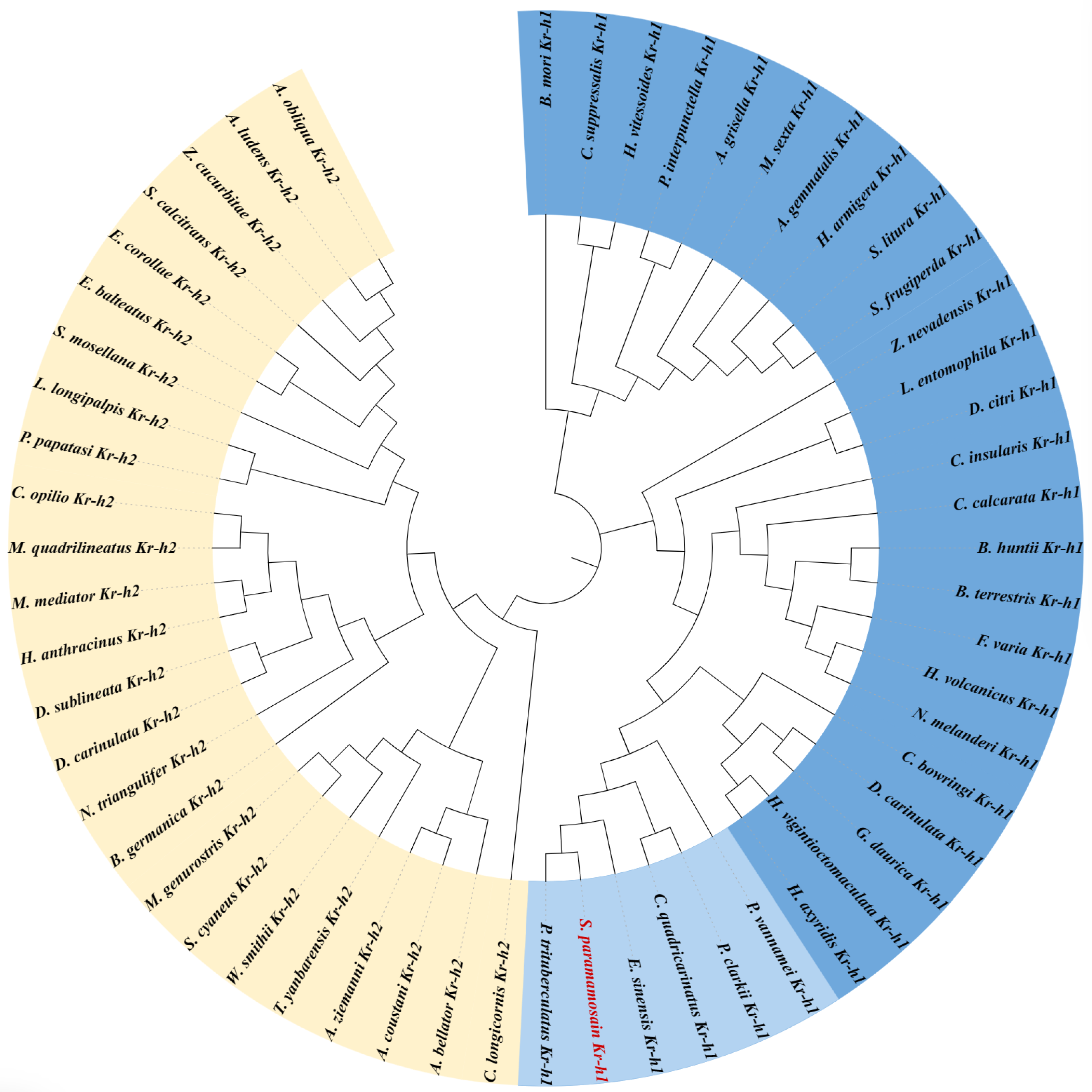
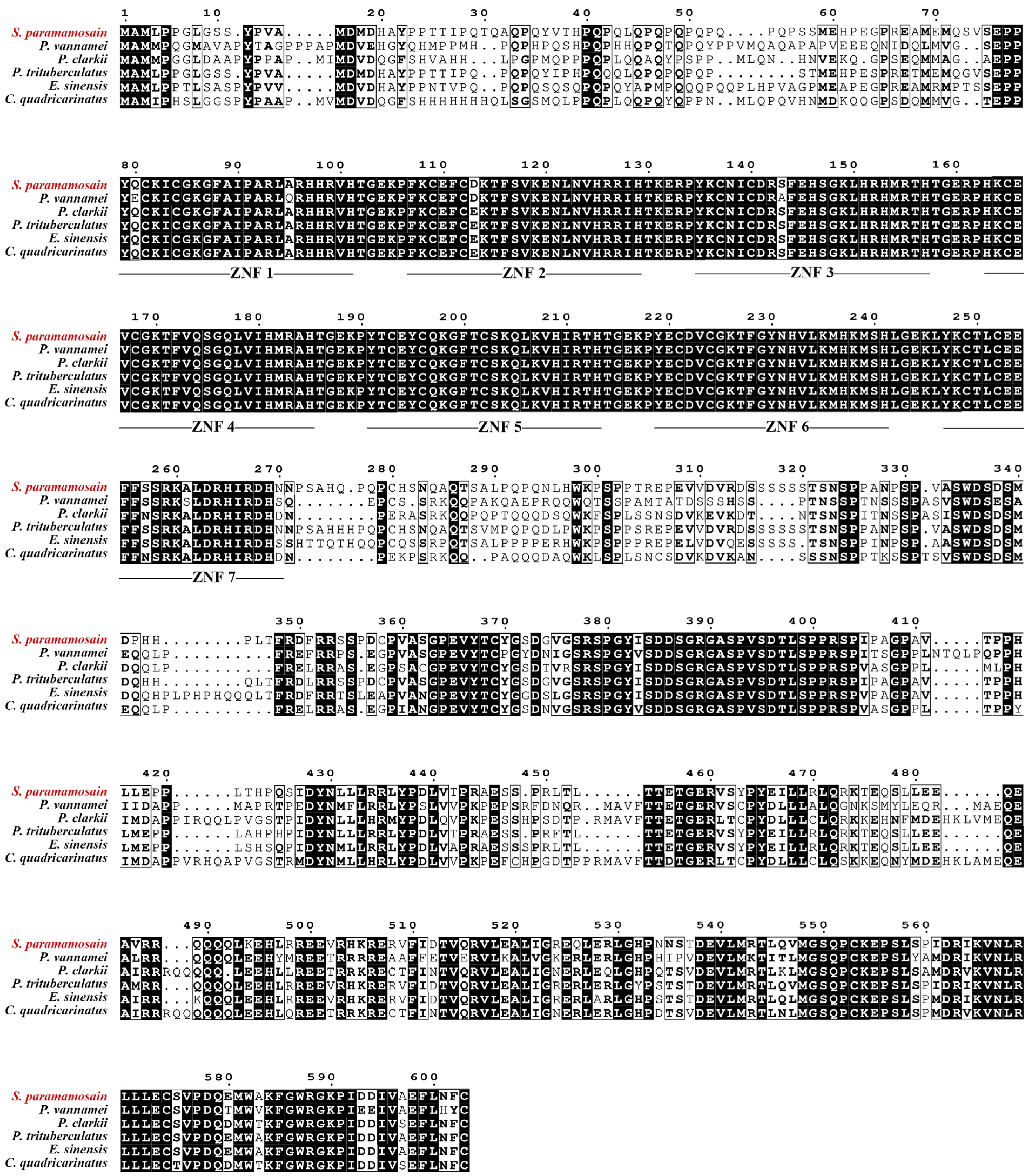
3.1. cDNA Cloning and Sequence Analysis

In the study, we obtained the coding sequence of Sp-Kr-h1 from the mud crab through transcriptome sequencing and PCR-based cloning technology. The ORF of Sp-Kr-h1 was 1812 bp (base pair) long, encoding a 603 aa precursor protein containing seven C2H2 Zinc-finger domains. The phylogenetic tree showed that the family of Kr-h protein was categorized as Kr-h1 and Kr-h2, and the crustaceans Kr-h1 fit into the same clade with the aligned Kr-h1 sequences from insects (Figure A1). Additionally, sequence analysis suggested that Sp-Kr-h1 exhibited a high identity with the Kr-h1 reported previously in other decapod crustaceans (Figure A2), including the swimming crab Poriunus trituberculatus (92.55%), the Chinese mitten crab Eriocheir sinensis (84.6%), the red claw crayfish Cherax quadricarinatus (67.88%), the red swamp crayfish P. clarkii (67.46%), and the whiteleg shrimp Penaeus vannamei (65.5%).

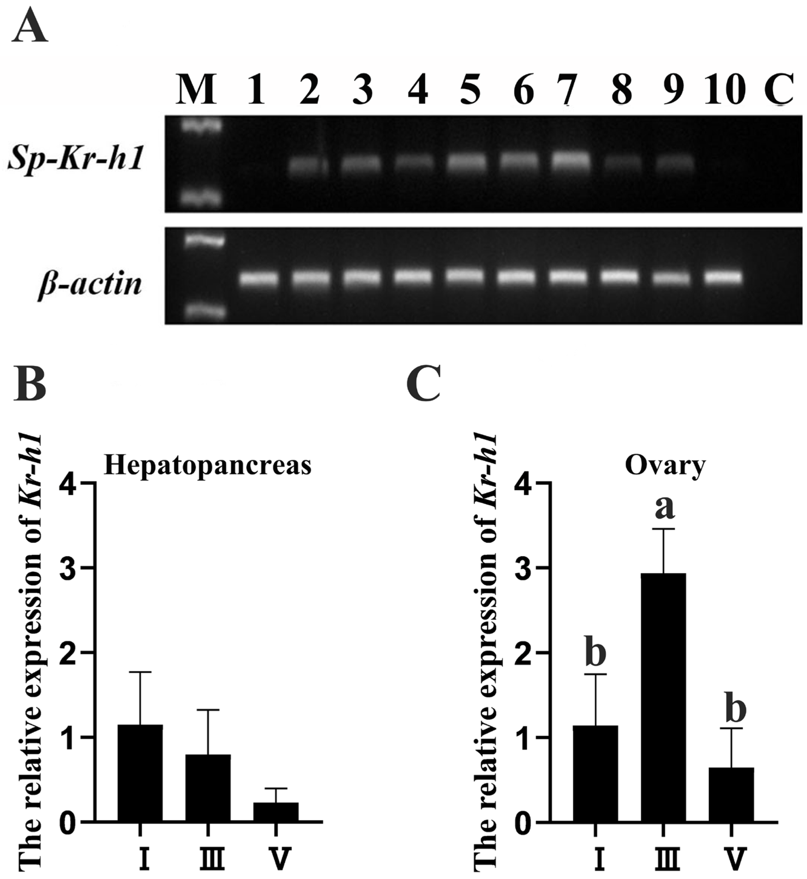
3.2. Expression Profiles of Kr-h1 in the Female Mud Crab

The tissue expression profiles of Sp-Kr-h1 in the female mud crab were detected by RT-PCR and qRT-PCR. It revealed that Sp-Kr-h1 exhibited a wide expression in the detected tissues except for the eyestalk ganglion and M-organ (Figure 1A). In addition, the qRT-PCR result showed that the level of Sp-Kr-h1 expression in the hepatopancreas was not significantly downregulated during ovarian development (ANOVA F(2,13) = 2.827, p = 0.112) (Figure 1B). Meanwhile, in the ovary, the expression of Sp-Kr-h1 reached its highest level at stage III and significantly declined at stage IV (ANOVA F(2,13) = 25.793, p < 0.05) (Figure 1C).

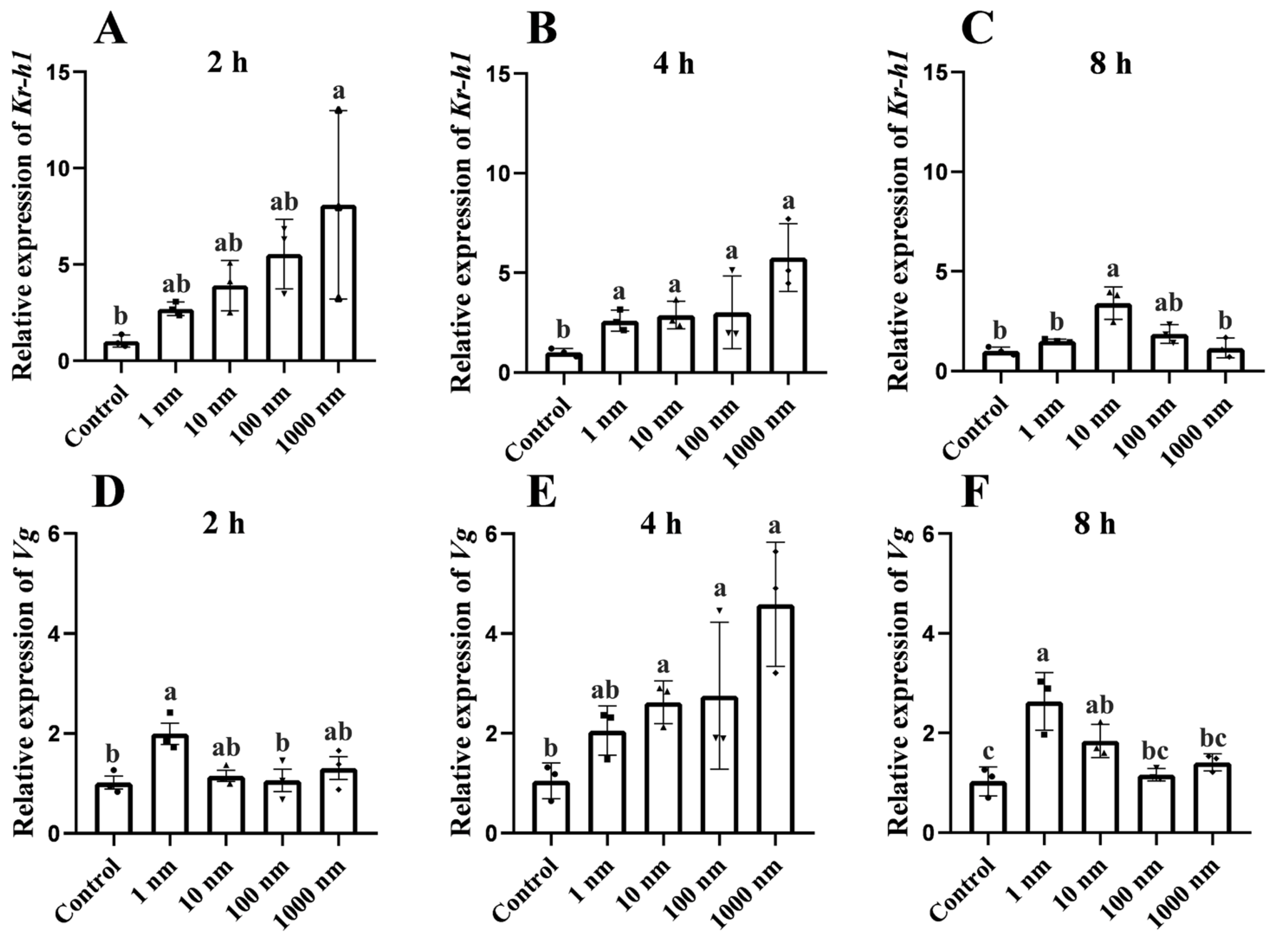
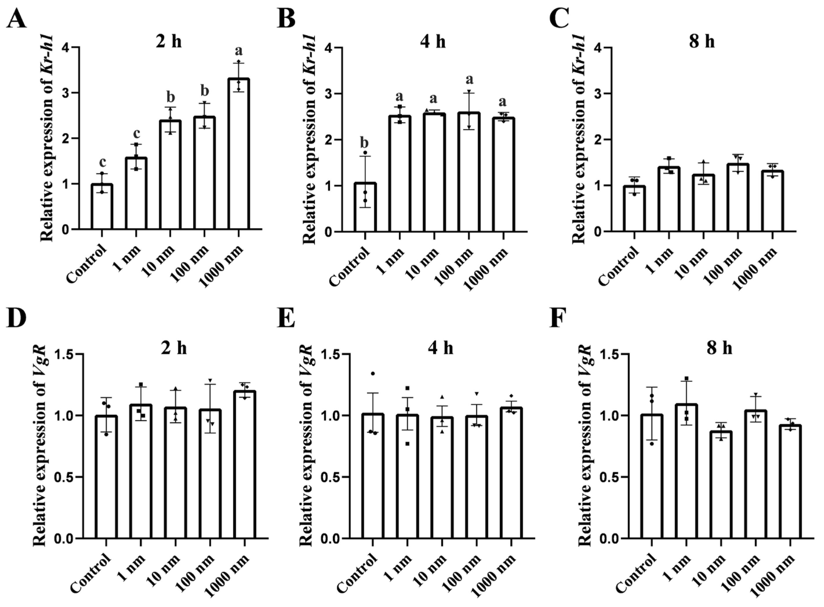
3.3. In Vitro Effect of MF on Kr-h1 Expression and Vitellogenesis of the Mud Crab

To investigate the impact of MF on vitellogenesis and the involvement of Sp-Kr-h1 in this progress, the in vitro experiments were conducted by the addition of MF into the hepatopancreatic and ovarian explants. This showed that the level of Sp-Kr-h1 transcript in the hepatopancreatic explants was significantly induced by the 2 h (ANOVA F(4,14) = 3.785, p = 0.040) (Figure 2A) and 4 h (ANOVA F(4,14) = 10.954, p = 0.001) (Figure 2B) addition of MF at a concentration of 1000 nM and 8 h of MF treatment with a concentration of 10 nM (ANOVA F(4,14) = 9.769, p = 0.002) (Figure 2C). In addition, the expression of vitellogenin (Vg) was significantly upregulated by 2 h of MF treatment at a concentration of 1 nM (ANOVA F(4,14) = 4.524, p = 0.024) (Figure 2D), 4 h of MF addition at a concentration of 1000 nM (ANOVA F(4,14) = 7.599, p = 0.004) (Figure 2E), and 8 h of MF incubation at concentrations of 1 and 10 nM (ANOVA F(4,14) = 10.174, p = 0.001) (Figure 2F).
Meanwhile, the Sp-Kr-h1 expression in ovarian explants could also been significantly induced in response to 2 h (Figure 3A) (ANOVA F(4,14) = 32.559, p < 0.05) and 4 h (Figure 3B) (ANOVA F(4,14) = 12.999, p = 0.001) MF stimulation. However, 8 h of MF treatment with all concentrations did not significantly influence Sp-Kr-h1 expression (Figure 3C) (ANOVA F(4,14) = 3.182, p = 0.063). However, the expression of VgR gene in ovarian explants was not significantly influenced by the addition of 2 h (Figure 3D) (ANOVA F(4,14) = 0.853, p = 0.524), 4 h (Figure 3E) (ANOVA F(4,14) = 0.078, p = 0.987), and 8 h (ANOVA F(4,14) = 1.270, p = 0.344) MF (Figure 3F).

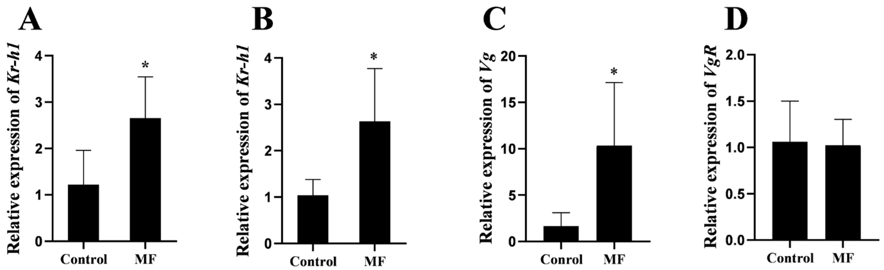
3.4. Effect of MF Injection on Kr-h1 Expression and Vitellogenesis of the Mud Crab

A further in vivo experiment by injection of MF into female mud crabs at stage II was performed to confirm the participation of Sp-Kr-h1 in vitellogenesis. This revealed that the expression of Sp-Kr-h1 in the hepatopancreas (Figure 4A) (Unpaired t-test, t = 2.650, p = 0.0330) and ovary (Figure 4B) (Unpaired t-test, t = 2.999, p = 0.0171) was significantly upregulated by MF. Accompanied by the upregulation of Sp-Kr-h1, the expression of Vg in the hepatopancreas was correspondingly increased (Unpaired t-test, t = 3.113, p = 0.0144) (Figure 4C) while the expression of VgR was not significantly changed (Unpaired t-test, t = 0.1910, p = 0.8517) (Figure 4D).

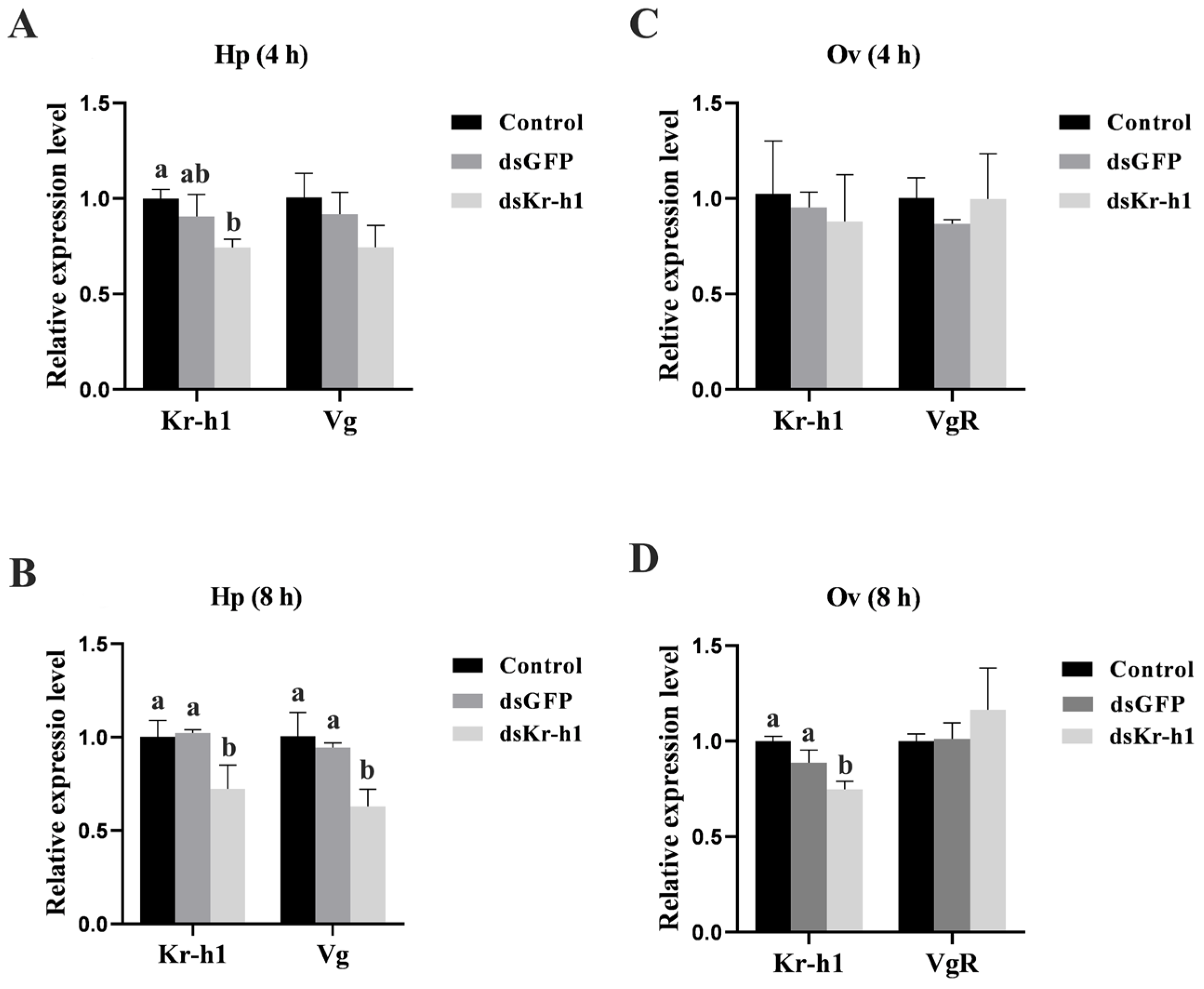
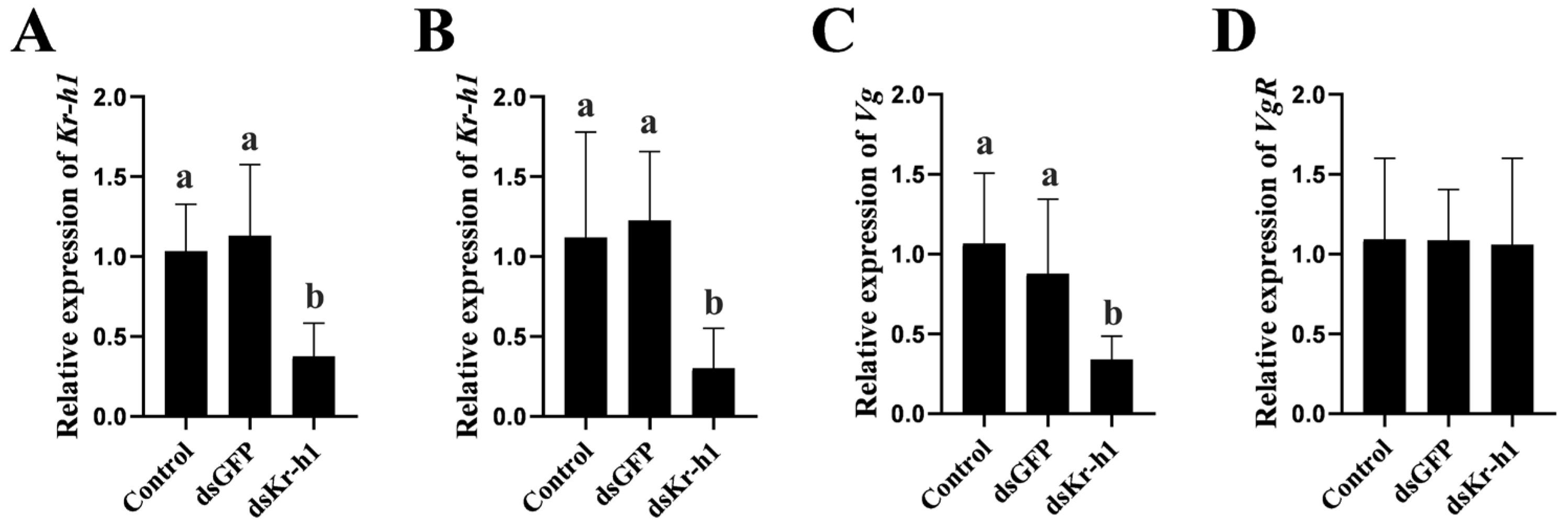
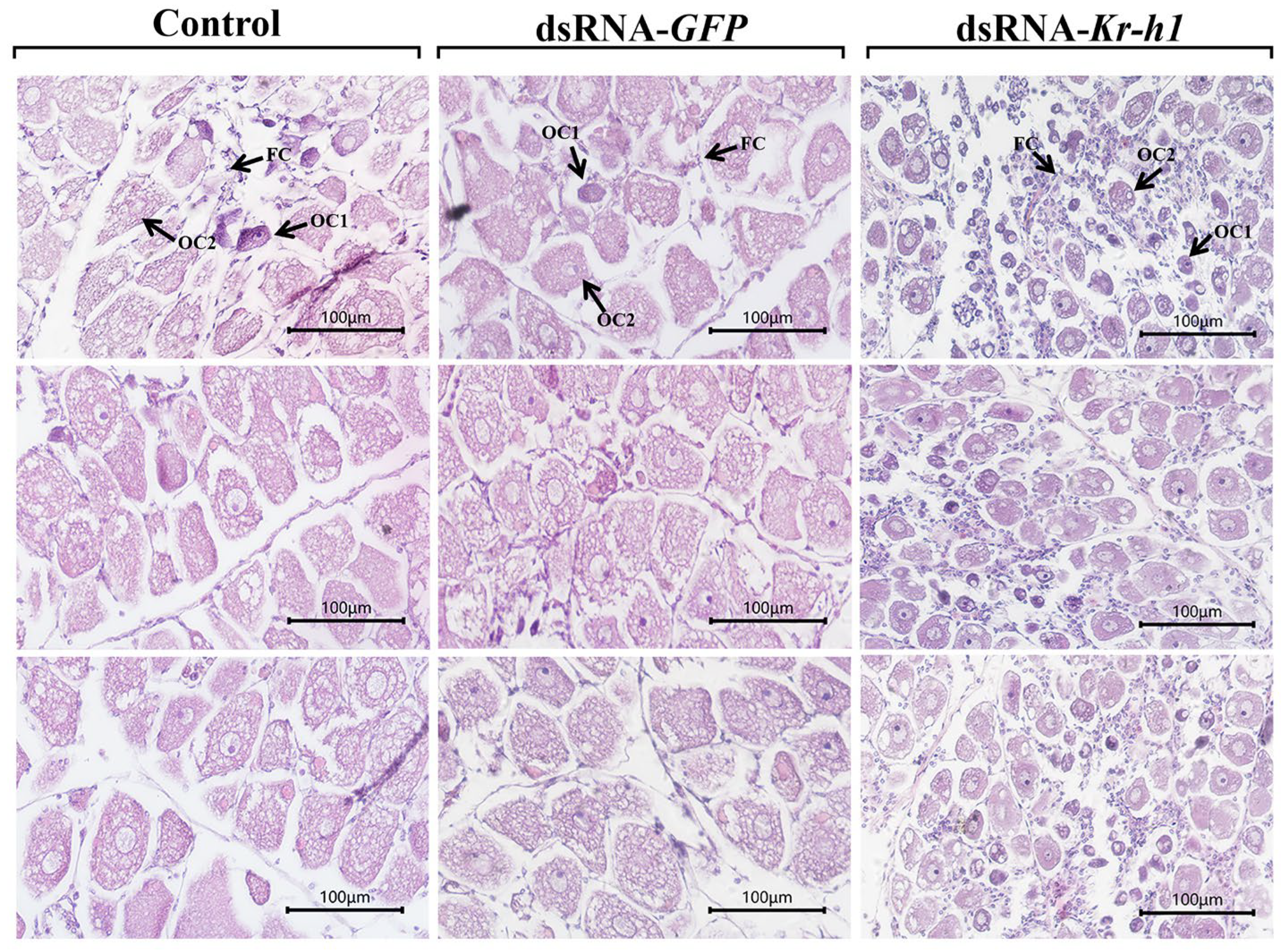
3.5. Effect of Kr-h1 Silencing on Ovarian Development in the Mud Crab

To establish the precise role of Sp-Kr-h1 in ovarian development in S. paramamosain, both in vitro and in vivo silence of Sp-Kr-h1 experiments were performed on female crabs at stage III. The qRT-PCR results revealed that the expression of Sp-Kr-h1 in the hepatopancreatic explants was significantly reduced after the 4 and 8 h incubation of Sp-Kr-h1-targeted dsRNA by 25.7% (ANOVA F(2,8) = 8.990, p = 0.016) and 27.9% (ANOVA F(2,8) = 10.490, p = 0.011), respectively (Figure 5A,B). Accordingly, the level of Sp-Vg transcript was significantly decreased at the 8th hour (ANOVA F(2,8) = 14.507, p = 0.005) (Figure 5B) while remaining unchanged at the 4th hour (ANOVA F(2,8) = 3.735, p = 0.088) (Figure 5A).
Instead, 4 h of Sp-Kr-h1-targeted dsRNA incubation neither suppressed Sp-Kr-h1 expression (ANOVA F(2,8) = 0.329, p = 0.732), nor did it influence Sp-VgR expression in the ovarian explants (ANOVA F(2,8) = 0.770, p = 0.504) (Figure 5C). In addition, the level of Sp-Kr-h1 transcript was knocked down after 8 h of Sp-Kr-h1-targeted dsRNA incubation (ANOVA F(2,8) = 21.169, p = 0.002) (Figure 5D). In response to the silence of Sp-Kr-h1, the expression of Sp-VgR in ovarian explants was not significantly influenced (ANOVA F(2,8) = 1.330, p = 0.333) (Figure 5D).
In the in vivo experiments, it was revealed that the expression of Sp-Kr-h1 in the hepatopancreas and ovary was significantly decreased in response to the prolonged injection of Sp-Kr-h1-targeted dsRNA. Sp-Kr-h1 expression levels in the hepatopancreas and ovary decreased by 63.75% (ANOVA F(2,20) = 10.927, p = 0.001) and 73.01% (ANOVA F(2,20) = 7.845, p = 0.004) when compared to the control, respectively (Figure 6A,B). Additionally, the level of Sp-Vg transcript in the hepatopancreas was significantly decreased (ANOVA F(2,20) = 6.951, p = 0.006) (Figure 6C), while the expression of Sp-VgR in the ovary was not influenced by the prolonged silence of Sp-Kr-h1 (ANOVA F(2,20) = 0.010, p = 0.990) (Figure 6D).
The histological changes in the ovaries were investigated in response to the prolonged injection of Sp-Kr-h1-targeted dsRNA. The results showed that in the control GFP-targeted dsRNA groups, the most prominent germ cells in the ovary were early-vitellogenic oocytes, and a number of previtellogenic oocytes were also observed. In the Sp-Kr-h1-targeted dsRNA group, a large number of previtellogenic oocytes appeared in the ovary, and the diameter of the oocytes and the number of yolk granules were reduced after the knockdown of Sp-Kr-h1 in female S. paramamosain (Figure 7). Oocyte diameter measurements showed that oocytes in the Sp-Kr-h1-targeted dsRNA group (mean size: 28.60 ± 3.60 μm) were significantly reduced by 41.9% and 42.4% (ANOVA F(2,20) = 85.298, p < 0.05) in the control group (mean size: 49.20 ± 3.99 μm) and GFP-targeted dsRNA groups (mean size: 49.62 ± 2.58 μm), respectively.

4. Discussion

MF is a counterpart to JH, which plays key roles in metamorphosis and ovarian development [30]. Currently, there are few reports on the MF signaling pathway in crustaceans. In this study, we verified the CDS of Sp-Kr-h1, assigned its function in vitellogenesis mediated by MF, and clarified the molecular mechanism of Kr-h1 in ovarian development in female crabs. This has laid a theoretical foundation for the development of the aquaculture technology of the mud crab industry. Future investigations might focus on the following: (1) identifying upstream regulators of Kr-h1; (2) deciphering crosstalk between the MF signaling pathway and other endocrine systems; and (3) employing CRISPR-Cas9-mediated gene knockout models to validate long-term reproductive phenotypic consequences of Kr-h1 manipulation. These proposed directions will provide innovative strategies for aquatic breeding programs.
Kr-h1 is a juvenile hormone transcription factor containing a C2H2 zinc finger structure, which is downstream of the juvenile hormone receptor Met. In this study, Sp-Kr-h1 was cloned from S. paramamosain, and sequence analysis showed that Sp-Kr-h1 contains seven C2H2 zinc finger domains and one helically coiled domain. It is worth mentioning that in D. melanogaster, B. mori, and other insects, Kr-h1 contains eight C2H2 zinc finger domains [16,31]. However, Kr-h1 in crustaceans, as in the whiteleg shrimp P. vannamei and swimming crab P. trituberculatus contain only seven C2H2 zinc finger domains [27,32]. According to the report, Kr-h1 in crustaceans lacks a zinc finger structure compared to insects, leading to the speculation that this may be related to specific physiological and ecological adaptations [27]. At present, there is no definite conclusion on this structural difference, which may be the result of the comprehensive action of gene mutation, gene recombination, gene loss, and other mechanisms in the long evolutionary process.
RT-PCR revealed that Sp-Kr-h1 was widely expressed in the detected tissues except for the eyestalk ganglion and M-organ (Figure 1A). It also found that Sp-Kr-h1 is significantly upregulated in different stages of ovarian development (Figure 1C). In the locust L. migratoria, the expression of Kr-h1 is induced about three times during yolk formation [33]. Similar expression patterns have been found in other crustaceans during ovarian development, including P. trituberculatus and E. sinensis [27,28]. These findings show that Kr-h1 may be involved in ovarian development in crustaceans.
Ovarian development in crustaceans is characterized by the production of complex yolk proteins to satisfy the substrate and energy requirements of embryonic development. This involves a large amount of vitellogenin to synthesize yolk as the end product, which is a complex biological process [34]. It was reported that the hepatopancreas and ovary are the main tissues that express Vg in E. sinensis [35]. Undoubtedly, Vg synthesized in the hepatopancreas is secreted into hemolymph and sequestered by VgR into developing oocytes through receptor-mediated endocytosis [36,37,38]. Treatment with VgR-siRNA suggested that VgR block can inhibit oocyte growth in P. vannamei [39]. Therefore, Vg and VgR play a crucial role in ovarian development. In this investigation, we discovered that Sp-Kr-h1 and Sp-Vg were significantly upregulated in the hepatopancreas when female mud crabs were treated by MF in vitro and in vivo. Similar conclusions have been published in the pest Chilo suppressalis [40], suggesting that Kr-h1 may induce Vg expression through the MF signaling pathway. In P. trituberculatus, Kr-h1 expression was not changed in the ovary, but after MF incubation, the expression in hepatopancreas was upregulated [27]. This suggests that Sp-Kr-h1 may be involved in the regulation of Vg transcription or accumulation during ovarian development and that it may be regulated by the MF signaling pathway. In this study, even exogenous MF at very high concentrations did not induce Sp-VgR transcription in the ovaries of mud crabs (Figure 3D–F). The results were the same in the in vivo and in vitro experiments; the transcription of Sp-VgR may not be directly induced by MF, suggesting the existence of other regulatory mechanisms.
To reveal the effect of Sp-kr-h1 in the ovarian development of mud crabs, we used RNAi technology to explore their physiological functions. Compared with female mud crabs injected with dsRNA-GFP, dsRNA-Kr-h1 significantly decreased the expression of Sp-Kr-h1 in the hepatopancreas and ovary. In the locust L. migratoria and the pest Helicoverpa armigera, the Kr-h1 gene has been confirmed to be involved in yolk formation and oocyte maturation [20,41]. In most of the insects studied, Kr-h1 gene silencing leads to the expression of Vg being decreased and impaired oocyte growth [25]. In female B. mori, the silence of Bm-Kr-h1 could inhibit Bm-VgR expression, resulting in reduced Bm-Vg deposition in oocytes [42]. In the female Liposcelis entomophila (Enderlein), LeKr-h1 was demonstrated to regulate the juvenile hormone-mediated vitellogenesis of female L. entomophila (end.) [43]. The knockdown of ApKr-h1 significantly decreased the fecundity of the parthenogenetic pea aphid Acyrthosiphon pisum [44]. Also, the silencing of SfKr-h1 inhibited the transcription of SfVg and SfVgR, resulting in blocked ovarian development and a significant decrease in the reproduction of Spodoptera frugiperda (JE Smith) [45]. However, in the present experiment, the reduction in Sp-Kr-h1 and MF stimulation did not affect the transcription of VgR in the ovary, suggesting that the Kr-h1-mediated MF signaling pathway may not be involved in the transcription of VgR in S. praramamosian. In the hemiptera flightless bug Pyrrhocoris apterus and bed bug Cimex lectularius, interestingly, silencing the expression of Kr-h1 gene neither decreased the expression of Vg, nor did it affect ovarian development, while the silencing of Met or absence of JH affected ovarian development and Vg gene expression [46,47]. In this study, the silence of Sp-Kr-h1 delayed oocyte growth. The oocyte diameter and yolk particle number decreased in ovaries injected with dsRNA-Kr-h1 compared with the dsRNA-GFP group. Consistent with this, in P. vannamei, a lack of Vg accumulation, resulting in oocyte nutrient deficiency, caused the ovary to be thin and elastic and to no longer be transparent and yellow [39]. Thus, MF might promote ovarian development via increasing the production and secretion of Vg in hepatopancreas rather than the internalization of Vg in the ovary. This suggests that the Kr-h1-mediated MF signaling pathway plays a role in vitellogenesis and thus regulates the ovarian development of mud crabs.

Author Contributions

Conceptualization, Y.L. and H.Y.; methodology, L.L. and S.G.; software, Y.L. and S.G.; validation, Y.L.; formal analysis, L.L.; investigation, Y.L., L.L. and S.G.; resources, H.Y.; data curation, Y.L. and S.G.; writing—original draft preparation, Y.L.; writing—review and editing, Y.L., L.L., F.L. and H.Y.; visualization, Y.L.; supervision, H.Y.; project administration, H.Y.; funding acquisition, H.Y. All authors have read and agreed to the published version of the manuscript.

Funding

This research was funded by the National Natural Science Foundation of China, grant number 32273113.

Institutional Review Board Statement

The study was conducted in accordance with the Declaration of Helsinki and approved by the Animal Care and Use Committee of the Fisheries College of Jimei University (Approval Code: 2021-04; Approval Date: 22 January 2021).

Informed Consent Statement

Not applicable.

Data Availability Statement

The datasets presented in this study can be found in online repositories. The names of the repository/repositories and accession number(s) can be found in the article and online resources.

Acknowledgments

We thank all laboratory members for their constructive suggestions and discussions. We are also grateful to the reviewers for their valuable suggestions.

Conflicts of Interest

The authors declare no conflicts of interest. The funders had no role in the design of the study; in the collection, analyses, or interpretation of data; in the writing of the manuscript; or in the decision to publish the results.

Abbreviations

The following abbreviations are used in this manuscript:
Kr-h1Krüppel homolog 1
JHjuvenile hormone
MFmethyl famesoate
MetMethoprene-tolerant
ORFopen reading frame
RT-PCRreverse transcription PCR
CPScrustacean physiological saline
PFAparaformaldehyde

Appendix A

Figure A1. Phylogenetic analysis of Kr-h1 and Kr-h2 in arthropods. Kr-h2 is highlighted with the earthy yellow background. Insects Kr-h1 are backgrounded by blue while crustaceans are in baby blue. Scylla paramamosain Kr-h1 (UIX26759.1); Portunus trituberculatus Kr-h1 (XP 045107010.1); Eriocheir sinensis Kr-h1 (XP 050720161.1); Cherax quadricarinatus Kr-h1 (XP 053650988.1); Procambarus clarkii Kr-h1 (XP 045624433.1); Penaeus vannamei Kr-h1 (XP 027230918.1); Harmonia axyridis Kr-h1 (QCC26696.1); Chelonus insularis Kr-h1 (XP 034936811.1); Henosepilachna vigintioctomaculata Kr-h1 (UZM28241.1); Zootermopsis nevadensis Kr-h1 (BAR92641.1); Diaphorina citri Kr-h1 (XP 026675827.1); Galeruca daurica Kr-h1 (QQM99835.1); Frieseomelitta varia Kr-h1 (XP 043509956.1); Liposcelis entomophila Kr-h1 (UYP39500.1); Bombus terrestris Kr-h1 (NP 001267850.1); Nomia melanderi Kr-h1 (XP 031829250.1); Ceratina calcarata Kr-h1 (XP 017885185.1); Colaphellus bowringi Kr-h1 (UPN66598.1); Diorhabda carinulata Kr-h1 (XP 057667963.1); Hylaeus volcanicus Kr-h1 (XP 053973998.1); Bombus huntii Kr-h1 (XP 050479257.1); Blattella germanica Kr-h2 (PSN35929.1); Chionoecetes opilio Kr-h2 (KAG0710283.1); Anopheles bellator Kr-h2 (XP 058062858.1); Topomyia yanbarensis Kr-h2 (XP 058819103.1); Neocloeon triangulifer Kr-h2 (XP 059488007.1); Malaya genurostris Kr-h2 (XP 058446389.1); Anopheles ziemanni Kr-h2 (XP 058168340.1); Anopheles coustani Kr-h2 (XP 058121298.1); Microplitis mediator Kr-h2 (XP 057319779.1); Lutzomyia longipalpis Kr-h2 (XP 055680517.1); Stomoxys calcitrans Kr-h2 (XP 013101323.2); Diorhabda sublineata Kr-h2 (XP 056632000.1); Diorhabda carinulata Kr-h2 (XP 057662124.1); Eupeodes corollae Kr-h2 (XP 055923697.1); Phlebotomus papatasi Kr-h2 (XP 055712661.1); Episyrphus balteatus Kr-h2 (XP 055852447.1); Wyeomyia smithii Kr-h2 (XP 055549549.1); Condylostylus longicornis Kr-h2 (XP 055387403.1); Sitodiplosis mosellana Kr-h2 (XP 055295984.1); Anastrepha obliqua Kr-h2 (XP 054742223.1); Macrosteles quadrilineatus Kr-h2 (XP 054261717.1); Zeugodacus cucurbitae Kr-h2 (XP 028900875.1); Hylaeus anthracinus Kr-h2 (XP 053999747.1); Anastrepha ludens Kr-h2 (XP 053959178.1); Sabethes cyaneus Kr-h2 (XP 053694493.1); Achroia grisella Kr-h1 (XP 059060437.1); Plodia interpunctella Kr-h1 (XP 053607016.1); Spodoptera frugiperda Kr-h1 (XP 035446317.1); Manduca sexta Kr-h1 (XP 030034444.1); Heortia vitessoides Kr-h1 (QEV83947.1); Spodoptera litura Kr-h1 (XP 022820948.1); Chilo suppressalis Kr-h1 (QHI01626.1); Anticarsia gemmatalis Kr-h1 (UBY12694.1); Helicoverpa armigera Kr-h1 (XP 021191291.1); Bombyx mori Kr-h1 (BAL04727.1).
Fishes 10 00103 g0a1
Figure A2. Sequence alignment of Kr-h1 in crustacean species. Sequences are listed below: Scylla paramamosain Kr-h1 (UIX26759.1); Penaeus vannamei Kr-h1 (XP 027230918.1); Procambarus clarkii Kr-h1 (XP 045624433.1); Portunus trituberculatus Kr-h1(XP 045107010.1); Eriocheir sinensis Kr-h1(XP 050720161.1); Cherax quadricarinatus Kr-h1(XP 053650988.1). S. paramamosain Kr-h1 is shown in red. ZNF: C2H2-type zinc finger domain.
Fishes 10 00103 g0a2

References

  1. Roy, S.; Saha, T.T.; Zou, Z.; Raikhel, A.S. Regulatory Pathways Controlling Female Insect Reproduction. Annu. Rev. Entomol. 2018, 63, 489–511. [Google Scholar] [CrossRef] [PubMed]
  2. Jindra, M.; Palli, S.R.; Riddiford, L.M. The juvenile hormone signaling pathway in insect development. Annu. Rev. Entomol. 2013, 58, 181–204. [Google Scholar] [CrossRef] [PubMed]
  3. Qu, Z.; Bendena, W.G.; Tobe, S.S.; Hui, J.H.L. Juvenile hormone and sesquiterpenoids in arthropods: Biosynthesis, signaling, and role of MicroRNA. J. Steroid. Biochem. Mol. Biol. 2018, 184, 69–76. [Google Scholar] [CrossRef] [PubMed]
  4. Hatem, N.E.; Wang, Z.; Nave, K.B.; Koyama, T.; Suzuki, Y. The role of juvenile hormone and insulin/TOR signaling in the growth of Manduca sexta. BMC Biol. 2015, 13, 44. [Google Scholar] [CrossRef]
  5. Ferreira, H.M.; Da Silva, R.C.; Do Nascimento, F.S.; Wenseleers, T.; Oi, C.A. Reproduction and fertility signalling under joint juvenile hormone control in primitively eusocial Mischocyttarus wasps. Chemoecology 2022, 32, 105–116. [Google Scholar] [CrossRef]
  6. Shianiou, G.; Teloni, S.; Apidianakis, Y. Intestinal immune deficiency and juvenile hormone signaling mediate a metabolic trade-off in adult Drosophila females. Metabolites 2023, 13, 340. [Google Scholar] [CrossRef]
  7. Laufer, H.; Borst, D.; Baker, F.C.; Reuter, C.C.; Tsai, L.W.; Schooley, D.A.; Carrasco, C.; Sinkus, M. Identification of a juvenile hormone-like compound in a crustacean. Science 1987, 235, 202–205. [Google Scholar] [CrossRef]
  8. Jo, Q.T.; Laufer, H.; Biggers, W.J.; Kang, H.S. Methyl farnesoate induced ovarian maturation in the spider crab, Libinia emarginata. Invertebr. Reprod. Dev. 1999, 36, 79–85. [Google Scholar] [CrossRef]
  9. Borst, D.W.; Laufer, H.; Landau, M.; Chang, E.S.; Hertz, W.A.; Baker, F.C.; Schooley, D.A. Methyl farnesoate and its role in crustacean reproduction and development. Insect Biochem. 1987, 17, 1123–1127. [Google Scholar] [CrossRef]
  10. Abdu, U.; Takac, P.; Laufer, H.; Sagi, A. Effect of methyl farnesoate on late larval development and metamorphosis in the prawn Macrobrachium rosenbergii (Decapoda, Palaemonidae): A juvenoid-like effect? Biol. Bull. 1998, 195, 112–119. [Google Scholar] [CrossRef]
  11. Rodríguez, E.M.; Greco, L.S.L.; Medesani, D.A.; Laufer, H.; Fingerman, M. Effect of methyl farnesoate, alone and in combination with other hormones, on ovarian growth of the red swamp crayfish, Procambarus clarkii, during vitellogenesis. Gen. Comp. Endocrinol. 2002, 125, 34–40. [Google Scholar] [CrossRef] [PubMed]
  12. Reddy, P.R.; Nagaraju, G.P.C.; Reddy, P.S. Involvement of methyl farnesoate in the regulation of molting and reproduction in the freshwater crab Oziotelphusa senex senex. J. Crustac. Biol. 2004, 24, 511–515. [Google Scholar] [CrossRef]
  13. Arath Raghavan, S.D.; Ayanath, A. Effect of methyl farnesoate administration on ovarian growth and maturation in the freshwater crab Travancoriana schirnerae. Egypt. J. Aquat. Biol. Fish. 2019, 22, 257–271. [Google Scholar] [CrossRef]
  14. Olmstead, A.W.; Leblanc, G.A. Juvenoid hormone methyl farnesoate is a sex determinant in the crustacean Daphnia magna. J. Exp. Zool. 2002, 293, 736–739. [Google Scholar] [CrossRef]
  15. Mu, X.; Leblanc, G.A. Cross communication between signaling pathways: Juvenoid hormones modulate ecdysteroid activity in a crustacean. J. Exp. Zool. Part A Comp. Exp. Biol. 2004, 301, 793–801. [Google Scholar] [CrossRef]
  16. Minakuchi, C.; Zhou, X.; Riddiford, L.M. Krüppel homolog 1 (Kr-h1) mediates juvenile hormone action during metamorphosis of Drosophila melanogaster. Mech. Dev. 2008, 125, 91–105. [Google Scholar] [CrossRef]
  17. Minakuchi, C.; Namiki, T.; Shinoda, T. Krüppel homolog 1, an early juvenile hormone-response gene downstream of Methoprene-tolerant, mediates its anti-metamorphic action in the red flour beetle Tribolium castaneum. Dev. Biol. 2009, 325, 341–350. [Google Scholar] [CrossRef]
  18. Belles, X.; Santos, C.G. The MEKRE93 (Methoprene tolerant-Krüppel homolog 1-E93) pathway in the regulation of insect metamorphosis, and the homology of the pupal stage. Insect Biochem. Mol. Biol. 2014, 52, 60–68. [Google Scholar] [CrossRef]
  19. Miyakawa, H.; Watanabe, M.; Araki, M.; Ogino, Y.; Miyagawa, S.; Iguchi, T. Juvenile hormone-independent function of Krüppel homolog 1 in early development of water flea Daphnia pulex. Insect Biochem. Mol. Biol. 2018, 93, 12–18. [Google Scholar] [CrossRef]
  20. Song, J.; Wu, Z.; Wang, Z.; Deng, S.; Zhou, S. Krüppel-homolog 1 mediates juvenile hormone action to promote vitellogenesis and oocyte maturation in the migratory locust. Insect Biochem. Mol. Biol. 2014, 52, 94–101. [Google Scholar] [CrossRef]
  21. Lozano, J.; Belles, X. Conserved repressive function of Krüppel homolog 1 on insect metamorphosis in hemimetabolous and holometabolous species. Sci. Rep. 2011, 1, 163. [Google Scholar] [CrossRef] [PubMed]
  22. Saha, T.T.; Roy, S.; Pei, G.; Dou, W.; Zou, Z.; Raikhel, A.S. Synergistic action of the transcription factors Krüppel homolog 1 and Hairy in juvenile hormone/Methoprene-tolerant-mediated gene-repression in the mosquito Aedes aegypti. PLoS Genet. 2019, 15, e1008443. [Google Scholar] [CrossRef] [PubMed]
  23. Lin, X.; Yao, Y.; Wang, B. Methoprene-tolerant (Met) and Krüpple-homologue 1 (Kr-h1) are required for ovariole development and egg maturation in the brown plant hopper. Sci. Rep. 2015, 5, 18064. [Google Scholar] [CrossRef]
  24. Kayukawa, T.; Jouraku, A.; Ito, Y.; Shinoda, T. Molecular mechanism underlying juvenile hormone-mediated repression of precocious larval–adult metamorphosis. Proc. Natl. Acad. Sci. USA 2017, 114, 1057–1062. [Google Scholar] [CrossRef]
  25. He, Q.; Zhang, Y. Kr-h1, a cornerstone gene in insect life history. Front. Physiol. 2022, 13, 905441. [Google Scholar] [CrossRef]
  26. Kayukawa, T.; Tateishi, K.; Shinoda, T. Establishment of a versatile cell line for juvenile hormone signaling analysis in Tribolium castaneum. Sci. Rep. 2013, 3, 1570. [Google Scholar] [CrossRef]
  27. Xie, X.; Liu, M.; Jiang, Q.; Zheng, H.; Zheng, L.; Zhu, D. Role of Kruppel homolog 1 (Kr-h1) in methyl farnesoate-mediated vitellogenesis in the swimming crab Portunus trituberculatus. Gene 2018, 679, 260–265. [Google Scholar] [CrossRef]
  28. Li, X.; Chen, T.; Jiang, H.; Huang, J.; Huang, M.; Xu, R.; Xie, Q.; Zhu, H.; Su, S. Effects of methyl farnesoate on Krüppel homolog 1 (Kr-h1) during vitellogenesis in the Chinese mitten crab (Eriocheir sinensis). Anim. Reprod. Sci. 2021, 224, 106653. [Google Scholar] [CrossRef]
  29. Huang, X.; Ye, H.; Huang, H.; Yang, Y.; Gong, J. An insulin-like androgenic gland hormone gene in the mud crab, Scylla paramamosain, extensively expressed and involved in the processes of growth and female reproduction. Gen. Comp. Endocrinol. 2014, 204, 229–238. [Google Scholar] [CrossRef]
  30. Shinoda, T. Methyl farnesoate. In Handbook of Hormones, 2nd ed.; Hironori, A., Ed.; Academic Press: New York, NY, USA, 2021; pp. 991–992. [Google Scholar]
  31. Kayukawa, T.; Murata, M.; Kobayashi, I.; Muramatsu, D.; Okada, C.; Uchino, K.; Sezutsu, H.; Kiiuchi, M.; Tamura, T.; Hiruma, K.; et al. Hormonal regulation and developmental role of Krüppel homolog 1, a repressor of metamorphosis, in the silkworm Bombyx mori. Dev. Biol. 2014, 388, 48–56. [Google Scholar] [CrossRef]
  32. Zheng, Z.; Liu, S.; Lin, Z.; Aweya, J.J.; Zheng, Z.; Zhao, Y.; Chen, X.; Li, S.; Zhang, Y. Krüppel homolog 1 modulates ROS production and antimicrobial peptides expression in shrimp hemocytes during infection by the Vibrio parahaemolyticus strain that causes AHPND. Front. Immunol. 2023, 14, 1246181. [Google Scholar] [CrossRef] [PubMed]
  33. Dale, J.F.; Tobe, S.S. Biosynthesis and titre of juvenile hormone during the first gonotrophic cycle in isolated and crowded Locusta migratoria females. J. Insect Physiol. 1986, 32, 763–769. [Google Scholar] [CrossRef]
  34. Cai, S.; Liu, H. Biological characteristics and synthesis of vitellogenin in Decapoda, crustacean. J. Fish Chin 2017, 41, 150–158. [Google Scholar]
  35. Li, K.; Chen, L.; Zhou, Z.; Li, E.; Zhao, X.; Guo, H. The site of vitellogenin synthesis in Chinese mitten-handed crab Eriocheir sinensis. Comp. Biochem. Physiol. Part B Biochem. Mol. Biol. 2006, 143, 453–458. [Google Scholar] [CrossRef]
  36. Schneider, W.J. Vitellogenin receptors: Oocyte-specific members of the low-density lipoprotein receptor supergene family. Int. Rev. Cytol. 1996, 166, 103–137. [Google Scholar]
  37. Sappington, T.W.; Raikhel, A.S. Molecular characteristics of insect vitellogenins and vitellogenin receptors. Insect Biochem. Mol. Biol. 1998, 28, 277–300. [Google Scholar] [CrossRef]
  38. Willnow, T.E. The low-density lipoprotein receptor gene family: Multiple roles in lipid metabolism. J. Mol. Med. 1999, 77, 306–315. [Google Scholar] [CrossRef]
  39. Ruan, Y.; Wong, N.K.; Zhang, X.; Luo, P.; Chen, T. Vitellogenin receptor (VgR) mediates oocyte maturation and ovarian development in the Pacific white shrimp (Litopenaeus vannamei). Front. Physiol. 2020, 11, 529982. [Google Scholar] [CrossRef]
  40. Kuang, S.; Tang, Y.; Gao, Q.; He, H.; Ding, W.; Xue, J.; Li, Y.; Qiu, L. Identification of Potential Target Transcription Factor Genes Regulated by Krüppel Homolog 1 in Chilo suppressalis (Lepidoptera: Crambidae). J. Entomol. Sci. 2023, 58, 318–334. [Google Scholar] [CrossRef]
  41. Zhang, W.N.; Ma, L.; Liu, C.; Chen, L.; Xiao, H.J.; Liang, G.M. Dissecting the role of Krüppel homolog 1 in the metamorphosis and female reproduction of the cotton bollworm, Helicoverpa armigera. Insect Mol. Biol. 2018, 27, 492–504. [Google Scholar] [CrossRef]
  42. Zhu, Z.; Tong, C.; Qiu, B.; Yang, H.; Xu, J.; Zheng, S.; Song, Q.; Feng, Q.; Deng, H. 20E-mediated regulation of BmKr-h1 by BmKRP promotes oocyte maturation. BMC Biol. 2021, 19, 39. [Google Scholar] [CrossRef] [PubMed]
  43. Yang, B.B.; Miao, S.Y.; Lu, Y.J.; Wang, S.S.; Wang, Z.Y.; Zhao, Y.R. Involvement of Methoprene-tolerant and Kruppel homolog 1 in juvenile hormone-mediated vitellogenesis of female Liposcelis entomophila (end.) (Psocoptera: Liposcelididae). Arch. Insect Biochem. Physiol. 2022, 112, e21973. [Google Scholar]
  44. Song, Z.C.; Gao, Y.Q.; Yan, Z.Q.; Zhao, Y.F.; Zhang, A.Y.; Liu, F.H.; Sun, J.H.; Xu, Y.Y.; Tian, H.G.; Li, S.; et al. Krüppel-homolog 1 is key in development and parthenogenetic reproduction in pea aphids. Entomol. Gen. 2025; early access. [Google Scholar] [CrossRef]
  45. Han, S.P.; Wang, X.Q.; Han, H.; Wang, D.; He, Y.Z. Hairy and Kruppel homolog 1 Comediate the Action of Juvenile Hormone/Methoprene-Tolerant Signaling Pathway in Vitellogenesis of Spodoptera frugiperda (JE Smith). J. Agric. Food Chem. 2025, 73, 1122–1130. [Google Scholar] [CrossRef]
  46. Smykal, V.; Bajgar, A.; Provaznik, J.; Fexova, S.; Buricova, M.; Takaki, K.; Hodkova, M.; Dolezel, D. Juvenile hormone signaling during reproduction and development of the linden bug, Pyrrhocoris apterus. Insect Biochem. Mol. Biol. 2014, 45, 69–76. [Google Scholar] [CrossRef]
  47. Gujar, H.; Palli, S.R. Juvenile hormone regulation of female reproduction in the common bed bug, Cimex lectularius. Sci. Rep. 2016, 6, 35546. [Google Scholar] [CrossRef]
Figure 1. Expression profiles of Sp-Kr-h1 in female S. paramamosain. (A) Tissue expression of Sp-Kr-h1 in a female crab at stage II. M: DNA marker; 1: eyestalk ganglion; 2: cerebral ganglion; 3: thoracic ganglion; 4: hepatopancreas; 5: ovary; 6: middle gut; 7: heart; 8: muscle; 9: Y-organ; 10: M-organ; C: amplification of deionized water as the negative control. Expression profile of Sp-Kr-h1 in the hepatopancreas (B) and ovary (C) of female mud crab at three different stages. The data are represented as mean ± SD (n = 5) (“a and b”, p < 0.05, one-way ANOVA followed by Tukey’s multiple range tests; n = 4–5).
Fishes 10 00103 g001
Figure 2. Effect of MF on Kr-h1 and Vg expression in the hepatopancreatic explants in vitro. (AC): Expression of Kr-h1 in the hepatopancreatic explants after 2 (A), 4 (B), and 8 (C) h of MF treatment. (DF): Expression of Vg in the hepatopancreatic explants after 2 (D), 4 (E), and 8 (F) h of MF treatment. The data are represented as mean ± SD (“a, b and c”, p < 0.05, one-way ANOVA followed by Tukey’s multiple range tests; n = 3).
Fishes 10 00103 g002
Figure 3. Effect of MF on Kr-h1 and VgR expression in the ovarian explants in vitro. (AC): Expression of Kr-h1 in the ovarian explants after 2 (A), 4 (B), and 8 (C) h of MF treatment. (DF): Expression of VgR in the ovarian explants after 2 (D), 4 (E), and 8 (F) h of MF treatment. Data were shown as mean ± SD (“a, b and c”, p < 0.05, one-way ANOVA followed by Tukey’s multiple range tests; n = 3).
Fishes 10 00103 g003
Figure 4. Effect of MF injection on Kr-h1 and vitellogenesis-related gene expression in the ovary and hepatopancreas. (A,B): Expression of Kr-h1 (A) and Vg (C) in the hepatopancreas in response to 12 h of MF injection. (C,D): Expression of Kr-h1 (B) and VgR (D) in the ovary in response to 12 h of MF injection. Data were shown as mean ± SD (Unpaired t-test, with * p < 0.05; n = 6).
Fishes 10 00103 g004
Figure 5. In vitro effect of dsRNA addition on Kr-h1 and vitellogenesis-related genes expression in the hepatopancreas and ovary. (A,B): Expression of Kr-h1 and Vg in the hepatopancreatic explants after 4 (A) and 8 (B) hours of dsRNA addition. (C,D): Expression of Kr-h1 and VgR in the ovarian explants after 4 (C) and 8 (D) hours of dsRNA addition. Data were shown as mean ± SD (“a and b”, p < 0.05, one-way ANOVA followed by Tukey’s multiple range tests; n = 7).
Fishes 10 00103 g005
Figure 6. Effects of prolonged knockdown of Kr-h1 on vitellogenesis in female S. paramamosain. A and B: Expression of Kr-h1 in the hepatopancreas (A) and ovary (B) in response to prolonged injection of Kr-h1-targeted dsRNA. (C): Expression of Vg in the hepatopancreas in response to prolonged knockdown of Kr-h1. (D): Expression of VgR in the ovary in response to prolonged knockdown of Kr-h1. Data were shown as mean ± SD (“a and b”, p < 0.05, one-way ANOVA followed by Tukey’s multiple range tests; n = 7).
Fishes 10 00103 g006
Figure 7. Effect of prolonged silence of Kr-h1 on ovarian development in S. paramamosain. Control: crab saline control group; dsRNA-GFP: GFP dsRNA group; dsRNA-kr-h1: Kr-h1-targeted dsRNA group; FCs: follicular cells; OC1: previtellogenic oocyte; OC2: early-vitellogenic oocyte.
Fishes 10 00103 g007
Table 1. Summary of primers used in this study.
PrimerPrimer Sequence (5′-3′)Application
Sp-Kr-h1-OFATGGCCATGCTGCCGcDNA cloning
Sp-Kr-h1-ORCTAGCAGAAGTTGAGGAACTCcDNA cloning
Sp-Kr-h1-QFCTGTGCGAGGAGTTCTTCAGqRT-PCR
Sp-Kr-h1-QRTCACGAAATGTCAGCGGATGqRT-PCR
Sp-Vg-QFGAGTGATGATGGAGGTGTCCTGqRT-PCR
Sp-Vg-QRGACCTTGAGCGATTCTGGTGACGAqRT-PCR
Sp-VgR-QFTTCTATACCAGGCCACTACCqRT-PCR
Sp-VgR-QRTTTTCACTCCAAGCACACTCqRT-PCR
β-actin-QFGAGCGAGAAATCGTTCGTGACqRT-PCR
β-actin-QRGGAAGGAAGGCTGGAAGAGAGqRT-PCR
dsKr-h1-FTAATACGACTCACTATAGGGGGAAGGGCTTTGCTATCCCRNAi
dsKr-h1-RTAATACGACTCACTATAGGAGCACGTGAACCCCTTCTGRNAi
GFP-FCACAAGTTCAGCGTGTCCGRNAi
GFP-RAGTTCACCTTGATGCCGTTCRNAi
Disclaimer/Publisher’s Note: The statements, opinions and data contained in all publications are solely those of the individual author(s) and contributor(s) and not of MDPI and/or the editor(s). MDPI and/or the editor(s) disclaim responsibility for any injury to people or property resulting from any ideas, methods, instructions or products referred to in the content.

