Next Article in Journal
Menopausal Symptoms: Prediction of Quality of Marital Relationship Among Couples
Previous Article in Journal
Diet and Food Chemicals Increasing the Risk of Colorectal Cancer—Literature Review
 
 
Journal of Mind and Medical Sciences is published by MDPI from Volume 12 Issue 1 (2025). Previous articles were published by another publisher in Open Access under a CC-BY (or CC-BY-NC-ND) licence, and they are hosted by MDPI on mdpi.com as a courtesy and upon agreement with Valparaiso University (ValpoScholar).
Article

The Comparison of Local Tumor Control After Microwave Ablation, Surgical Resection and Combined Treatment for Colorectal Liver Metastases

by
Fatma Zeynep Güngören
1,2,*,
Cengiz Erol
1,
Ahmet Bilici
3,
Murat Dayangaç
4,
Mehmet Şeker
1,
Ömer Fatih Ölmez
3,
Onur Yaprak
4,
Özcan Yıldız
3 and
Mustafa Öncel
4
1
Department of Radiology, Istanbul Medipol University, Istanbul, Turkey
2
Department of Radiology, Istanbul Medipol University Hospital, TEM Avrupa Otoyolu Göztepe Çıkışı No:1, Bağcılar, 34214 Istanbul, Turkey
3
Department of Oncology, Istanbul Medipol University, Istanbul, Turkey
4
Department of General Surgery, Istanbul Medipol University, Istanbul, Turkey
*
Author to whom correspondence should be addressed.
J. Mind Med. Sci. 2022, 9(1), 125-132; https://doi.org/10.22543/7674.91.P125132
Submission received: 14 February 2022 / Revised: 15 February 2022 / Accepted: 24 March 2022 / Published: 10 April 2022

Abstract

:
Aim. We aimed to compare the local therapeutic efficiency of microwave ablation (MWA), surgical resection, and combined treatment, assess the outcomes, and identify predictive factors for local treatment response in colorectal liver metastases (CLMs). Methods. From March 2013 to September 2019, a total of 54 patients with 302 CLMs were enrolled in this retrospective study. Eleven patients (20.4%) were treated with MWA, 9 patients (16.7%) with surgery, and 34 patients (63%) with the combined method. Univariate and multivariate analyses were performed to investigate overall survival (OS) and hepatic progression-free survival (HPFS) using the Cox proportional hazard regression model. The logistic regression analysis was used to identify the predictive factors for the local treatment response. Results. Total treatment response was achieved in 46.3% (n = 25) of the patients. Local tumor progression was seen in 7.4% (n = 4) of the patients, and the rate of intrahepatic distal recurrence was 46.3% (n = 25). There were no significant differences in HPFS and OS between the three groups (p = 0.56 and 0.90, respectively). Younger age (<60), smaller (?2 cm) or fewer (?3) liver metastases, and wild-type RAS were predictive for higher rates of local treatment response (OR 0.22, 95% CI 0.15–0.93, p = 0.04; OR 1.12, 95% CI 0.54–2.12, p = 0.029; OR 1.37, 95% CI 0.97–2.37, p = 0.035; OR 0.23, 95% CI 0.10–0.85, p = 0.028, respectively). Conclusions. The results of this study reveal that the use of MWA, alone or combined with resection, may achieve high local treatment response and similar survival rates compared to patients undergoing resection, suggesting that MWA could potentially be preferred over surgical procedures.

Introduction

Colorectal cancer (CRC) is the third leading cause of death worldwide [1]. At the time of the diagnosis, almost 25% of the patients present with synchronous liver metastases [2]. Only 10% of the CRC patients with liver metastases are candidates for surgical resection due to late-stage cancer, anatomic restrictions, comorbidities, or limited liver reserve [3]. Several guidelines consider surgery as the first-line treatment for colorectal liver metastases (CLMs) and recommend the use of ablation techniques as a suitable option for unresectable tumors. In contrary to the surgical resection, ablation techniques enable the preservation of the liver volume and, therefore, allow re-intervention in the setting of any local tumor progression (LTP) or intrahepatic distant recurrence (IDR) [4,5,6]. Because of the aging patient population with more comorbidities and possible liver damage due to chemotherapy, less invasive ablative treatments including radiofrequency ablation (RFA) and microwave ablation (MWA) are gaining more importance in the curative-intent treatment.
Various studies in the literature investigate the efficiency of different thermal ablation techniques in heterogeneous patient populations, mostly with hepatocellular carcinoma or miscellaneous metastases [7,8,9,10,11]. This study focusing solely on MWA of CLMs regarding the thermal ablation method differs from prior studies. Our aim is to compare the overall survival (OS), and the hepatic progression-free survival (HPFS) of MWA, surgical resection, and combined method, and also to determine predictive factors for local treatment response.

Materials and Methods

The choice of treatment was made by a multidisciplinary tumor board in a case-based manner. The local non-interventional clinical research ethics board approved the study (decision number 749, date of approval 12.19.2018). The informed consent was obtained from all individual participants included in the study.

Inclusion criteria and data collection

From March 2013 to September 2019, the data from patients with CLMs were retrospectively analyzed. After excluding nine patients lost to follow-up, fifty-four patients with 302 CLMs were included in the study.
Patients with advanced-stage cancer, poor liver reserve, anatomic restrictions, severe comorbidities, or refusing the surgical resection were deemed MWA candidates. Moreover, patients having less than 25 CLMs with a maximum diameter smaller than 5 cm were considered suitable for intraoperative MWA. In the combined treatment, MWA was preferred in small lesions located deep in the hepatic parenchyma or near vasculo-biliary structures, which were difficult to reach in surgery.
The patients’ data including age, gender, type of surgery, histopathology, stage, ECOG (Eastern Cooperative Oncology Group) performance status, Rat sarcoma viral oncogene homologous (RAS) mutation status, the number and diameter of liver metastases, the systemic chemotherapy and/or radiation therapy, the responses to treatment, and the survival rate were retrospectively collected from the patients’ charts.
CRCs were initially diagnosed by pathological evaluation of endoscopic biopsy or surgical resection specimens, whereas the diagnosis of liver metastases was made according to the characteristic imaging features. RAS mutation status was investigated only in primary CRC tumor specimens.

Procedures

Percutaneous MWA was performed under conscious sedation, whereas general anesthesia was preferred in surgical treatments. The surgical resection comprised either partial hepatectomy or metastasectomy. Microwave ablation was performed with Acculis/Solero Microwave Tissue Ablation System (Angiodynamics, New York, USA), including generator and 15.5 G internally cooled antenna. An energy output between 60-140 W for a period of 2-6 min was preferred to ablate the lesion with at least 5 mm of the surrounding liver parenchyma. US (LOGIQ S8, Waukesha, Wisconsin, USA) enabled real-time monitoring of the antenna position and the size of the ablation zone. IOUS was performed routinely in surgery and combined treatment to identify metastases not detected preoperatively and to confirm the relationship with the adjacent vasculo-biliary structures. At the end of the session, the applicator track was ablated to prevent any tumor seeding. All IOUS and MWA were performed by a single interventional radiologist with 15 years of experience (CE).

Follow-up imaging

All patients were followed up with 256-slice CT (Brilliance iCT, Philips, Eindhoven, The Netherlands) or MRI (Ingenia 1.5T, Philips, Best, The Netherlands and Achieva 3.0T-x, Philips, Best, The Netherlands) one month after the treatment, every 3 months for the first year and every 6 months thereafter. The median follow-up time of the study was 27.7 months (ranging from 8.2 to 126 months). Contrast enhanced CT or MRI were utilized to assess the treatment efficiency. According to the Cardiovascular and Interventional Radiological Society of Europe (CIRSE) standards, a non-enhancing area larger than the index tumor with or without peripheral rim-like enhancement following ablation was regarded as complete ablation, whereas scattered, nodular or peripheral rim enhancement was categorized as incomplete ablation [12]. LTP was defined as the presence of a new tumor at the border of the ablation on the follow-up imaging. IDR represented a new tumor elsewhere in the liver. Local tumor control and local tumor response were used interchangeably referring to the absence of any LTP or IDR following a complete ablation.
According to the Society of Interventional Radiology guidelines, complications resolving spontaneously without treatment were classified as minor, while those requiring treatment or hospitalization were defined as major complications [13]. The OS was calculated from the day of the primary tumor diagnosis until death or the day of the last follow-up. HPFS was defined as the time between the local treatment procedure and the detection of any LTP/ IDR or the last follow-up.

Statistical analysis
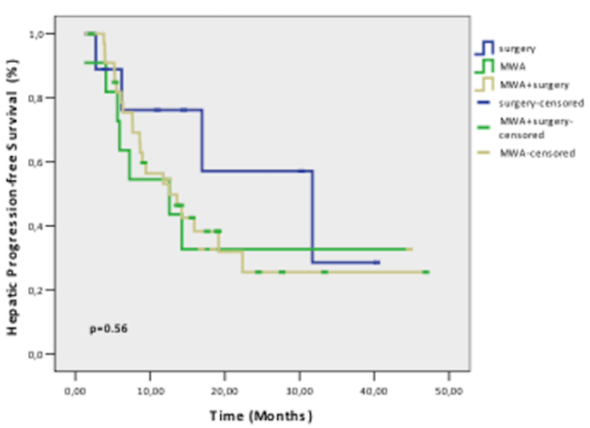
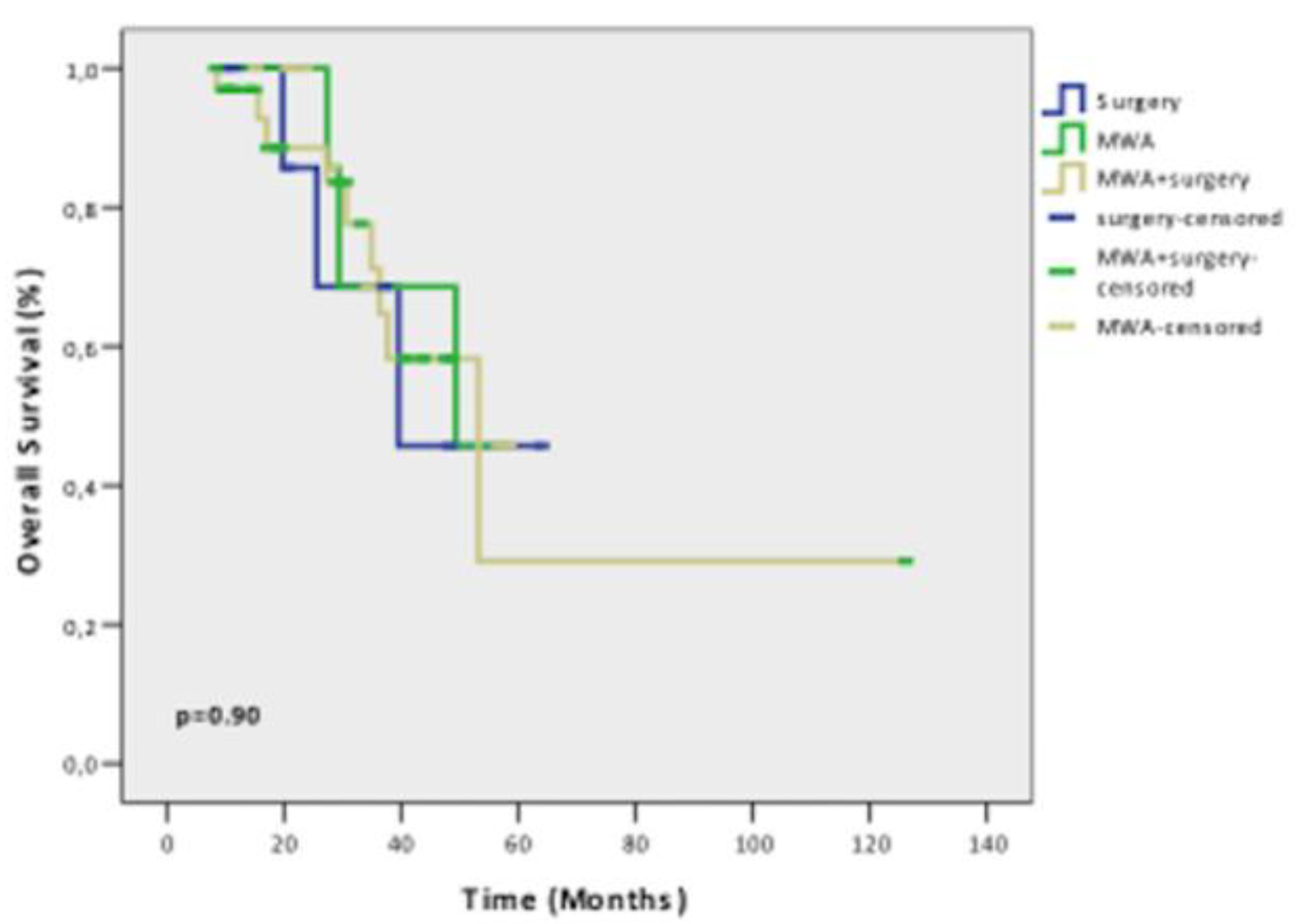
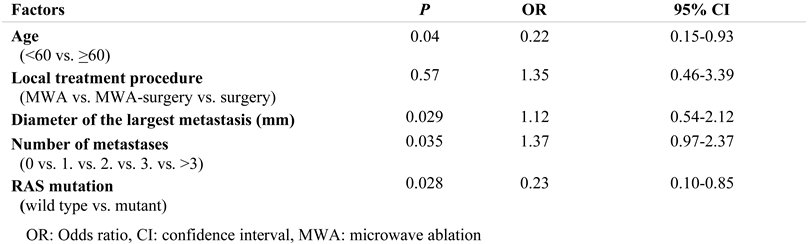
All statistical analyses were conducted using SPSS 22.0 (SPSS Inc., Chicago, IL, USA). The relationship between local treatment modalities and clinico-pathological factors was analyzed with the chi-square and the Fischer’s exact test. Univariate and multivariate analyses were performed to assess the overall and hepatic progression-free survival using the Cox proportional hazard regression model. Logistic regression analysis was used to identify the predictive factors for the local treatment response. Both OS and HPFS were analyzed using the Kaplan-Meier estimates, and comparisons were performed using the log rank test. All statistical tests were two-sided, and p values <0.05 were considered statistically significant.

Results

In the study population, 27.8% were women (n=15) and 72.2% were (n=39) men. The patients’ ages ranged from 30 to 83 and the median age was 62. The number of CLMs varied from 1 to 27 and the median lesion number was 4. The median size of metastases was 20 mm (range 2-80 mm). The original tumor was colon cancer in 32 (59.3%) of the patients and rectal cancer in 22 (40.7%) of them. All patients were treated with systemic chemotherapy plus targeted agents. A total of 17 (31.5%) patients received neoadjuvant chemotherapy regimen, whereas 2 (3.7%) patients received neoadjuvant radiotherapy for the primary tumor and 10 (18.5%) patients underwent chemo-radiotherapy.
In 20.4% of the patients (n=11) MWA was performed, in 16.7% (n=9) of the patients, surgical resection was performed, and in 63% (n=34) of them, combined treatment was conducted. In 6 patients, partial hepatectomy was performed, while in 39 patients metastasectomy was preferred. A total of 3 (7%) of the MWA sessions were performed under CT/US-guidance and 42 (93%) sessions were conducted with IOUS. IOUS was performed either during metastasectomy or colectomy.
The relationship between local treatment modalities and clinicopathological factors are summarized in Table 1. Our study groups showed similar baseline characteristics, with the exception of the primary tumor location, the number of metastases and the extrahepatic progression following the treatment. Major complications including biloma, hepatic abscess and pleural effusion occurred in 20% (n=25) of the patients, whereas minor complications were encountered in 17% (n=9) of the patients.
No incomplete ablation was observed in the first month follow-up after MWA. The rate of LTP after treatment was 7.4% (n=4) and the rate of IDR was 46.3% (n=25). In 46.3% (n=25) of the patients, total response was achieved (Figure 1).
The median OS was 49.2 months after MWA, 39.4 months after surgery, and 53.1 months after the combined treatment. The median HPFS was 14.2 months (95% CI 10.0-18.3). There were no significant differences in HPFS and OS between the three groups (p=0.56 and 0.90, respectively) (Figure 2 and Figure 3).
The univariate analysis revealed that patients with ECOG performance status 2, RAS mutation, three or more liver metastases, and those treated with percutaneous MWA had significantly worse HPFS (p=0.041, 0.035, 0.026 and 0.001, respectively). On the multivariate analysis, the time of liver metastasis occurrence (synchronous or metachronous) and the number of CLMs (<3 or ≥ 3) were independent prognostic factors for HPFS (HR 3.31, 95% CI 0.97-4.26, p=0.035; HR 1.56, 95% CI 0.99-2.46, p=0.031, respectively). On the univariate analysis, the significant factors associated with better OS were the absence of extrahepatic progression (p=0.006) and the local treatment response (p=0.005), while the extrahepatic progression following the treatment remained an independent prognostic factor on multivariate analysis (HR 5.74, 95% CI 1.35-10.4, p=0.017). The logistic regression analysis revealed that patients younger than 60-years old, with smaller or fewer metastases, and wild-type RAS showed significantly higher rates of local treatment response (OR 0.22, 95% CI 0.15-0.93, p=0.04; OR 1.12, 95% CI 0.54-2.12, p=0.029; OR 1.37, 95% CI 0.97-2.37, p=0.035; OR 0.23, 95% CI 0.10-0.85, p=0.028, respectively) (Table 2).

Discussion

Our study demonstrates the non-inferiority of MWA compared with surgical resection and combined treatment. Additionally, younger age (<60), smaller (≤ 2 cm) or fewer (≤3) CLMs, and the presence of wild-type RAS mutation were proven to be predictive factors for local tumor control.
Despite the fact that the surgical resection has been considered the sole curative treatment for CLMs, only 10% of the patients are eligible for surgery even with the progress in systemic chemotherapy regimens and surgical techniques, and five-year OS after surgery has been reported as 25-46% [2,14,15]. Herein, thermal ablation techniques including cryoablation, RFA, and MWA have emerged as an alternative. Their main advantages include the preservation of the liver volume, repeatability, real-time imaging, low morbidity and mortality rates, and low costs. Moreover, they do not require hospitalization and enable combined treatment with other modalities. Due to their minimally invasive nature, these methods could potentially be applied to a larger patient population [5,6].
In this aspect RFA is the most investigated thermal ablation technique, but MWA offers several advantages over RFA, as it is not affected by desiccation or charring, provides larger and more homogenous ablation area more rapidly, does not require grounding pads, and enables the use of multiple antenna simultaneously [16,17].
The relatively higher LTP/IDR rate is the main drawback limiting ablation as a first-line curative means [7,10,18]. The LTP rates of MWA in heterogeneous study populations with primary and secondary liver malignancies vary between 2.9- 34% [11,19]. Our results revealing a relatively lower LTP rate (7.4%) gives hope for the future.
Most of the studies in the existing literature focus on both primary and metastatic hepatic tumors. Hence, it is hard to make an exact comparison regarding the clinical outcome [7,8,9,10,20]. Therefore, the results of our study focusing only on CLMs and comparing different treatment modalities, and analyzing distinct prognostic and predictive factors for local treatment response provide a significant contribution to the literature.
Although there are many studies in the existing literature comparing different ablation methods and defining the factors affecting the clinical outcome, none of them set a cut-off value for the patients’ age regarding the local treatment response [2,8,11,12,19,20,21,22,23]. Therefore, one of the main strengths of our study is the correlation of the outcomes with the patients’ age.
The results of a study evaluating the safety and the efficiency of MWA in a heterogeneous population including patients with hepatocellular carcinoma and CLMs revealed that the tumor size did not affect the local treatment response [9]. However, in our study, three or fewer lesions and lesions smaller than two cm in the largest diameter resulted in higher rates of local treatment response.
In a prospective randomized trial, namely the COLLISION trial, investigating the non-inferiority of thermal ablation techniques compared with surgical resection, the patient population was divided into two groups, i.e. the surgical and the thermal ablation group. The latter included patients undergoing either RFA or MWA. Their report revealed that surgery should not be regarded as superior to ablation techniques. In lesions with a diameter ≤3 cm, ablation techniques provided similar results to surgery. Furthermore, they emphasized lower mortality and morbidity rates, shorter hospitalization duration and lower costs of ablation therapies. This study does not completely reflect the results of the MWA group due to the heterogeneity of the ablation group including both RFA and MWA [6]. Our study with a wider range of lesions regarding the largest diameter also revealed that there was no significant difference between MWA and surgery in local treatment response, OS, and HPFS.
There is only one prospective randomized trial focusing solely on MWA as the thermal ablation method in the literature. The study population comprised 30 patients with multiple CLMs and it was divided into MWA only and hepatic resection only groups. The 1-, 2- and 3-year survival rates and the mean survival times were reported as 71%, 57%, 14% and 27 months, respectively, for the MWA group and 69%, 56%, 23% and 25 months for patients undergoing hepatectomy. No significant difference was found between the two groups (p=0.65). As a result of this study, MWA is proved to be as effective as surgical resection in the treatment of less than ten CLMs [21]. A study with 53 patients undergoing either hepatectomy (37 patients) or MWA plus hepatectomy (16 patients) investigated the treatment efficiency and compared the results including survival and recurrence rates. The results revealed no significant difference between the groups in OS, HPFS, and disease-free survival (p=0.43, 0.54, and 0.86, respectively) [2]. They emphasized the role of ablation methods in expanding the indications of surgery in patients with multiple CLMs. Our study involved an additional group undergoing only MWA and so enabled a comparison of the results from MWA, surgery, and combined treatment in a wide range of lesions regarding the number and the largest diameter of the metastasis, which adds a primary contribution to the literature.
A multi-institutional study evaluating the survival rates after MWA and combined treatment revealed no significant differences in disease-free survival and OS between the groups (p=0.525 and 0.132, respectively) [23]. Several cohort studies reported 3-, 4- and 5-year OS after MWA between 35-79%, 35-58% and 17-18% [11,19,21].
These results are also compatible with our study, showing no significant differences in HPFS and OS between the MWA alone, surgery alone and combined treatment groups (p=0.56 and 0.90, respectively).
We conducted a case-based approach in the treatment of CLMs, in accordance with the guidelines of CIRSE and the consensus report of COLLISION trial [6,12]. The choice of the treatment modality was decided by a multidisciplinary board according to the number, the size, the location (proximity to the vasculo-biliary structures or deep in the parenchyma) of the CLM. This approach might be beneficial for customized treatment strategies. The results of our study revealing no significant difference in survival rates and local treatment response between the groups might reflect the successful assignment of the patients to different treatment options.
Patients with a poor ECOG performance status and three or more liver metastases showed worse HPFS (p=0.041 and 0.026, respectively) in our study. This can be explained by the fact that these patients have a higher tumor burden. The number of CLMs was also an independent prognostic factor for HPFS (p=0.031). As expected, achieving complete ablation and the absence of extrahepatic progression were associated with better OS (p=0.006 and 0.005, respectively). A report from the literature suggests that a tumor size of three cm or more was a predictive factor for HPFS in patients with hepatic malignancies [8]. In a more recent study, the maximum diameter of CLMs and patients’ response to pre-ablation systemic chemotherapy were independent risk factors for both OS and HPFS [22]. In our study, the diameter of the largest metastasis was an independent predictive factor for local treatment response (p=0.029).
According to a recent study, prehepatectomy carcinoembryonic antigen concentration in serum served as an independent prognostic factor for survival, which did not differ between the resection and the combined treatment group (p=0.02) [2]. Likewise, RAS mutation, a genetic biomarker, was also found to be the main predictive factor for the local treatment response (p=0.028) in our study. These results are compatible with the studies in the literature reporting lower HPFS rates of mutant RAS compared to wild-type RAS [24,25].
A meta-analysis comparing ablation therapies with surgical procedures for CLMs evaluated 75 studies and concluded that ablative therapies offer significantly higher survival rates and lower complication rates [11]. Major complication rates of MWA varies between 2.6% and 16% in the literature [11,20,26]. In our study, no major complication occurred following MWA alone. A total of 9 patients (26.5%) in the combined treatment group and 2 (22.2%) in the surgery alone group experienced major complications such as abscess, biloma and pleural effusion requiring drainage. In this regard, MWA could be regarded as a safe technique.
The limitations of our study include retrospective design, potential population bias, a relatively small sample size and a limited follow-up interval. The main reason why there were considerably fewer patients in the only-surgery group was the widely accepted use of combined treatment with ablation methods. Even though patients with more advanced disease and metastases in proximity to vasculo-biliary structures were referred to MWA, and more favorable patients underwent surgical resection, MWA achieved similar outcomes, either alone or as adjunct to surgery. Finally, one of the main disadvantages of our study is the lack of ability to compare systemic treatment response. Although the results of our study reveal promising results regarding the role of MWA, studies with larger sample sizes and longer follow-up duration are required for further validation.

Conclusions

In conclusion, MWA as a minimally invasive and easily repeatable curative method might achieve similar survival rates compared to surgical resection and provide high local tumor control in the treatment of patients with CLMs whose life expectancy has been increased. In all these aspects, we believe that MWA will be preferred over surgical procedures in the near future.

Conflicts of Interest disclosure

There are no known conflicts of interest in the publication of this article. The manuscript was read and approved by all authors.

Compliance with ethical standards

Any aspect of the work covered in this manuscript has been conducted with the ethical approval of all relevant bodies and that such approvals are acknowledged within the manuscript.

References

  1. Siegel, R.L.; Miller, K.D.; Jemal, A. Cancer statistics, 2020. CA Cancer J Clin. 2020, 70, 7–30. [Google Scholar] [CrossRef] [PubMed]
  2. Tanaka, K.; Shimada, H.; Nagano, Y.; Endo, I.; Sekido, H.; Togo, S. Outcome after hepatic resection versus combined resection and microwave ablation for multiple bilobar colorectal metastases to the liver. Surgery. 2006, 139, 263–273. [Google Scholar] [CrossRef]
  3. Stättner, S.; Primavesi, F.; Yip, V.S.; Jones, R.P.; Öfner, D.; Malik, H.Z.; Fenwick, S.W.; Poston, G.J. Evolution of surgical microwave ablation for the treatment of colorectal cancer liver metastasis: review of the literature and a single centre experience. Surg Today. 2015, 45, 407–415. [Google Scholar] [CrossRef]
  4. Meijerink, M.R.; Puijk, R.S.; van Tilborg, A.A.J.M.; Henningsen, K.H.; Fernandez, L.G.; Neyt, M.; Heymans, J.; Frankema, J.S.; de Jong, K.P.; Richel, D.J.; Prevoo, W.; Vlayen, J. Radiofrequency and Microwave Ablation Compared to Systemic Chemotherapy and to Partial Hepatectomy in the Treatment of Colorectal Liver Metastases: A Systematic Review and Meta-Analysis. Cardiovasc Intervent Radiol. 2018, 41, 1189–1204. [Google Scholar] [CrossRef] [PubMed]
  5. Benson, A.B.; Venook, A.P.; Al-Hawary, M.M.; Arain, M.A.; Chen, Y.J.; Ciombor, K.K.; Cohen, S.; Cooper, H.S.; Deming, D.; Farkas, L.; et al. Colon Cancer, Version 2.2021, NCCN Clinical Practice Guidelines in Oncology. J Natl Compr Canc Netw. 2021, 19, 329–359. [Google Scholar] [CrossRef]
  6. Nieuwenhuizen, S.; Puijk, R.S.; van den Bemd, B.; et al. Resectability and Ablatability Criteria for the Treatment of Liver Only Colorectal Metastases: Multidisciplinary Consensus Document from the COLLISION Trial Group. Cancers (Basel) 2020, 12, 1779. [Google Scholar] [CrossRef]
  7. Leung, U.; Kuk, D.; D'Angelica, M.I.; Kingham, T.P.; Allen, P.J.; DeMatteo, R.P.; Jarnagin, W.R.; Fong, Y. Long-term outcomes following microwave ablation for liver malignancies. Br J Surg. 2015, 102, 85–91. [Google Scholar] [CrossRef] [PubMed]
  8. Groeschl, R.T.; Pilgrim, C.H.; Hanna, E.M.; Simo, K.A.; Swan, R.Z.; Sindram, D.; Martinie, J.B.; Iannitti, D.A.; Bloomston, M.; Schmidt, C.; Khabiri, H.; Shirley, L.A.; Martin, R.C.; Tsai, S.; Turaga, K.K.; Christians, K.K.; Rilling, W.S.; Gamblin, T.C. Microwave ablation for hepatic malignancies: a multiinstitutional analysis. Ann Surg. 2014, 259, 1195–1200. [Google Scholar] [CrossRef]
  9. Alexander, E.S.; Wolf, F.J.; Machan, J.T.; Charpentier, K.P.; Beland, M.D.; Iannuccilli, J.D.; Haas, R.H.; Dupuy, D.E. Microwave ablation of focal hepatic malignancies regardless of size: A 9-year retrospective study of 64 patients. Eur J Radiol. 2015, 84, 1083–1090. [Google Scholar] [CrossRef]
  10. Takahashi, H.; Kahramangil, B.; Berber, E. Local recurrence after microwave thermosphere ablation of malignant liver tumors: results of a surgical series. Surgery. 2018, 163, 709–713. [Google Scholar] [CrossRef]
  11. Pathak, S.; Jones, R.; Tang, J.M.; Parmar, C.; Fenwick, S.; Malik, H.; Poston, G. Ablative therapies for colorectal liver metastases: a systematic review. Colorectal Dis. 2011, 13, e252–e265. [Google Scholar] [CrossRef] [PubMed]
  12. Crocetti, L.; de Baére, T.; Pereira, P.L.; Tarantino, F.P. CIRSE Standards of Practice on Thermal Ablation of Liver Tumours. Cardiovasc Intervent Radiol. 2020, 43, 951–962. [Google Scholar] [CrossRef] [PubMed]
  13. Ahmed, M.; Solbiati, L.; Brace, C.L.; et al.; International Working Group on Image-guided Tumor Ablation; Interventional Oncology Sans Frontières Expert Panel; Technology Assessment Committee of the Society of Interventional Radiology; Standard of Practice Committee of the Cardiovascular and Interventional Radiological Society of Europe Image-guided tumor ablation: standardization of terminology and reporting criteria--a 10-year update. Radiology. 2014, 273, 241–260. [Google Scholar] [CrossRef] [PubMed]
  14. Gomez, D.; Sangha, V.K.; Morris-Stiff, G.; Malik, H.Z.; Guthrie, A.J.; Toogood, G.J.; Lodge, J.P.; Prasad, K.R. Outcomes of intensive surveillance after resection of hepatic colorectal metastases. Br J Surg. 2010, 97, 1552–1560. [Google Scholar] [CrossRef]
  15. Camacho, J.C.; Petre, E.N.; Sofocleous, C.T. Thermal Ablation of Metastatic Colon Cancer to the Liver. Semin Intervent Radiol. 2019, 36, 310–318. [Google Scholar] [CrossRef]
  16. Simon, C.J.; Dupuy, D.E.; Mayo-Smith, W.W. Microwave ablation: principles and applications. Radiographics. 2005, 25 (Suppl. 1), S69–S83. [Google Scholar] [CrossRef]
  17. Song, P.; Sheng, L.; Sun, Y.; An, Y.; Guo, Y.; Zhang, Y. The clinical utility and outcomes of microwave ablation for colorectal cancer liver metastases. Oncotarget. 2017, 8, 51792–51799. [Google Scholar] [CrossRef]
  18. Urbonas, T.; Anderson, E.M.; Gordon-Weeks, A.N.; Kabir, S.I.; Soonawalla, Z.; Silva, M.A.; Gleeson, F.V.; Reddy, S. Factors predicting ablation site recurrence following percutaneous microwave ablation of colorectal hepatic metastases. HPB (Oxford). 2019, 21, 1175–1184. [Google Scholar] [CrossRef]
  19. Puijk, R.S.; Ruarus, A.H.; Vroomen, L.G.P.H.; et al.; COLLISION Trial Group Colorectal liver metastases: surgery versus thermal ablation (COLLISION)—A phase III single-blind prospective randomized controlled trial. BMC Cancer. 2018, 18, 821. [Google Scholar] [CrossRef]
  20. Liang, P.; Wang, Y.; Yu, X.; Dong, B. Malignant liver tumors: treatment with percutaneous microwave ablation-- complications among cohort of 1136 patients. Radiology. 2009, 251, 933–940. [Google Scholar] [CrossRef]
  21. Shibata, T.; Niinobu, T.; Ogata, N.; Takami, M. Microwave coagulation therapy for multiple hepatic metastases from colorectal carcinoma. Cancer 2000, 89, 276–284. [Google Scholar] [CrossRef] [PubMed]
  22. Zhou, F.; Yu, X.; Liang, P.; Han, Z.; Cheng, Z.; Yu, J.; Liu, F.; Hu, Y. Does primary tumor location impact the prognosis of colorectal liver metastases patients after microwave ablation? - Lessons from 10 years' experience. Oncotarget. 2017, 8, 100791–100800. [Google Scholar] [CrossRef] [PubMed]
  23. Philips, P.; Groeschl, R.T.; Hanna, E.M.; Swan, R.Z.; Turaga, K.K.; Martinie, J.B.; Iannitti, D.A.; Schmidt, C.; Gamblin, T.C.; Martin, R.C. Single-stage resection and microwave ablation for bilobar colorectal liver metastases. Br J Surg. 2016, 103, 1048–1054. [Google Scholar] [CrossRef]
  24. Odisio, B.C.; Yamashita, S.; Huang, S.Y.; Harmoush, S.; Kopetz, S.E.; Ahrar, K.; Shin Chun, Y.; Conrad, C.; Aloia, T.A.; Gupta, S.; Hicks, M.E.; Vauthey, J.N. Local tumour progression after percutaneous ablation of colorectal liver metastases according to RAS mutation status. Br J Surg. 2017, 104, 760–768. [Google Scholar] [CrossRef] [PubMed]
  25. Calandri, M.; Yamashita, S.; Gazzera, C.; Fonio, P.; Veltri, A.; Bustreo, S.; et al. Ablation of colorectal liver metastasis: Interaction of ablation margins and RAS mutation profiling on local tumour progression-free survival. Eur Radiol. 2018, 28, 2727–2734. [Google Scholar] [CrossRef]
  26. Livraghi, T.; Meloni, F.; Solbiati, L.; Zanus, G.; Collaborative Italian Group using AMICA system. Complications of microwave ablation for liver tumors: results of a multicenter study. Cardiovasc Intervent Radiol. 2012, 35, 868–874. [Google Scholar] [CrossRef]
Figure 1. Local tumor progression in a 65-year-old patient with colon cancer. (a) T2-weighed image shows a hyperintense metastasis with a diameter of 3 cm in segment V near the gallbladder bed and portal vein branch, which was treated with microwave ablation therapy. (b) Three metastases in the left lobe (black asterisks), which were resected via lobectomy, are seen on T2-weighed image. (c) At 1-year follow-up, a local tumor progression (white asterisk) adjacent to a portal vein branch (arrow) is encountered on postcontrast T1-weighed image.
Jmms 09 00012 g001
Figure 2. Hepatic progression-free survival of patients with colorectal liver metastases after microwave ablation (MWA), surgery, and combined treatment.
Jmms 09 00012 g002
Figure 3. Overall survival of patients with hepatic metastases from colorectal cancer following surgery, microwave ablation (MWA), and combined treatment.
Jmms 09 00012 g003
Table 1. The relationship between local treatment modalities and clinicopathological factors.
Jmms 09 00012 i001
Table 2. Logistic regression analysis of the predictive factors for local treatment response.
Jmms 09 00012 i002

