Next Article in Journal
Carbamazepine-Induced Dress Syndrome: A Case Report
Previous Article in Journal
Clinical Features, Pathology, and Surgical Management of Periocular Giant Basal Cell Carcinoma
 
 
Journal of Mind and Medical Sciences is published by MDPI from Volume 12 Issue 1 (2025). Previous articles were published by another publisher in Open Access under a CC-BY (or CC-BY-NC-ND) licence, and they are hosted by MDPI on mdpi.com as a courtesy and upon agreement with Valparaiso University (ValpoScholar).
Case Report

When Myocardial Infarction Is Choosing Young Victims

by
Cristina Nedelcu
1,
Mihaela Ionescu
2,*,
Paris Ionescu
2,
Vitalie Morosanu
3,
Gabriel Smarandache
4,
Dumitru Cristinel Badiu
4,
Florentina Gherghiceanu
4,
Vlad Denis Constantin
4,
Emanuele Maria Merlo
5 and
Daniel Niţă
6
1
Army Center for Cardiovascular Diseases, Constanta, Romania
2
Faculty of Medicine, Ovidius University, Mamaia Blvd. 124, 900527 Constanta, Romania
3
County Clinical Emergency Hospital of Constanta, Constanta, Romania
4
Carol Davila University of Medicine and Pharmacy, Bucharest, Romania
5
Department of Cognitive Sciences, Psychology, Educational and Cultural Studies (COSPECS), University of Messina, Italy
6
Army Center for Cardiovascular Diseases, Bucharest, Romania
*
Author to whom correspondence should be addressed.
J. Mind Med. Sci. 2020, 7(2), 233-238; https://doi.org/10.22543/7674.72.P233238
Submission received: 14 June 2020 / Revised: 18 August 2020 / Accepted: 18 August 2020 / Published: 29 September 2020

Abstract

:
We present the case of a 31-year-old patient, without cardiovascular risk factors, without significant pathological or family history of cardiovascular disease, who was diagnosed with severe coronary artery disease, left main bifurcation localization, which required surgical coronary revascularization. The angiographic and intraoperative aspect excluded the most common causes of non-atherosclerotic causes of coronary lesions. Vasculitis was another possible etiology but was also excluded based on the normal clinical examination, negative inflammation markers, lack of diffuse vascular impairment, TPHA and negative VDRL. Prolonged mental stress can lead to accelerated progression of atherosclerosis by inducing endothelial dysfunction, our patient describing a “burnout syndrome” in the last few months. The particularity of the presented case is the severe coronary artery disease in a young patient without discovering the main etiology of the advanced atherosclerotic process.

Introduction

Coronary heart disease (CHD) continues to be the leading cause of death worldwide, as World Health Organization (WHO) reports. Although it is considered that acute myocardial infarction (AMI) among young adults is relatively rare, it still seems that 4% to 10% of all heart attacks occur before age 45, and most of these cases involve men [1].
The primary cause of CHD in young adults-as well as in the elderly—is atherosclerosis, accounting for about 80% of heart attacks [2]. The other causes should also be considered, like: congenital malformations of the coronary anatomy, embolic coronary disease (4% of case), blood clotting disorders, coronary spasm-illicit drug abuse (especially cocaine and amphetamines), radiotherapy for chest tumors, trauma or inflammation of the arteries. Unfortunately, the disease carries a significant morbidity, psychological distress, financial burden for the patient but also for the patient’s family and for the entire society, especially when it occurs at a young age [3].
The “Burnout Syndrome” is an extreme response to a job-related chronic stress and is defined as high levels of physical and mental exhaustion that marks the need of the organism to face a threat to its internal homeostasis [4].
Although chronic stress is a known risk factor for CHD, due to the continuous activation of the autonomic nervous system and neuroendocrine axis, situations of intensive acute stress can be also associated with an increased prevalence of cardiovascular events [5].
These young patients have also a certain cardiovascular risk profile, as well as particular clinical presentation and prognosis in comparison with the elderly, which has to be taken into consideration when managing with CHD [6].
However, the vast majority of the young patients have atherosclerosis as the main etiology of CHD, accelerated by the conventional cardiovascular risk factors—but withsome different features [7]. Smoking is the most common risk factor among the young CHD patients, as Zimmerman et al. found a prevalence of 92% [8]. Mukherjee et al. concluded that the prevalence of smoking was higher in those less than 40 years old, compared to those above 60 years old (58.7 versus 43%) [9]. The other major CV risk factors among youngsters are hypertension, high triglycerides, low HDL-cholesterol and obesity.
Myocardial infarction in individuals less than 40 years of age is almost exclusively seen in male patients. Fortunately, the younger patient usually has a single vessel disease and therefore a better prognosis [10].

Case Presentation

We present the case of a seemingly healthy 31-year-old patient, without cardiovascular risk factors, without significant family history of cardiovascular disease, who was admitted to the hospital for a retrosternal chest pain–with the first episode occurring three weeks before admission. Pain was self-limited, with no relation to the effort, with a maximum duration of 5 minutes.
The clinical examination did not reveal pathological elements, blood pressure = 130/80 mmHg equal in both arms, ventricular allure = 65 beats per minute (bpm), rhythmic cardiac noises, without cardiac or vascular murmurs, and peripheral pulse symmetrically present.
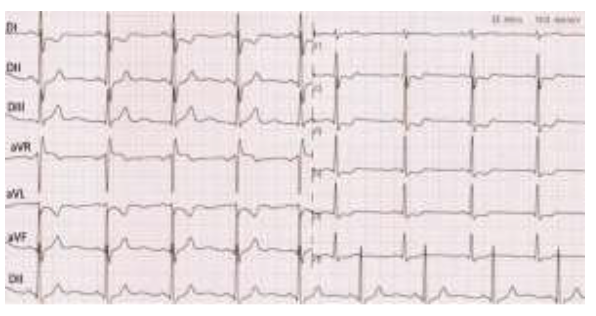
The initial electrocardiogram (ECG) showed: sinus rhythm, 65 beat per minute, QRS axis + 75 degrees, incomplete right bundle branch block, T wave flattened in DI and negative in AVL (Figure 1).
From the laboratory blood results at admission, we noted: troponin T 0.045 ng/mL (cut-off value 0.020 ng/mL), CK-MB within normal limits, serum transaminases (ALT/AST) elevated 3 times the upper limit of normal; normal markers of inflammation, serum lipids within normal ranges; in dynamics, 6 hours after the first measurement, troponin T increased at 0.085 ng/ml, subsequently descending.
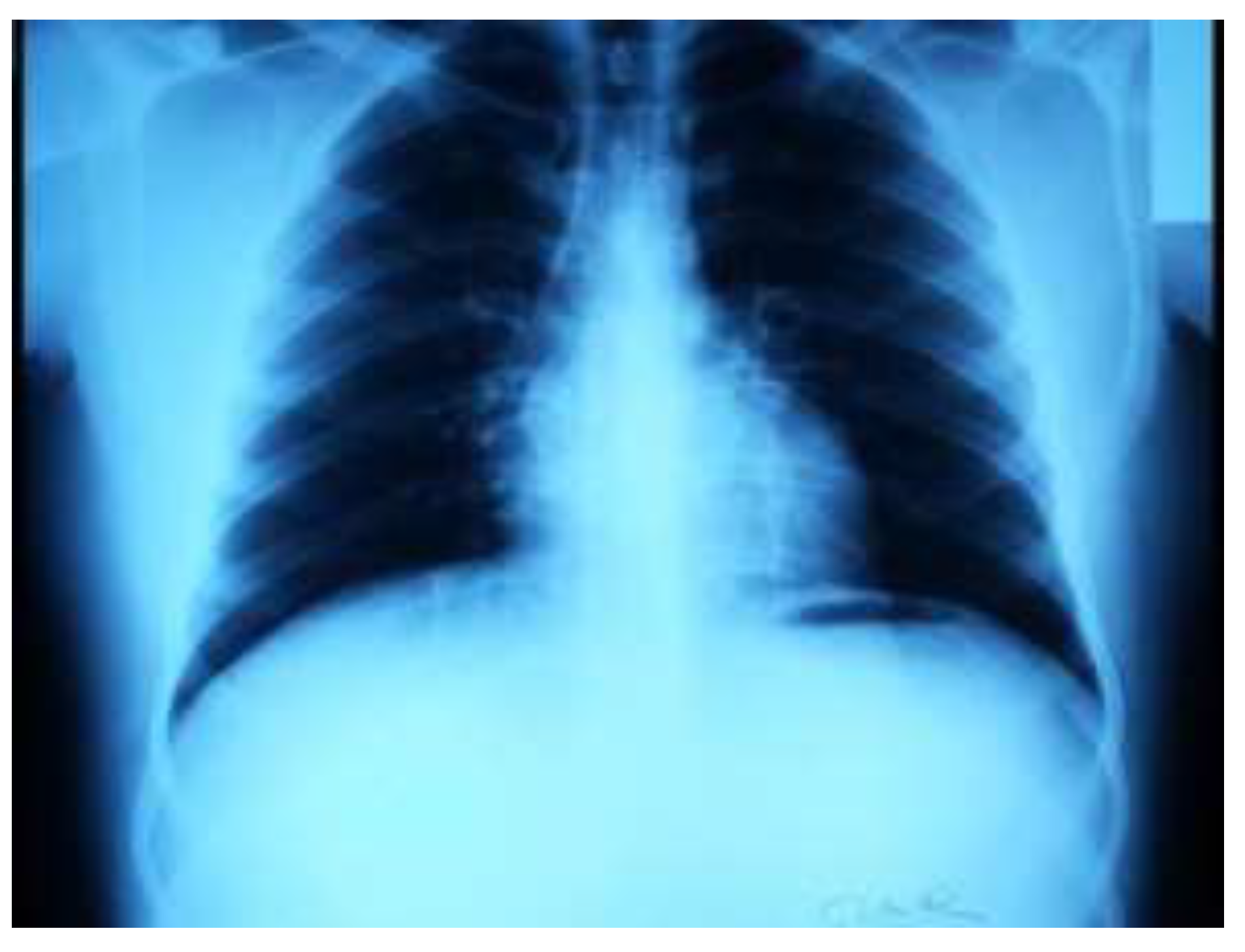
Cardiopulmonary radiography showed a heart within normal limits, without pleuro-pulmonary or chest wall injuries (Figure 2).
Standard transthoracic echocardiography revealed a normal-sized left ventricle, with normal global and segmental systolic function, normal diastolic profile, minimal mitral regurgitation, straight cavities with normal dimensions, free pericardium, left ventricular ejection fraction 60% (Simpson method).
Within the hospital stay, electrocardiograms were recorded during chest pain, and changes were noticed compared to the baseline electrocardiogram: ST segment depression in both leads DI and DIII, of maximum 1.5 mm with negative T-waves, ST segment depression in V2–V5 of maximum 1 mm, as well as ST segment elevation of 1 mm in aVR (findings that may suggest severe coronary artery disease – left main lesion or triple vessel disease) (Figure 3).
Considering the ECG changes during chest pain episodes in association with the dynamics of cardiac biomarkers, we have made the diagnosis of acute non-ST segment elevation myocardial infarction, and the specific treatment was instituted: dual antiplatelet therapy, low molecular weight heparin, high-dose of statin, beta-blocker, angiotensin-converting enzyme inhibitor, and sublingual nitroglycerin as needed [11,12,13]. The coronary evaluation confirmed the diagnosis, revealing a sub- occlusion that involved the bifurcation of the common trunk, involving the ostium of the anterior descending artery and the intermediate branch (ramus intermedius)—a large artery calibre.
Because of the lesions location, difficult to approach in the case of the percutaneous angioplasty, the heart team decided to perform the surgical coronary revascularization with double coronary bypass grafting using the left internal mammary artery in the middle segment of the anterior descending artery and the right internal breast artery on the intermediate branch. Our patient had favourable postoperative evolution, without repeated chest pain.
The final diagnosis was:
  • Surgical coronary revascularization - double coronary bypass graft using left internal mammary artery–on the anterior descending artery and right internal—on the intermediate artery.
  • Sub-occlusion of anterior descending artery and intermediate branch.
  • Non-ST segment elevation myocardial infarction.
  • Incomplete right bundle branch block.

Discussions

In the context of severe coronary artery disease in a young patient without cardiovascular risk factors, we aimed to investigate the existence of a non-atherosclerotic etiology of coronary lesions [14]. A series of necropsy studies in subjects with sudden cardiac death showed the presence of coronary anomalies in 60% of cases in children, while in adults coronary atherosclerosis was involved in 90–95% of cases [15]. It is reported in the literature that between 4 and 7% of patients with fatal myocardial infarction have had non-atherosclerotic coronary lesions at necropsy [16].
Of the non-atherosclerotic causes of coronary lesions, we list in order of frequency reported in a series of necropsy studies made in patients with sudden cardiac death: congenital anomalies of coronary arteries, acute coronary artery dissection, vasculitis affecting coronary circulation, fibromuscular dysplasia [17,18,19]. Variations in the origin, pathway, or distribution of coronary arteries are found in 1–2% of the population, but certain coronary abnormalities (ostial lesions, passage of an important branch between the trunk of the pulmonary artery and aorta, the origin of an important branch in the trunk of the pulmonary artery, myocardial decks) presents a higher risk of ischemia [20,21]. Coronary aneurysms, which can be congenital or acquired, single or multiple, can lead to thrombosis due to turbulent blood flow and increased parietal stress, sometimes associated with an abnormal structure of the arterial wall, with possible occlusion of the vessel and myocardial infarction [22]. The incidence of coronary aneurysms is about 1.5% of the patients studied at the autopsy; the etiology of acquired aneurysms is: atherosclerosis, trauma, angioplasty, arteritis (including syphilis), fungal, viral embolism, dissection [23].
The coronary angiography report and later the intraoperative assessment have excluded: the aberrant origin of the coronary arteries, the muscular bridges, the spontaneous coronary dissection, the embolism or the coronary spasm. Another possible etiology is the inflammation of the arteries, namely vasculitis, but it has been excluded based on the following data: normal clinical examination, negative inflammation markers, lack of diffuse vascular impairment, TPHA and negative VDRL.
Specific laboratory tests were performed for hypercoagulable states which revealed the mutation of the MTHFR gene C677T in the heterozygous form, and a slight deficiency of protein S, mutations that are common in the general population and not in the heterozygous per se.
It should be noted that when resuming the anamnesis, the patient describes a period of psychic stress and exhaustion at work, including night shifts; several studies pointed out that mental stress, overwork and deprivation of sleep at night decrease the DNA repair capacity, and may induce endothelial dysfunction which in its turn promotes the process of atherosclerosis [24].
In this case, the coronary angiography and later the intraoperative assessment excluded: the origin, route and aberrant distribution of coronary arteries, muscle bridges, spontaneous coronary dissection, embolism, coronary spasm. The following etiology is represented by vasculitis (Takayasu disease, systemic lupus erythematosus, rheumatoid arthritis, syphilis, polyarteritis nodosa etc.) which was excluded based on the clinical and paraclinical data mentioned above. As a non-atherosclerotic cause of coronary artery disease, some metabolic disorders have been described to accumulate certain substances in the walls of large or small coronary arteries, which may lead to myocardial infarction. These include: mucopolysaccharidosis (eg. Hunter disease, Hurler disease), gangliosidosis, and Fabry disease [25,26,27,28].
Because a clear non-atherosclerotic cause could not be identified, and given that atherosclerosis is a process beginning in childhood [29,30,31], we considered this etiology as the most plausible. It is reported that acute coronary syndrome in patients under 30 years is due to atherosclerosis in 60% of cases [32]. In autopsy series performed on male subjects aged 20–25 who died from traumatic causes, it was shown that anterior descending artery stenosis of over 40% (as a marker of atherosclerotic impairment) was present in 3-4% of cases. Similar studies have correlated coronary atherosclerotic impairment with cardiovascular risk factors, demonstrating that at this age the same cardiovascular risk factors apply as in adulthood.
When resuming the anamnesis, the patient describes a period of intense mental stress and intellectual overload, as well as sleep deprivation through work in night guards, which we call “burnout syndrome” [33,34,35,36]. Many studies done on healthy people without cardiovascular risk factors showed that prolonged mental stress can lead to accelerated progression of atherosclerosis by inducing endothelial dysfunction [37,38]. The mechanism consisted of vasoconstriction due to sympathetic activation and alteration of nitric oxide production and action [39,40]. Endothelial dysfunction after psychic stress appears to be due to oxidative stress and release of vasoconstrictive substances, such as endothelin and angiotensin II [41,42,43,44]. Catecholamines do not directly affect endothelial function, although they influence it indirectly by increasing blood pressure [45].

Conclusions

The particularity of the presented case is the severe coronary artery disease with left main bifurcation localization, which required surgical revascularization, in a young patient without cardiovascular risk factors.

Conflict of interest disclosure

There are no known conflicts of interest in the publication of this article. The manuscript was read and approved by all authors.

Compliance with ethical standards

Any aspect of the work covered in this manuscript has been conducted with the ethical approval of all relevant bodies and that such approvals are acknowledged within the manuscript. Verbal informed consent was obtained from the patient(s) for their anonymized information to be published in this article.

References

  1. Sanchis-Gomar, F.; Perez-Quilis, C.; Leischik, R.; Lucia, A. Epidemiology of coronary heart disease and acute coronary syndrome. Ann Transl Med. 2016, 4, 256. [Google Scholar] [CrossRef] [PubMed]
  2. Yazdanyar, A.; Newman, A.B. The burden of cardiovascular disease in the elderly: Morbidity, mortality, and costs. Clin Geriatr Med. 2009, 25, 563-vii. [Google Scholar] [CrossRef]
  3. Egred, M.; Viswanathan, G.; Davis, G.K. Myocardial infarction in young adults. Postgrad Med J. 2005, 81, 741–745. [Google Scholar] [CrossRef]
  4. Innstrand, S.T.; Langballe, E.M.; Falkum, E.; Aasland, O.G. Exploring within- and between-gender differences in burnout: 8 different occupational groups. Int Arch Occup Environ Health. 2011, 84, 813–824. [Google Scholar] [CrossRef] [PubMed]
  5. Kivimaki, M.; Nyberg, S.T.; Fransson, E.I.; Heikkila, K.; Alfredsson, L.; Casini, A.; et al. Associations of job strain and lifestyle risk factors with risk of coronary artery disease: A meta-analysis of individual participant data. CMAJ. 2013, 185, 763–769. [Google Scholar] [PubMed]
  6. Egred, M.; Viswanathan, G.; Davis, G.K. Myocardial infarction in young adults. Postgrad Med J. 2005, 81, 741–745. [Google Scholar] [CrossRef]
  7. Bhardwaj, R.; Kandoria, A.; Sharma, R. Myocardial infarction in young adults-risk factors and pattern of coronary artery involvement. Niger Med J. 2014, 55, 44–47. [Google Scholar] [CrossRef]
  8. Zimmerman, F.H.; Cameron, A.; Fisher, L.D.; Ng, G. Myocardial infarction in young adults: Angiographic characteristics, risk factors and prognosis, coronary artery surgery study register (CASS). J Am Coll Cardiol. 1995, 26, 654–61. [Google Scholar]
  9. Mukherjee, D.; Hsu, A.; Moliterno, D.J.; Lincoff, A.M.; Goormastic, M.; Topol, E.J. Risk factors for premature coronary artery disease and determinants of adverse outcomes after revascularization in patients less than 40 years old. Am J Cardiol. 2003, 92, 1465–7. [Google Scholar]
  10. Deshmukh, P.P.; Singh, M.M.; Deshpande, M.A.; Rajput, A.S. Clinical and angiographic profile of very young adults presenting with first acute myocardial infarction: Data from a tertiary care center in Central India. Indian Heart J. 2019, 71, 418–421. [Google Scholar] [CrossRef]
  11. Laslo, C.L.; Pantea Stoian, A.; Socea, B.; et al. New oral anticoagulants and their reversal agents. J Mind Med Sci. 2018, 5, 195–201. [Google Scholar] [CrossRef]
  12. Diaconu, C.; Paraschiv, B.; Stanescu, A.M.A.; et al. Arterial Hypertension, a Frequent Comorbidity in Diabetes: The Perioperative Management. In Proceedings of the 35th Balkan Medical Week on Healthy Ageing-An Endless Challenge, Athens, Greece, 25–27 September 2018; pp. 15–20. [Google Scholar]
  13. Cozma, M.A.; Gaman, M.A.; Dobrica, E.C.; et al. Beta-Blockers in the Treatment of Chronic Heart Failure: Did Gender Make a Difference? In Proceedings of the 35th Balkan Medical Week on Healthy Ageing-An Endless Challenge, Athens, Greece, 25–27 September 2018; pp. 21–26. [Google Scholar]
  14. Waller, B.F. Schlant, R.C., Alexander, R.W., O’Rourke, R.A., Roberts, R., Sonnenblick, E.H., Eds.; Nonatherosclerotic causes of coronary artery narrowing. In The Heart, 8th ed.; Mc Graw Hill, 1994; pp. 1239–1261. [Google Scholar]
  15. McGill, H.C., Jr.; McMahan, C.A.; Herderick, E.E.; Malcom, G.T.; Tracy, R.E.; Strong, J.P. Origin of atherosclerosis in childhood and adolescence. Am J Clin Nutr. 2000, 72 (Suppl. S5), 1307S–1315S. [Google Scholar] [PubMed]
  16. Egred, M.; Viswanathan, G.; Davis, G.K. Myocardial infarction in young adults. Postgrad Med J. 2005, 81, 741–745. [Google Scholar]
  17. Chen, L.; Chester, M.; Kaski, J.C. Clinical factors and angiographic features associated with premature coronary artery disease. Chest 1995, 108, 364–369. [Google Scholar]
  18. Mazilu, L.; Stanculeanu, D.L.; Gheorghe, A.D.; Voinea, F.; et al. Incidenced of Chemotherapy Induced Paripheral Neuropathy in Cancer Patients in clinical Practice. Farmacia 2018, 66, 904–908. [Google Scholar]
  19. Tuta, L.A.; Iorga, I.; Azis, O.; Voinea, F. End-of-life Care in Erderly Patients with End-Stage Renal Disease–Ethical and Clinical Issues. In SGEM 2015, Book 1: Psychology and Psyhiatry, Sociology and Healthcare, Education Conference Proceedings; 2015; pp. 487–493. [Google Scholar]
  20. Ong, P.; Athanasiadis, A.; Hill, S.; Vogelsberg, H.; Voehringer, M.; Sechtem, U. Coronary artery spasm as a frequent cause of acute coronary syndrome: The CASPAR (Coronary Artery Spasm in Patients With Acute Coronary Syndrome) Study. J Am Coll Cardiol. 2008, 52, 523–527. [Google Scholar]
  21. Fulga, I.; Georgescu, C.; Ardeleanu, V. Single arterial trunk arising from the aortic arch with aortic coarctation. Report of a case. Embryological considerations. Rom J Morphol Embryol. 2014, 55, 165–169. [Google Scholar]
  22. Settineri, S.; Frisone, F.; Alibrandi, A.; Pino, G.; Lupo, N.J.; Merlo, E.M. Psychological Types and Learning Styles. Mediterr. J. Clin. Psychol. 2018, 6. [Google Scholar] [CrossRef]
  23. Suceveanu, A.I.; Mazilu, L.; Voinea, C.; Suceveanu, A.P. Concomitant Serum Presence of Hepatitis B Surface Antigen (HBsAg) and High Titers of Hepatitis B Surface Antibodies (Anti-HBsAb) in a Patient with Chronic Hepatitis B (HBV) Genotype D from Black Sea Coast Region: A Case Report. Hepat. Mon. 2018, 18, e60156. [Google Scholar]
  24. Tobaldini, E.; Fiorelli, E.M.; Solbiati, M.; Costantino, G.; Nobili, L.; Montano, N. Short sleep duration and cardiometabolic risk: From pathophysiology to clinical evidence. Nat Rev Cardiol. 2019, 16, 213–224. [Google Scholar] [CrossRef]
  25. Blieden, L.C.; Moller, J.H. Cardiac involvement in inherited disorders of metabolism. Prog Cardiovasc Dis. 1974, 16, 615–631. [Google Scholar] [CrossRef] [PubMed]
  26. Moraru, D.; Suceveanu, A.P.; Pantea Stoian, A.; Nitipir, C.; Pituru, S.; Voinea, F.; Timofte, D.; Suceveanu, A.I. Amyloidosis–the importance of an early diagnosis. In Proceeding of 35 Balkan Medical Week, Athens, Greece, 25–27 September; pp. 182–6.
  27. Leblebicioglu, H.; Arama, V.; Causse, X.; et al. Predictors associated with treatment initiation and switch in a real- world chronic hepatitis B population from five European countries. J Viral Hepat. 2014, 21, 662–670. [Google Scholar] [CrossRef] [PubMed]
  28. Suceveanu, A.I.; Mazilu, L.; Tomescu, D.; Ciufu, N.; Parepa, I.R.; Suceveanu, A.P. Screening of hepatopulmonary syndrome (HPS) with CEUS and pulse-oximetry in liver cirrhosis patients eligible for liver transplant. Chirurgia (Bucur). 2013, 108, 684–688. [Google Scholar]
  29. Tofolean, D.E.; Mazilu, L.; Stăniceanu, F.; et al. Clinical presentation of a patient with cutis laxa with systemic involvement: A case report. Rom J Morphol Embryol. 2015, 56, 1205–1210. [Google Scholar]
  30. Parepa, I.R.; Suceveanu, A.I.; Mazilu, L.; Mohamed, A.; Niţă, D.; Tuţă, L.A. Preventing cardiac complications after non-cardiac non-vascular surgery by using perioperative statin therapy–A prospective study in Constanţa County, Romania. Farmacia 2017, 65, 120–124. [Google Scholar]
  31. Motofei, I.G. A dual physiological character for cerebral mechanisms of sexuality and cognition: Common somatic peripheral afferents. BJU Int. 2011, 108, 1634–1639. [Google Scholar] [CrossRef]
  32. Brill, I.C.; Brodeur, M.T.H.; Oyama, A.A. Myocardial infarction in two sisters less than 20 years old. J Am Med Assoc. 1971, 217, 1345–1348. [Google Scholar]
  33. De Vente, W.; Olff, M.; Van Amsterdam, J.G.; Kamphuis, J.H.; Emmelkamp, P.M. Physiological differences between burnout patients and healthy controls: Blood pressure, heart rate, and cortisol responses. Occup Environ Med. 2003, 60 (Suppl. S1), i54–i61. [Google Scholar]
  34. Merlo, E.M. Opinion Article: The role of psychological features in chronic diseases, advancements and perspectives. Mediterr. J. Clin. Psychol. 2019, 7. [Google Scholar] [CrossRef]
  35. Bargellini, A.; Barbieri, A.; Rovesti, S.; Vivoli, R.; Roncaglia, R.; Borella, P. Relation between immune variables and burnout in a sample of physicians. Occup. Environ. Med. 2000, 57, 453–457. [Google Scholar] [CrossRef]
  36. Dimitriu, M.C.T.; Pantea-Stoian, A.; Smaranda, A.C.; et al. Burnout syndrome in Romanian medical residents in time of the COVID-19 pandemic. Med Hypotheses. 2020, 144, 109972. [Google Scholar] [CrossRef] [PubMed]
  37. Mazilu, L.; Parepa, I.R.; Suceveanu, A.I.; Suceveanu, A.P.; Baz, R.; Catrinoiu, D. Venous thromboembolism: Secondary prevention with dabigatran vs. acenocumarol in patients with paraneoplastic deep vein thrombosis. Results from a small prospective study in Romania. Cardiovasc. Res. J. 2014, 103 (Suppl. S1), S39. [Google Scholar] [CrossRef]
  38. Izawa, S.; Sugaya, N.; Yamamoto, R.; Ogawa, N.; Nomura, S. The cortisol awakening response and autonomic nervous system activity during nocturnal and early morning periods. Neuro Endocrinol Lett. 2010, 31, 685–689. [Google Scholar] [PubMed]
  39. Stefano, G.B.; Kream, R.M. Reciprocal regulation of cellular nitric oxide formation by nitric oxide synthase and nitrite reductases. Med Sci Monit. 2011, 17, RA221–RA226. [Google Scholar] [CrossRef]
  40. Esch, T.; Stefano, G.B.; Fricchione, G.L.; Benson, H. Stress-related diseases-a potential role for nitric oxide. Med Sci Monit. 2002, 8, RA103–RA118. [Google Scholar]
  41. Coco, H.; Oliveira, A.M. Endothelial Dysfunction Induced by Chronic Psychological Stress: A Risk Factor for Atherosclerosis. Cardiovasc. Pharmacol Open Access 2015, 4, 5. [Google Scholar] [CrossRef]
  42. Chen, H.; Zhang, L.; Zhang, M.; et al. Relationship of depression, stress and endothelial function in stable angina patients. Physiol Behav. 2013, 118, 152–158. [Google Scholar] [CrossRef]
  43. Parepa, I.R.; Stoian, A.P.; Radulescu, A.M.; Tica, I.; Mazilu, L.; Stanculeanu, A.L.; Suceveanu, A.P.; Serafinceanu, C.; Suceveanu, A.I. Involvement of Adiponectin in Early Phase of Acute Myocardial Infarction with ST- Segment Elevation (STEMI). REV CHIM 2019, 70, 522–526. [Google Scholar] [CrossRef]
  44. Mazilu, L.; Suceveanu, A.I.; Tomescu, D.; et al. Optimizing the indication for breast-conservative surgery (BCS) in patients with locally-advanced breast cancer. Chirurgia 2013, 108, 478–481. [Google Scholar]
  45. Huang, C.J.; Webb, H.E.; Zourdos, M.C.; Acevedo, E.O. Cardiovascular reactivity, stress, and physical activity. Front Physiol. 2013, 4, 314. [Google Scholar] [CrossRef]
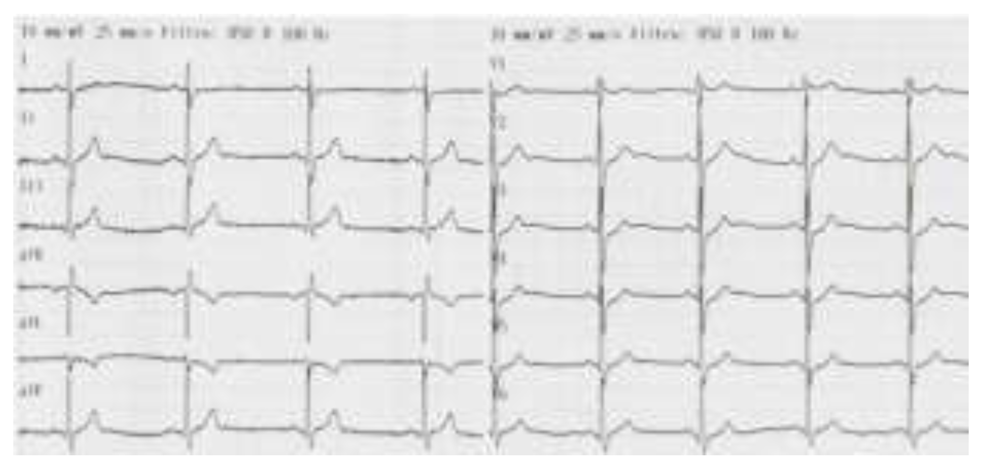
Figure 1. Electrocardiogram at admission showed sinus rhythm, 60 b/min, QRS axis at 75 degrees, incomplete right bundle branch block, the T wave flattened in DI and negative in AVL.
Jmms 07 00036 g001
Figure 2. Cardio-pulmonary radiography, postero-anterior view: heart in normal limits, without pleuro-pulmonary or thoracic wall lesions.
Jmms 07 00036 g002
Figure 3. Electrocardiogram recorded during chest pain: sinus rhythm, ventricular allure = 65/min, incomplete right bundle branch block, ST segment sub-level in DI, aVL, V2–V5 of maximum 1.5 mm, negative T-wave in DI, aVL and ST segment elevation 1mm in aVR.
Jmms 07 00036 g003