Next Article in Journal
Duodenal Carcinoid Tumour—A Case Report
Previous Article in Journal
Therapeutic Approach for Amyand’s Hernia; A Case Report
 
 
Journal of Mind and Medical Sciences is published by MDPI from Volume 12 Issue 1 (2025). Previous articles were published by another publisher in Open Access under a CC-BY (or CC-BY-NC-ND) licence, and they are hosted by MDPI on mdpi.com as a courtesy and upon agreement with Valparaiso University (ValpoScholar).
Case Report

Extra Medullar Granulocytic Sarcoma: A Case Report of an Exceptional Localization

by
Bouhani Malek
1,*,
Sakhri Saida
1,
Jaidane Olfa
1,
Adouni Olfa
2,
Chargui Riadh
1 and
Rahal Khaled
1
1
Department of Oncologic Surgery, Salah Azaiz Institute, Boulevard 9th April, Tunis 1009, Tunisia
2
Department of Anatomo-Pathology, Salah Azaiz Institute, Boulevard 9th April, Tunis 1009, Tunisia
*
Author to whom correspondence should be addressed.
J. Mind Med. Sci. 2020, 7(1), 110-113; https://doi.org/10.22543/7674.71.P110113
Submission received: 21 October 2019 / Revised: 10 January 2020 / Accepted: 10 January 2020 / Published: 20 April 2020

Abstract

:
Granulocytic sarcoma is a rare type of tumor composed of extramedullary immature cells. The breast location is very rare; it accounts for less than 8% of cases. The present study reports the case of a 36-year-old female with a medical history of myelodysplastic syndrome. She was referred because of a lump in the left breast. We have diagnosed a case of granulocytic sarcoma of the breast by core biopsy. Histology and immunohistochemistry showed hypercellular smears with immature myeloid cells. The blast cells were myeloperoxidase positive. The patient underwent a lumpectomy. Five months later, she developed a contralateral recurrence, treated by lumpectomy and radiotherapy. Three years later, she developed a recurrence in the left knee. We report this case for its rarity and as a note of caution to a physician to consider myeloid sarcoma in the differential diagnosis of a breast lump, to provide the correct diagnosis and avoid incorrect treatment of a curable disease.

Introduction

Granulocytic sarcoma of the breast (GS), also known as myeloid or chloroma sarcoma, is a rare extramedullary manifestation of acute myeloid leukemia (AML) and myelodysplastic syndrome (MDS). It accounts for less than 8% of cases. It was first described by Burns in 1811. It is characterized by an unusual type of tumor composed of immature cells outside the bone marrow. The tumor can arise de-novo or in association with hematological malignancies [1,2,3,4,5]. Here we present an isolated recurrence of GS in the breast, with treatment outcomes.

Case presentation

A 36-year-old woman presented with the clinical history of MDS occurring four years earlier. She had undergone allogeneic bone marrow transplantation. The patient was free of disease, but she was subsequently undergoing therapy for graft-versus-host disease reaction. She was referred to our institute with complaints of a painful mass in the left breast that had been apparent for one month. On physical exam, we found a well-defined mass located in the upper outer quadrant of the left breast, measuring 40 mm, without skin modification.
The complete blood count and peripheral blood examination were normal. Mammography showed an asymmetric opacity in the periareolar area without microcalcifications. Breast ultrasound showed a heterogenous well-circumscribed mass in the left breast measuring 40 mm without posterior acoustic shadowing and internal vascularity (Figure 1).
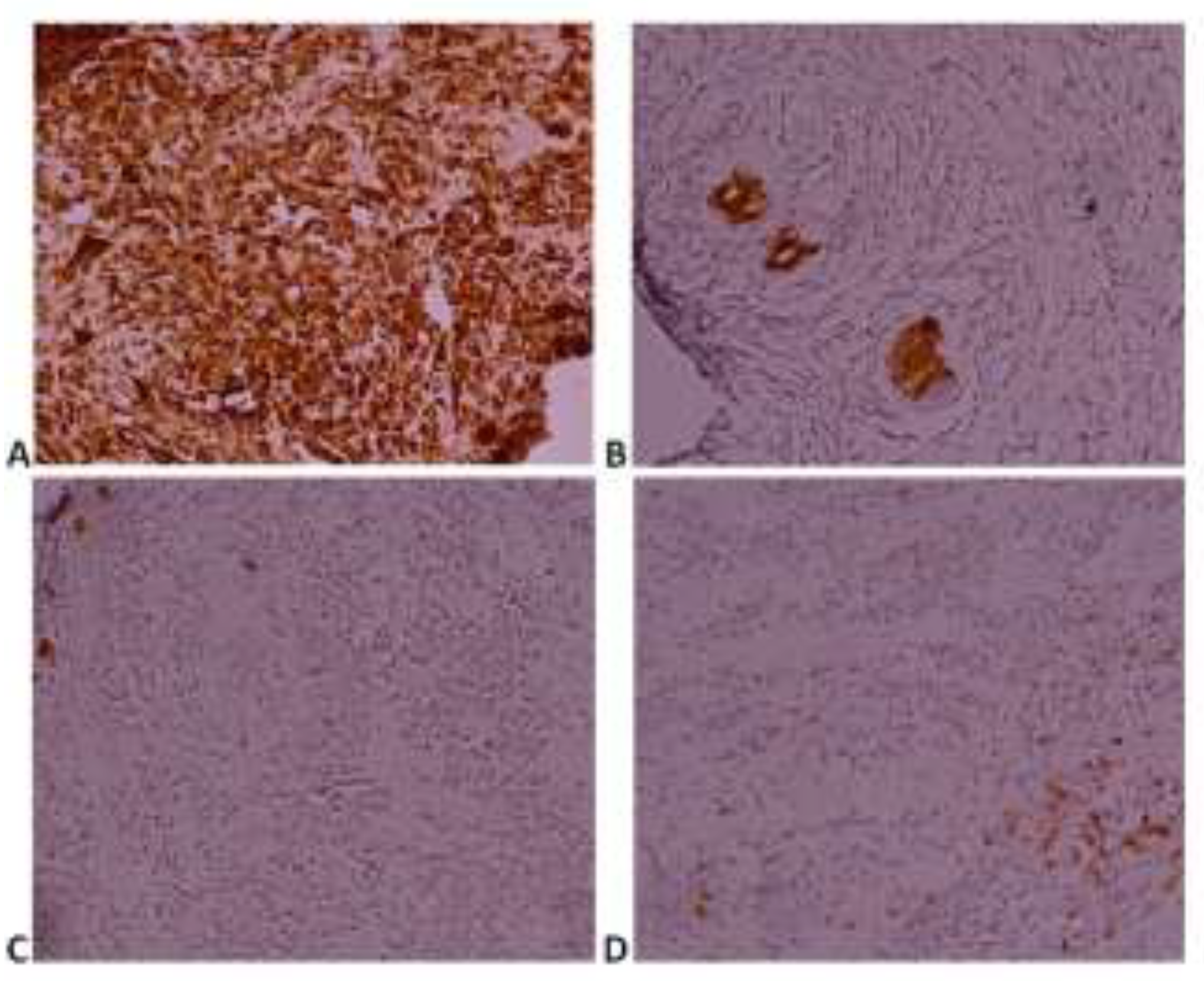
A core biopsy of the mass was performed. Histology and immunohistochemistry revealed hypercellular smears with immature myeloid cells few neutrophils and many large round cells with the high nuclear-cytoplasmic ratio, prominent nucleoli, and a moderate amount of granular cytoplasm (Figure 2). The blast cells were positive for myeloperoxidase enzyme (MPO), vimentin, and CD117 and negative for Pan B, and Pan T (Figure 3). The diagnostic was a breast relapse for her MDS, known as GS of the breast.
The patient underwent a left breast lumpectomy. In the macroscopic examination, we found a grayish mass measuring 50 mm, and the margins were free of disease. Radiotherapy was proposed for the patient, but she failed to follow up. Five months later, she consulted for contralateral breast mass. Upon examination we found a well-defined mass, measuring 15 mm in the right submammary fold. She underwent a local excision. The histologic findings showed a GS of the right breast, with free margins.
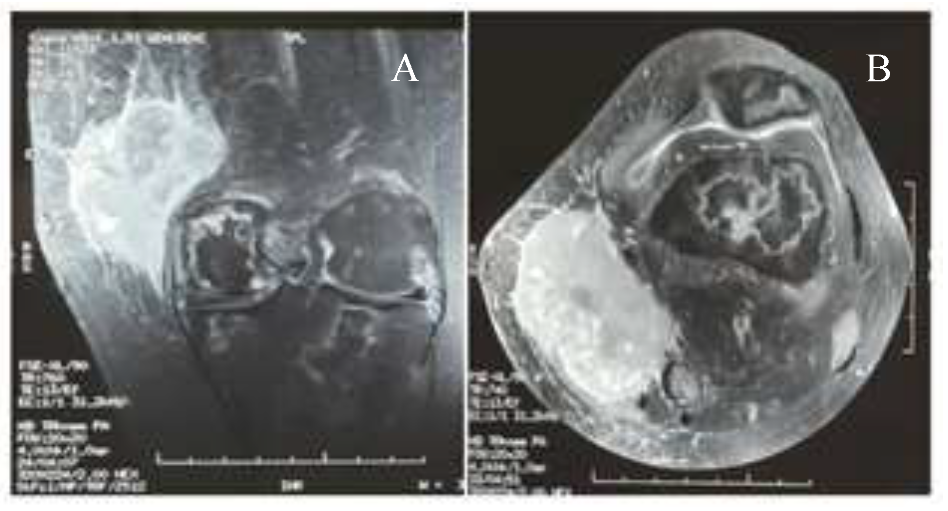
The patient received adjuvant radiotherapy for the left breast, and contact radiotherapy for the right breast. The total dose was 36 Gy for both breasts. Three years later she presented an isolated recurrence in her left knee. The clinical exam showed a hard mass, measuring 40 mm. The MRI showed a suspected mass measuring 88×32 mm with cribriform appearance, infiltrating the ligament and the tendon of the left knee (Figure 4). The core biopsy and histologic findings confirmed the recurrence of her anterior GS. The multidisciplinary committee outlined surgery, but the patient refused the operation. Chemotherapy has been challenged due to localized disease. The patient was then lost again to follow up, though in good general condition when released.

Discussions

GS of the breast most commonly occurs in patients with previously diagnosed AML or MDS. The breast is an uncommon site for presentation of this tumor. It is characterized by the formation of tumors containing immature myeloid cells [1,2,5,6,7]. GS can arise in any part of the human body, but bones, lymph nodes, soft tissues, and skin are among the most common sites of presentation other than the urogenital tract [1,4,6].
The rarity of breast GS makes the diagnosis difficult, often being misdiagnosed as lymphoma, sarcoma, or breast carcinoma, especially in the absence of bone marrow invasion. In this case, the patient had a medical history of the hematological disease, even though with complete remission [4,7,8].
The clinical manifestations of the GS of the breast are not specific. It often manifests as a palpable mass that can be either painless or painful. Other symptoms can be found such as skin involvement and axillary lymph node, although nipple retraction or discharge or inversion is not common [1,4,7].
Due to their rarity of the condition, radiological findings are variable. It is difficult to differentiate GS from benign lesions or other malignant lesions. The ultrasonography showed a regular or micolobulated mass with well-defined margins, irregularly shaped, heterogeneous or hypoechoic with visible posterior acoustic shadows. Sometimes, it appears with spiculated or microlobulated margins [5,7].
On the mammography, the lesions can have regular or irregular limits usually without microcalcifications (2, 8). Sometimes MRI is used to eliminate diagnostic errors, but results are often indistinguishable from benign and malignant lesions; GS appears on T2 as hyperintense, heterogeneous, well-defined [4,7]. The diagnosis is confirmed only by core biopsy and histological examination.
Lumpectomy combined with systemic chemotherapy results in a good outcome. Some studies had demonstrated that the overall survival was longer in patients treated by chemotherapy compared with those who did not receive chemotherapy [5,6,7]. However, because of the limited number of GS cases in the literature, therapeutic approaches remain controversial.
The pillar of treatment is systemic chemotherapy with regimens normally utilized for AML [1,8]. Several studies and reviews have featured the significance of early systemic therapy in order to accomplish a long, disease-free survival and to avoid AML development or relapse [9,10,11,12,13].
There is an absence of clear indication for combination chemotherapy with radiotherapy since survival rates appear to be equal, despite the fact that it may be a valuable device for symptomatic relief or in refractory disease. In addition, several retrospective studies have shown a high rate of AML development in patients with isolated GS experiencing radiotherapy or surgery alone. Our case was unique due to the unusual treatment management; we performed lumpectomy and radiotherapy without chemotherapy with good outcomes.
There is no clear consensus whether or not bone marrow transplantation has an effect even though recent studies have shown a significant survival benefit [10,11,13]. Finally, promising targeted therapies currently in trial for AML, such as FLT3 inhibitors, farnesyltransferase inhibitors and histone deacetylase inhibitors, might be an effective option against GS [10].
Histopathology and the immunohistochemistry confirm the diagnostic. Macroscopically the tumors had a green color, due to the MPO present in the myeloid cells [3,14]. Histological examination usually shows the presence of the myeloblasts, these cells appeared pale on hematoxylin and eosin stain, the nucleus was irregular with finely granular chromatin with scanty cytoplasm [3].
Immunohistochemical usually confirms the diagnosis. There is a large array of cytogenetic tests and flow cytometry to distinguish GS from lymphoma [6,7]. The tumor is often positive for MPO, CD117, CD99, CD68, lysozyme, and nucleophosmin. The most frequent cytogenetic and biochemical abnormalities carry t(8; 21), inv [14], and cytoplasmic expression of NPM and FLT3-ITD [10].
Prognosis depends on some factors such as cytogenetic profiles and age of onset. Some studies have shown that the prognosis in isolated disease is better than GS without medullary involvement [1,5].

Highlights

Extramedullary manifestation of acute myeloid leukemia are various, the breast mass in this context is very rare, the aim of this report to keep on mind the diagnosis of granulocytic sarcoma even the patient was in complete remission.

Conclusions

GS of the breast is a rare manifestation of MDS. The diagnosis is thus difficult to establish. GS can mimic other neoplasms like lymphoma and breast carcinoma, so it can be easily misdiagnosed without the immunohistochemistry which uses a vast panel of markers. The management of this disease remains unclear; some studies have concluded that lumpectomy combined with systemic chemotherapy may give good results.

Compliance with ethical standards

Our institution does not require ethical approval for reporting individual cases or case series. Verbal informed consent was obtained from the patient(s) for their anonymized information to be published in this article.

Conflict of interest disclosure

There are no known conflicts of interest in the publication of this article. The manuscript was read and approved by all authors.

References

  1. Fernandes Vieira, V.; Vo, Q.D.; Bouquet de la Jolinière, J.; Khomsi, F.; Feki, A.; Hoogewoud, H.-M. Granulocytic Sarcoma Presenting as a Palpable Breast Lump. Front Surg. 2017, 3, 67. [Google Scholar] [CrossRef] [PubMed]
  2. Viani, L.; Zannoni, M.; Luzietti, E.; Caramatti, C.; Martella, E.M.; et al. Primitive Myeloid Sarcoma of the Breast: A Case Report. J Clin Case Rep. 2016, 6, 671. [Google Scholar] [CrossRef]
  3. Kudva, R.; Monappa, V.; Solanke, G.; Valiathan, M.; Rao, A.C.K.; Geetha, V. Myeloid sarcoma: A clinicopathological study with emphasis on diagnostic difficulties. J Cancer Res Ther. 2017, 13, 989–993. [Google Scholar] [PubMed]
  4. Huang, X.E.; Li, Y.J.; Zhou, X.D. Granulocytic sarcoma of the breast: A case report. Oncol Lett. 2015, 10, 2447–2449. [Google Scholar] [CrossRef] [PubMed]
  5. Ozsoy, A.; Dolek, B.A.; Barca, N.; Aktas, H.; Araz, L.; Kulacoglu, S. Ultrasound Findings in a Case of Myeloid Sarcoma of the Breast. J Belg Soc Radiol. 2016, 100, 15. [Google Scholar] [CrossRef] [PubMed]
  6. Girshova, L.; Romanova, E.; Kholopova, I.; Nikulina, T.; Mirolubova, J.; Zaritskey, A. Isolated Myeloid Sarcoma Involving the Breast. Blood. 2012, 120, 4345. [Google Scholar] [CrossRef]
  7. Zhai, J.; Kong, X.; Yang, X.; Gao, J.; Xuan, L.; Wang, X.; Wang, J.; et al. An uncommon granulocytic sarcoma of the breast: a case report and literature review. Onco Targets Ther. 2018, 11, 3685–3690. [Google Scholar] [CrossRef] [PubMed]
  8. Valbuena, J.R.; Admirand, J.H.; Gualco, G.; Medeiros, L.J. Myeloid Sarcoma Involving the Breast. Arch Pathol Lab Med. 2005, 129, 32–38. [Google Scholar] [CrossRef] [PubMed]
  9. Pileri, S.A.; Ascani, S.; Cox, M.C.; Campidelli, C.; Bacci, F.; Piccioli, M.; et al. Myeloid sarcoma: clinico-pathologic, phenotypic and cytogenetic analysis of 92 adult patients. Leukemia. 2007, 21, 340–350. [Google Scholar] [CrossRef] [PubMed]
  10. Yilmaz, A.F.; Saydam, G.; Sahin, F.; Baran, Y. Granulocytic sarcoma: a systematic review. Am J Blood Res. 2013, 3, 265–270. [Google Scholar] [PubMed]
  11. Bakst, R.L.; Tallman, M.S.; Douer, D.; Yahalom, J. How I treat extramedullary acute myeloid leukemia. Blood. 2011, 118, 3785–3793. [Google Scholar] [CrossRef] [PubMed]
  12. Cunningham, I. A clinical review of breast involvement in acute leukemia. Leuk Lymphoma. 2006, 47, 2517–2526. [Google Scholar] [CrossRef] [PubMed]
  13. Avni, B.; Koren-Michowitz, M. Myeloid sarcoma: current approach and therapeutic options. Ther Adv Hematol. 2011, 2, 309–316. [Google Scholar] [CrossRef] [PubMed]
  14. Nishida, H.; Kinoshita, T.; Yashiro, N.; Ikeda, Y.; O’Uchi, T. MR findings of granulocytic sarcoma of the breasts. Br J Radiol. 2006, 79, e112–e115. [Google Scholar] [CrossRef] [PubMed]
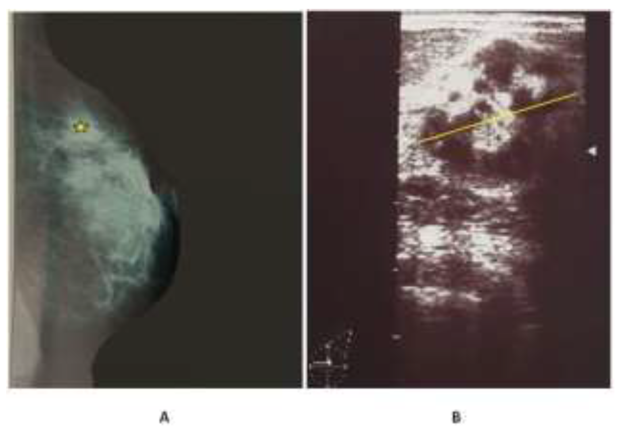
Figure 1. (A) hyperdense opacity in the periareolar area without microcalcifications on mammography (yellow asterisk); (B) a heterogenous well-circumscribed mass, measured 40 mm in the left breast without posterior acoustic on ultrasound.
Jmms 07 00017 g001
Figure 2. Progressive magnification in Hematoxylineosin staining: (A) ×5; (B) × 10; (C) ×20; (D) ×40; showing hypercellular smears with immature myeloid cells few neutrophils, megakaryocytes and many large round cells with the high nuclear-cytoplasmic ratio.
Jmms 07 00017 g002
Figure 3. Immunohistochemistry features, (A) Myeloperoxidase; (B) vimentin; (C) and (D) CD117. Magnification: 20×.
Jmms 07 00017 g003
Figure 4. MRI sequence T1-weighted with Gadolinium showed a suspected mass measuring 88×32 mm with cribriform appearance, infiltrating the ligament and the tendon of the left knee.
Jmms 07 00017 g004