Next Article in Journal
Ethical Aspects in Managing Patients Diagnosed with Digestive Cancers; A Review of Literature
Previous Article in Journal
Determination of Pyrrolizidine Alkaloids in Dietary Sources Using a Spectrophotometric Method
 
 
Journal of Mind and Medical Sciences is published by MDPI from Volume 12 Issue 1 (2025). Previous articles were published by another publisher in Open Access under a CC-BY (or CC-BY-NC-ND) licence, and they are hosted by MDPI on mdpi.com as a courtesy and upon agreement with Valparaiso University (ValpoScholar).
Case Report

Hepatocarcinoma with Tumor Thrombus Occupying the Right Atrium and Portal Vein in a Patient with Hereditary Hemochromatosis and Liver Cirrhosis

by
Olga Hilda Orasan
1,*,†,
Andreea Maria Stefan
2,†,
Laura Urian
3,†,
Anca Mihailov
4,
Ovidiu Fabian
5,
Flaviu Muresan
5,
Iulia Breaban
6,
Sorina Cezara Secara
6,
Simina Tarmure Sarlea
1,
Vasile Negrean
1,
Dorel Sampelean
1 and
Angela Cozma
1
1
4th Department of Internal Medicine, Iuliu Hatieganu University of Medicine and Pharmacy Cluj-Napoca, 18 Republicii Street, Cluj-Napoca, Romania
2
Regional Institute of Gastroenterology and Hepatology, Prof. Dr. Octavian Fodor, Cluj-Napoca, Romania
3
Department of Hematology, Iuliu Hatieganu University of Medicine and Pharmacy Cluj-Napoca, Cluj- Napoca, Romania
4
4th Surgical Department, Railway Hospital, Cluj-Napoca, Romania
5
4th Department of Surgery, Iuliu Hatieganu University of Medicine and Pharmacy Cluj-Napoca, Cluj- Napoca, Romania
6
Prof. Dr. Octavian Fodor Regional Institute of Gastroenterology and Hepatology, Cluj-Napoca, Romania
*
Author to whom correspondence should be addressed.
Authors with equal contribution.
J. Mind Med. Sci. 2018, 5(2), 300-304; https://doi.org/10.22543/7674.52.P300304
Submission received: 31 July 2018 / Revised: 31 August 2018 / Accepted: 30 September 2018 / Published: 3 October 2018

Highlights

  • Hepatocellular carcinoma with right atrium invasion is a rare condition, especially in hereditary hemochromatosis.
  • All first-degree relatives of a patient diagnosed with type 1 hereditary hemochromatosis should be genetically tested for HFE mutations and monitored in order to prevent iron overload.

Abstract

:

Highlights

  • Hepatocellular carcinoma with right atrium invasion is a rare condition, especially in hereditary hemochromatosis.
  • All first-degree relatives of a patient diagnosed with type 1 hereditary hemochromatosis should be genetically tested for HFE mutations and monitored in order to prevent iron overload.

Abstract

We present the case of a 46-year old patient with Child-Pugh class C cirrhosis with MEDL-Score 16, and hepatocellular carcinoma invading the inferior vena cava and the right atrium. The etiology of cirrhosis is type 1 hereditary hemochromatosis with positive HFE C282Y/C282Y and H63D/H63D mutations. A systematic review of the literature was performed and only 30 cases of hepatocellular carcinoma with tumor thrombosis extending into the right atrium have been described. To our knowledge, this is the first case that evidences the presence in hereditary hemochromatosis of hepatocellular carcinoma with atypical invasion into the right atrium. Screening of patients with a family history of hereditary hemochromatosis allows detection of the disease in the asymptomatic phase, allowing initiation of early therapy and improved prognosis.

Introduction

Vascular invasion into the portal vein (PV) occurs most frequently in hepatocellular carcinoma (HCC) [1,2]. Inferior vena cava (IVC) invasion is rarely found in medical practice and is associated with a poorer prognosis [3].
Hereditary hemochromatosis (HH) is a genetic disease with an autosomal recessive pattern of inheritance. Mutations in the genes encoding the proteins involved in iron metabolism induce increased intestinal absorption of iron and its excessive deposition in organs, causing their malfunction. The most frequent HFE gene mutations in type 1 HH are C282Y and H63D. The majority of patients are homozygous for the C282Y mutation or compound heterozygous C282Y/H63D [4]. In HH, there is a 20-fold higher risk for developing HCC compared to the general population. HCC occurs in about 10% of patients with HH [5], with half of the patients with HH dying from HCC [6]. Ye et al. demonstrated that HFE C282Y may increase the odds of developing HCC, while HFE H63D is more likely associated with susceptibility to HCC without cirrhosis in the African population [7].

Case report

We report the case of a 46-year-old man who presented for the first time to a medical service for scleral jaundice, low protein leg edema, and increased abdominal volume due to ascites. Under drug treatment with diuretics and albumin supplements, edematous syndrome remitted in three weeks, but low-intensity pain occurred in the right hypochondrium associated with asthenia, fatigue, weight loss of about three kilos over the last month, and polyarthralgia.
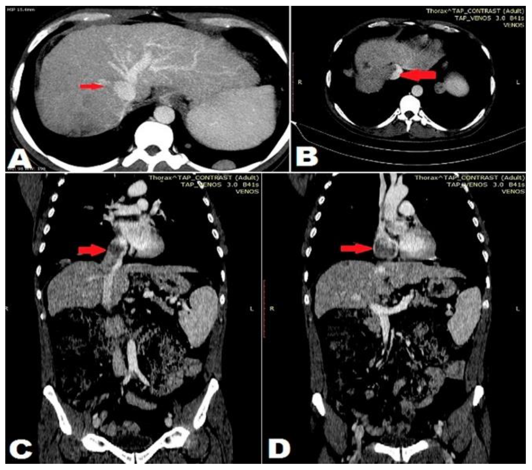
The patient reported a mean alcohol consumption of 50 g/day over the past three years, under the threshold of hepatotoxicity. The patient’s family history evidenced that two brothers (38 and 42-year-old) and an aunt (51-year-old) had died from cirrhosis at young ages. nodular masses having a maximum diameter of 83mm, in segment VII, suggestive of multicentric HCC. Doppler examination detected right suprahepatic vein and IVC thrombosis. The PV had a maximum diameter of 15 mm and blood flow was hepatoportal. The spleen was increased in volume (longitudinal axis length of 174mm) and homogeneous in structure. The splenic vein had a maximum diameter of 13mm. A large amount of ascites was evidenced at the first examination and disappeared after treatment. Contrast-enhanced thoracoabdominal CT revealed a liver reduced in size, with irregular outline and a highly inhomogeneous structure, with multiple hypodense, high-uptake masses in the arterial phase with newly formed vessels from the hepatic artery, the largest mass being 78mm in size, in segments VI-VII (Figure 1). The tumor invaded the right hepatic vein, with extension of the tumor thrombus into the IVC, RA, and the segmental branches of the right PV (Figure 2); interaortocaval, retrocaval, celiac, and hepatic hilar adenopathies, as well as multiple bilateral pulmonary nodular lesions up to 3cm in size having a computed tomographic appearance typical of metastasis (Figure 3).
The physical exam revealed generalized skin hyperpigmentation; scleral jaundice; hippocratic fingers and toes; bilateral leg edema; bilateral diminished pectoral sound, sonority alternating with sub-dullness, bilateral diminished vesicular murmur; distended abdomen due to ascites, umbilical scar protrusion, and the presence of cavo-cave collateral circulation.
Abdominal ultrasound detected a liver reduced in size, with irregular outline and a highly inhomogeneous structure, with inhomogeneous, confluent echogenic
Blood tests showed hypersplenism with pancytopenia, macrocytosis, cholestatic syndrome (alkaline phosphatase=652 U/L, gamma-GT=84 U/L), mixed hyperbilirubinemia (total bilirubin=4.1 mg/dl, direct bilirubin=2 mg/dl), hepatic cytolysis syndrome (aspartate-aminotransferase=339 U/L, alanine- aminotransferase=239 U/L), hepatic synthetic dysfunction with spontaneously prolonged INR (1.55), serum albumin and serum creatinine within normal limits (albumin=3 g/dl, creatinine = 0.9 mg/dl), and hypersideremia (205=μg/dl).
The diagnosis of Child-Pugh class C cirrhosis with a MEDL-Score 16, HCC invading the right suprahepatic vein, IVC, RA and the right PV, lung and lymph node metastases was established.
The viral etiology of chronic liver disease was excluded by negative HBs Ag and anti-HCV antibody. Given the patient’s family history, cirrhosis of uncertain etiology with alcohol consumption below the threshold of toxicity, hypersideremia, generalized melanoderma, and the sudden onset of liver cirrhosis at the stage of complications, assessing the etiology of cirrhosis in relation to hereditary causes, particularly HH, was considered necessary.
The diagnosis of type 1 HH was confirmed by increased serum ferritin levels (1492 ng/ml) and transferrin saturation (85.61%), the patient being homozygous for both C282Y and H63D HFE mutations.
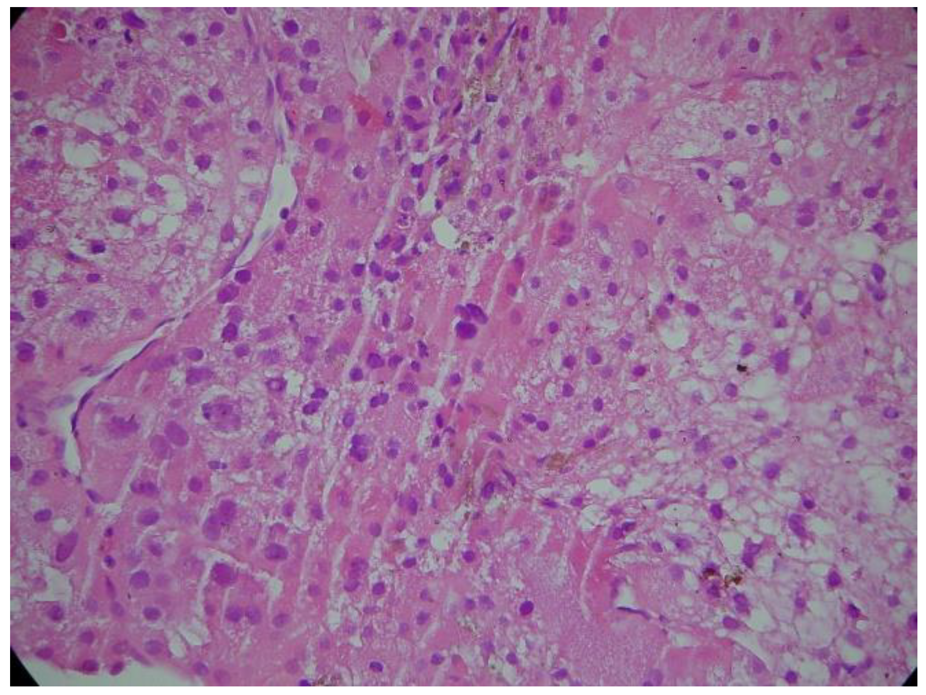
For the characterization of the liver tumor mass, the following tumor markers were measured: alpha- fetoprotein= 24.81 ng/ml (upper normal value: 9 ng/ml) and CA 19.9, which ranged within normal limits. Given the slightly increased level of alpha-fetoprotein, the diagnosis of HCC was confirmed by liver puncture biopsy (Figure 4).
Intracardiac tumor extension was evaluated by transthoracic echocardiography, which evidenced a polylobulated tumor mass 27/43 mm in size, occupying the entire RA and compressing the tricuspid annulus (Figure 5). No signs of heart failure were detected.
On discharge, palliative therapy was recommended, which included low molecular weight heparin, tyrosine kinase inhibitors, hepatoprotective drugs, loop diuretics, and anti-aldosterone drugs. Etiological treatment with systematic phlebotomy and iron chelators was not initiated due to the presence of hypersplenism with pancytopenia and end-stage liver disease. Death from liver failure with grade IV hepatoportal encephalopathy occurred after 8 weeks.
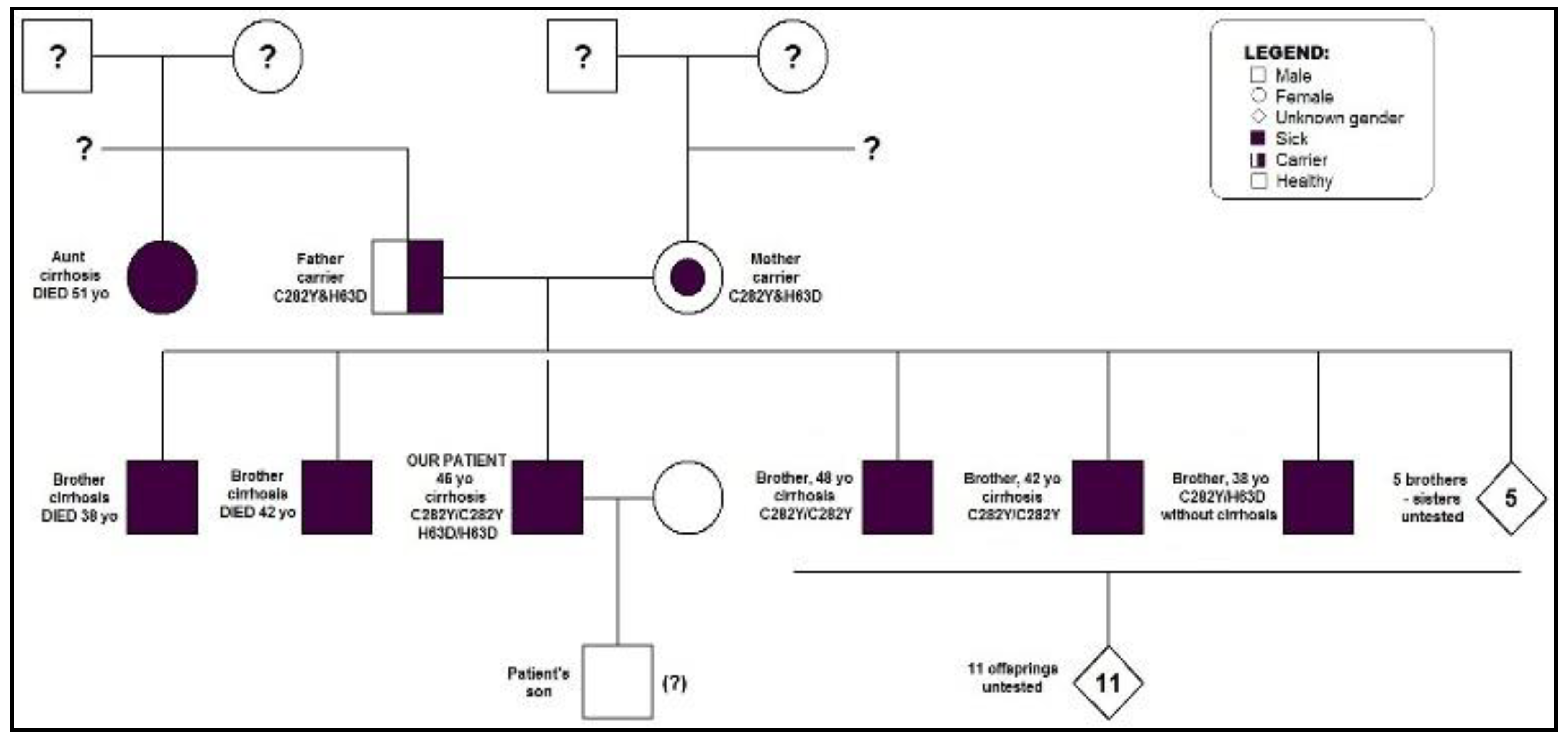
Screening of patients with a family history of HH allows for early initiation of specific therapy and improved prognosis. We recommended screening for the patient’s 8 living brothers and their 11 offspring. So far, the patient’s son and 3 other brothers, aged 38, 42 and 48 years, respectively, have been evaluated clinically, biologically, and genetically. Two brothers aged 42 and 48 years have cirrhosis evidenced by ultrasound. The 42-year-old brother has the C282Y/C282Y HFE mutation, serum ferritin=1207 ng/ml and transferrin saturation=76.57%, consequently a diagnosis of certainty of hemochromatosis. The recommended treatment consists of weekly phlebotomy and iron chelation therapy during the first 2 weeks. The two other brothers, aged 48 and 38 years respectively, are carriers of the C282Y/C282Y HFE mutation, and of the C282Y/H63D HFE mutation respectively. The 48 year-old brother has serum ferritin=657 ng/ml and transferrin saturation=65%. He was recommended weekly phlebotomy. The 38 year-old brother with serum ferritin=254 ng/ml and transferrin saturation=37% will be evaluated annually in order to detect the optimal time for initiation of phlebotomy. The genetic testing of the patient’s son did not reveal HFE gene mutations (Figure 6).

Discussion

HCC cases with tumor thrombosis extending to the RA are extremely rare, only 30 cases have been described. None of these was documented in HH [3]. Intracardiac thrombosis is associated with an increased risk of sudden death from pulmonary thromboembolism or acute heart failure, and more frequent and more extensive lung metastasis. Liver resection and surgical removal of the thrombus have proved to be the most effective approach in the case of patients with resectable liver tumors and sufficient residual hepatic reserve, with a mean survival of 19 months after surgery [8]. In the case of our patient, multiple liver tumors, metastatic adenopathies, and secondary lung tumors contraindicated a curative or palliative surgical approach.
Screening is an important component of preventive care. It consists of the identification of an asymptomatic disease, harmful condition, or risk factor, with the overall aim that the condition be caught early before it causes harm to the affected individual. Since HH is present at birth, its cause cannot be removed, and primary prevention is not practical. However, secondary prevention, which aims to detect early disease when it is asymptomatic and when treatment might stop its progression, is a viable option [9,10].
Screening is performed for all first-degree relatives of patients with HH and includes HFE genotyping and annual monitoring by determination of serum ferritin and transferrin saturation (TS). The ideal age to start HH screening is between 18 and 30 years, when signs of the disease caused by iron deposition in organs occur. Phlebotomy is initiated in the case of asymptomatic patients with a family history of HH who have a progressive increase in serum ferritin and transferrin saturation, as well as symptomatic patients, with severe organ damage. Phlebotomy is initiated at serum ferritin values >500 ng/ml (serum ferritin > 1000 ng/ml shows that the complications of excessive iron deposition are already present) and at a TS >50%. One phlebotomy session is conducted every 1-2 weeks, during which 500 ml blood are removed. Phlebotomy is performed weekly until serum ferritin is <50 ng/ml and TS is <50%. This is followed by a maintenance period, during which phlebotomy is performed every 2-4 months so that the serum ferritin level is maintained between 50-100 ng/ml. Iron chelators are preferred only when phlebotomy is contraindicated (severe/moderate anemia), is not well tolerated (hemodynamic instability, post-procedural symptomatic hypovolemia), or is ineffective (aceruloplasminemia). Use of iron chelation has been suggested in conjunction with phlebotomy in patients with massive iron overload and organ damage, in which rapid removal of the iron burden may be required.

Conclusions

We presented the case of a young patient diagnosed with multicentric HHC at the metastatic stage, with tumor thrombosis extending to the RA, developed in cirrhosis of hereditary etiology, type 1 HH. Genetic counseling in HH is extremely important for early diagnosis of the affected offspring and initiation of monitoring and specific therapy in order to improve their prognosis.
Abbreviations: HCC: hepatocellular carcinoma; HH: hereditary hemochromatosis; IVC: inferior vena cava; PV: portal vein; RA: right atrium; TS: transferrin saturation

References

  1. Jemal, A.; Bray, F.; Center, M.M.; Ferlay, J.; Ward, E.; Forman, D. Global cancer statistics. CA Cancer J Clin. 2011, 61, 69–90. [Google Scholar] [CrossRef] [PubMed]
  2. Okuda, K. Hepatocellular carcinoma: clinicopathological aspects. J Gastroenterol Hepatol. 1997, 12, S314–318. [Google Scholar] [CrossRef] [PubMed]
  3. Luo, X.; Zhang, B.; Dong, S.; Zhang, B.; Chen, X. Hepatocellular carcinoma with tumor thrombus occupying the right atrium and portal vein. A case report and literature review. Medicine. 2015, 94, e1049. [Google Scholar] [CrossRef] [PubMed]
  4. Balaceanu, A.; Diaconu, C.; Aron, G. Budd-Chiari syndrome as an initial presentation of hepatocellular carcinoma – a case report. Med Ultrason. 2014, 16, 172–174. [Google Scholar] [CrossRef] [PubMed]
  5. Beutler, E.; Gelbart, T.; West, C.; Lee, P.; Adams, M.; Blackstone, R.; Pockros, P.; Kosty, M.; Venditti, C.P.; Phatak, P.D.; Seese, N.K.; Chorney, K.A.; Ten Elshof, A.E.; Gerhard, G.S.; Chorney, M. Mutation analysis in hereditary hemochromatosis. Blood Cells Mol Dis. 1996, 22, 187–94. [Google Scholar] [CrossRef] [PubMed]
  6. Elmberg, M.; Hultcrantz, R.; Ekbom, A.; Brandt, L.; Olsson, S.; Olsson, R.; Lindgren, S.; Lööf, L.; Stål, P.; Wallerstedt, S.; Almer, S.; Sandberg-Gertzén, H.; Askling, J. Cancer risk in patients with hereditary hemochromatosis and in their first-degree relatives. Gastroenterology. 2003, 125, 1733–41. [Google Scholar] [CrossRef] [PubMed]
  7. Fracanzani, A.L.; Conte, D.; Fraquelli, M.; Taioli, E.; Mattioli, M.; Losco, A.; Fargion, S. Increased cancer risk in a cohort of 230 patients with hereditary hemochromatosis in comparison to matched control patients with non-iron-related chronic liver disease. Hepatology. 2001, 33, 647–51. [Google Scholar] [CrossRef] [PubMed]
  8. Ye, Q.; Qian, B.X.; Yin, W.L.; Wang, F.M.; Han, T. Association between the HFE C282Y, H63D polymorphisms and the risks of non-alcoholic fatty liver disease, liver cirrhosis and hepatocellular carcinoma: an updated systematic review and meta- analysis of 5,758 cases and 14,741 controls. PLOS One. 2016, 11, e0163423. [Google Scholar] [CrossRef] [PubMed]
  9. Diaconu, C.; Bălăceanu, A.; Moroşan, E. Sepsis biomarkers: past, present and future. Farmacia 2015, 63, 811–815. [Google Scholar]
  10. Wang, Y.; Yuan, L.; Ge, R.L.; Sun, Y.; Wei, G. Survival benefit of surgical treatment for hepatocellular carcinoma with inferior vena cava/right atrium tumor thrombus: results of a retrospective cohort study. Ann Surg Oncol. 2013, 20, 914–22. [Google Scholar] [CrossRef] [PubMed]
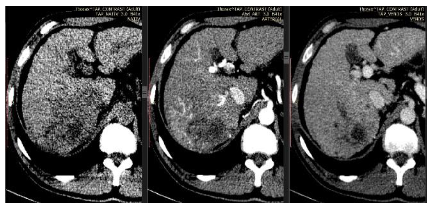
Figure 1. Computed tomography, transverse section showing tumor behavior in the native, arterial and venous phase.
Jmms 05 00044 g001
Figure 2. Computed tomography, tumor thrombus extension to the right hepatic vein (A), inferior vena cava (B-axial, C-coronal), up to the right atrium, which it occupies entirely (D).
Jmms 05 00044 g002
Figure 3. Computed tomography, 3D MIP reconstruction, 59.2 mm thickness, coronal section showing multiple lung metastases.
Jmms 05 00044 g003
Figure 4. Hematoxylin-eosin stain showing hepatocellular carcinoma.
Jmms 05 00044 g004
Figure 5. Transthoracic echocardiography, 4- chamber apical view, showing a 27/43 mm hyperechoic thrombus in the right atrium.
Jmms 05 00044 g005
Figure 6. Genetic test results within patient’s family.
Jmms 05 00044 g006