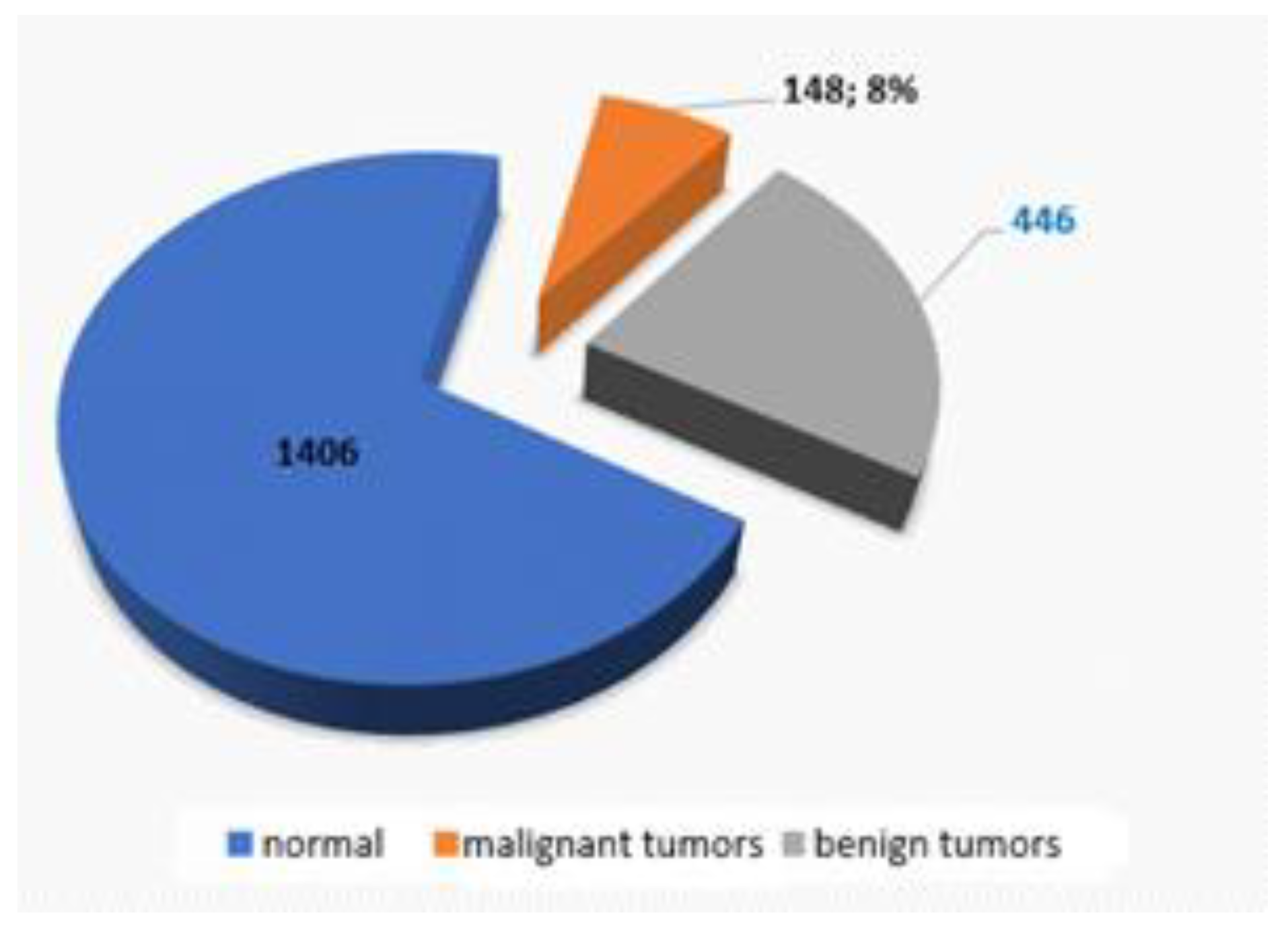
Next Article in Journal
Pancreatogenic Type 3C Diabetes
Previous Article in Journal
Can Non-Small Cell Lung Cancer Histologic Subtypes Predict Survival? A Single Institution Experience
 
 
Journal of Mind and Medical Sciences is published by MDPI from Volume 12 Issue 1 (2025). Previous articles were published by another publisher in Open Access under a CC-BY (or CC-BY-NC-ND) licence, and they are hosted by MDPI on mdpi.com as a courtesy and upon agreement with Valparaiso University (ValpoScholar).
Article

A Proposed Therapeutic Algorithm for Colorectal Cancer Prevention, Based on Endoscopic Polypectomies in Patients with Multiple Colonic Polyps

by
Alexandru Septimiu
1,
Florin Dan Ungureanu
2,
Kutasi Incze Reka
3 and
Alec Cosmin Moldovan
1,2,*
1
Department of General Surgery, Witting Clinical Hospital, Bucharest, Romania
2
Faculty of Medicine, Titu Maiorescu University of Bucharest, 67A Gheorghe Pătrașcu Street, Bucharest, Romania
3
Faculty of Medicine, Târgu Mureș University of Medicine and Pharmacy, Târgu Mureș, Romania
*
Author to whom correspondence should be addressed.
J. Mind Med. Sci. 2018, 5(2), 261-269; https://doi.org/10.22543/7674.52.P261269
Submission received: 31 July 2018 / Revised: 31 August 2018 / Accepted: 30 September 2018 / Published: 3 October 2018

Highlights

  • This paper presents an algorithm/ guideline for performing endoscopic polypectomies in the cases of patients with multiple polyps.
  • Such stepwise algorithm-based approach in the clinical management of patients with multiple polyposis can lead to a substantial decrease of unnecessary colectomies.

Abstract

:

Highlights

  • This paper presents an algorithm/guideline for performing endoscopic polypectomies in the cases of patients with multiple polyps.
  • Such stepwise algorithm-based approach in the clinical management of patients with multiple polyposis can lead to a substantial decrease of unnecessary colectomies.

Abstract

Introduction. Results from single medical centers having large numbers of patients with multiple colonic polyps that have undergone colonoscopy management are rare. Material and method. The present study is based on 2,000 cases enrolled during 2009 and 2017, including patients that underwent colonoscopy for various reasons in the Department for Upper and Lower Endoscopy of Witting Clinical Hospital, Bucharest, Romania. Results. Of the 2,000 patients examined during 2009–2017, 594 tumor formations were detected; 148 (24.9%) patients had colorectal malignancies, and 446 (75%) polypoid formations in 313 patients who had one or more colon polyps were found. Discussions. After performing statistical analyses on several general parameters (such as age, sex, overall clinical outcome of the patient) and polyp-related characteristics (such as: dimensions, location, histological type), we developed a stepwise algorithm for clinical management (with endoscopic polypectomy) of patients with multiple polyposis, with the goal of reducing unnecessary colorectal surgeries. Conclusions. We believe that this type of stepwise algorithm-based approach in the clinical management of patients with multiple polyposis can lead to a substantial decrease in unnecessary colectomies (no matter the approach, via laparotomy or laparoscopic procedures), with the accompanying benefit of avoiding the complications and negative long-life impact that they impose.

Introduction

The issue of polyps and polypectomy in gastroenterology has been at the forefront of many scientific papers over the past years [1,2], but few reports have addressed the issue from the general surgeon`s perspective [3].
The present study represents the experience of a single center for Upper and Lower Endoscopy Department from Witting Clinical Hospital in Bucharest, Romania. The purpose of this study is not only to present a unified conclusion, in the form of a stepwise decisional algorithm based both on 2,000 endoscopic procedures and on personal direct surgical experience, but also to take into account the extensive literature regarding the efficacy of medical procedures designed to prevent colorectal cancer.
With the general objective of providing evidence regarding the usefulness of polypectomies, this paper assumes the surgeon`s point of view—a clinician that understands both the needs and outcomes in terms of physical and mental postoperative challenges—and considers the perspective of a patient that has been subjected to a surgical procedure for colorectal cancer.

Materials and Methods

This longitudinal observational study was carried out on 2,000 patients admitted and monitored to the General Surgery Department of Witting Clinical Hospital, Bucharest, Romania, from January 2009 to January 2017. Selection of subjects was carried out to include patients with a variety of symptoms that required a colonoscopy in order to establish a diagnosis, regardless of gender or age, but who had not undergone colonoscopy investigation prior to admission in our unit. Inclusion was done as randomly as possible, such that the reference group could be considered representative for any case admitted to a clinic with a similar profile. The actual number of total colonoscopies was much higher than 2,000 due to the fact that, for each patient enrolled in the study, we had to perform several colonoscopies in succession, both for control or therapeutic purposes.
The equipment used for endoscopies is a complete line from Pentax™ consisting of an endoscopy kit and the ERBE ICC200™ with Endo-Cut electro-coagulation and cutting unit. All images were recorded with conventional PC-based digital video systems. Histopathological examinations were processed in the hospital`s pathology unit. Of the 2,000 enrolled patients, only 73% (323) of the polyps had full histopathological panel workup; for the remainder, this information was not available.
After a successful polypectomy, patients were followed by yearly-spaced controls for at least three years. For patients requiring multiple polypectomies, treatments were performed at 3-6 month intervals, and, after completion of the established number of polypectomies, they entered an annual follow-up program. The results from the analysis of colonoscopies were recorded in a standard Microsoft Excel™ database, which we later used for statistical analyses.
In the first phase of the study, we highlighted the number of polyp formations discovered globally in a sample drawn from a population that presented to the hospital for a variety of symptoms, as the study was performed on patients admitted to the hospital and not on a population assumed to be healthy, as is the case with screening projects. Of the total number of tumor formations found, the study attempted to establish the ratio between cases with malignant tumors (found in evolution) and pre-malignant lesions that can later be removed to prevent the onset of neoplasia.
The second phase of the study aimed to identify the profile of the therapeutic interventions performed in relation to the characteristics of this type of polyp, in order to establish a protocol to identify high priority dysplasia patients with multiple polyps requiring multiple therapeutic sessions to completely remove existing polyps. Data collection, storage, and analysis were performed according to the protocol of a descriptive statistic study, uniquely and multi-variate. For quantitative variables, means and standard deviations (SD) were generated as central trend and dispersion indicators. The t-test for independent means and/or ANOVAs were used to compare groups, with statistical significance set at p < 0.05. For qualitative variables, distribution indicators (absolute and relative frequency) and structural indicators were used. For comparison, the ƛ2 test was used with p <0.05. Interpretation of the results was done with a confidence interval (CI) of 95%. Analyses were carried out using Excel (Microsoft Office™ suite) and EpiInfo™.

Results

Of the 2,000 patients examined during 2009 - 2017, 594 tumor formations were detected, 148 (24.9%) of patients with colorectal malignancies, and 446 (75%) polypoid formations in 313 patients who had one or more colon polyps (Figure 1).
The number of detected tumor formations varied significantly (p = 0.000) in relation to the sex of the investigated subjects, being higher for men, 395, than women, 199. For the tumor type, polyploid formations were dominant over the malignant tumors in both sexes (Table 1).
As indicated in Figure 2, a higher frequency of colon cancer was detected in women, 67 cases in199 tumor formations (34%), compared men, 81 cases in 395 tumor formations (21%) (Figure 2).
The age of patients with CRC (colorectal cancer) was significantly higher (67.5 years, 32-89 years) compared to those with polyps (61.7 years, 27-87 years) (p = 0.000); the statistical significance (p = 0.036) was maintained even when the gender factor was considered. In the CRC group, the men were older than the women (about 69 years for male vs 66 years for female), compared to the group with polyps in which men were younger than women (about 61 years for males vs. 64 years for females) (Table 2).
For both sexes the occurrence of cancer takes place at a higher age than the appearance of polyps, suggesting that early detection and removal of polyps may prevent their malignancy; in men, the spread is about 8 years (61 years polyps vs 69 years CRC) and in women less than 2 years (64 years polyps vs 66 years CRC). Figure 3 illustrates the variation of the “age” parameter in relation to the presence of CRC and the sex of the patients, observing the higher age difference in male patients [1] with malignant tumors versus the polyps and female patients [2] where the difference between the presence of CRC and polyps is much smaller (Figure 3).
Regarding the polypectomies, as an endoscopic maneuver, of the 2,000 patients, we performed 446 polypectomies, of which 314 (70.4%) were on men and 132 (29.6%) were on women. The distribution of polypectomies in relation to the sex of the patients is presented in the following Figure 4.
The age of patients undergoing polypectomy ranged from 27 to 87 years with an average of 61.6 ± 11.8 years; the age of women was slightly higher than that of men (64 ± 12.1 years F versus 61 ± 11.5 years M) but did not reach statistical significance. Regarding the number of polyps, 232 patients (74%) had single procedures, 59 patients (19%) underwent dissection of two polyps, 16 patients (5%) three polyps, and 6 patients four or more polyps (Figure 4).
Patients with multiple polyps dominated the lot, with the association of two polyps, a situation found in 59 patients out of a total of 81 (73%) (Table 3).
Regarding excised polyps, most of the polypectomies were performed for sessile formations, 361 polyps representing 81%, and 84 pediculous forms, representing 19% (Figure 5).
Regarding size, the polyps were classified as follows: below 5 mm very small polyps, between 5 and 10 mm small polyps, and over 10 mm large polyps. As a frequency, small and very small polyps prevailed, accounting for about 59% of the total (Figure 6).
Size of the polyps varied between 2 and 80 mm, with an average size of 10.2 ± 7 mm, with no significant differences between sexes (p> 0.05). Regarding location, polyps prevailed in the left half of the colon (355 polyps, 79.6%). Regarding position in relation to the colon segments, most were in the sigmoid colon (132 cases), followed by those in the lower colon (118 cases) and rectum (105 cases) (Table 4).
Regarding histological type, 95% of the polyps were of neoplastic, predominantly tubulo-adenomatous polyps (162 polyps, 36%), followed by tubulo-viloid polyps (142 polyps, 32%) (Table 5). However, only 323 (73%) of extirpated polyps had associated anatomopathological studies.
The outcome of the anatomo-pathological examination revealed, in most cases, changes in low dysplasia (Table 6), although 22 cases with high dysplasia polyps were found.
Regarding the ratio of polyp size and dysplasia, the smallest dimension (7.8 ± 6.9 mm) was recorded in low dysplasia polyps and the highest in those with high dysplasia (32.2 ± 20.5 mm), demonstrating the direct ratio between size and dysplasia, with 91% of the high dysplasia polyps being considered to be large (over 10 mm) (Table 7; Figure 7 and Figure 8).
The most common low dysplasia polyps were located in the sigmoid area (29%) and the descending colon (about 28%). The most common high dysplasia polyps were in the rectum (about 46%) (Table 8 and Figure 9).
Analysis of polyp histology and degree of polyp dysplasia reveals that for all histological types, low dysplasia polyps were excised; only tubulo-adenomatous and tubulo-villous polyps were extruded as were polyps with a high degree of dysplasia (Table 9).
The study also found that the percentage of high- displacement tubulo-villous polyps was higher than tubulo-adenomatous polyps, which were numerically more numerous (Figure 10).
Analysis of the polyps and their degree of dysplasia indicate that the sessile polyps show the highest rate of dysplasia and high (Table 10, Figure 11).

Discussions

Considering that the reviewed literature [4,5] did not contain specific guidelines for practicing repeated polypectomies in patients with multiple polyps, and given the results of our observations, we developed an algorithm that can be applied to cases of serial polypectomies in patients with multiple polyps. The goal is to implement uniform practice in gastroenterology and surgery that can provide meaningful and systematized results for future studies in this field and to resolve as many polyps with high dysplasia at the first examinations.
Thus, review of our findings revealed a direct correlation between the larger size of the polyps and the higher degree of high dysplasia, highlighting that the most frequent presence of high dysplasia polyps are rectal, followed by a gradual decrease with ascension toward the rest of the colon segments [6,7,8].
Consequently, we have chosen the following characteristics of the polyps as key-points for the algorithm: size, shape, location, and histological type, thus determining which polyps will be resected first and in the precise order of conducting the following procedures [9,10].
As such the decisional algorithm is based on several layers, starting from:
  • o Dimension (in mm): polypectomies will start with the largest polyps first. The basis for this decision relies on data depicted in Figure 12;
  • o Morphology: if several polyps are present and they all are roughly the same size, polypectomies should start from the sessile ones and then to the pediculate (based on data from Figure 13);
  • o Location: polypectomies should start from the left colon and proceed to the right, respectively from the rectum (based on data available in Figure 14);
  • o Histological type: if previous histological results are available, the villous polyps should be resected first and then the other forms would be considered (as laid out in Figure 15).

Conclusions

Taking into consideration all our analyses, we developed an algorithm for polypectomies in cases of multiple polyps, an algorithm that can be interpreted as a guideline for managing such cases both in a surgical ward as well as in private clinics with endoscopic departments.
The algorithm, presented in Figure 16, is easy to understand, useful, and begins with the positive identification, during endoscopy, of polyps. For solitary ones, excision is the way to proceed. For multiple polyps, size is the determining factor: Large ones should be excised first, then small ones. If both forms are present, the sessile formations should go first and the pediculate after. For sessile polyps located all over the colon, ones located in the left colon are excised first, then proceeding toward the right segments of the colon. If the patient has previous histopathological investigations (such as a patient already enrolled in a monitoring study), villous types of polyps are excised first.
We believe that this kind of a stepwise algorithm- based approach in the clinical management of patients with multiple polyposis can lead to a substantial decrease of unnecessary colectomies (no matter the approach, via laparotomy or laparoscopic procedures) and will increase the number of targeted and timed surgical interventions for malignant colorectal pathology, thus decreasing hospitalization time and offering a significantly better life expectancy and quality of life for this category of patients.

Acknowledgments

This material is the result of a PhD studies thesis, developed by Septimiu Cristian Alexandru, M.D., Ph.D. Candidate at Titu Maiorescu University of Bucharest, Faculty of Medicine, with Univ. Professor Ungureanu Florin Dan, M.D., Ph. D., as thesis coordinator. The authors declare no conflict of interests and state that no financial support has been received in the form of grants from any third parties during this entire study. All authors had equal contribution to the development of this research.

Conflicts of Interest

The authors declare that there are no conflicts of interest to be disclosed for this article.

References

  1. Omundsen, M.; Lam, F.F. The other colonic polyposis syndromes. ANZ J Surg. 2012, 82, 675–681. [Google Scholar] [CrossRef] [PubMed]
  2. Talseth-Palmer, B.A. The genetic basis of colonic adenomatous polyposis syndromes. Hered Cancer Clin Pract. 2017, 15, 5. [Google Scholar] [CrossRef] [PubMed]
  3. Kaz, A.M.; Dominitz, J.A. Can individuals with colonic polyposis be managed safely with colonoscopic surveillance? Endoscopy 2016, 48, 7–8. [Google Scholar] [CrossRef] [PubMed]
  4. Dawwas, M.F.; Ibrahim, A.K.; Lee, Y.M. Colonic polyposis: More than meets the eye. Metastatic melanoma. Gastroenterology 2010, 139, 1097. [Google Scholar] [CrossRef] [PubMed]
  5. D'Mello, A.; Deshpande, R.B.; Desai, D.; Shah, S.R. Colonic polyposis. Ann Gastroenterol. 2014, 27, 168. [Google Scholar] [PubMed]
  6. Ellis, C.N. Colonic adenomatous polyposis syndromes: Clinical management. Clin Colon Rectal Surg. 2008, 21, 256–262. [Google Scholar] [CrossRef] [PubMed]
  7. Aslan, D.; Bordea, A.; Burcoș, T. Anastomotic leakage after sphincter-sparing surgery in a young woman diagnosed with low rectal cancer - case report. J Clin Invest Surg. 2017, 2, 45–53. [Google Scholar] [CrossRef]
  8. Blajin, A.; Dan, C.; Sălcianu, I.; Welt, L.; Scăunaşu, R.V.; Berevoescu, N.; Blajin, C.; Burcoş, T. The diagnostic and therapeutic management of a peculiar case of rectal submucosal adenocarcinoma. J Clin Invest Surg. 2018, 3, 47–52. [Google Scholar] [CrossRef]
  9. He, E.Y.; Wyld, L.; Sloane, M.A.; Canfell, K.; Ward, R.L. The molecular characteristics of colonic neoplasms in serrated polyposis: A systematic review and meta-analysis. J Pathol Clin Res. 2016, 2, 127–137. [Google Scholar] [CrossRef] [PubMed]
  10. Sreedharanunni, S.; Paulose, R.; Jojo, A.; Dhar, P.; Gangadharan, P. Colonic polyposis syndromes--an experience from a tertiary centre in South India. Indian J Gastroenterol. 2015, 34, 233–239. [Google Scholar] [CrossRef] [PubMed]
Figure 1. Distribution of malignant and benign tumors as for the entire lot of colonoscopies.
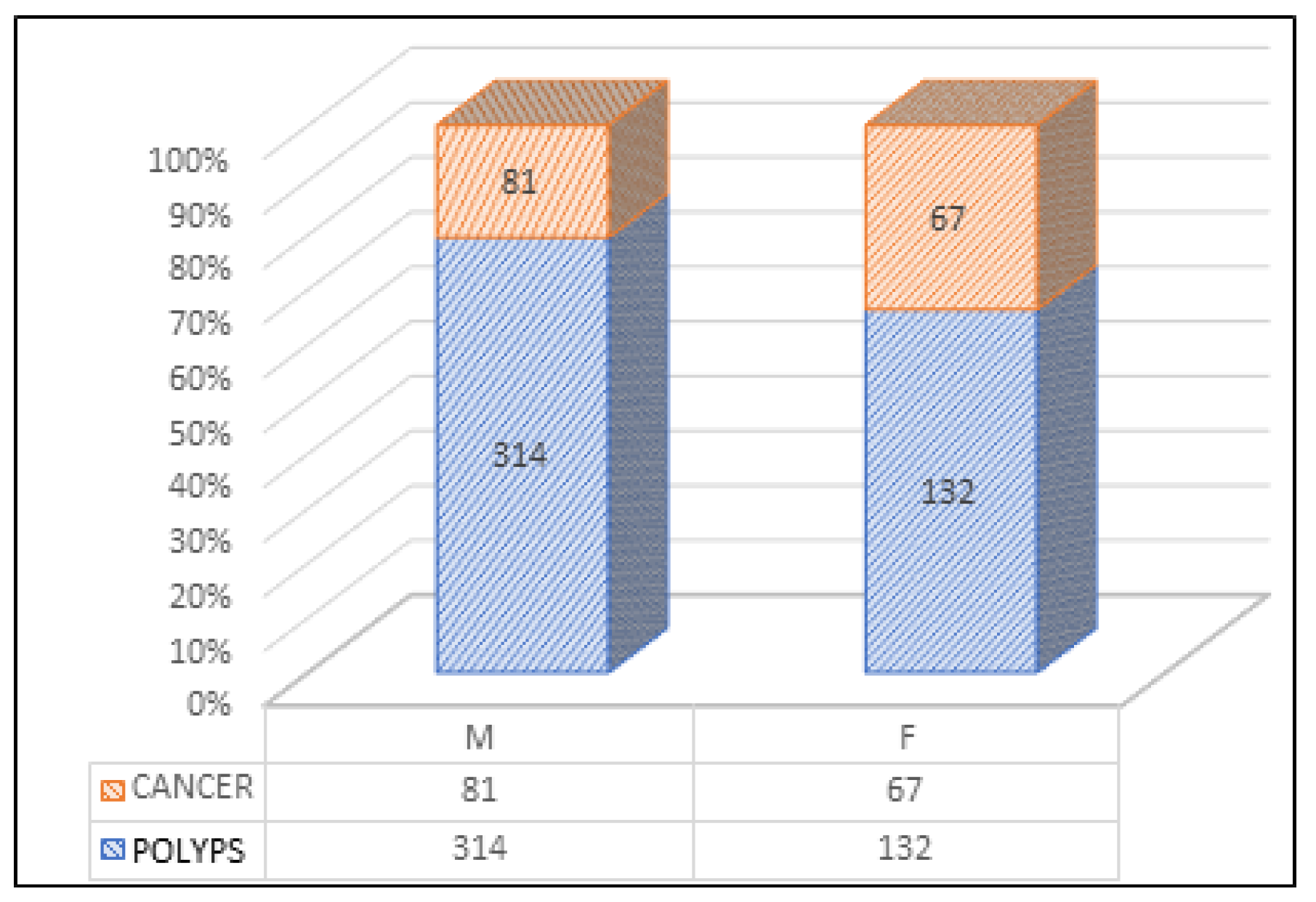
Jmms 05 00039 g001
Figure 2. Distribution of colonoscopies by sex and the type of tumor formations detected.
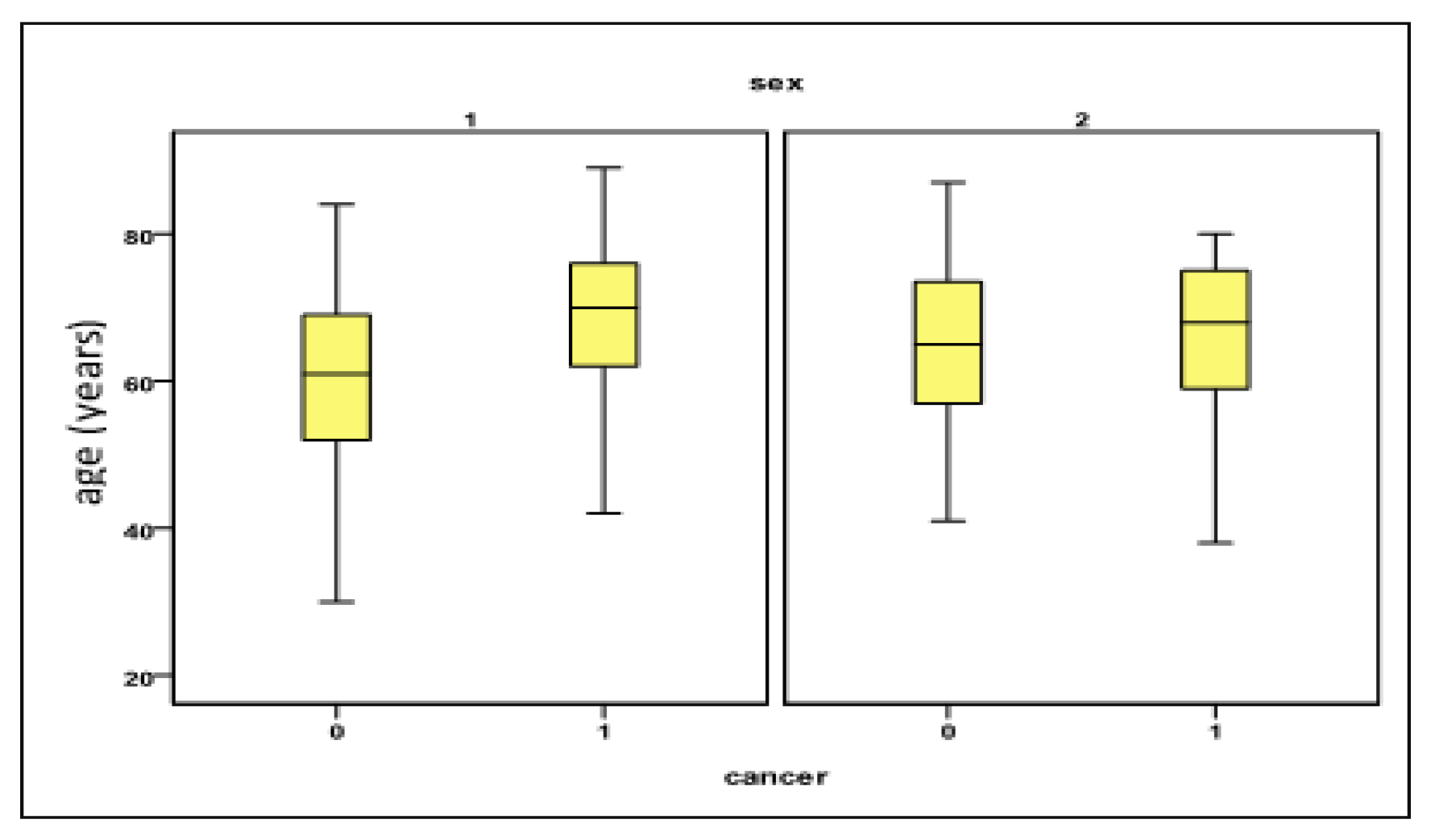
Jmms 05 00039 g002
Figure 3. Variation of the “age” parameter in relation to the presence of CRC and the sex of the investigated patients. Legend Sex 1 = male; 2 = female; Cancer 0 = absent (polyps present); 1 = present.
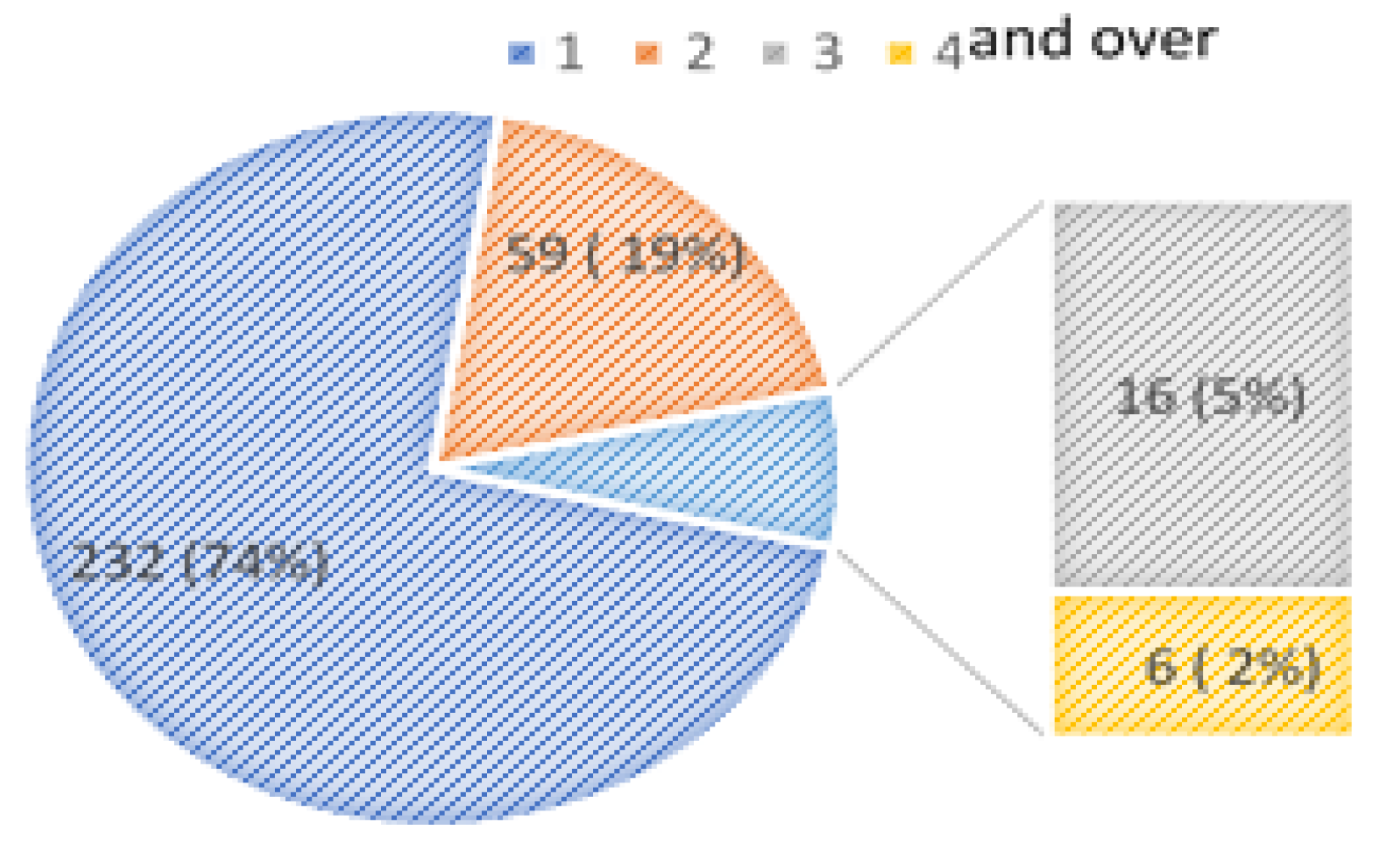
Jmms 05 00039 g003
Figure 4. Distribution of patients in relation to the number of polyps researched per session.
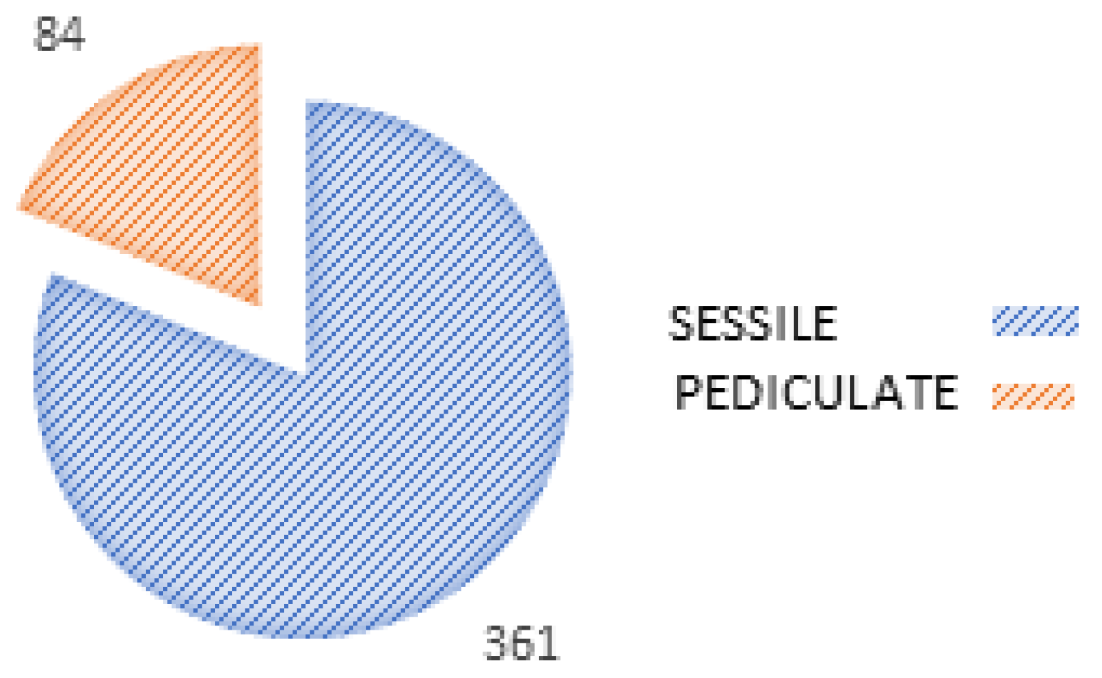
Jmms 05 00039 g004
Figure 5. Distribution of polypectomies in relation to the form of polyps resected.
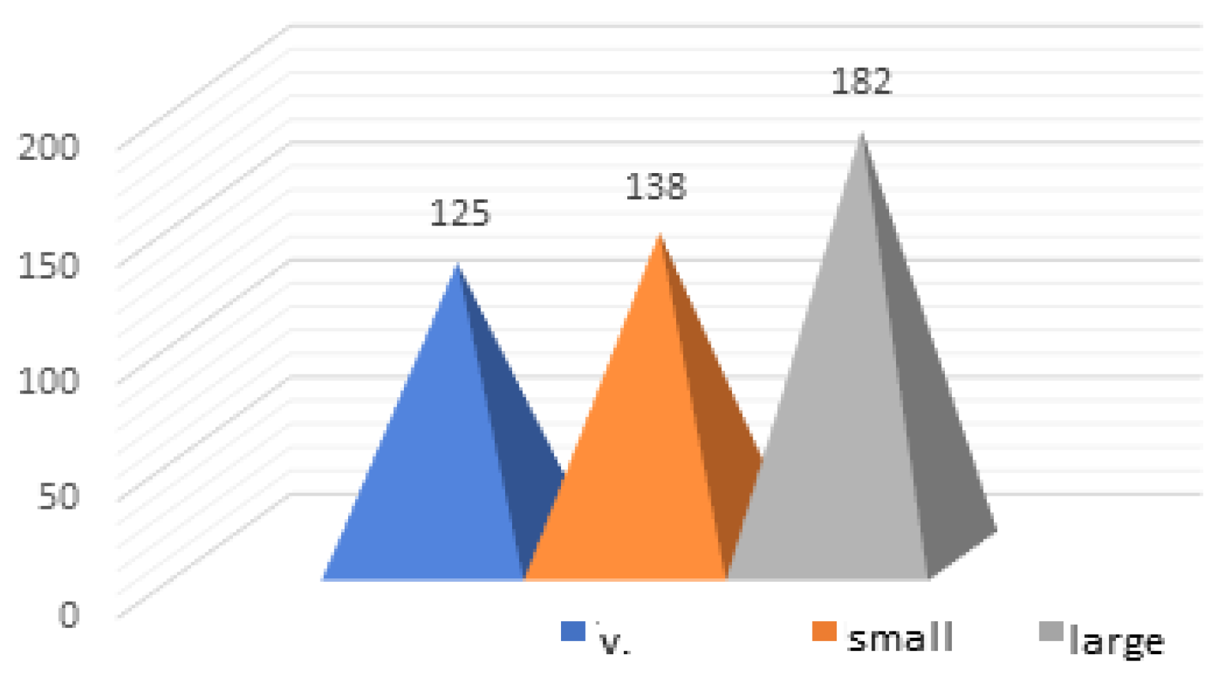
Jmms 05 00039 g005
Figure 6. Distribution of polypectomies in relation to the size of the resected polyps.
Jmms 05 00039 g006
Figure 7. The variation of the "polyp size" parameter (mm) in relation to the degree of malignancy of the polyps removed.
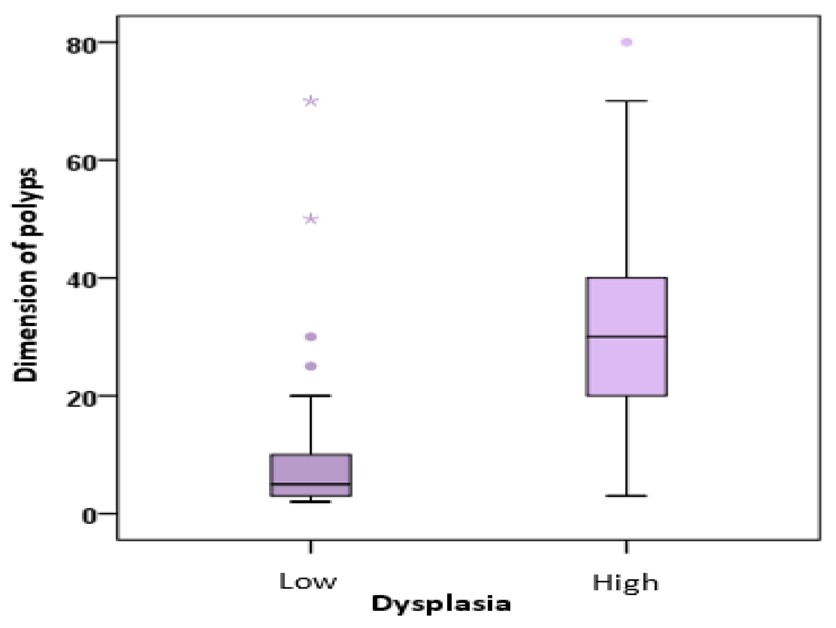
Jmms 05 00039 g007
Figure 8. Distribution of polyps in relation to the size of the polyps and their degree of dysplasia. Legend: 1= low, 2= high.
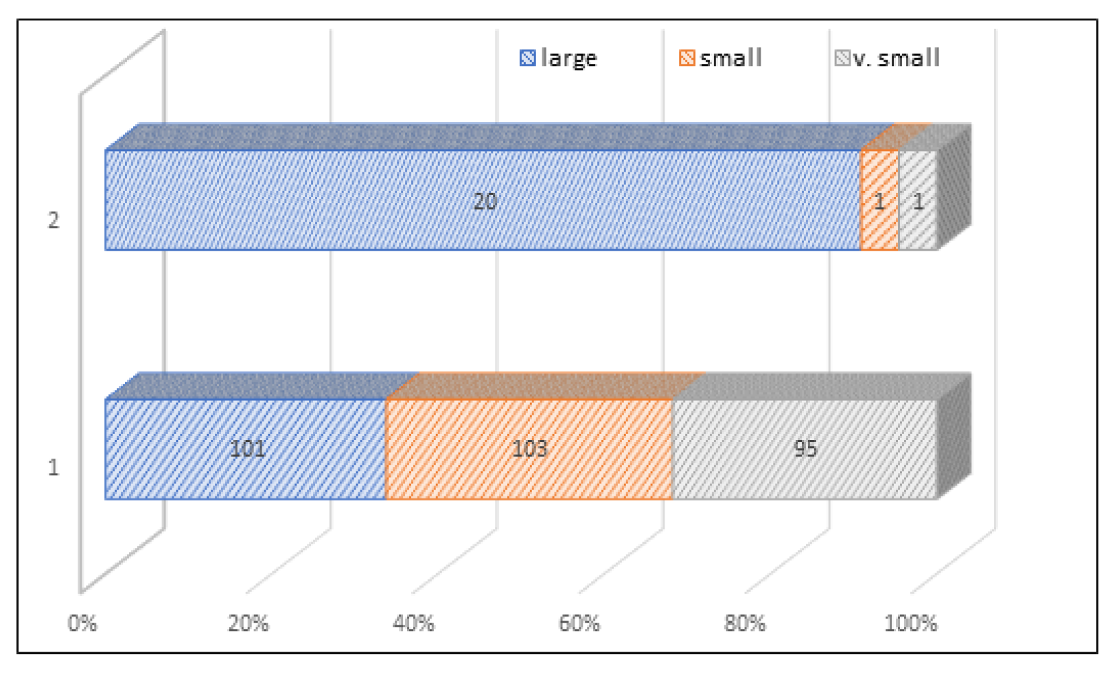
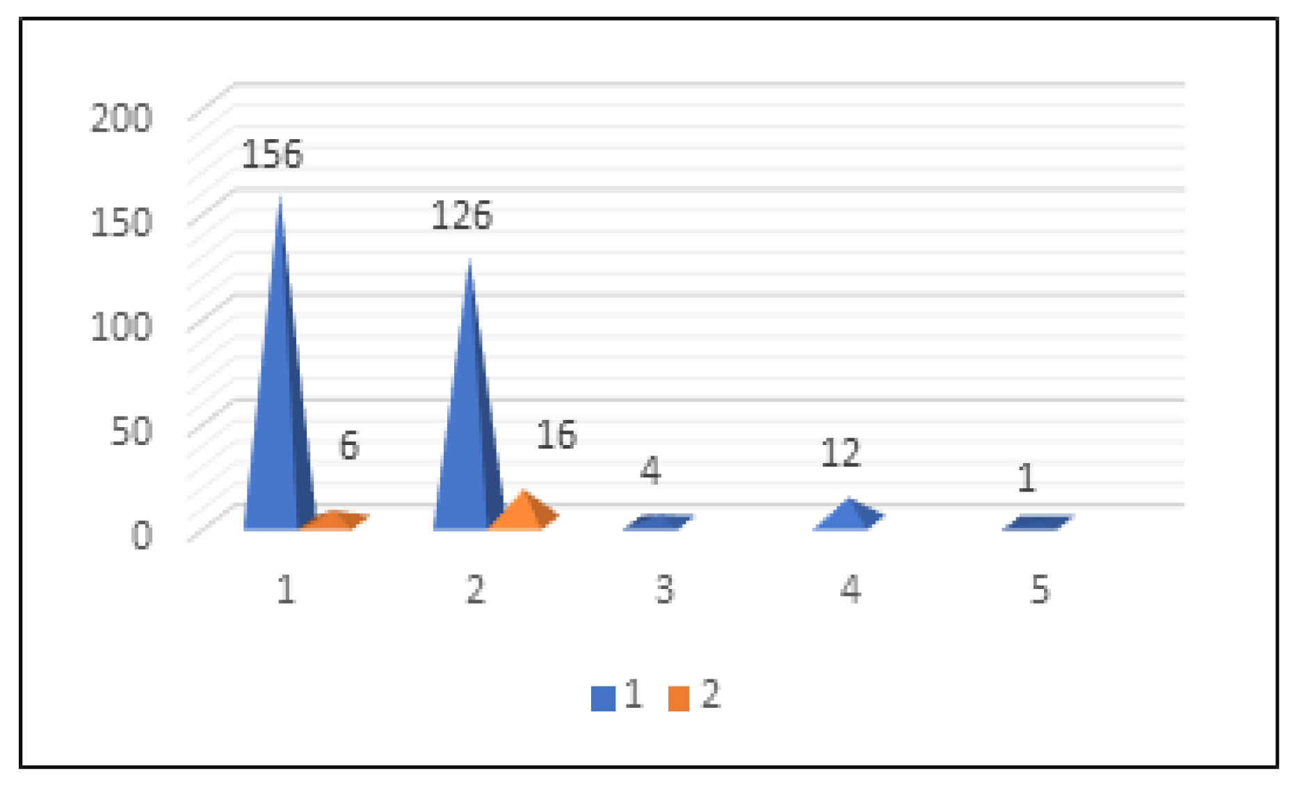
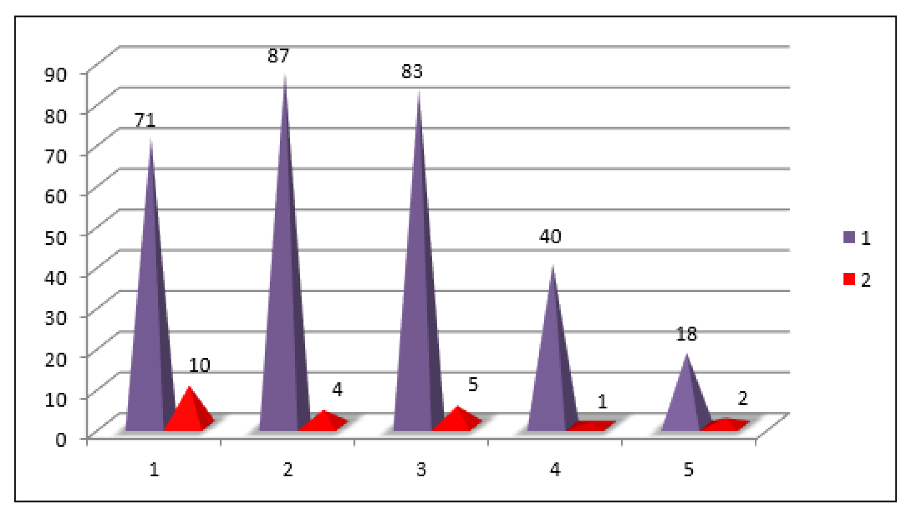
Jmms 05 00039 g008
Figure 9. Distribution of polypectomies in relation to the location of polyps and their degree of dysplasia. Legend. Location: 1= rect, 2= sigmoid, 3= descendant, 4= transverse, 5= ceco-ascendent | Type of dysplasia: 1= low, 2= high.
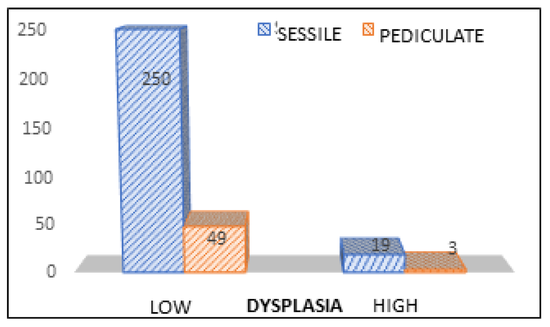
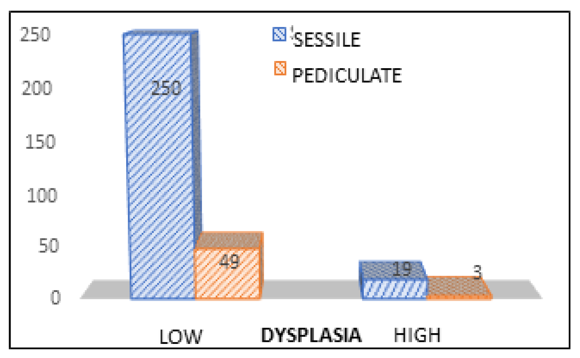
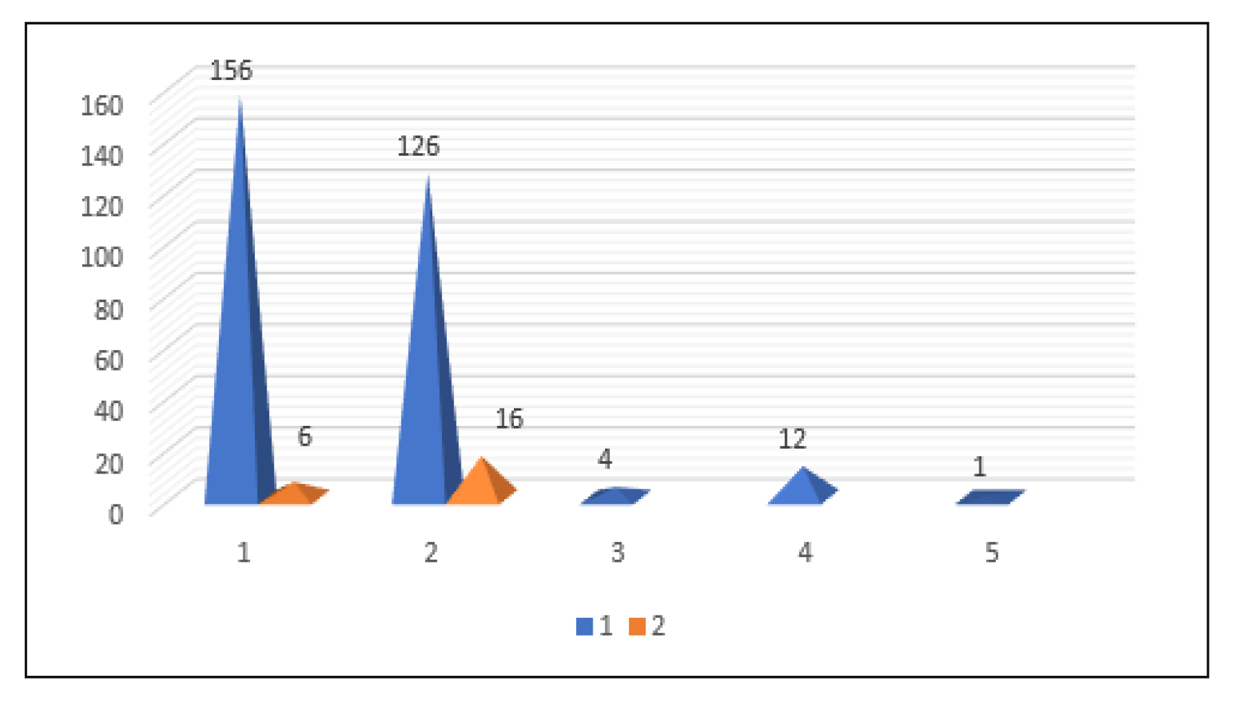
Jmms 05 00039 g009
Figure 10. Distribution of polypectomies in relation to the histological type of polyps and their degree of dysplasia. Legend. Type of dysplasia: 1= low, 2= high, Histology: 1= tubulo-adenomatous, 2= tubulo-villous, 3= serous, 4= mucous, 5= leiomyoma.
Jmms 05 00039 g010
Figure 11. Distribution of polyps in relation to existing form and degree of dysplasia.
Jmms 05 00039 g011
Figure 12. Types of dysplasia correlated with types of polyps.
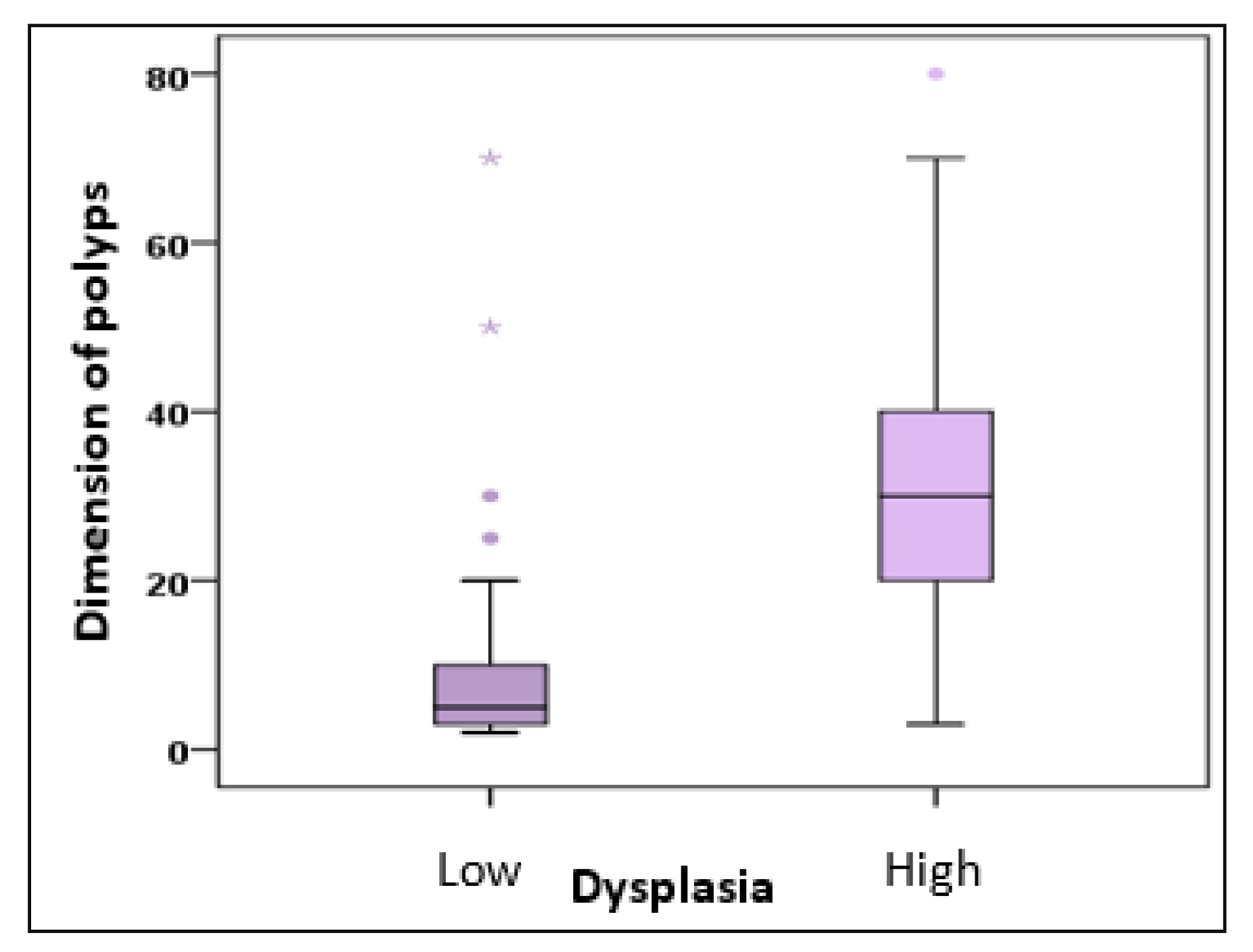
Jmms 05 00039 g012
Figure 13. Types of dysplasia correlated dimensions of the polyps.
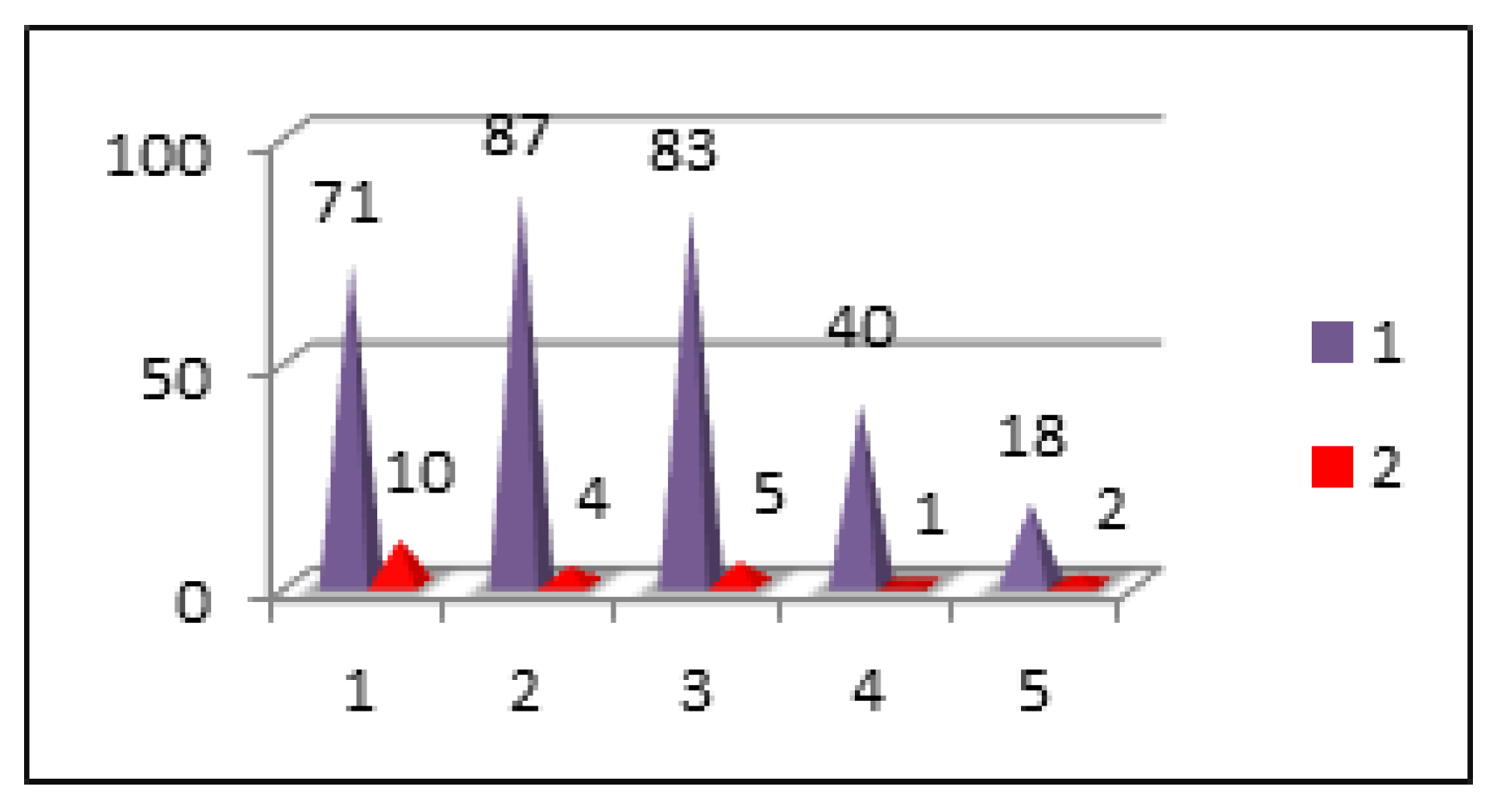
Jmms 05 00039 g013
Figure 14. Location of the polyps correlated with grade of dysplasia. Legend. Location: 1= rectum, 2= sigmoid, 3= descendant, 4= transverse, 5= ceco-ascendent. Types of dysplasia: 1= low, 2= high.
Jmms 05 00039 g014
Figure 15. Histology of the polyps correlated with the grade of dysplasia.
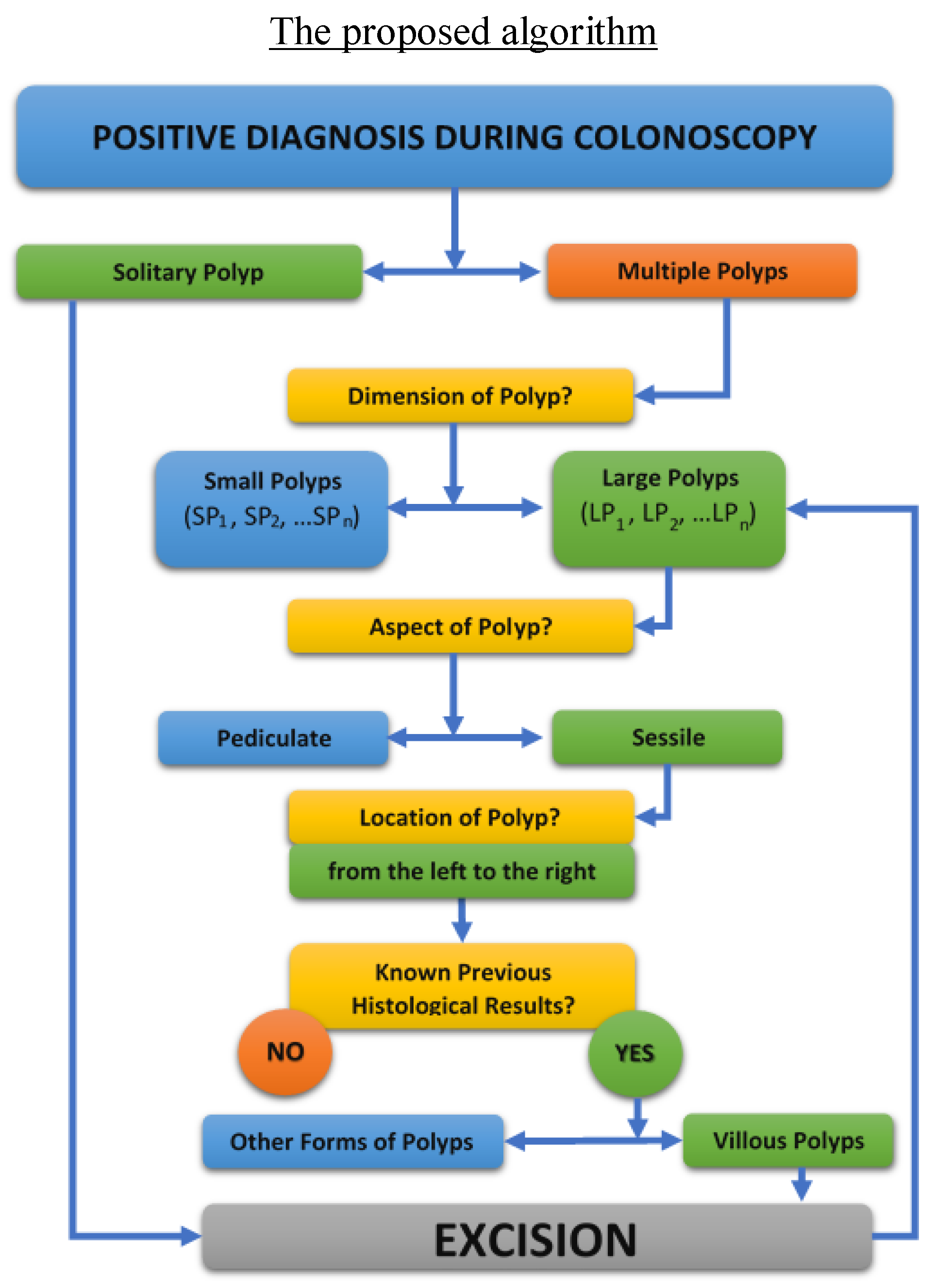
Jmms 05 00039 g015
Figure 16. The complete proposed algorithm for the clinical management of patients with multiple polyposis.
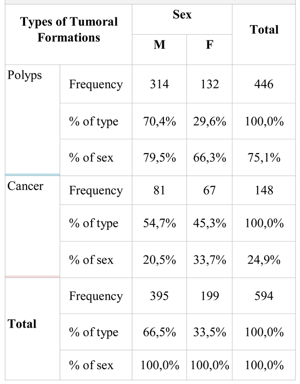
Jmms 05 00039 g016
Table 1. Distribution of colonoscopies in relation with the type of tumor formations detected and sex of investigated patients.
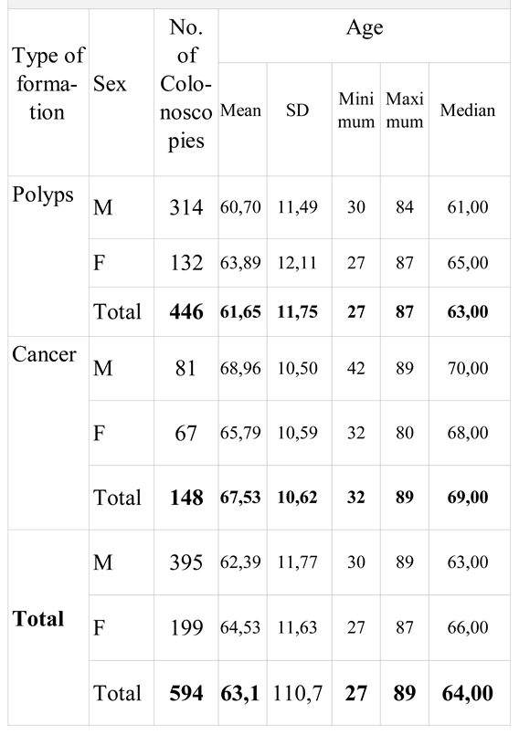
Jmms 05 00039 i001
Table 2. Statistical interpretation of “age” parameter in relation to the tumor type of the identified formations and sex of the investigated patients.
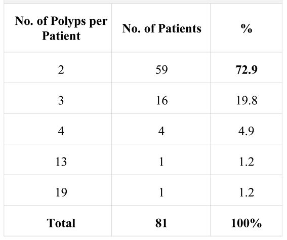
Jmms 05 00039 i002
Table 3. Distribution of patients with multiple polyposis.
Jmms 05 00039 i003
Table 4. Distribution of polypectomies in relation to the location of polyps resected on segments. Legend. Location: 1= rectal, 2= sigmoid, 3= descending, 4= transverse, 5= ceco-ascendent colon. nd= no data.
Jmms 05 00039 i004
Table 5. distribution of polypectomies relative to the histological type of polyps. Legend. Histology: 1= tubulo-adenomatous, 2= tubulo-villous, 3= serosa, 4= mucosal, 5= leiomyoma | nd = no data available.
Jmms 05 00039 i005
Table 6. Distribution of polypectomies in relation to the degree of dysplasia of polyps.
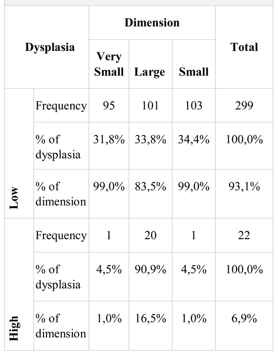
Jmms 05 00039 i006
Table 7. Distribution of polyps in relation to size and degree of dysplasia.
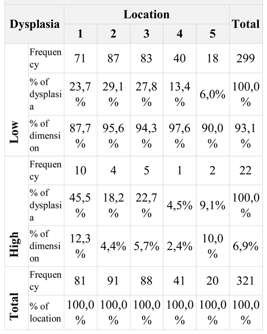
Jmms 05 00039 i007
Table 8. Associative distribution of polypectomies in relation to polyp location and dysplasia. Legend. Location: 1= rectal, 2= sigmoid, 3= descendant, 4= transverse, 5= ceco-ascendent.
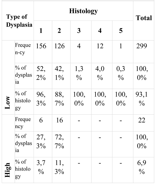
Jmms 05 00039 i008
Table 9. Associative distribution of polypectomies in relation to dysplasia and histology.
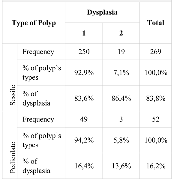
Jmms 05 00039 i009
Table 10. Associative distribution of polypectomies in polyp type ratio and degree of dysplasia. Legend. Type of dysplasia: 1= low, 2= high.
Jmms 05 00039 i010

