Next Article in Journal
Can Non-Small Cell Lung Cancer Histologic Subtypes Predict Survival? A Single Institution Experience
Previous Article in Journal
HPTLC Assay of Nicotine and Cotinine in Biological Samples
 
 
Journal of Mind and Medical Sciences is published by MDPI from Volume 12 Issue 1 (2025). Previous articles were published by another publisher in Open Access under a CC-BY (or CC-BY-NC-ND) licence, and they are hosted by MDPI on mdpi.com as a courtesy and upon agreement with Valparaiso University (ValpoScholar).
Article

Reconstruction of Heel Soft Tissue Defects Using Sensate Medial Plantar Flap

by
Mihaela Pertea
1,2,
Natalia Velenciuc
1,3,
Oxana Grosu
2,
Bogdan Veliceasa
1,4,
Vladimir Poroch
1,5,* and
Sorinel Lunca
1,3
1
Grigore T. Popa University of Medicine and Pharmacy, St. Spiridon Emergency County Hospital, Iasi, Romania
2
Department of Plastic Surgery and Reconstructive Microsurgery, St. Spiridon Emergency County Hospital, Iasi, Romania
3
Department of Surgery, Regional Institute of Oncology, Iasi, Romania
4
Department of Orthopedics and Traumatology, St. Spiridon Emergency County Hospital, Iasi, Romania
5
Department of Palliative Care, Regional Institute of Oncology, Iasi, Romania
*
Author to whom correspondence should be addressed.
J. Mind Med. Sci. 2018, 5(2), 250-254; https://doi.org/10.22543/7674.52.P250254
Submission received: 31 July 2018 / Revised: 31 August 2018 / Accepted: 30 September 2018 / Published: 3 October 2018

Highlights

  • Reconstruction of calcaneal soft tissue defects implies a special technique, using a skin highly resistant to pressure and well-innervated.
  • Sensate medial plantar fasciocutaneous flap can be a good solution for reconstruction of such calcaneal soft parts defects.

Abstract

:

Highlights

  • Reconstruction of calcaneal soft tissue defects implies a special technique, using a skin highly resistant to pressure and well-innervated.
  • Sensate medial plantar fasciocutaneous flap can be a good solution for reconstruction of such calcaneal soft parts defects.

Abstract

Introduction. Reconstruction of heel soft tissue defects represents a true challenge for any surgeon due to the particularities of this anatomical region. The tissue used to reconstruct the heel area must be resistant, innervated, and adapted to take over the body weight. Innervated medial plantar fasciocutaneous flap is one of the best solutions to cover defects at the heel level. Materials and Methods. We studied 5 patients, 4 males and one female, aged 42 to 67 years, who presented heel soft tissue defects of various etiologies. In all cases, the used reconstructive method was an insular innervated medial plantar fasciocutaneous flap. Results. Immediate and late outcomes were good. No immediate complications of necrosis type were recorded in any of the cases, and 2 years postoperatively there was no evidence of ulceration or other type of flap injury. The socio-professional reintegration of the patients was relatively rapid and their satisfaction was high. Donor area morbidity was minimal. Conclusions. Sensate medial plantar fasciocutaneous flap represents the first choice for the reconstruction of the heel soft tissue defects when patients’ local and general status allows it.

Introduction

Soft tissue defects of the heel have different causes. Among the most common are traumas of any kind, metabolic and vascular diseases, infections, and postsurgical effects of removal of benign or malignant tumors of the heel. The coverage of heel soft tissue defects is a challenge for the surgeon due to the structural and functional peculiarities of the heel and sole skin. The thickness of the skin, presence of septa that provide resistance and protection to support the whole weight of the body during passive orthostatism and movement make it difficult to choose a surgical technique by which to bring a skin with a structure similar to the initial one with a good vascular supply, and which ensures a proper sensitivity and resistance.
Over time, various techniques have been proposed, starting with the use of skin grafts, the cross-leg flap to free tissue transfer flaps, but none of these techniques was fully satisfactory from the point of view of immediate and late outcomes [1]. Harrison and Morgan in 1981 described a fasciocutaneous flap harvested from the plantar arch supplied by the medial plantar artery, which could provide protective sensitivity [2]. Subsequently, studies have shown that the use of the medial plantar flap for the reconstruction of calcaneal defects is one of the best solutions, the flap providing skin of similar structure and texture pressure-resistant, with minimal donor morbidity. This procedure requires a complex technique but nevertheless has a high success rate [3]. We present our experience and outcomes with sensate medial plantar flap in the reconstruction of calcaneal defects. No complications (necrosis) were recorded. Reintegration with full weight bearing and normal shoe wearing was possible 2 months postoperatively.

Materials and Methods

The study included 5 patients, 4 men and one woman, aged 42 to 67 years. Three patients presented trophic lesions as a result of neurological disorders (lumbar spine surgery) initially neglected by the patient. These lesions progressed for 2 to 6 years. The fourth case was a calcaneal defect following surgical removal of an ulcerated basal cell carcinoma and in case 5 the calcaneal defect had posttraumatic etiology being associated with a comminuted calcaneal fracture with exposed bone.
In all 5 cases preoperative preparation included investigation of patient's vascular status by Doppler examination.
Case 1, a 44-year-old male patient presented with a 5-year ulcerative lesion in the calcaneal region, painless both spontaneously and on palpation with a perilesional hyperkeratotic area. Past medical history revealed that the patient underwent a lumbar spine surgery for a herniated disc, resulting in dysesthesias in the right pelvic limb. One year after neurosurgical intervention, he developed ulceration on the right calcaneus, for which the patient did not seek medical help and underwent no treatment. The evolution was slow, declaratively with no significant changes in lesion appearance and symptoms. X ray examination did not reveal any bone injury and the echo-Doppler examination showed a normal arterial flow and a medial plantar artery with normal anatomy and physiological flow. As a result of physical examination and laboratory investigations, it was decided to cover the residual defect left after removal of the calcaneal ulceration with a medial plantar flap.
Case 2, a 56-year-old female patient with a 2-year ulcerated lesion and worsening of symptoms over time; the lesion increased in size, was bleeding, and caused marked functional impotence. X ray examination did not reveal any heal bone injury.
Cases 3 and 4, 65-and 67-year-old men, respectively, presented minor cardiac disorders and calcaneal ulcerations due to neurological disorders with a history of 2 and 4 years and no increase in size of the ulcerations.
Case 5, a 50-year-old male patient, was the victim of a domestic accident resulting in a comminuted calcaneal fracture with soft tissue loss and bone exposure.
In all cases, it was decided to cover the calcaneal defects with a sensate medial plantar fasciocutaneous flap.
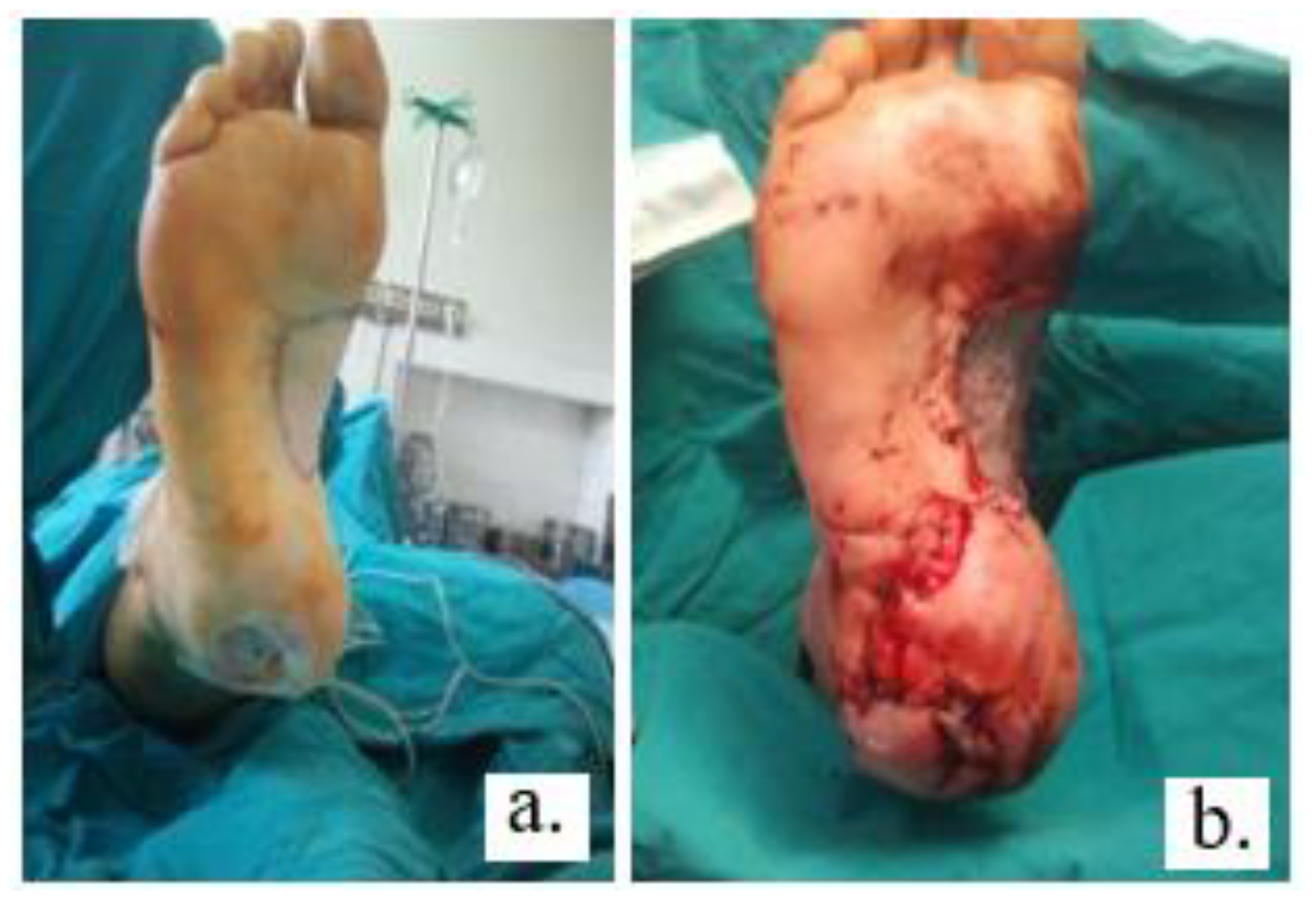
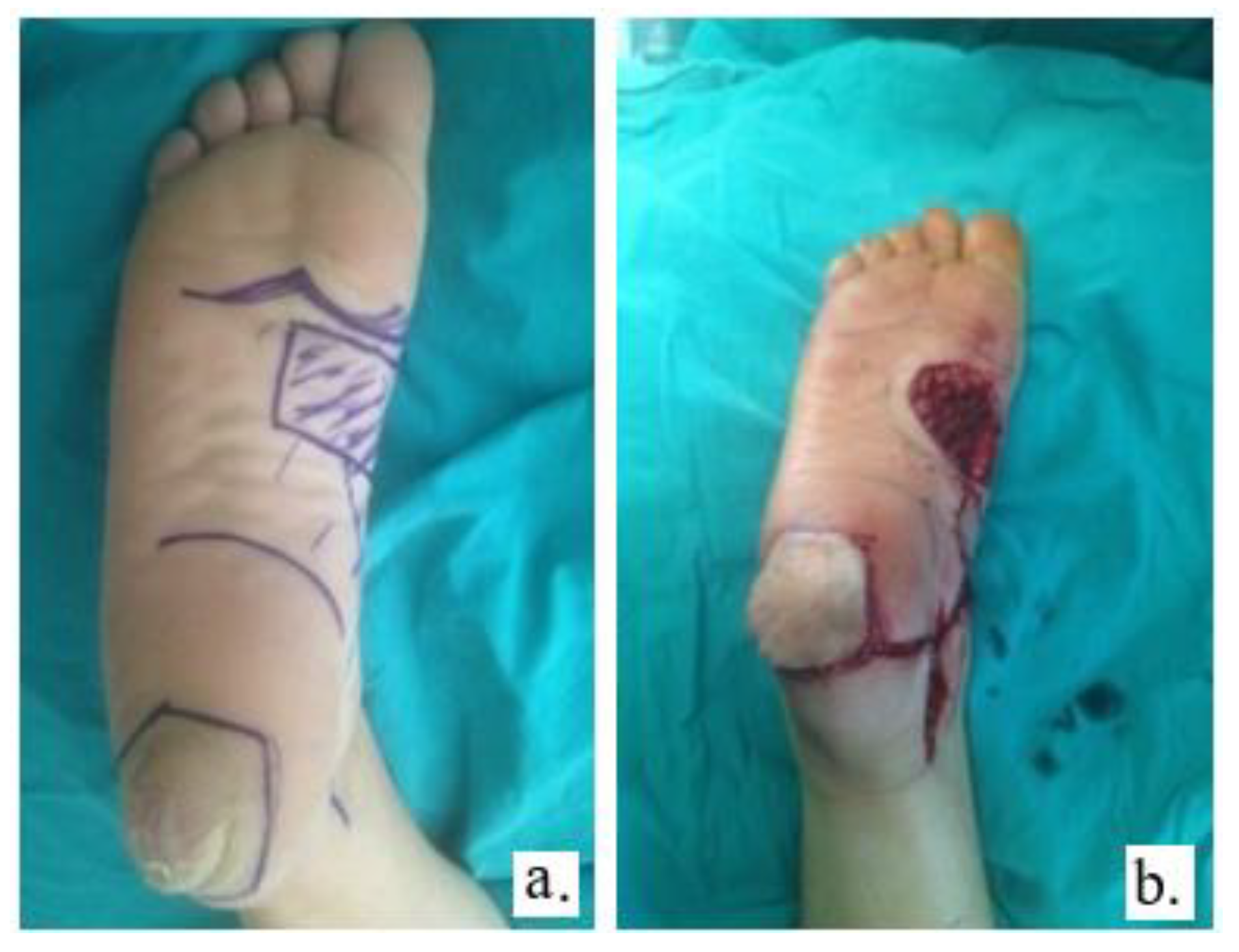
Surgical interventions were performed under spinal anesthesia in a bloodless field obtained by placing a tourniquet at the root of the thigh. According to the preoperative design, we performed the excision of the calcaneal lesion tangential to the bone plane and of the perilesional hyperkeratosis area, where the clinical appearance required it. The size of the post-excisional defects ranged from 4/3 cm and 5/4 cm. The harvesting of the internal plantar flap began with the exposure of the posterior tibial bundle and incision of flap edges, drawn preoperatively up to the level of plantar aponevrosis which was included into the flap (Figure 1).
The medial plantar artery was exposed by cutting the abductor hallucis muscle and later ligated to the distal edge of the flap (Figure 1). The plantar medial nerve was discovered and dissected to be included into the flap, thus preserving its sensitivity. The flap was elevated from distal to proximal with the inclusion of the neurovascular pedicle. The continuity of abductor hallucis muscle was restored. In all cases, the donor sites were covered by split-thickness free skin plasty and the flap was positioned at the defect level by tunneling in 3 cases and in the other two cases by skin incision and sutured to the defect edges with separate stitches (Figure 2 and Figure 3).

Results

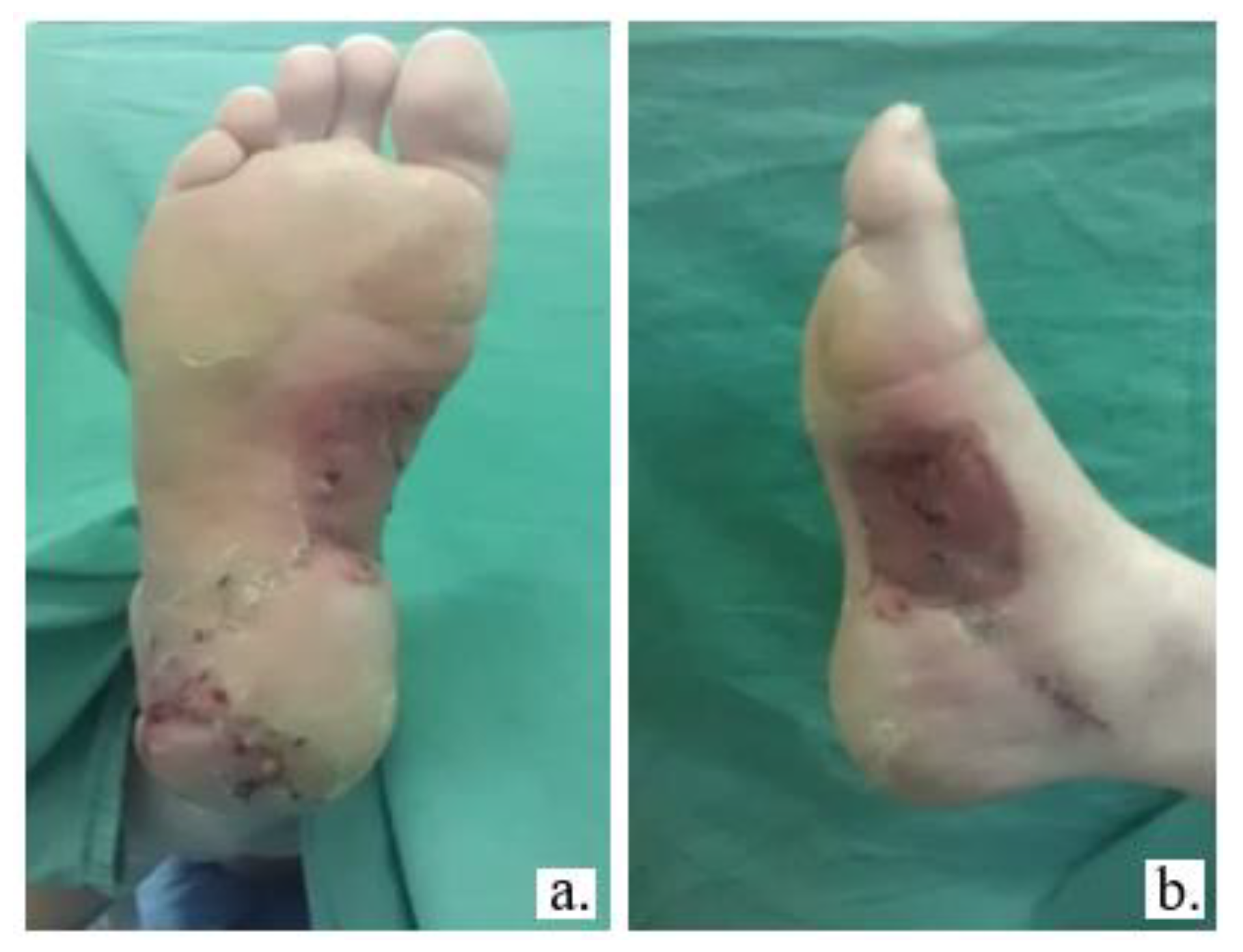
In our study no complications (necrosis of the flap) were recorded. Hospital stay ranged from 7 to 14 days. In all cases we reported good results at 4 weeks after surgery (Figure 4). The morbidity of the donor site was minimal.
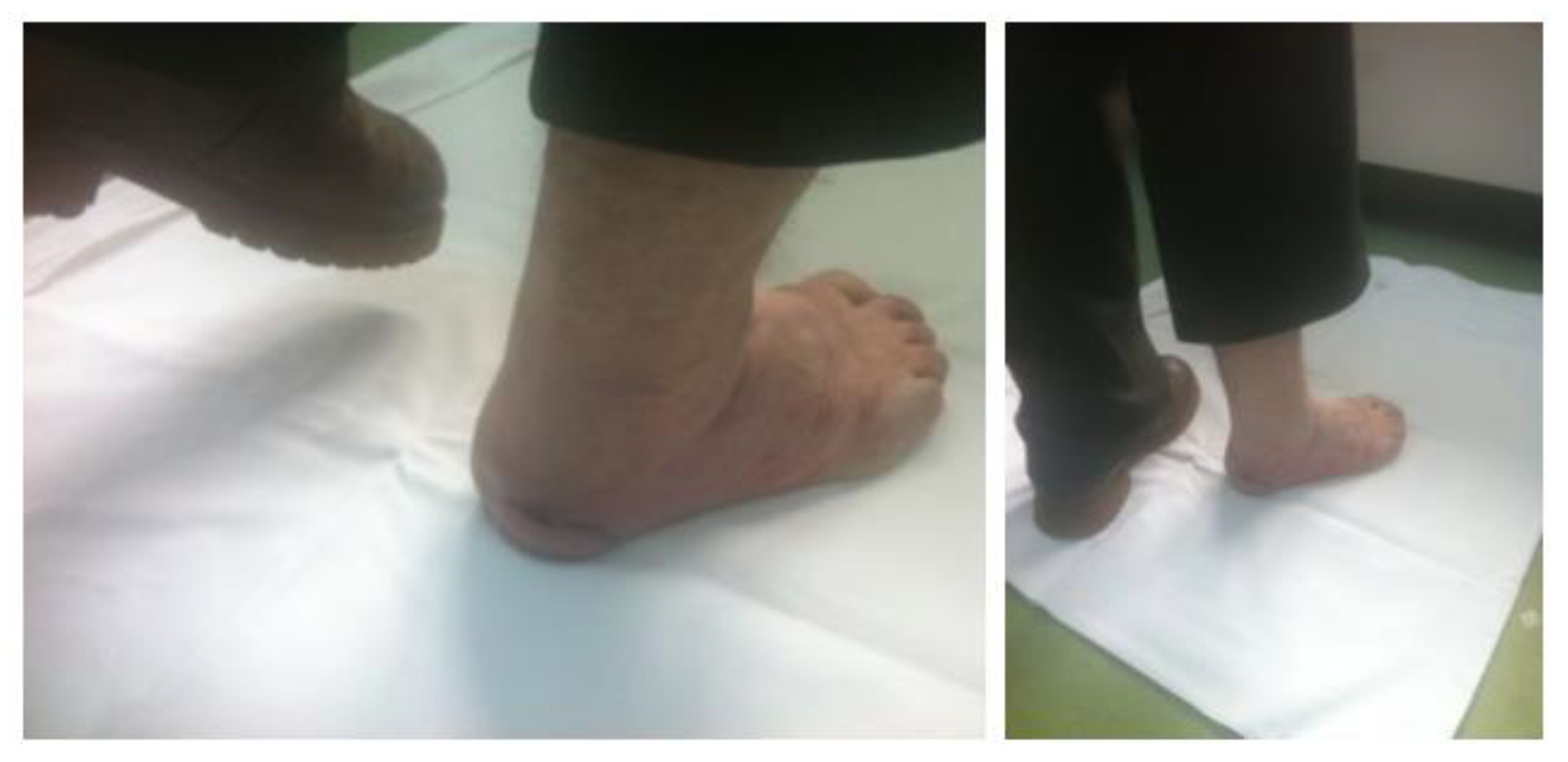
Reintegration with full weight bearing and normal shoe wearing was possible 2 months postoperatively (Figure 5). There was no evidence of ulcerative or other lesions at flap level 2 years postoperatively.

Discussions

Over time, the reconstruction of heel soft tissue defects has posed problems to surgeons due to the structural peculiarities of the skin at this level adapted to withstand the whole-body pressure and weight [4]. In standing position, the heel takes approximately 80% of body weight [5], shock resistance, the resistance at this level being possible due to the presence of vertical fibrous septa between the fascia and dermis [6].
Due to these unique properties of plantar skin and especially of the skin at calcaneal level, the reconstruction of soft tissue defects at this level is difficult, especially with respect to the “like-with-like” principle [7]. Seeking solutions for the coverage of soft tissue defects at calcaneal level, Shanahan and Gingrass studied in 1979 the anatomy of the medial plantar artery [8], and then described the vascularized and innervated medial plantar flap. In 1981 Harrison and Morgan described the fasciocutaneous island flap on the plantar medial artery [2]. Studies have shown that the innervated plantar medial fasciocutaneous flap has become the first choice for the reconstruction of the calcaneal region due to its versatility, the resistant skin it provides, the well-represented vascular bundle, and a moderately complex surgical technique [9].
It should also be noted that the use of this flap has good functional and esthetic results, important being also the fact that it is innervated, thus having a protective sensitivity [10]. This flap has been the subject of study for a long time in different centers, demonstrating its efficiency and viability. Thus, Benito-Ruiz et al. carried out a study in 2004 in which six medial plantar flaps were performed with 83% survival, complete necrosis being recorded in only one case [1]. Also, in 2004, Rashid et al. reported 20 medial plantar flaps with 100% survival [11], and in 2005 Schwarz and Negrini studied 51 flaps carried out in 48 patients and reported a total flap survival of 98%, one case of complete necrosis and 4 cases with delayed healing, the recurrence of ulceration being recorded in 7 cases [12].
In 2006 Mourougayan performed 12 medial plantar flaps to cover plantar defects of various causes: traumatic, infectious, or after tumor resection, reporting a 100% viability of the flaps without any immediate or late postoperative complication [13]. Also studied was the effectiveness of medial plantar flap compared to sural flap in a group of 80 patients, the sural flap being used in 50 cases [14]. In the plantar medial flap group, a single case of flap necrosis was recorded (29 cases with flap survival), and in the sural flap group there were 3 cases of marginal flap necrosis and 2 cases with complete necrosis. A more rapid adaptation of the medial plantar flap has been reported with regard to taking over the body weight during walking [14].
In our study on 5 patients, no complete or partial necrosis of the medial plantar flap was recorded. Being innervated, the plantar medial flap provides the necessary sensitivity to the plantar area. In 2014, Trevatt et al. demonstrated by two-point discrimination testing that the sensitivity of medial plantar flap is almost normal 1 year postoperatively, sensitivity being recovered starting with 4 months postoperatively [15].
Regarding the surgical technique, it is more favorable to bring the flap from the donor site to the receptor site via tunneling, thus avoiding the occurrence of vascular kinking and compression of the future scar over the vascular pedicle [16]. Donor site morbidity was minimal with the medial plantar flap, in only a few cases the sectioning of abductor hallucis muscle being necessary in order to expose the vascular pedicle [17,18]. In our study we undertook flap tunneling in 3 cases and in the remaining 2 cases skin incision to bring the flap to the receptor site. In none of these cases complications due to pedicle compression were recorded.
There was no difference in subsequent scarring in the cases in which skin incision was used. To determine the donor site morbidity, the 17-item Foot Function Index (FFI) [19] and 8-item Emeking Questionnaire were used [20]. In our study of 5 patients, a patient satisfaction questionnaire was used, satisfaction being maximum in all cases. Thus, we conclude that the innervated medial plantar fasciocutaneous flap is the best choice for reconstruction of the soft tissue defects of the heel, being resilient, innervated, with an area-like texture capable of taking over and supporting body weight, and having minimal donor site morbidity [4].

Conclusions

For the reconstruction of calcaneal soft tissue defects, surgeons should choose a technique that can provide a pressure resistant and innervated skin in order to prevent the secondary posttraumatic lesions with minimal donor site morbidity and a good esthetic result. These conditions are met by the sensate medial plantar fasciocutaneous flap, which is the first option for the reconstruction of calcaneal soft parts defects of different etiologies.

Conflicts of Interest

There are no known conflicts of interest in the publication of this article, and there was no financial support that could have influenced the outcomes. The manuscript was read and approved by all authors.

Compliance with ethical standards

Any aspect of the work covered in this manuscript that has involved human patients has been conducted with the ethical approval of all relevant bodies and that such approvals are acknowledged within the manuscript.

References

  1. Benito-Ruiz, J.; Yoon, T.; Guisantes-Pintos, E.; Monner, J.; Serra-Renom, J.M. Reconstruction of soft- tissue defects of the heel with local fasciocutaneous flaps. Ann Plast Surg. 2004, 52, 380–384. [Google Scholar] [CrossRef] [PubMed]
  2. Harrison, D.H.; Morgan, B.D. The instep island flap to resurface plantar defects. Br J Plast Surg. 1981, 34, 315–318. [Google Scholar] [CrossRef] [PubMed]
  3. Macedo, J.L.S.; Rosa, S.C.; Neto, A.V.R.F.; Silva, A.A.D.; Amorim, A.C.S. Reconstruction of soft-tissue lesions of the foot with the use of the medial plantar flap. Rev Bras Ortop. 2017, 52, 699–704. [Google Scholar] [CrossRef] [PubMed]
  4. Paget, J.T.; Izadi, D.; Haj-Basheer, M.; Barnett, S.; Winson, I.; Khan, U. Donor site morbidity of the medial plantar artery flap studied with gait and pressure analysis. Foot Ankle Surg. 2015, 21, 60–66. [Google Scholar] [CrossRef] [PubMed]
  5. Ishikawa, K.; Isshiki, N.; Suzuki, S.; Shimamura, S. Distally based dorsalis pedis island flap for coverage of the distal portion of the foot. Br J Plast Surg. 1987, 40, 521–525. [Google Scholar] [CrossRef] [PubMed]
  6. Baker, G.L.; Newton, E.D.; Franklin, J.D. Fasciocutaneous island flap based on the medial plantar artery: Clinical applications for leg, ankle, and forefoot. Plast Reconstr Surg 1990, 85, 47–58. [Google Scholar] [PubMed]
  7. Gillies, H.; Millard, D. The principles and art of plastic surgery; Butterworth: London, UK, 1957; Volume 2, pp. 368–384. [Google Scholar]
  8. Shanahan, R.E.; Gingrass, R.P. Medial plantar sensory flap for coverage of heel defects. Plast Reconstr Surg. 1979, 64, 295–298. [Google Scholar] [PubMed]
  9. Wan, D.C.; Gabbay, J.; Levi, B.; Boyd, J.B.; Granzow, J.W. Quality of innervation in sensate medial plantar flaps for heel reconstruction. Plast Reconstr Surg. 2011, 127, 723–730. [Google Scholar] [CrossRef] [PubMed]
  10. Weinzweig, N.; Davies, B.W. Foot and ankle reconstruction using the radial forearm flap: A review of 25 cases. Plast Reconstr Surg. 1998, 102, 1999–2005. [Google Scholar] [CrossRef] [PubMed]
  11. Rashid, M.; Hussain, S.S.; Aslam, R.; Illahi, I. A comparison of two fasciocutaneous flaps in the reconstruction of defects of the weight-bearing. J Coll Physicians Surg Pak. 2003, 13, 216–218. [Google Scholar] [PubMed]
  12. Schwarz, R.J.; Negrini, J.F. Medial plantar artery island flap for heel reconstruction. Ann Plast Surg. 2006, 57, 658–661. [Google Scholar] [CrossRef] [PubMed]
  13. Mourougayan, V. Medial plantar artery (instep flap) flap. Ann Plast Surg. 2006, 56, 160–163. [Google Scholar] [CrossRef] [PubMed]
  14. Alius, C.; Oprescu, S.; Balalau, C.; Nica, A.E. Indocyanine green enhanced surgery; principle, clinical applications and future research directions. J Clin Invest Surg. 2018, 3, 1–8. [Google Scholar] [CrossRef]
  15. Trevatt, A.E.; Filobbos, G.; UIHaq, A.; Khan, U. Long- term sensation in the medial plantar flap: A two- centre study. Foot Ankle Surg. 2014, 20, 166–169. [Google Scholar] [CrossRef] [PubMed]
  16. Gu, J.X.; Huan, A.S.; Zhang, N.C.; Liu, H.J.; Xia, S.C.; Regmi, S.; Yang, L. Reconstruction of Heel Soft Tissue Defects Using Medial Plantar Artery Island Pedicle Flap: Clinical Experience and Outcomes Analysis. J Foot Ankle Surg. 2017, 56, 226–229. [Google Scholar] [CrossRef] [PubMed]
  17. Oh, S.J.; Moon, M.; Cha, J.; Koh, S.H.; Chung, C.H. Weight-bearing plantar reconstruction using versatile medial plantar sensate flap. J Plast Reconstr Aesthet Surg. 2011, 64, 248–254. [Google Scholar] [CrossRef] [PubMed]
  18. Yang, D.; Yang, J.F.; Morris, S.F.; Tang, M.; Nie, C. Medial plantar artery perforator flap for soft-tissue reconstruction of the heel. Ann Plast Surg. 2011, 67, 294–298. [Google Scholar] [CrossRef] [PubMed]
  19. Budiman-Mak, E.; Conrad, K.J.; Roach, K.E. The Foot Function Index: A measure of foot pain and disability. J Clin Epidemiol. 1991, 44, 561–570. [Google Scholar] [CrossRef] [PubMed]
  20. Enneking, W.F.; Dunham, W.; Gebhardt, M.C.; Malawar, M.; Pritchard, D.J. A system for the functional evaluation of reconstructive procedures after surgical treatment of tumors of the musculoskeletal system. Clin Orthop Relat Res. 1993, 286, 241–246. [Google Scholar] [CrossRef] [PubMed]
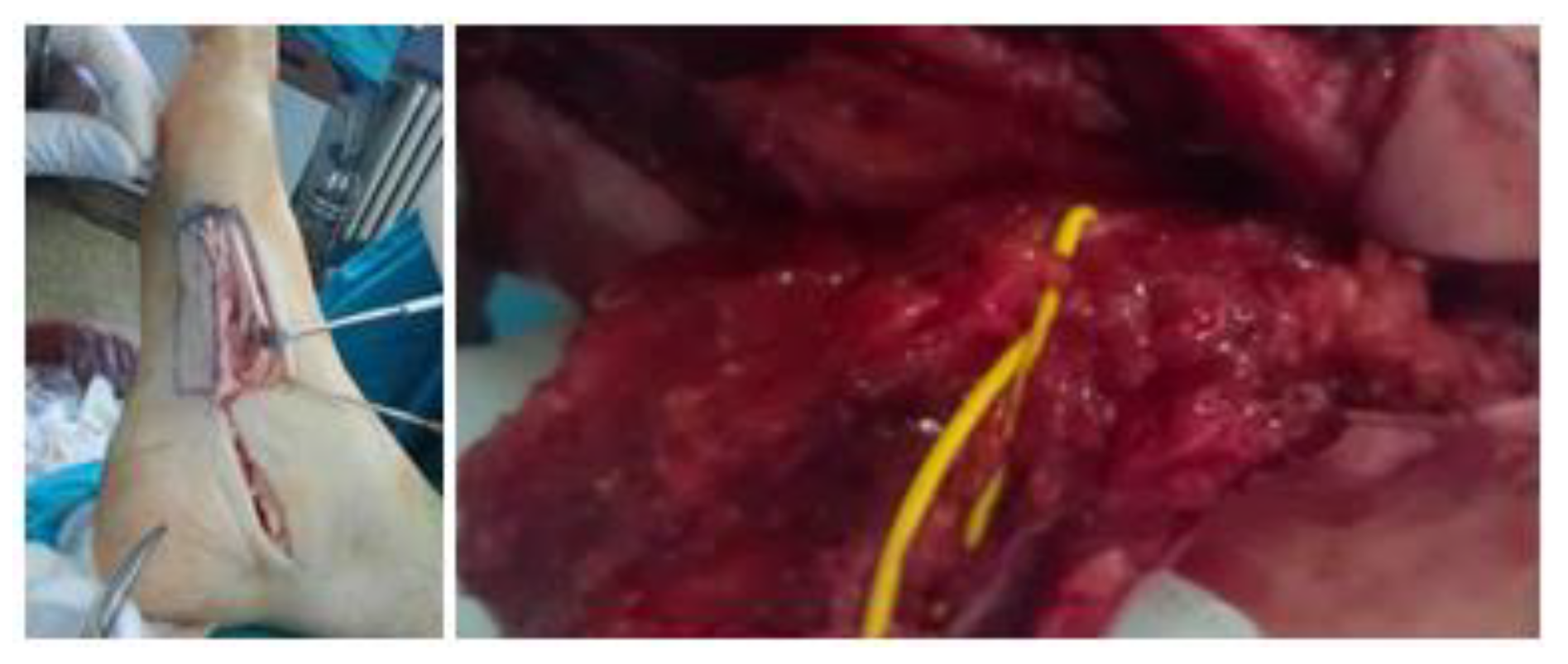
Figure 1. Elevation of medial plantar flap.
Jmms 05 00037 g001
Figure 2. Calcaneal defect (neurologic-type ulceration)- a. flap design, b. defect coverage with sensate insular medial plantar fasciocutaneous flap – immediate postoperative result.
Jmms 05 00037 g002
Figure 3. Calcaneal defect following tumor removal – a. flap design, b. flap elevation and placement on the defect.
Jmms 05 00037 g003
Figure 4. Result at 4 weeks postoperatively: a. flap, b. donor site.
Jmms 05 00037 g004
Figure 5. Outcome 2 month postoperatively – full weight bearing.
Jmms 05 00037 g005