Next Article in Journal
Necrotizing Fasciitis of the Neck: Case Presentation
Previous Article in Journal
Therapeutic Challenges in a Case of Psoriasis with Nail Onset
 
 
Journal of Mind and Medical Sciences is published by MDPI from Volume 12 Issue 1 (2025). Previous articles were published by another publisher in Open Access under a CC-BY (or CC-BY-NC-ND) licence, and they are hosted by MDPI on mdpi.com as a courtesy and upon agreement with Valparaiso University (ValpoScholar).
Case Report

Zenker’s Diverticulum and Squamous Esophageal Cancer: A Case Report

by
Ion Dina
1,
Octav Ginghina
2,
Corina D. Toderescu
3,
Cristian Bălălău
4,*,
Bianca Galateanu
5,
Carolina Negrei
6 and
Claudia Iacobescu
1
1
Department of Gastroenterology, St. Ioan Clinical Hospital, Carol Davila University, Bucharest, Romania
2
Department of Surgery, St. Ioan Clinical Hospital, Carol Davila University, Bucharest, Romania
3
Faculty of General Medicine, Vasile Goldis Western University of Arad, Arad, Romania
4
Department of Surgery, St. Pantelimon Hospital, Carol Davila University, Bucharest, Romania
5
Department of Biochemistry and Molecular Biology, University of Bucharest, Bucharest, Romania
6
Department of Toxicology, Carol Davila University, Bucharest, Romania
*
Author to whom correspondence should be addressed.
J. Mind Med. Sci. 2017, 4(2), 193-197; https://doi.org/10.22543/7674.42.P193197
Submission received: 1 August 2017 / Revised: 1 September 2017 / Accepted: 1 October 2017 / Published: 5 October 2017

Abstract

:
Zenker’s diverticulum represents a rare esophageal lesion developed especially in the elderly population due to herniation of esophageal mucosa above the cricopharyngeus muscle. The condition leads to food retention, regurgitation, aspiration, and dysphagia in affected patients. Progressive dysphagia also characterizes malignant diseases of the esophagus like squamous esophageal carcinoma that typically appears in male patients in the seventh decade of life, with a history of cigarette smoking and alcohol abuse. We report a case of a male patient who presented with dysphagia for both solids and liquids along with significant weight loss, and who was diagnosed with medium esophageal cancer associated with Zenker’s diverticulum.

Introduction

Zenker’s diverticulum is a benign esophageal condition, quite rarely encountered in clinical practice with an annual incidence of two per 100,000 persons per year [1]. The condition typically affects the elderly population in the seventh decade of life, being uncommon before the age of 40, and it is more frequently described in males than females [1,2]. In fact, Zenker’s diverticulum or pharyngeal pouch is a pulsion diverticulum that results from the herniation of the esophageal mucosa on the posterior wall between the cricopharyngeus muscle and the inferior pharyngeal constrictor muscle [3].
Zenker’s diverticulum is a false diverticulum, lined with stratified squamous epithelium and without any muscular layer. Morton and Bartney have devised a classification of diverticulosis based of dimension: small pouch (less than 2 cm), medium (between 2 cm and 4 cm), and large (more than 4 cm) [1,4]. The majority of diverticula exhibit no symptoms and are discovered incidentally [5]. On the other hand, the presence of an esophageal diverticulum is associated with characteristic symptoms with variable degree of severity. The most common symptom is dysphagia which affects 80-90% of patients and shows a progressive increase [4,6]. Food retention within the diverticulum may predispose to regurgitation and aspiration into the bronchial tree with subsequent dyspnea [4]. Typical complaints for this type of pouch are loud swallowing of liquids, hoarseness, cough, and halitosis [4]. Diagnostic tools used routinely consist of barium swallow studies and esophagoscopy.
A marked worsening of symptoms in a patient diagnosed with Zenker’s pouch may announce the presence of malignancy within the diverticulum [7]. Alarm symptoms as presence of blood into regurgitate, weight loss, odynophagia, melena, or progressive dysphagia should promptly alert the physician to the need to investigate the possibility of a superimposed cancer [8]. The risk of cancer development within the diverticulum is around 0,3-7% [8]. The mechanism involved is probably related to inflammatory processes induced by repeated diverticulitis attacks that could lead to cellular changes. Risk factors for malignancy in diverticulum are: older age, larger size diverticula, and a long course of the disease [8].
The management strategy of Zenker’s diverticulum is mainly based on two options: open surgical procedures represented by pouch removal, and endoscopic technique consisting of cricopharyngeus muscle myotomy [9,10]. Endotherapy appears to be the future standard of care for patients with Zenker’s diverticulum, both replacing surgical procedures and showing promising results concerning dysphagia relief on one hand, and low complication rates on the other [11].
Squamous esophageal cancer is a devastating disease that occurs more frequently during the sixth and seventh decades of life and carries a poor prognosis [12]. Esophageal cancer represents the sixth cause of cancer-related death worldwide and generally affects male patients more than females [12]. Squamous type of carcinoma is the predominant form, accounting for 95% of all esophageal carcinomas. The main symptom that dominates the clinical picture is progressive dysphagia associated with neoplastic signs. The current management of esophageal cancer consists of a multidisciplinary approach – surgery, chemotherapy, and radiation, taking into account the tumoral stage. Early stages, like mucosal cancer, are amenable to endoscopic treatment, while advanced tumors typically benefit from combined therapies [12].

Case Report

A 56-years old male was admitted to our clinic for progressive dysphagia that began four months prior to presentation. Initially, dysphagia was reported to solid aliments, but subsequently became total, both to solids and liquids, resulting in a significant weight loss of 15 kg. Additional complaints included heartburn and postprandial regurgitation of undigested food. His past medical history as well as his family history was uneventful. The patient reported heavy smoking (around 35 cigarettes per day since the age of 20) and occasional alcohol consumption. Physical inspection showed an ill-appearing patient, thin, with a low BMI of 17kg/m2.
Abdominal examination revealed a nontender, nondistended abdomen, with slight epigastric sensitivity, without palpable masses or organomegalia. The rest of the clinical examination was unremarkable. The routine laboratory studies including cell blood count and biochemistry were within normal limits.
A barium swallow test evidenced the presence of a large diverticulum lying above the cricopharyngeus associated with a stricture of the esophageal lumen, suggesting a malignant stenosis involving the middle esophagus (Figure 1).
Endoscopic assessment confirmed the presence of a wide ostium localized at 17 cm from the dental arcade; biopsy samples were taken for histopathological examination which revealed squamous esophageal cancer (Figure 2).
A contrast enhanced CT scan of the chest and abdomen failed to detect any distant metastases and also did not reveal involvement of peri-esopageal lymph nodes. CT examination provided additional information concerning the length of the esophageal tumor, around 15 cm, and excluded the presence of eso-tracheal fistulas (Figure 3).
Taking into account the coexistence of two pathologies, Zenker’s diverticulum and squamous cancer, the appropriate therapeutic approach was rather difficult to define. We choose to dilate with Savary bougies the stenotic esophageal process in order to pass the endoscope into the stomach (which presented a normal endoscopic appearance) and subsequently to refer the patient for surgical intervention.

Discussion

Dysphagia is an important symptom that should be urgently investigated because it could announce a benign as well a malignant disease. The most aggressive esophageal disease associated with the presence of dysphagia is esophageal cancer that carries a poor prognosis because it is usually discovered in advanced stages, either locally advanced or with distant metastasis [13].
The most common type of esophageal cancer worldwide is squamous cell carcinoma, smoking and alcohol consumption being major risk factors (and factors also encountered in our patient) [12,13]. We considered that the principal culprit for dysphagia development in this case was the esophageal tumor due to its obstructive nature, knowing that the endoscope cannot be passed through the tumor. On the other hand, the other pathology represented by Zenker’s pouch was also responsible for the swallowing difficulties of the patient.
The coexistence of two diseases, both of them causing dysphagia, is quite interesting and challenging from therapeutic perspective. Our patient met the criterion regarding the age pattern for esophageal malignancy, which is the sixth decade of life, but not for Zenker’s diverticulum, the pouch being discovered frequently after the age of sixty. The development of cancer within the diverticulum, “intradiverticular cancer,” although a well-recognized phenomenon, is encountered very infrequently in this type of patient.
The other aspect that we reported here, the association of two different diseases consisting in esophageal pouch and middle esophageal carcinoma, were found to be completely coincidental (distinct conceptual profiles, with no relationship between the two pathologies) [14]. We chose to dilate the malignant stricture using Savary bougies for two reasons. On the one hand, a correct evaluation of the upper digestive tract, including stomach and inferior esophagus, was mandatory to assess the stricture length, and on the other hand, an improvement of the patient’s nutritional status before surgical intervention was needed. Taking into account the absence of distant metastasis as well as of lymph nodes invasion, we considered the esophageal tumor potentially resectable and we referred the patient to the surgery department.

Conclusions

Three distinct treatment options are currently available for the therapeutic approach to Zenker's diverticulum: open surgery, treatment via rigid endoscopy, or treatment through flexible endoscopy. All three methods improve dysphagia, with success rates near 90%.
The open surgical treatment generally has the highest complication rate (being the most invasive procedure), so that alternative therapies through endoscopy should be considered whenever possible. Rigid endoscopy is well established especially within Europe, having multiple advantages, such as short hospitalization and relatively low rates of recurrences and complications. Flexible endoscopy (mucomyotomy and other techniques) also has a high success rate, the percentage of patients without symptoms after the therapy varying markedly from sample to sample according to accuracy of questioning.

References

  1. Albers, D.V.; Kondo, A.; Bernardo, W.M.; Sakai, P.; Moura, R.N.; Silva, G.L.; Ide, E.; Tomishige, T.; de Moura, E.G. Endoscopic versus surgical approach in the treatment of Zenker's diverticulum: systematic review and meta-analysis. Endosc Int Open. 2016, 4, E678–86. [Google Scholar] [CrossRef] [PubMed]
  2. Dzeletovic, I.; Ekbom, D.C.; Baron, T.H. Flexible endoscopic and surgical management of Zenker's diverticulum. Expert Rev Gastroenterol Hepatol. 2012, 6, 449–65, quiz 466. [Google Scholar] [CrossRef] [PubMed]
  3. Anagiotos, A.; Preuss, S.F.; Koebke, J. Does the hypopharyngeal cavernous body protect the development of Zenker's diverticulum? Auris Nasus Larynx. 2013, 40, 93–7. [Google Scholar] [CrossRef] [PubMed]
  4. Beard, K.; Swanström, L.L. Zenker's diverticulum: flexible versus rigid repair. J Thorac Dis. 2017, 9, S154–S162. [Google Scholar] [CrossRef] [PubMed]
  5. Goelder, S.K.; Brueckner, J.; Messmann, H. Endoscopic treatment of Zenker's diverticulum with the stag beetle knife (sb knife) - feasibility and follow-up. Scand J Gastroenterol. 2016, 51, 1155–8. [Google Scholar] [CrossRef] [PubMed]
  6. Ferreira, L.E.; Simmons, D.T.; Baron, T.H. Zenker's diverticula: pathophysiology, clinical presentation, and flexible endoscopicmanagement. Dis Esophagus 2008, 21, 1–8. [Google Scholar] [CrossRef] [PubMed]
  7. Scher, R.L. Zenker Diverticulum and Clinical Controversies in Otolaryngology: Which Surgical Approach Is Superior? JAMA Otolaryngol Head Neck Surg. 2016, 142, 403–4. [Google Scholar] [CrossRef] [PubMed]
  8. Herbella, F.A.; Dubecz, A.; Patti, M.G. Esophageal diverticula and cancer. Dis Esophagus. 2012, 25, 153–8. [Google Scholar] [CrossRef] [PubMed]
  9. Vogelsang, A.; Schumacher, B.; Neuhaus, H. Therapy of Zenker’s Diverticulum. Dtsch Arztebl Int. 2008, 105, 120–6. [Google Scholar] [CrossRef] [PubMed]
  10. Gutschow, C.A.; Hamoir, M.; Rombaux POtte, J.B.; Goncette, L.; Collard, J.M. Management of pharyngoesophageal (Zenker's) diverticulum: which technique? Ann Thorac Surg. 2002, 74, 1677–82, discussion 1682-1683. [Google Scholar] [CrossRef] [PubMed]
  11. Vogelsang, A.; Preiss, C.; Neuhaus HSchumacher, B. Endotherapy of Zenker's diverticulum using the needle-knife technique: long-term follow-up. Endoscopy. 2007, 39, 131–6. [Google Scholar] [CrossRef] [PubMed]
  12. Higuchi, K.; Koizumi, W.; Tanabe SSasaki, T.; Katada, C.; Azuma, M.; Nakatani, K.; Ishido, K.; Naruke, A.; Ryu, T. Current Management of Esophageal Squamous- Cell Carcinoma in Japan and Other Countries. Gastrointest Cancer Res. 2009, 3, 153–61. [Google Scholar] [PubMed]
  13. Iwanski, G.B.; Block, A.; Keller GMuench, J.; Claus, S.; Fiedler, W.; Bokemeyer, C. Esophageal squamous cell carcinoma presenting with extensive skin lesions: a case report. J Med Case Rep. 2008, 2, 115. [Google Scholar] [CrossRef] [PubMed]
  14. Holeab, C.; Paunica, M.; Curaj, A. A complex method of semantic bibliometrics for revealing conceptual profiles and trends in scientific literature. The case of future-oriented technology analysis (FTA) science. Economic computation and economic cybernetics studies and research. 2017, 51, 23–37. [Google Scholar]
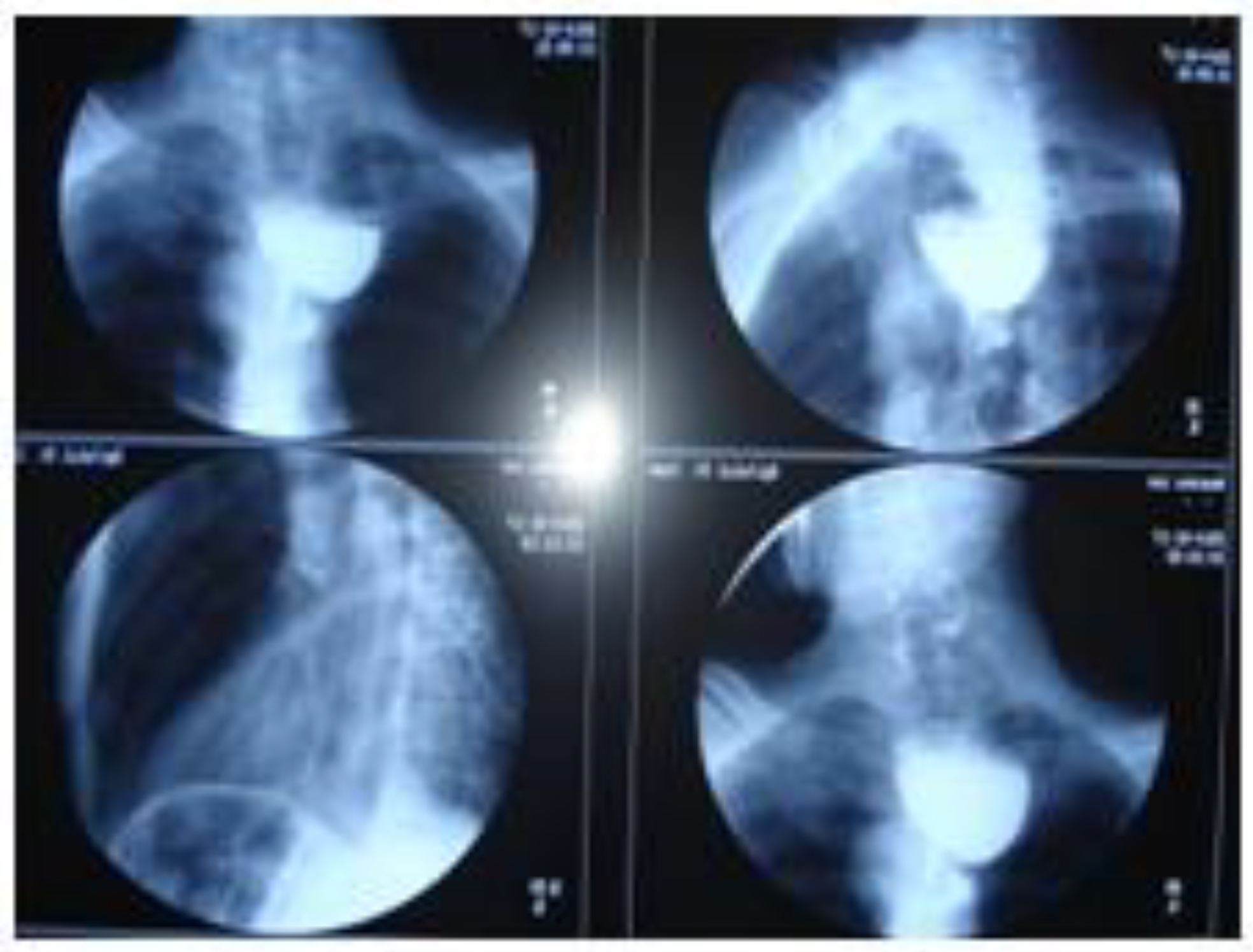
Figure 1. Zenker’s diverticulum and medium esophageal malignant stenosis.
Jmms 04 00026 g001
Figure 2. HEx40, squamous esophageal cancer.
Jmms 04 00026 g002
Figure 3. CT scan of the chest and abdomen.
Jmms 04 00026 g003