Next Article in Journal
The Psychological Influence of the Diagnosis of Breast Cancer on Therapeutic Options Selection
Previous Article in Journal
Cesarean Scar Defects and Placental Abnormalities—A 3 Year Survey Study
 
 
Journal of Mind and Medical Sciences is published by MDPI from Volume 12 Issue 1 (2025). Previous articles were published by another publisher in Open Access under a CC-BY (or CC-BY-NC-ND) licence, and they are hosted by MDPI on mdpi.com as a courtesy and upon agreement with Valparaiso University (ValpoScholar).
Article

Protective Role of S-Adenosylmethionine Against Fructose-Induced Oxidative Damage in Obesity

by
Kameliya Zh Bratoeva
1,*,
Mariya A. Radanova
2,
Albena V. Merdzhanova
3 and
Ivan S. Donev
4
1
Medical University of Varna, Faculty of Medicin, Department of Pathophysiology, Varna, Bulgaria
2
Medical University of Varna, Department of Biochemistry, Molecular Medicine and Nutrigenomics, Laboratory of Nutrigenomics, Functional Foods and Nutraceuticals, Faculty of Pharmacy, Varna, Bulgaria
3
Medical University of Varna, Department of Chemistry, Faculty of Pharmacy, Varna, Bulgaria
4
Medical University of Varna, Department of Propedeutics of Internal Diseases, Medical Oncology Clinic, University Hospital “St. Marina”, Varna, Bulgaria
*
Author to whom correspondence should be addressed.
J. Mind Med. Sci. 2017, 4(2), 163-171; https://doi.org/10.22543/7674.42.P163171
Submission received: 1 August 2017 / Revised: 1 September 2017 / Accepted: 1 October 2017 / Published: 5 October 2017

Abstract

:
Introduction. It has been shown that S-adenosylmethionine (S-AMe) stimulates glutathione synthesis and increases cell resistance to the cytotoxic action of free radicals and pro-inflammatory cytokines. The aim of this study was to determine the effect of S-adenosylmethionine on the oxidative stress in adipose tissue in a model of fructose-induced obesity. Methods. The study was performed on male Wistar rats divided into 3 groups: control, fructose fed (HFD) (35%, 16 weeks), and HFD + S-AMe (20 mg/kg). We examined the changes in the ratio of retroperitoneal adipose tissue weight / body weight; levels of reduced glutathione (GSH) and malondialdehyde (MDA) in the retroperitoneal adipose tissue, and serum levels of GSH and TNF-α. Results. Significant increases in the retroperitoneal adipose tissue, MDA, and serum TNF-α were identified, as well as decreased tissue and serum levels of GSH in rats fed with a high-fructose diet as compared with the control group. In the group fed with HFD and S-AMe, we found significant reduction in the retroperitoneal adipose tissue and decreased levels of MDA and serum TNF-α, as well as increased tissue and serum levels of GSH as compared with the group only on HFD. In conclusion, our results show that fructose-induced obesity causes oxidative stress in hypertrophic visceral adipose tissue. The administration of S-AMe improves the antioxidative protection of adipocytes, and reduces oxidative damage and excessive accumulation of lipids and inflammation.

Introduction

In the last decade, obesity has reached epidemic proportions and is considered a major factor in the development of metabolic syndrome, type 2 diabetes mellitus, non-alcoholic fatty liver disease, cardiovascular disease, etc. An important element in the cluster of risk factors includes the consumption of ready-made foods and beverages with high fructose content, produced by the food industry. In humans and in experimental models of metabolic syndrome, fructose causes obesity and related complications - oxidative stress, hyperglycemia, dyslipidemia, hepatic steatosis, and so on [1].
Obesity is a condition in which a significant increase of the adipose tissue in the body occurs due to disturbed energy balance, favoring a positive energy balance. The increase of adipose tissue in the conditions of over-nutrition and excess lipids leads to hypoxia and increased formation of reactive oxygen species (ROS) [2], which induces low-grade inflammation, impaired adipocyte differentiation, chronic remodeling of hypertrophic adipose tissue and dysfunction [3]. These changes are manifested by unregulated secretion of adipocytokines with pro-inflammatory, pro-thrombogenic, hypertensive and diabetogenic effects, affecting as well other tissues and organs such as endothelium, liver, pancreas, and muscles [4]. Some of the effects such as TNF-α, IL-6, MCP-1, CRP, etc. also induce oxidative stress outside the adipose tissue and create conditions for maintaining a systemic pro-inflammatory state manifested by insulin resistance, endothelial dysfunction and tissue injury [5,6]. Parallel to this, an inhibition in the synthesis of adiponectin is observed, which exhibits anti-inflammatory, antidiabetic and anti-obesity effects [7,8]. Therefore, a number of authors have determined oxidative stress and the impaired redox balance in the visceral adipose tissue to be key factors in the pathogenesis of metabolic complications associated with obesity [9].
In support of this assumption, low levels of GSH and antioxidant enzymes such as catalase and glutathione peroxidase have been found in the fatty tissue of obese people and experimental animals [10], a condition prerequisite for increased oxidative stress. Regardless of the fact that adipose tissue serves as a depot for storage of natural antioxidants (vitamins A, vitamin E and carotenoids), in obese humans there are low serum levels of these vitamins [11]. Based on the research literature, it can be concluded that oxidative stress in adipose tissue as an adjunct to the early metabolic damage in obesity, defines a new approach for the treatment and development of therapies.
In this study, we showed the influence of S-adenosylmethionine (S-AMe) on oxidative damage in an experimental model of metabolic syndrome using rats fed with HFD. S-AMe is a methyl group donor and a glutathione precursor that is metabolized mainly in the liver. S-AMe stimulates GSH synthesis and increases cell resistance to the cytotoxic action of free radicals and pro-inflammatory cytokines [12]. Regarding its therapeutic effects, literature data support predominantly antioxidative, anti-steatotic, and anti-apoptotic effects in various liver diseases. The influence of S-AMe on fructose-induced obesity and oxidative stress in visceral adipose tissue has not been studied, which defines the goal of this investigation.

Materials and Methods

Animal models

Male albino Wistar rats were housed at 20 ± 2ºC room temperature under a standard 12 h light/dark cycle. All animals received a standard diet and water ad libitum. The standard diet was composed of starch- 50%, protein – 20%, fat – 4.5%, 5% cellulose, standard vitamins and mineral mix. At the beginning of the experiment the body weight of rats was 140 - 180 g. After acclimation (two weeks), animals were randomly assigned to three groups of seven each: HFD group (35% glucose-fructose corn syrup in drinking water for 16 weeks), HFD+S-AMe group (20 mg/kg b.w. in drinking water for 16 weeks), and a control group (water drinking). Fructose (Amilym, Bulgaria) and S-AMe (NOW Foods, USA) were used. Food intake was recorded daily and weights were monitored weekly. At the end of the experiment, rats were euthanized with a lethal dose of Thiopental. All manipulations were performed at 4-8oC. Analysis was performed immediately after thawing of the samples. The experimental procedures were approved by the Home Office for the Care and Use of Laboratory Animals and performed with strong consideration for the ethics of animal experimentation according to the International Guiding Principles for Animal Research approved in Bulgaria.

Blood and tissue collection

At the termination of the experiment, blood was collected from the tail vein of rats after pre-narcosis with Thiopental at a dose of 15mg / kg i.p. Serum (subject to rules of good practice Lab) was used to determine some biochemical indicators on the same day, and the remainder serum was frozen. Under general anesthesia with Thiopental 30mg/kg (rapid intravenous), a laparotomy was conducted. We removed various organs for research, prepared them as a homogenate, and the quickly dissected and froze them. The weight of retroperitoneal fat was measured. Tissue extracts were prepared by homogenization with buffer PBS-50mM, pH=7.4 (1 g tissue, 9ml of buffer) on ice using a Teflon glass homogenizer (2000 rpm / 3 min). Homogenates were spun in a refrigerated centrifuge (4000 rpm/ 10min). The supernatant was used for analysis after having identified the concentration of protein.

Biochemical Analyses

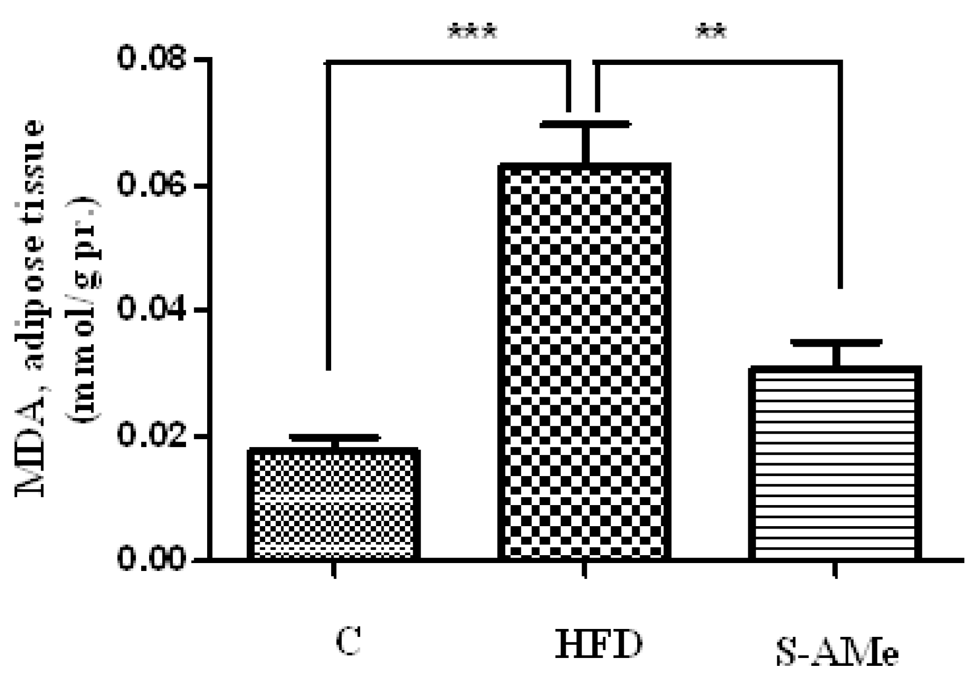
  • Determination of lipid peroxides. Membrane lipid peroxidation was assayed by MDA (malondialdehyde) measured by its thiobarbituric acid (TBA) (Merk, Germany) reactivity in adipose tissue homogenates using the method detailed by Porter et al. [13]. Results were expressed in nmol MDA per g tissue and were determined using the extinction coefficient of MDA–TBA complex at 532 nm= 1.56 – 10-5 cm-1 M-1 solution.
  • Determination of total thiols. GSH levels in adipose tissue and sera were defined as the total number of sulfhydryl groups (SH groups) in the sample. SH groups were determined by the method of Hu [14], based on the absorption of the color complex between thiol groups and 5,5'-dithiobis-(2- nitrobenzoic acid (DTNB) (Merk, Germany) at 412 nm. Standard solutions of reduced glutathione were used to calculate the concentration of thiol groups.
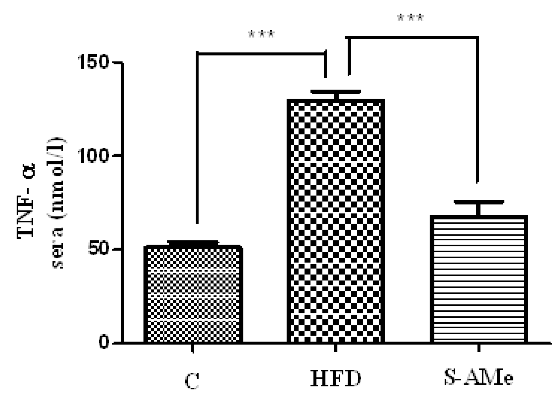
  • Determination of TNF-α levels. Concentrations of TNF-α cytokine (IBL International) were measured in sera using the commercially available ELISA kit.

Statistical analysis

An analysis of variance (ANOVA) was used to compare the differences in the treatment of experimental groups using the .05 significance level. A t-test was used to compare the mean values of two groups when monitoring the effect of S-AMe on treated and healthy rats, with differences between groups significant at p ≤ 0.05. Statistical analysis of the data was performed by using the GraphPad Prism 5.0 software. Graphs were drawn with the GraphPad Prism 5.0 graphing program.

Results

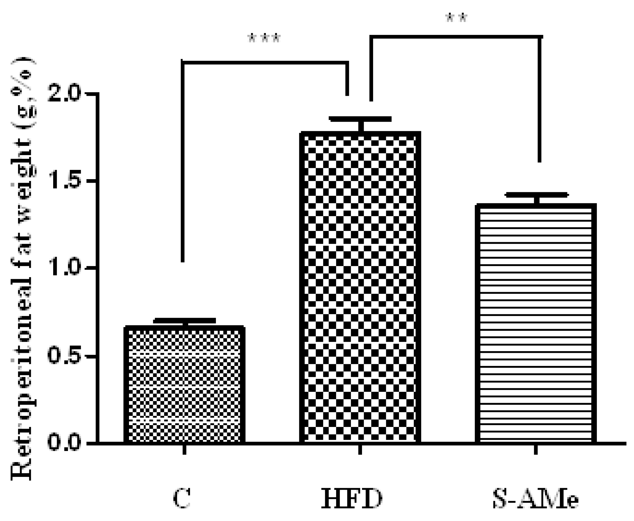
Changes in body weight and weight of visceral adipose tissue.

The body weight of the experimental animals was measured weekly, with consistent increases in all groups through the end of the experiment. Results showed a significant progression in obesity in the HFD group compared with the control group, with more than 50% increase in body weight and a 200% increase in retroperitoneal adipose tissue (p <0.0001). In the group taking S-AMe, the increase in body weight relative to that of the control animals was over 65%, and by more than 15% than the HFD group. However, despite the increase in final body weight in the S-AMe group, the weight of retroperitoneal adipose tissue was statistically reduced relative to the HFD group (p < 0.005). Therefore, changes in the weight of retroperitoneal adipose tissue have been reported as a percentage of the total body weight (Figure 1).

Markers of oxidative stress - levels of MDA and SH groups.

The levels of lipid peroxides were identified in a homogenate from visceral adipose tissue as amount of MDA per gram of protein from the sample. The results showed significantly elevated levels of MDA in the HFD group (p < 0.0005) relative to the control group. In the group fed with S-AMe, lower levels of MDA were found compared with the HFD group (p <0.005) and these levels were near those of the control group (Figure 2).
GSH levels in adipose tissue and sera were defined as the total number of sulfhydryl groups (SH groups) in the sample. The results indicate significantly decreased levels of SH groups in the retroperitoneal adipose tissue in the HFD group (p <0.01) versus the control group. In the S-AMe -fed group, the levels of SH groups were higher than those in the HFD group (p< 0.05), Figure 3A. Levels of SH groups in sera showed a significant reduction in the HFD group (over 50%) relative to controls (p <0.0005), whereas in the S-AMe group, levels were significantly higher (p <0.05) as compared with the HFD group, Figure 3B.

3. Marker of inflammation.

Changes in serum TNF-α levels. We found significantly elevated levels of TNF-α in the HFD group versus the control (p < 0.0005). In the S-AMe group, lower levels of pro-inflammatory cytokine were recorded as compared with the HFD group (p <0.0005), see Figure 4.

Discussion

Increased oxidative stress in visceral adipose tissue and the subsequent inflammatory processes are key pathogenic factors associated with the metabolic complications of obesity-insulin resistance, dyslipidemia, endothelial dysfunction, fatty liver degeneration and others. Expansion of fructose consumption is an important factor in the pathogenesis of obesity as it is associated with increased calorie intake and a positive energy balance [15,16]. Our results indicate that the high fructose diet causes a significant increase in weight of the retroperitoneal adipose tissue that is closely related to the tested markers of oxidative stress and inflammation, whereas S-AMe intake results in reduction of adipose tissue, MDA, and serum TNF- α, and significantly elevates the levels of reduced glutathione in fatty tissue and serum.
It has been shown that fructose is a highly lipogenic nutrient due to its conversion into fatty acids and activation of de novo lipogenesis in tissues, especially in enterocytes and the liver [17,18]. In addition, its metabolism in the liver leads to hyperglycemia, regardless of the energy state of cells and serum glucose levels [19]. Taken together, these effects lead to obesity and hypertrophy of adipose tissue. The main mechanisms involved in generating ROS in fatty tissue in a fructose-rich diet are adipocyte hypoxia, increased uric acid formation, and activation of leukocytes and endothelial cells [20]. Adipocyte hypoxia takes part in the attraction and retention of macrophages within it (important sources of ROS by activation of NADPH-oxidase, myeloperoxidase and xanthine oxidase) [21] and in the inhibition of adipocyte differentiation, which is the cause of ectopic lipid deposition and subsequent metabolic injury [22]. For example, Furukawa et al. 2004 have established that inhibition of NADPH oxidase activity results in decreased generation of ROS and TNF-α in white adipose tissue and increased levels of adiponectin [9].
In addition, HFD leads to increased uric acid formation [23] and superoxide radicals, and decreased expression of antioxidative enzymes [24]. In a number of studies, hyperuricemia has been shown to cause systemic oxidative stress, suppress adiponectin secretion, reduce endothelial NO levels, and increase plasma triglycerides, which is a prerequisite for the development of insulin resistance, endothelial dysfunction, and hypertension [25]. The ROS generated during these reactions induces the activation of lipid peroxidation, whose products are cytotoxic and deplete the cellular levels of reduced glutathione, thus potentiating the effects of oxidative stress [26].
Reduced glutathione as a SH group donor is the main endogenous non-enzymatic antioxidant and modulator of redox status in tissue cells throughout the body [27]. The liver is the major source of reduced glutathione and, at the same time, most frequently attacked in case of oxidative stress. Oxidative injuries of the liver disrupt its normal secretion and homeostasis in the entire organism [28]. Decreased tissue levels are a common indicator of chronic oxidative stress (due to depletion) or impaired synthesis. Our data also confirm the presence of lipid peroxidation in adipose tissue and an imbalance in the anti-oxidative protection, as per the significantly higher levels of MDA and low levels of SH groups in the HFD versus control groups.
Our results also showed a significant loss of serum SH groups in the fructose-fed group. In obesity and under conditions of insulin resistance, hypertrophic adipocytes are sources of free fatty acids that have a direct lipotoxic effect and maintain a pro-oxidant state throughout the body and especially in the liver, where the major amount of S-AMe is metabolized. S-AMe is the main endogenous donor of cysteine, which is required for glutathione synthesis. In another of our studies, we found that S-AMe administration increased the levels of SH groups in the liver under the conditions of oxidative stress, and it is probably through this mechanism that it protects against systemic oxidative stress [29].
We assume that chronically increased ROS formation and suppression of anti-oxidative protection in hypertrophic fatty tissue induces oxidative damage and subsequent inflammation, which is the cause of elevated serum levels of TNF-α in the fructose-fed group, which were proportional to the increase in retroperitoneal adipose tissue and oxidative injury. The administration of S-AMe reduces these negative effects. In humans and rodents, macrophage infiltration into adipose tissue and serum levels of inflammatory mediators such as TNF-α, IL-6, and CRP have been shown to rise proportionally with the increase of Body Mass Index, body fat, and adipocyte hypertrophy, a reversible process upon loss of body weight [30]. Both macrophages and adipocytes are sensitive to the effects of TNF-α, which, by activating the inflammatory IKK/NF- κB signaling pathway, also increases the expression of other mediators of inflammation such as acute - phase proteins, interleukin-6, etc., resulting in a self-perpetuating cycle of increased release of pro-inflammatory cytokines [31]. S-AMe has been shown to inhibit in vitro the kinase activity of IKK-b of the pro-inflammatory IKK-b / NF-kb signaling pathway in 3T3-L1 adipocytes and to suppress the TNF-α – induced insulin resistance [32]. These findings suggest that, in addition to its antioxidant activity, S-AMe also exhibits an anti-inflammatory effect in vivo in a model of HFD obesity, which we report for the first time in this study.
Our data also indicate that the exogenous application of S-AMe results in reduction of visceral adipose tissue. There is no evidence in the literature of the effect of S-AMe on the accumulation of triglycerides in adipose tissue. In addition to being a glutathione precursor, S-AMe is the active form of methionine that takes part in the methylation reactions of a large number of substrates - proteins, lipids and nucleic acids. Transmethylation of nucleic acids determines the epigenetic profile and transcriptional activity of key genes involved in cellular metabolism in all cell types [33]. We presume that the adipose tissue reduction by means of S-AMe is possibly due to epigenetic changes in genes associated with the activity of lipid metabolism and energy balance. For example, hypermethylation of DNA (Deoxyribonucleic acid) promoter regions in FASN (a fatty acid synthesis regulating gene) reduces TG deposition in the liver [34].

Conclusions

In conclusion, our study shows the relationship between the level of hypertrophy in visceral adipose tissue and elevated markers of oxidative stress and inflammation in HFD. We presume that in obesity, a pro-oxidant state is maintained that is largely responsible for the development of subsequent metabolic complications.
To our knowledge, our research is the first to show that S-AMe exhibits anti-oxidative and anti-inflammatory activity in vivo by reducing fructose-induced hypertrophy and oxidative and inflammatory damage in adipose tissue. These effects are likely to be essential for the normal functioning of adipocytes by reducing the risk of developing insulin resistance and hence the metabolic complications of obesity.

References

  1. Oron-Herman, M.; Kamari, Y.; Grossman, E.; Yeger, G.; Peleg, E.; Shabtay, Z.; Shamiss, A.; Sharabi, Y. Metabolic syndrome: comparison of the two commonly used animal models. Am J Hypertens. 2008, 21, 1018–22. [Google Scholar] [CrossRef] [PubMed]
  2. Rudich, A.; Kanety, H.; Bashan, N. Adipose stress- sensing kinases: linking obesity to malfunction. Trends Endocrinol Metab. 2007, 18, 291–9. [Google Scholar] [CrossRef] [PubMed]
  3. Huang, R.C.; Mori, T.A.; Burke, V.; Newnham, J.; Stanley, F.J.; Landau, L.I.; Kendall, G.E.; Oddy, W.H.; Beilin, L.J. Synergy between adiposity, insulin resistance, metabolic risk factors, and inflammation in adolescents. Diabetes Care. 2009, 32, 695–701. [Google Scholar] [CrossRef] [PubMed]
  4. Sabio, G.; Das, M.; Mora, A.; Zhang, Z.; Jun, J.Y.; Ko, H.J.; Barrett, T.; Kim, J.K.; Davis, R.J. A stress signaling pathway in adipose tissue regulates hepatic insulin resistance. Science 2008, 322, 1539–43. [Google Scholar] [CrossRef] [PubMed]
  5. Day, C.P. From fat to inflammation. Gastroenterology 2006, 130, 207–10. [Google Scholar] [CrossRef] [PubMed]
  6. Plomgaard P, Bouzakri K, Krogh-Madsen R, Mittendorfer B, Zierath JR, Pedersen BK. Tumor necrosis factor-α induces skeletal muscle insulin resistance in healthy human subjects via inhibition of Akt substrate 160 phosphorylation. Diabetes 2005, 54, 2939–45. [CrossRef] [PubMed]
  7. Wolf, A.; Wolf, D.; Rumpold, H.; Enrich, B.; Tilg, H. Adiponectin induces the anti-inflammatory cytokines IL-10 and IL-1RA in human leukocytes. Biochem Biophys Res Commun. 2004, 323, 630–5. [Google Scholar] [CrossRef] [PubMed]
  8. Weyer, C.; Funahashi, T.; Tanaka, S.; Hotta, K.; Matsuzawa, Y.; Pratley, R.E.; Tataranni, P.A. Hypoadiponectinemia in obesity and type 2 diabetes: close association with insulin resistance and hyperinsulinemia. J Clin Endocrinol Metab. 2001, 1930–5. [Google Scholar] [CrossRef] [PubMed]
  9. Furukawa, S.; Fujita, T.; Shimabukuro, M.; Iwaki, M.; Yamada, Y.; Nakajima, Y.; Nakayama, O.; Makishima, M.; Matsuda, M.; Shimomura, I. Increased oxidative stress in obesity and its impact on metabolic syndrome. J Clin Invest. 2004, 114, 1752–61. [Google Scholar] [CrossRef] [PubMed]
  10. Neuhouser, M.L.; Rock, C.L.; Eldridge, A.L.; Kristal, A.R.; Patterson, R.E.; Cooper, D.A.; Neumark-Sztainer, D.; Cheskin, L.J.; Thornquist, M.D. Serum concentrations of retinol, alpha-tocopherol and the carotenoids are influenced by diet, race and obesity in a sample of healthy adolescents. J Nutr. 2001, 131, 2184–91. [Google Scholar] [CrossRef] [PubMed]
  11. Nicolae, A.C.; Drăgoi, C.M.; Ceaușu, I.; Poalelungi, C.; Iliescu, D.; Arsene, A.L. Clinical implications of the indolergic system and oxidative stress in physiological gestational homeostasis. Farmacia. 2015, 63, 46–51. [Google Scholar]
  12. Andringa, K.K.; King, A.L.; Eccleston, H.B.; Mantena, S.K.; Landar, A.; Jhala, N.C.; Dickinson, D.A.; Squadrito, G.L.; Bailey, S.M. Analysis of the liver mitochondrial proteome in response to ethanol and S- adenosylmethionine treatments: novel molecular targets of disease and hepatoprotection. Am J Physiol Gastrointest Liver Physiol. 2010, 298, G732–45. [Google Scholar] [CrossRef] [PubMed]
  13. Porter, N.A.; Nixon, J.; Isaac, J. Cyclic peroxidase and thiobarbituric assay. Biochim Biophys Acta 1976, 441, 596–599. [Google Scholar] [PubMed]
  14. Hu, M. Measurment of protein thiol groups and glutathione in plasma. Methods Enzymol. 1994, 233, 380–5. [Google Scholar] [PubMed]
  15. Melanson, K.J.; Angelopoulos, T.J.; Nguyen, V.; Zukley, L.; Lowndes, J.; Rippe, J.M. High-fructose corn syrup, energy intake, and appetite regulation. Am J Clin Nutr. 2008, 88, 1738S–1744S PMID: 19064539. [Google Scholar] [CrossRef] [PubMed]
  16. Basciano, H.; Federico, L.; Adeli, K. Fructose, insulin resistance, and metabolic dyslipidemia. Nutr Metab. 2012, 37, 233–40. [Google Scholar] [CrossRef] [PubMed]
  17. Botezelli, J.D.; Mora, R.F.; Dalia, R.A.; Moura, L.P.; Cambri, L.T.; Ghezzi, A.C.; Voltarelli, F.A.; Mello, M.A. Exercise counteracts fatty liver disease in rats fed on fructose-rich diet. Lipids Health Dis. 2010, 9, 116. [Google Scholar] [CrossRef] [PubMed]
  18. Busserolles, J.; Gueux, E.; Rock, E.; Mazur, A.; Rayssiguier, Y. High fructose feeding of magnesium deficient rats is associated with increased plasma triglyceride concentration and increased oxidative stress. Magnes Res. 2003, 16, 7–12. [Google Scholar] [PubMed]
  19. Cortez-Pinto, H.; Chatham, J.; Chacko, V.P.; Arnold, C.; Rashid, A.; Diehl, A.M. Alterations in liver ATP homeostasis in human nonalcoholic steatohepatitis: a pilot study. JAMA 1999, 282, 1659–64. [Google Scholar] [CrossRef] [PubMed]
  20. Nakagawa, T.; Hu, H.; Zharikov, S.; Tuttle, K.R.; Short, R.A.; Glushakova, O.; Ouyang, X.; Feig, D.I.; Block, E.R.; Herrera-Acosta, J.; Patel, J.M.; Johnson, R.J. A causal role for uric acid in fructose-induced metabolic syndrome. Am J Physiol Renal Physiol. 2006, 290, F625–31. [Google Scholar] [CrossRef] [PubMed]
  21. Talior I, Tennenbaum T, Kuroki T. Eldar-Finkelman H. PKC-delta-dependent activation of oxidative stress in adipocytes of obese and insulin-resistant mice: role for NADPH oxidase. Am J Physiol Endocrinol Metab. 2005, 288, E405–11. [Google Scholar] [CrossRef] [PubMed]
  22. Yun, Z.; Maecker, H.L.; Johnson, R.S.; Giaccia, A.J. Inhibition of PPAR gamma 2 gene expression by the HIF-1-regulated gene DEC1/Stra13: a mechanism for regulation of adipogenesis by hypoxia. Dev Cell. 2002, 2, 331–41. [Google Scholar] [CrossRef] [PubMed]
  23. Ackerman, Z.; Oron-Herman, M.; Grozovski, M.; Rosenthal, T.; Pappo, O.; Link, G.; Sela, B.A. Fructose- induced fatty liver disease: hepatic effects of blood pressure and plasma triglyceride reduction. Hypertension. 2005, 45, 1012–8. [Google Scholar] [CrossRef] [PubMed]
  24. Kanellis, J.; Watanabe, S.; Li, J.H.; Kang, D.H.; Li, P.; Nakagawa, T.; Wamsley, A.; Sheikh-Hamad, D.; Lan, H.Y.; Feng, L.; Johnson, R.J. Uric acid stimulates monocyte chemoattractant protein-1 production in vascular smooth muscle cells via mitogen-activated protein kinase and cyclooxygenase-2. Hypertension 2003, 41, 1287–93. [Google Scholar] [CrossRef] [PubMed]
  25. Kang, D.H.; Park, S.K.; Lee, I.K.; Johnson, R.J. Uric acid- induced C-reactive protein expression: implication on cell proliferation and nitric oxide production of human vascular cells. J Am Soc Nephrol. 2005, 16, 3553–62. [Google Scholar] [CrossRef] [PubMed]
  26. Seki, S.; Kitada, T.; Yamada, T.; Sakaguchi, H.; Nakatani, K.; Wakasa, K. In situ detection of lipid peroxidation and oxidative DNA damage in non-alcoholic fatty liver diseases. J Hepatol. 2002, 37, 56–62. [Google Scholar] [CrossRef] [PubMed]
  27. Forman, H.J.; Zhang, H.; Rinna, A. Glutathione: overview of its protective roles, measurement, and biosynthesis. Mol Aspects Med. 2009, 30, 1–12. [Google Scholar] [CrossRef] [PubMed]
  28. Lu, S.C. Regulation of glutathione synthesis. Mol Aspects Med. 2009, 30, 42–59. [Google Scholar] [CrossRef] [PubMed]
  29. Bekyarova, G.; Tzaneva, M.; Bratoeva, K.; Kotzev, I.; Radanova, M. Heme-oxygenase-1 upregulated by s- adenosylmethionine. Potential protection against non-alcoholic fatty liver induced by high fructose diet. Farmacia 2017, 6, 262–7. [Google Scholar]
  30. Gustafson, B. Adipose tissue, inflammation and atherosclerosis. J Atheroscler Thromb. 2010, 17, 332–41. [Google Scholar] [CrossRef] [PubMed]
  31. Hotamisligil, G.S.; Shargill, N.S.; Spiegelman, B.M. Adipose expression of tumor necrosis factor-alpha: direct role in obesity-linked insulin resistance. Science 1993, 259, 87–91. [Google Scholar] [CrossRef] [PubMed]
  32. Moon MK, Kim M, Chung SS, Lee HJ, Koh SH, Svovoda P, Jung MH, Cho YM, Park YJ, Choi SH, Jang HC, Park KS, Lee HK. S-Adenosyl-l-methionine ameliorates TNFalpha-induced insulin resistance in 3T3-L1 adipocytes. Exp Mol Med 2010, 42, 345–352. [CrossRef] [PubMed]
  33. Bekyarova, G.; Atanasova, M.; Tzaneva, M.; Dimitrova, A. Melatonin modulates inflammatory response and suppresses burn-induced apoptotic injury. J Mind Med Sci. 2017, 4, 59–66. [Google Scholar] [CrossRef]
  34. Cordero, P.; Gomez-Uriz, A.M.; Campion, J.; Milagro, F.I.; Martinez, J.A. Dietary supplementation with methyl donors reduces fatty liver and modifies the fatty acid synthase DNA methylation profile in rats fed an obesogenic diet. Genes Nutr. 2013, 8, 105–13. [Google Scholar] [CrossRef] [PubMed]
Figure 1. Weight of retroperitoneal adipose tissue expressed as % of total rat body weight. ***p< 0.05 HFD vs. control group; **p < 0.05 S-AMevs. HFD group
Jmms 04 00022 g001
Figure 2. Effect of SAMe on HFD – induced changes in adipose tissue MDA in rats. .***p < 0.05 HFD vs. control group; **p < 0.05 S-AMevs. HFD group.
Jmms 04 00022 g002
Figure 3. Effect of SAMe on HFD – induced changes in SH groups in rats. A ) SH groups sera: ***p < 0.05 HFD vs. control group; *p < 0.05 S-AMe vs. HFD group B ) SH groups adiposse tissue: **p < 0.05 HFD vs. control group; *p < 0.05 S-AMe vs. HFD group.
Jmms 04 00022 g003
Figure 4. Effect of SAMe on HFD – induced changes in TNF-α levels in rats ***p < 0.05 HFD vs. control group; ***p < 0.05 S-AMe vs. HFD group.
Jmms 04 00022 g004

